Venous Occlusive Diseases
|
|
|
- Wilfrid Mosley
- 5 years ago
- Views:
Transcription


1 Venous Occlusive Diseases Bruce R. Saran, MD Adjunct Assistant Clinical Professor of Medicine Scheie Eye Institute University of Pennsylvania School of Medicine Philadelphia, PA -a division of:
2 RVO Demographics } prevalence of RVOs worldwide is about 0.5% } estimated to affect more than 16 million people worldwide in 2008 } BRVO occurs 6-7 times more than CRVO } African Americans have a similar incidence of CRVO to white Americans
3 RVO Demographics } 2.14 cases/1000 > 40 years } 5.36 cases/1000 > 64 years } males slightly > females } No racial preference } Second eye 7-11%
4 RVO Risk Factors } CRVO in one eye has a 1% risk per year of CRVO developing in the fellow eye } CRVO is associated with glaucoma } Risk of BRVO is more likely associated with local vascular factors (arterial-venous crossing changes) rather than local ocular factors
5 RVO Risk Factors for BRVO } Arterial hypertension } Hyperlipidemia } Diabetes } Coronary artery disease

6 CRVO-risk factors Lower Risk } Increased physical activity } alcohol } exogenous estrogens
7 CRVO-medical workup: } Blood pressure } Erythrocyte sedimentation rate (ESR) } Full blood count (FBC) } Random blood glucose } Random total and HDL cholesterol } Urea, electrolytes and creatinine (renal disease in association with hypertension) } Thyroid function tests (associated with dyslipidemia) } EKG (left ventricular hypertrophy secondary to hypertension)
8 CRVO-medical workup: patients<50 or bilateral } Chest X-ray (sarcoidosis, tuberculosis) } C-reactive protein (CRP) } "Thrombophilia screen" (thrombin time, prothrombin time and activated partial thromboplastin time, antithrombin functional assay, protein C, protein S, activated protein C resistance, factor V Leiden mutation, prothrombin G20210A mutation; anticardiolipin antibody, lupus anticoagulant) } Autoantibodies (RF, ANA, anti-dna antibody) } Serum angiotensin-converting enzyme (ACE) } Fasting plasma homocysteine level } Treponemal serology } Carotid duplex imaging
9 CRVO: differential diagnosis } A. Diabetic retinopathy } B. Radiation retinopathy } C. Conditions leading to anemia or low platelets, i.e., blood dyscrasias such as leukemia } D. Ocular ischemia from carotid disease } E. Malignant Hypertension
10 CRVO-classification Non-ischemic } venous stasis retinopathy Ischemic } hemorrhagic retinopathy } CVO study P } CVO study N } visual acuity typically 20/200 or better } visual acuity typically 20/400 or worse

11 CRVO- Non-ischemic classification } may present with good vision } few retinal hemorrhages } Few or no cotton-wool spots } no APD } Fluorescein angiography shows preserved perfusion } may resolve or progress to ischemic

12 CRVO-Ischemic classification } severe visual loss 20/400 or worse } extensive retinal hemorrhages & cotton-wool spots } Positive APD } Fluorescein angiography shows poor perfusion } Extinguished /depressed ERG } neovascular glaucoma and/or proliferative retinopathy
13 RVO Treatment: guided by clinical trials } Laser studies } 1980s Branch Vein Occlusion Study (BVOS) } 1990s Central Vein Occlusion Study (CVOS) } 2009 Steroid studies } SCORE Study } Ozurdex Trials: Geneva } 2010 Anti-VEGF Ranibizumab studies } BRAVO and CRUISE } 2012 Anti-VEGF Aflibercept studies } Copernicus and Galileo

14 CVOS Central Vein Occlusion Study Group - established guidelines for treating CRVO } 725 patients, followed for 3 years } Visual outcome = dependent on initial vision } 65% had VA > 20/40
15 CVOS Central Vein Occlusion Study Group intermediate VA (20/50-20/200) VA < 20/200 } 19% improved >20/50 } 44% stayed in group } 80% chance of not changing VA whether perfused or nonperfused } 37% decreased < 20/200
16 CVOS Central Vein Occlusion Study Group } First 4 months: 15% ischemic } Next 32 months:19% ischemic = 34% at 3 years } INV/ANV occurred in 16% total-35% in non-perfused on fluorescein angiography and only 10% perfused } poor VA and non-perfusion on fluorescein angiography associated with INV/ANV Rule of 3rds: 1/3 gets better, 1/3 same, 1/3 worse

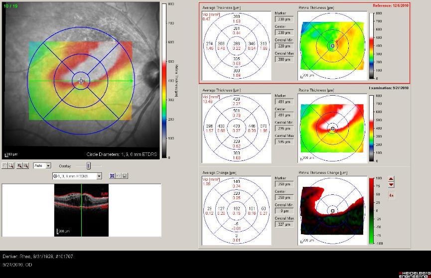
17 Non-ischemic Central Retinal Vein Occlusion } optic disc edema may be present } conversion to ischemic 5%-34% } Late visual loss from CME & pigmentary changes

 and INL with SRF in acute")
18 Non-ischemic Central Retinal Vein Occlusion } IVFA leakage from veins, disc and macular edema } ERG is normal } IOP may be low } OCT with edema in Henle s layer (OPL) and INL with SRF in acute disease

19 Non-Ischemic CRVO 42 y WF PPLOV VA=20/200 OD


20 intravitreal Avastin given 1 month s/p Avastin VA=20/40

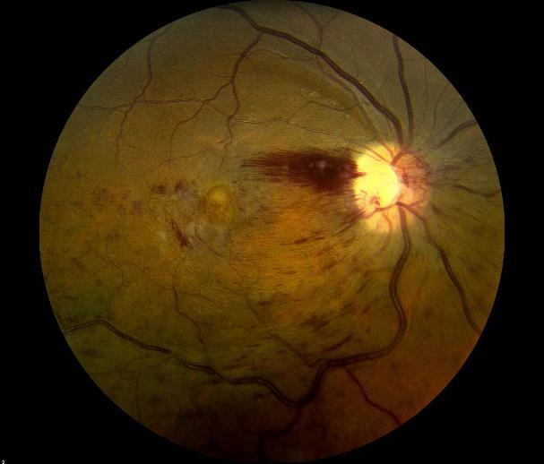
21 Ischemic Central Retinal Vein Occlusion } sudden, painless decrease in VA } 20/400 or worse } x=68.5 years (older) } Blurred/obscured disc margin, dilated precapillary arterioles

22 Ischemic Central Retinal Vein Occlusion } confluent retinal hemorrhages, flame shaped } dilated, tortuous veins } cotton-wool spots } macular edema or macular hemorrhage pronounced
23 Ischemic-CRVO general course (pre anti-vegf era) } 58 % iris neovascularization } 47 % angle neovascularization } 33 % neovascular glaucoma } 8 % NVE
24 Ischemic Central Retinal Vein Occlusion- IVFA } CVO study : VA < 20/200 or non-perfusion predictive of NVI } NVG 10% with 10 DD nonperfusion } NVG 50% % with 80 DD or more

25 Ischemic Central Retinal Vein Occlusion } NVI highly associated with NVG } non-perfusion or NVI requires aggressive PRP and anti-vegf treatments } follow-up interval is increased *this is the classic 90 day glaucoma associated with CRVO


26 CRVO treatment: laser } Focal laser ineffective: reduces anatomic macular edema with little effect on visual acuity } Panretinal photocoagulation effective for ischemia and NVI / ANV

27 CRVO treatment: new paradigm } Avastin } Lucentis } Eylea } Ozurdex
28 CRVO treatment: Ranibizumab (Lucenits) [Cruise Study] } 0.5-mg Ranibizumab gained a mean of 14.9 letters } 0.8 letters in the sham group } 47.7% of ranibizumab gained 15 letters, >3 lines compared with 16.9% of sham } rapid improvement in BCVA with rapid reduction in central foveal thickness after treatment with ranibizumab at day 7 compared with no reduction in the sham group
29 CRVO treatment: Aflibercept (Eylea) [Copernicus/Galileo Study] } 2 mg Aflibercept gained a mean of 18 letters } 3.3 letters in the sham group } % of ranibizumab gained 15 letters, >3 lines compared with % of sham } rapid improvement in BCVA with rapid reduction in central foveal thickness after treatment with ranibizumab at day 7 compared with no reduction in the sham group
 (dexamethasone) Intravitreal Implant [Geneva Study] } NOVADUR solid polymer delivery system through 22 gauge needle } 30% achieved 15 or more letters of improvement after 60 days } 28%")
30 OZURDEX 0.7 mg (700 µg) (dexamethasone) Intravitreal Implant [Geneva Study] } NOVADUR solid polymer delivery system through 22 gauge needle } 30% achieved 15 or more letters of improvement after 60 days } 28% gained 15 or more letters at 2 months } 45% at 6 months } 39% at 1 year } 32% achieved 15 or more letters of improvement 60 days after 2 nd TX improvement (3 lines of vision) occurred 1-2 weeks after implantation in 20-30% } duration of effect persisted 1-3 months after onset

31 CRVO CASE STUDY : Acute VA=20/400


32 CRVO s/p Lucentis X 1 CRVO CASE STUDY : Acute VA=20/80


33 CASE STUDY 2: Bilateral VA 20/150 reduced to CF-treated with Avastin then Eylea. Ozurdex offered


34 2009 VA=CF 2011 VA=20/150 CRVO s/p Ozurdex X 1 CASE STUDY : post tx VA=20/150 at 6 weeks
35 HRVO

36
37 HRVO } CRVO with two trunks behind lamina cribosa (20%) } Thought to be variant of CRVO but with complications and findings of both types of vein occlusions } Also divided into ischemic and nonischemic
38 CRVO Anatomy
39 Anatomy of HRVO
40 HRVO/CRVO Similarities } Hayreh found BRVO artery crosses over vein in 91% whereas in HRVO only 1/3 showed this and >1/3 had no crossing } Collateral vessels in BRVO feed at crossing whereas in CRVO they form at disc or more posterior as found in HRVO } In HRVO about 1/3 showed increased IOP as did CRVO unlike BRVO where increased IOP not > general population } ON edema seen in HRVO/CRVO but not usually in BRVO
41 Treatment Implications } HRVO (ischemic) 13% NVI (Hayreh): NVI > BRVO but < CRVO } Non-ischemic showed no neovascularization } NVD 29% and NVE 42% in ischemic: HRVO > CRVO and BRVO

42

43 BRVO
44 Branch Retinal Vein Occlusion-BRVO } Leber occlusion at the A/V crossing } 3/4 in the superior retina } share a common adventitial sheath } venous lumen diminished by atherosclerosis } artery caliber increases with arterial media thickening

45 Branch Retinal Vein Occlusion-BRVO } collaterals develop as vein-to-vein channels } maybe only indicator of old disease } do not leak on IVFA
46 BRVO-risk factors Increased } Hypertension } Glaucoma } CAD Decreased } higher HDL levels } alcohol consumption } Hyperopia } higher levels Alpha-2 globulin

47 BRVO- classification } non-ischemic } Indeterminate } ischemic > 5 DD capillary non-perfusion

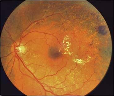
48 BRVO- characteristics } unilateral and segmental distribution nerve fiber hemorrhages } intraretinal hemorrhages } cotton-wool spots } retinal edema
49 BRVO- characteristics } 41% visual acuity of 20/20 to 20/50 } 25% VA of 20/60 to 20/200 } 32% VA of 20/200 or worse } 60%-100% have macular edema at some point in their clinical course } 30% with persistent macular edema after 1 year, can improve over 6-12 months

50 BRVO- characteristics retinal hemorrhages and venous tortuousity } gradually decrease 6- to 12-month } microaneurysms characteristic of acute recovery } narrowed arterioles with venous occlusion/sheathing

51 BRVO- characteristics late changes } RPE proliferation } residual macular edema } macular cysts and holes } macular pucker } persistent macular hemorrhages and microaneurysms


52 BRVO- characteristics late changes } subretinal fibrosis } neovascularization of the disc and retina
53 BRVO: differential diagnosis } A. Diabetic retinopathy } B. Radiation retinopathy } C. Hemi central retinal vein occlusion } D. Juxtafoveal telangiectasia } E. Epiretinal membrane } H. Hypertensive retinopathy
54 BRVO: treatment } Initial observation for MANY patients } ischemic type of BRVO, NVE or hemorrhage would warrant scatter photocoagulation (in distribution of BRVO) } observation 3-6 months, patients with persistent macular edema and/or visual acuity 20/40 or worse, should consider focal laser or intravitreal medication } 1. Good blood pressure control } 2. Treatment of dyslipidemia } 3. Treatment of diabetes mellitus, if present
55 BRVO- Treatment from BVOS FOCAL LASER } 65% treated gained 2 or more lines -compared to 37% controls } 60% treated eyes had VA > 20/40 -compared to 34% controls } 12% treated eyes had VA < 20/200 -compared to 23% controls } 12% treated lost 2 or more lines -compared to 17% controls

56 BRVO- Treatment from BVOS sectoral PRP* to prevent the retinal neovascularization } reduced neovascularization from 22% to 12% } ischemic occlusion more than 5 disc diameters of capillary nonperfusion
57 New Treatments: BRVO Anti-VEGF Surgery } Avastin } Lucentis } Arterial-Venous Delamination } Eylea Steroids: } Ozurdex } Kenalog

58 SCORE : IVTA BRVO } 1mg 29% } 4mg 26% } STD 27% achieved vision endpoint } rates of elevated IOP & cataract were similar for the standard care and 1-mg groups, but higher in the 4-mg group } Conclusions NO difference identified in visual acuity at 12 months for the standard care group compared with the triamcinolone groups } rates of adverse events (particularly elevated intraocular pressure and cataract) were highest in the 4-mg group

59 Ozurdex + Avastin for Macular Edema Due to BRVO } 23% lasted to 6 months without additional therapy } +10 letters at 2 weeks } +14 letters at month 1 and 2 } +12 letters at month 4 } > +3 line VA in 28% at week 2 to 45%, at months 4
60 Lucentis (Ranibizumab) for BRVO BRAVO study } Efficacy } Ranibizumab treatment groups } Rapid improvement in visual and OCT by Day 7 and sustained through Month 6 with monthly treatment } With initiation of PRN treatment at Month 6, gains in BCVA were maintained, on average, through Month 12 } 18.3 letter gain with ranibizumab vs. 7.3 letter gain in shame } 54.5% of the sham group requiring rescue laser } only 19.8% ranibizumab required rescue laser
61 Eylea (Aflibercept) for BRVO: Vibrant Study } Eylea } mean change of BCVA was 17.1 letter gain with Eylea vs 12.2 in grid laser group at 52 weeks } rescue treatment was required in 10% of drug group and in 80% eyes in the laser/drug group Avastin (Bevacizumab): BERVOLT and multiple studies } BERVOLT retrospectively studied efficacy and safety of bevacizumab for macular edema due to BRVO and CRVO } Efficacy similar to Eylea and Lucentis
62 Case 1 PPLOV X 3 months VA 20/60

63

64 1 month s/p intravtreal Lucentis
65 1 month s/p intravtreal Lucentis VA 20/60 to 20/25

66 Case 2 BRVO VA 20/200 s/p Lucentis 6 months previous
67 Case 2 BRVO VA 20/200 s/p Lucentis 6 months previous
68 Case 2 BRVO VA 20/200 s/p Lucentis 6 months previous

69
70 Case 2

71 BRVO VA 20/200 s/p Lucentis 6 months previous

72 3 months s/p Lucentis and peripheral PRP VA 20/40
73 Pre tx 2010 VA = 20/200 3 months post tx2010 VA = 20/40

74 6 months s/p Lucentis and peripheral PRP VA 20/30
75 2011 VA = 20/30

76 Macular Branch Retinal Vein Occlusion } small venous tributary } subgroup of BRVO } patients with macular branch vein occlusion complain of blurring or distortion of vision

77 Macular Branch Retinal Vein Occlusion } superior macular more common } 85% macular edema } small areas capillary nonperfusion in 20% } neovascularization typically is not seen


78 40 yo WM with PPLOV OD: 20/70 OS: 20/50


79 40 yo WM 3 months later OD: 20/20 OS: 20/20
80 Summary Ocular Complications Type NVD NVE NVI NVG % % % CRVO % Ischemic 15% HRVO (I) 11 (-29)% BRVO 10.6% Macular- 0% BRVO 8% 58% 33% 9 (-42)% 9 (-13)% 3% 20.7% 1.6% 0.8% 0% 0% 0%
81 Retinal Vein Occlusion Practice Guidelines } Symptoms <2 months without CME on OCT or loss of vision: observe monthly for 3 months and then quarterly } Symptoms> 2 months, CME, RPE changes, VA >20/40 consider treatment } Medical work up to PMD } Follow all patients monthly for first 3 months and then quarterly

82
Vascular Disease Ocular Manifestations of Systemic Hypertension
 Vascular Disease Ocular Manifestations of Systemic Hypertension Maynard L. Pohl, OD, FAAO Pacific Cataract & Laser Institute 10500 NE 8 th Street, Suite 1650 Bellevue, WA 98004 USA 425-462-7664 Cerebrovascular
OCCLUSIVE VASCULAR DISORDERS OF THE RETINA
 OCCLUSIVE VASCULAR DISORDERS OF THE RETINA Learning outcomes By the end of this lecture the students would be able to Classify occlusive vascular disorders (OVD) of the retina. Correlate the clinical features
4/27/2010 INTRODUCTION TO RETINAL VASCULAR DISEASE VENOUS/VENULAR CENTRAL RETINAL VEIN OBSTRUCTION / CRVO ADDITIONAL FEATURES /COMPLICATIONS
 INTRODUCTION TO RETINAL VASCULAR DISEASE VENOUS/VENULAR Leo Semes, OD Professor, UAB Optometry 2 CENTRAL RETINAL VEIN OBSTRUCTION CENTRAL RETINAL VEIN OBSTRUCTION / OCCLUSION (CRVO) obstruction of the
An Update on Branch Retinal Vein Occlusion Treatment Studies. Amiee Ho, O.D. Pacific University College of Optometry
 An Update on Branch Retinal Vein Occlusion Treatment Studies Amiee Ho, O.D. Pacific University College of Optometry Course Description This course focuses on current treatment options available for macular
Goals/Objectives. Disclosures. Risk Factors RAO and RVO. Risk Factors. Retinal Artery Occlusions Branch and Central
 Jeffrey D. Perotti, OD, MS Indiana University School of Optometry Goals/Objectives RETINAL VASCULAR OCCLUSIONS FOR THE PRIMARY CARE CLINICIAN Using cases as a framework, review current evaluation and management
The Era of anti- - - VEGF Kirk L. Halvorson, OD
 The Era of anti- - - VEGF Kirk L. Halvorson, OD Introduction: Anti- - - Vascular Endothelial Growth Factor (Anti- - - VEGF) medication is a relatively a new line of medications used in treating a variety
Retinal Vein Occlusion (RVO) Treatment pathway- Northeast England. Retinal Vein Occlusion (RVO) with Macular oedema (MO)
 Treatment pathway- Northeast England. Retinal Vein Occlusion (RVO) with Macular oedema (MO)") Retinal Vein Occlusion (RVO) Treatment pathway- Northeast England (Royal Victoria Infirmary, Sunderland Eye Infirmary, James Cook University Hospital, Darlington Memorial Hospital, University Hospital
Retinal Vein Occlusion (RVO) Treatment pathway- Northeast England (Royal Victoria Infirmary, Sunderland Eye Infirmary, James Cook University Hospital, Darlington Memorial Hospital, University Hospital
FA Conference. Lara Rosenwasser Newman, M.D. 10/2/14 University of Louisville Department of Ophthalmology and Visual Sciences
 FA Conference Lara Rosenwasser Newman, M.D. 10/2/14 University of Louisville Department of Ophthalmology and Visual Sciences Patient Presentation CC: (sent by optometrist) Blurry/foggy vision HPI: 62 yo
Macular edema (ME) is the most common
 is the most common") MANAGEMENT OF RETINAL VEIN OCCLUSIONS * Peter A. Campochiaro, MD ABSTRACT Macular edema (ME) is the most common cause of reduced vision in patients with retinal vein occlusions (RVOs). The primary cause
MANAGEMENT OF RETINAL VEIN OCCLUSIONS * Peter A. Campochiaro, MD ABSTRACT Macular edema (ME) is the most common cause of reduced vision in patients with retinal vein occlusions (RVOs). The primary cause
Retinal Vein Occlusion
 Retinal Update 2018 Retinal Vein Occlusion Case Presentations to Myself Branch Vein Occlusion What medical evaluation do you recommend for this 72 year old patient? Is there anything you ask of your medical
Diabetic Retinopatathy
 Diabetic Retinopatathy Jay M. Haynie, OD, FAAO Financial Disclosure I have received honoraria or am on the advisory board for the following companies: Carl Zeiss Meditec Arctic DX Macula Risk Advanced
From Outdated to Updated: A Review of Important Clinical Trials in Ocular Disease from 2014
 From Outdated to Updated: A Review of Important Clinical Trials in Ocular Disease from 2014 1. This course is designed to review the important ophthalmic literature that was released between October 2013
Optimal Treatment of Retinal Vein Occlusion: Canadian Expert Consensus
 Original Paper Received: December 9, 2014 Accepted: February 25, 2015 Published online: June 12, 2015 Optimal Treatment of Retinal Vein Occlusion: Canadian Expert Consensus Alan R. Berger a Alan F. Cruess
Clinically Significant Macular Edema (CSME)
") Clinically Significant Macular Edema (CSME) 1 Clinically Significant Macular Edema (CSME) Sadrina T. Shaw OMT I Student July 26, 2014 Advisor: Dr. Uwaydat Clinically Significant Macular Edema (CSME) 2
Clinically Significant Macular Edema (CSME) 1 Clinically Significant Macular Edema (CSME) Sadrina T. Shaw OMT I Student July 26, 2014 Advisor: Dr. Uwaydat Clinically Significant Macular Edema (CSME) 2
RETINAL VEIN OCCLUSIONS (RVO) PREFERRED PRACTICE PATTERNS (PPP) Philippines: 2016
 PREFERRED PRACTICE PATTERNS (PPP) Philippines: 2016") RETINAL VEIN OCCLUSIONS (RVO) PREFERRED PRACTICE PATTERNS (PPP) Philippines: 2016 The Retinal Vein Occlusions (RVO) Preferred Practice Patterns (PPP) Philippines: 2016 was prepared by the VitreoRetina
RETINAL VEIN OCCLUSIONS (RVO) PREFERRED PRACTICE PATTERNS (PPP) Philippines: 2016 The Retinal Vein Occlusions (RVO) Preferred Practice Patterns (PPP) Philippines: 2016 was prepared by the VitreoRetina
PART 1: GENERAL RETINAL ANATOMY
 PART 1: GENERAL RETINAL ANATOMY General Anatomy At Ora Serrata At Optic Nerve Head Fundoscopic View Of Normal Retina What Is So Special About Diabetic Retinopathy? The WHO definition of blindness is
Clinical Case Presentation. Branch Retinal Vein Occlusion. Sarita M. Registered Nurse Whangarei Base Hospital
 Clinical Case Presentation on Branch Retinal Vein Occlusion Sarita M. Registered Nurse Whangarei Base Hospital Introduction Case Study Pathogenesis Clinical Features Investigations Treatment Follow-up
Jay M. Haynie, O.D.; F.A.A.O. Olympia Tacoma Renton Kennewick Washington
 Jay M. Haynie, O.D.; F.A.A.O. Olympia Tacoma Renton Kennewick Washington I Jay M. Haynie, OD, FAAO have received honoraria from the following companies: Reichert Technologies Notal Vision Carl Zeiss Meditec
Comparison of BRVO and CRVO management
 Comparison of BRVO and CRVO management Francesco Bandello, MD, FEBO Department of Ophthalmology University Vita-Salute Scientific Institute San Raffaele Milan, Italy 1 Financial Disclosure Advisory Board
ZEISS AngioPlex OCT Angiography. Clinical Case Reports
 Clinical Case Reports Proliferative Diabetic Retinopathy (PDR) Case Report 969 PROLIFERATIVE DIABETIC RETINOPATHY 1 1-year-old diabetic female presents for follow-up of proliferative diabetic retinopathy
Diagnosis and treatment of diabetic retinopathy. Blake Cooper MD Ophthalmologist Vitreoretinal Surgeon Retina Associates Kansas City
 Diagnosis and treatment of diabetic retinopathy Blake Cooper MD Ophthalmologist Vitreoretinal Surgeon Retina Associates Kansas City Disclosures Consulted for Novo Nordisk 2017,2018. Will be discussing
Marie Tsaloumas Consultant Ophthalmic Surgeon Queen Elizabeth Hospital, Birmingham. bars 2014
 Marie Tsaloumas Consultant Ophthalmic Surgeon Queen Elizabeth Hospital, Birmingham bars 2014 Declaration of interest I have sat on Advisory boards for Novartis and Bayer Involved in Novartis sponsored
Diabetic Retinopathy
 Diabetic Retinopathy Diabetes can be classified into type 1 diabetes mellitus and type 2 diabetes mellitus, formerly known as insulin-dependent diabetes mellitus, and non-insulin diabetes mellitus, respectively.
Past ocular history. DME Case 1. Patient presents blurred VA. Hemoglobin A1c 11.5% -- patient states sugars have not been in good control
 Past ocular history Patient presents blurred VA DME Case 1 Hemoglobin A1c 11.5% -- patient states sugars have not been in good control PDR with macular edema OU Rishi Singh MD Cleveland Clinic OD OS 1
Diabetic Retinopathy: Managing the Extremes. J. Michael Jumper, MD West Coast Retina
 Diabetic Retinopathy: Managing the Extremes J. Michael Jumper, MD West Coast Retina Case 1: EC 65 y.o. HM No vision complaints Meds: Glyburide Metformin Pioglitazone Va: 20/20 OU 20/20 Case 2: HS 68 y.o.
The Foundation WHAT IS THE RETINA?
 The Foundation American Society of Retina Specialists Committed to improving the quality of life of all people with retinal disease. Branch Retinal Vein Occlusion Retinal vein occlusions occur when there
Diabetic Retinopathy. Barry Emara MD FRCS(C) Giovanni Caboto Club October 3, 2012
 Giovanni Caboto Club October 3, 2012") Diabetic Retinopathy Barry Emara MD FRCS(C) Giovanni Caboto Club October 3, 2012 Outline Statistics Anatomy Categories Assessment Management Risk factors What do you need to do? Objectives Summarize the
Diabetic Retinopathy Barry Emara MD FRCS(C) Giovanni Caboto Club October 3, 2012 Outline Statistics Anatomy Categories Assessment Management Risk factors What do you need to do? Objectives Summarize the
The Diabetic Retinopathy Clinical Research Network. Management of DME in Eyes with PDR
 The Diabetic Retinopathy Clinical Research Network Management of DME in Eyes with PDR 1 What Has Been Learned? Diabetic Retinopathy Treatment Protocol F: Results suggest that clinically meaningful differences
Chris Brown, M.D. Eye Specialty Group, PLC Continuing Education Series
 Chris Brown, M.D. Eye Specialty Group, PLC 2018 Continuing Education Series Disclaimer I have no financial interests in this lecture or any information discussed therein Objectives Fluorescein Angiogram
CENTENE PHARMACY AND THERAPEUTICS NEW DRUG REVIEW 2Q17 April May
 BRAND NAME Lucentis GENERIC NAME ranibizumab MANUFACTURER Genentech, Inc. DATE OF APPROVAL June 30, 2006 PRODUCT LAUNCH DATE July 13, 2006 REVIEW TYPE Review type 1 (RT1): New Drug Review Full review of
Christina L. Ryu, 1 Adrian Elfersy, 1 Uday Desai, 1 Thomas Hessburg, 1 Paul Edwards, 1 and Hua Gao 1,2. 1. Introduction
 Ophthalmology, Article ID 317694, 6 pages http://dx.doi.org/10.1155/2014/317694 Research Article The Effect of Antivascular Endothelial Growth Factor Therapy on the Development of Neovascular Glaucoma
EU Regulatory workshop Ophthalmology clinical development and scientific advice. Industry view on DME and macular edema secondary to RVO
 EU Regulatory workshop Ophthalmology clinical development and scientific advice. Industry view on DME and macular edema secondary to RVO Yehia Hashad, M.D. Vice President and Global Therapeutic Area Head
Marcus Gonzales, OD, FAAO Cedar Springs Eye Clinic
 Marcus Gonzales, OD, FAAO Cedar Springs Eye Clinic 25.6 million adults 11.3% of the adult population 10.9 million adults 65 years and older 26.9% of this age population 79 million people are Pre-diabetic!!
A Patient s Guide to Diabetic Retinopathy
 Diabetic Retinopathy A Patient s Guide to Diabetic Retinopathy 840 Walnut Street, Philadelphia PA 19107 www.willseye.org Diabetic Retinopathy 1. Definition Diabetic retinopathy is a complication of diabetes
Facts About Diabetic Eye Disease
 Facts About Diabetic Eye Disease Points to Remember 1. Diabetic eye disease comprises a group of eye conditions that affect people with diabetes. These conditions include diabetic retinopathy, diabetic
New Developments in the treatment of Diabetic Retinopathy
 New Developments in the treatment of Diabetic Retinopathy B. Jeroen Klevering University Medical Centre Nijmegen - The Netherlands Topics Management of diabetic retinopathy Interventions a. primary (prevention)
 measure of your overall performance. An isolated glucose test is helpful to let you know what your sugar level is at one moment, but it doesn t tell you whether or not your diabetes is under adequate control
Treatment of Retinal Vein Occlusion (RVO)
") Manchester Royal Eye Hospital Medical Retina Services Information for Patients Treatment of Retinal Vein Occlusion (RVO) What is a Retinal Vein Occlusion (RVO)? The retina is the light sensitive layer
Manchester Royal Eye Hospital Medical Retina Services Information for Patients Treatment of Retinal Vein Occlusion (RVO) What is a Retinal Vein Occlusion (RVO)? The retina is the light sensitive layer
Themes for conferences No 42
 Themes for conferences No 42 Systemic Arterial Hypertension Venous Occlusive Disease Arterial Occlusive Disease Ocular Ischemic Syndrome Med. pract. Anton R. Xavier Dr. med. Claudia Zawinka Dr. med. Stephan
Neuropathy (NAION) and Avastin. Clinical Assembly of the AOCOO-HNS Foundation May 9, 2013
 and Avastin. Clinical Assembly of the AOCOO-HNS Foundation May 9, 2013") Non Arteritic Ischemic Optic Neuropathy (NAION) and Avastin Shalom Kelman, MD Clinical Assembly of the AOCOO-HNS Foundation May 9, 2013 Anterior Ischemic Optic Neuropathy Acute, painless, visual loss,
Non Arteritic Ischemic Optic Neuropathy (NAION) and Avastin Shalom Kelman, MD Clinical Assembly of the AOCOO-HNS Foundation May 9, 2013 Anterior Ischemic Optic Neuropathy Acute, painless, visual loss,
Retinal Plaques. Prevalence RISK FACTORS. Prevalence. Retinal Plaques 1/16/19
 Re(nal Manifesta(ons of Systemic Disease Steven Ferrucci, OD, FAAO Chief, Optometry Sepulveda VA Professor, SCCO/MBKU Several different types of plaques can often be visualized in the retinal vasculature
Posterior Segment Macular Edema
 Posterior Segment Macular Edema Treatment of Macular Edema following Branch Retinal Vein Occlusion Raafay Sophie, MD 1 and Peter A Campochiaro, MD 2 1. Post-doctoral Fellow; 2. Eccles Professor of Ophthalmology
 Research Article http://www.alliedacademies.org/clinical-ophthalmology-and-vision-science/ A 2-year retrospective study of the treatment of retinal vein occlusion with dexamethasone 0.7 mg intravitreal
1/25/2018. Case Management Strategies in Diabetic Retinopathy. Case Study #1: Severe DME. DDOS: 3/31/2016 Va 20/400. Disclosures
 Case Management Strategies in Diabetic Retinopathy Disclosures No financial conflict of interest Will discuss off label use of intraocular Bevacizumab (Avastin) for Diabetic Retinopathy Sundeep Dev, MD
Applying New Data to Improve the Standard of Care in Retinal Disease. With articles by Gaurav K. Shah, MD Carl D. Regillo, MD.
 Supplement to July/August 2012 CME Activity Applying New Data to Improve the Standard of Care in Retinal Disease With articles by Gaurav K. Shah, MD Carl D. Regillo, MD Release date: August 2012. Expiration
Non-arteritic anterior ischemic optic neuropathy (NAION) with segmental optic disc edema. Jonathan A. Micieli, MD Valérie Biousse, MD
 with segmental optic disc edema. Jonathan A. Micieli, MD Valérie Biousse, MD") Non-arteritic anterior ischemic optic neuropathy (NAION) with segmental optic disc edema Jonathan A. Micieli, MD Valérie Biousse, MD A 75 year old white woman lost vision in the inferior part of her visual
Non-arteritic anterior ischemic optic neuropathy (NAION) with segmental optic disc edema Jonathan A. Micieli, MD Valérie Biousse, MD A 75 year old white woman lost vision in the inferior part of her visual
EyePACS Grading System (Part 2): Detecting Presence and Severity of Background (Non-Proliferative) Diabetic Retinopathy Lesion
: Detecting Presence and Severity of Background (Non-Proliferative) Diabetic Retinopathy Lesion") EyePACS Grading System (Part 2): Detecting Presence and Severity of Background (Non-Proliferative) Diabetic Retinopathy Lesion George Bresnick MD MPA Jorge Cuadros OD PhD Anatomy of the eye: 3 Normal Retina
EyePACS Grading System (Part 2): Detecting Presence and Severity of Background (Non-Proliferative) Diabetic Retinopathy Lesion George Bresnick MD MPA Jorge Cuadros OD PhD Anatomy of the eye: 3 Normal Retina
Diabetic Retinopathy
 Diabetic Retinopathy Diabetes mellitus is one of the leading causes of irreversible blindness worldwide. In the United States, it is the most common cause of blindness in people younger than 65 years.
RVO RETINAL VEIN OCCLUSION
 RVO RETINAL VEIN OCCLUSION A guide to understanding RVO Take some time to learn about RVO - it may help you hold on to your vision Retinal vein occlusion is a common disorder of the retina and a leading
aflibercept 40mg/mL solution for injection (Eylea ) SMC No. (1074/15) Bayer
 SMC No. (1074/15) Bayer") aflibercept 40mg/mL solution for injection (Eylea ) SMC No. (1074/15) Bayer 07 August 2015 The Scottish Medicines Consortium (SMC) has completed its assessment of the above product and advises NHS Boards
aflibercept 40mg/mL solution for injection (Eylea ) SMC No. (1074/15) Bayer 07 August 2015 The Scottish Medicines Consortium (SMC) has completed its assessment of the above product and advises NHS Boards
The Human Eye. Cornea Iris. Pupil. Lens. Retina
 The Retina Thin layer of light-sensitive tissue at the back of the eye (the film of the camera). Light rays are focused on the retina then transmitted to the brain. The macula is the very small area in
Serious Eye diseases, New treatments. Mr. M. Usman Saeed MBBS, FRCS, FRCOphth Consultant Ophthalmologist
 Serious Eye diseases, New treatments Mr. M. Usman Saeed MBBS, FRCS, FRCOphth Consultant Ophthalmologist 5 major causes of loss of vision Cataracts Glaucoma Macular degeneration Retinal Vein occlusions
NATIONAL INSTITUTE FOR HEALTH AND CARE EXCELLENCE. Health Technology Appraisal. Aflibercept for treating diabetic macular oedema.
 NATIONAL INSTITUTE FOR HEALTH AND CARE EXCELLENCE Health Technology Appraisal Aflibercept for treating diabetic macular oedema Final scope Final remit/appraisal objective To appraise the clinical and cost
Applying New Data to Improve the Standard of Care in Retinal Diseases Managing Macular Edema Associated With Retinal Venous Occlusions
 Supplement to November/December 2013 CME Activity Applying New Data to Improve the Standard of Care in Retinal Diseases Managing Macular Edema Associated With Retinal Venous Occlusions By Michael Singer,
Scott M. Pfahler D.O. Dayton Vitreo-Retinal Associates AOCOO-HNS Palm Springs, CA 2012
 Scott M. Pfahler D.O. Dayton Vitreo-Retinal Associates AOCOO-HNS Palm Springs, CA 2012 Proliferative Diabetic Retinopathy Laser Treatments Medical Treatment Surgical Treatment Diabetic Macular Edema Laser
Eyes on Diabetics: How to Avoid Blindness in Diabetic Patient
 Eyes on Diabetics: How to Avoid Blindness in Diabetic Patient Rova Virgana FK Unpad Pusat Mata Nasional RS Mata Cicendo Bandung Eye Center (Hospital and Clinic) PIT IDI Jabar 2018 Keys Facts from WHO
Retinal vein occlusions (RVO) are a heterogeneous group of
 are a heterogeneous group of") REVIEW ARTICLe Retinal Vein Occlusion Review Michael Ip, MD,* and Andrew Hendrick, MD Abstract: Retinal vein occlusions are a very common condition with great importance in ophthalmology clinical practice.
REVIEW ARTICLe Retinal Vein Occlusion Review Michael Ip, MD,* and Andrew Hendrick, MD Abstract: Retinal vein occlusions are a very common condition with great importance in ophthalmology clinical practice.
Update on the Use of Anti-VEGF Intravitreal Therapies for Retinal Vein Occlusion. Yi Jiang, MD, and William F. Mieler, MD
 REVIEW ARTICLe Update on the Use of Anti-VEGF Intravitreal Therapies for Retinal Vein Occlusion Yi Jiang, MD, and William F. Mieler, MD Abstract: The use of anti vascular endothelial growth factor (VEGF)
Retina Conference. Janelle Fassbender, MD, PhD University of Louisville Department of Ophthalmology and Visual Sciences 09/04/2014
 Retina Conference Janelle Fassbender, MD, PhD University of Louisville Department of Ophthalmology and Visual Sciences 09/04/2014 Subjective CC/HPI: 64 year old Caucasian female referred by outside ophthalmologist
Diabetic Macular Edema Treatment in the 21st Century
 Transcript Details This is a transcript of a continuing medical education (CME) activity accessible on the ReachMD network. Additional media formats for the activity and full activity details (including
Neovascular Glaucoma Associated with Cilioretinal Artery Occlusion Combined with Perfused Central Retinal Vein Occlusion
 Neovascular Glaucoma Associated with Cilioretinal Artery Occlusion Combined with Perfused Central Retinal Vein Occlusion Man-Seong Seo,* Jae-Moon Woo* and Jeong-Jin Seo *Department of Ophthalmology, Chonnam
OCT Angiography The Next Frontier
 Choroid Retina avascular 5/13/2017 OCT Angiography The Next Frontier Pierce Kenworthy OD, FAAO June 9, 2017 OCT Angiography (OCTA) 2016 Non-invasive, motion contrast imaging Represents erythrocyte movement
FRANZCO, MD, MBBS. Royal Darwin Hospital
 Diabetes and Eye By Dr. Nishantha Wijesinghe FRANZCO, MD, MBBS Consultant Ophthalmologist Royal Darwin Hospital 98% of Diabetics do not need to suffer from severe visual loss Yet Diabetic eye disease is
FROM OUTDATED TO UPDATED Eminence-Based Medicine
 FROM OUTDATED TO UPDATED Eminence-Based Medicine Evidence-Based Medicine A REVIEW OF KEY CLINICAL TRIALS Anthony DeWilde, OD FAAO 1 EMINENCE BASED MEDICINE 2 EVIDENCE BASED MEDICINE 3 4 CLINICAL TRIALS
Diabetic Retinopathy A Presentation for the Public
 Diabetic Retinopathy A Presentation for the Public Ray M. Balyeat, MD The Eye Institute Tulsa, Oklahoma The Healthy Eye Light rays enter the eye through the cornea, pupil and lens. These light rays are
Diabetic Management beyond traditional risk factors and LDL-C control: Can we improve macro and microvascular risks?
 Retinopathy Diabetes has a negative effect on eyes in many ways, increasing the risk of cataracts for example, but the most common and serious ocular complication of diabetes is retinopathy. Diabetic retinopathy
EFFICACY OF ANTI-VASCULAR ENDOTHELIAL GROWTH FACTOR AGENTS IN RETINAL DISORDER FOR BETTER VISUAL ACUITY
 EFFICACY OF ANTI-VASCULAR ENDOTHELIAL GROWTH FACTOR AGENTS IN RETINAL DISORDER FOR BETTER VISUAL ACUITY Diwakar chaudhary *1, 2, Hu shuqiong, Long Yuan and Xiong kun 1 Yangtze University, 1 Nanhuan Road
Systematic literature review of treatments for management of complications of ischemic central retinal vein occlusion
 Bradshaw et al. BMC Ophthalmology (2016) 16:104 DOI 10.1186/s12886-016-0282-5 RESEARCH ARTICLE Open Access Systematic literature review of treatments for management of complications of ischemic central
Ophthalmic VEGF Inhibitors. Eylea (aflibercept), Macugen (pegaptanib) Description
, Macugen (pegaptanib) Description") Federal Employee Program 1310 G Street, N.W. Washington, D.C. 20005 202.942.1000 Fax 202.942.1125 Subject: Ophthalmic VEGF Inhibitors Page: 1 of 5 Last Review Date: September 20, 2018 Ophthalmic VEGF Inhibitors
Federal Employee Program 1310 G Street, N.W. Washington, D.C. 20005 202.942.1000 Fax 202.942.1125 Subject: Ophthalmic VEGF Inhibitors Page: 1 of 5 Last Review Date: September 20, 2018 Ophthalmic VEGF Inhibitors
Diabetic maculopathy 11/ An update on. Miss Vasuki Sivagnanavel
 Miss Vasuki Sivagnanavel Consultant Ophthalmologist An update on Diabetic maculopathy Despite advances in the management of diabetes, diabetic retinopathy is already the commonest cause of blindness among
Recalcitrant Diabetic Macular Oedema: Therapeutic Options
 December 2007 A. Giridhar et al. - Recalcitrant DME 451 CONSULTATION S E C T I O N Recalcitrant Diabetic Macular Oedema: Therapeutic Options Dr. Cyrus M Shroff 1, Dr. N S Muralidhar 2, Dr. R Narayanan
Optical Coherence Tomography: Pearls for the Anterior Segment Surgeon Basic Science Michael Stewart, M.D.
 Optical Coherence Tomography: Pearls for the Anterior Segment Surgeon Basic Science Michael Stewart, M.D. Disclosure OCT Optical Coherence Tomography No relevant financial relationships I will refer to
Incorporating OCT Angiography Into Patient Care
 Incorporating OCT Angiography Into Patient Care Beth A. Steele, OD, FAAO OCT A: Introduction Isolates microvascular circulation from OCT image data Axial resolution = 5 microns (i.e. fine capillaries visible)
Retinal Complications of Obstructive Sleep Apnea A Growing Concern!
 Retinal Complications of Obstructive Sleep Apnea A Growing Concern! Jay M. Haynie, OD, FAAO Financial Disclosure I have received honoraria or am on the advisory board for the following companies: Carl
Paradigm Shift in the treatment of Diabetic Retinopathy. Haytham I. S. Salti, MD Associate Professor
 Paradigm Shift in the treatment of Diabetic Retinopathy Haytham I. S. Salti, MD Associate Professor Disclosure No financial interests related to the subject matter of this talk This presentation includes
Diabetic eye disease. Diabetic retinopathy. Sam S. Dahr, M.D. Retina Center of Oklahoma.
 Diabetic eye disease Sam S. Dahr, M.D. Retina Center of Oklahoma www.rcoklahoma.com Downloaded from: The Retina (on 28 May 2007 12:48 AM) 2007 Elsevier Diabetic retinopathy Downloaded from: The Retina
Research Article Differentiation between Good and Low-Responders to Intravitreal Ranibizumab for Macular Edema Secondary to Retinal Vein Occlusion
 Hindawi Publishing Corporation Journal of Ophthalmology Volume 2016, Article ID 9875741, 6 pages http://dx.doi.org/10.1155/2016/9875741 Research Article Differentiation between Good and Low-Responders
Supplement to March Ranibizumab: Expanding Horizons in Retinal Vein Occlusion Management. Sponsored by Novartis Pharma AG
 Supplement to March 2015 Ranibizumab: Expanding Horizons in Retinal Vein Occlusion Management Sponsored by Novartis Pharma AG Ranibizumab: Expanding Horizons in Retinal Vein Occlusion Management This supplement
Anti VEGF Agents in Retinal Disorders Current Scenario
 Retina Anti VEGF Agents in Retinal Disorders Current Scenario Charu Gupta MS Charu Gupta MS, Cyrus M. Shroff MD Shroff Eye Centre, New Delhi T is a group of proteins involved in the regulation of angiogenesis,
Ophthalmology Macular Pathways
 Ophthalmology Macular Pathways Age related Macular Degeneration Diabetic Macular Oedema Macular Oedema secondary to Central Retinal Macular Oedema secondary to Branch Retinal CNV associated with pathological
Brampton Hurontario Street Brampton, ON L6Y 0P6
 Diabetic Retinopathy What is Diabetic Retinopathy Diabetic retinopathy is one of the leading causes of blindness world-wide. Diabetes damages blood vessels in many organs of the body including the eyes.
ROLE OF LASER PHOTOCOAGULATION VERSUS INTRAVITREAL TRIAMCINOLONE ACETONIDE IN ANGIOGRAPHIC MACULAR EDEMA IN DIABETES MELLITUS
 ORIGINAL ARTICLE ROLE OF LASER PHOTOCOAGULATION VERSUS INTRAVITREAL TRIAMCINOLONE ACETONIDE IN ANGIOGRAPHIC MACULAR EDEMA IN DIABETES MELLITUS Aggarwal Somesh VP 1, Shah Sonali N 2, Bharwada Rekha M 3,
Outline. Preventing & Treating Diabetes Related Blindness. Eye Care Center Doctors. Justin Kanoff, MD. Eye Care Center of Northern Colorado
 Outline Preventing & Treating Diabetes Related Blindness Justin Kanoff, MD Eye Care Center of Northern Colorado 303 974 4302 Introduction to Eye Care Center of Northern Colorado How the eye works Eye problems
Drug Discoveries & Therapeutics. 2016; 10(3): An ultra-low-molecular-weight heparin, fondaparinux, to treat retinal vein occlusion
: An ultra-low-molecular-weight heparin, fondaparinux, to treat retinal vein occlusion") Brief Report Drug Discoveries & Therapeutics. 2016; 10(3):167-171. 167 DOI: 10.5582/ddt.2016.01037 An ultra-low-molecular-weight heparin, fondaparinux, to treat retinal vein occlusion Robert D Steigerwalt
Brief Report Drug Discoveries & Therapeutics. 2016; 10(3):167-171. 167 DOI: 10.5582/ddt.2016.01037 An ultra-low-molecular-weight heparin, fondaparinux, to treat retinal vein occlusion Robert D Steigerwalt
Optical Coherence Tomography in Diabetic Retinopathy. Mrs Samantha Mann Consultant Ophthalmologist Clinical Lead of SEL-DESP
 Optical Coherence Tomography in Diabetic Retinopathy Mrs Samantha Mann Consultant Ophthalmologist Clinical Lead of SEL-DESP Content OCT imaging Retinal layers OCT features in Diabetes Some NON DR features
COMPARISON OF INTRAVITREAL TRIAMCINOLONE INJECTION VS LASER PHOTOCOAGULATION IN ANGIOGRAPHIC MACULAR EDEMA IN DIABETIC RETINOPATHY
 Original Article COMPARISON OF INTRAVITREAL TRIAMCINOLONE INJECTION VS LASER PHOTOCOAGULATION IN ANGIOGRAPHIC MACULAR EDEMA IN DIABETIC RETINOPATHY Aggarwal Somesh V 1, Shah Sonali N 2, Bharwada Rekha
Update and review of central retinal vein occlusion Nikolas J.S. London and Gary Brown
 Update and review of central retinal vein occlusion Nikolas J.S. London and Gary Brown Retina Service, Wills Eye Institute, Thomas Jefferson University, Philadelphia, Pennsylvania, USA Correspondence to
Diabesity A Public Health Crisis: AOA Evidence Based Translation to Care Series
 Diabesity A Public Health Crisis: AOA Evidence Based Translation to Care Series Joseph J. Pizzimenti, OD, FAAO Associate Professor Nova Southeastern University The Eye Care Institute pizzimen@nova.edu
Diabetic Retinopathy Update 2017
 Diabetic Retinopathy Update 2017 Basis for DR management standards for past 40 years Diabetic Retinopathy Study (DRS) 1971-1989 and Early Treatment of Diabetic Retinopathy Study (ETDRS) 1979-1990 Sundeep
EyePACS Grading System (Part 3): Detecting Proliferative (Neovascular) Diabetic Retinopathy. George Bresnick MD MPA Jorge Cuadros OD PhD
: Detecting Proliferative (Neovascular) Diabetic Retinopathy. George Bresnick MD MPA Jorge Cuadros OD PhD") EyePACS Grading System (Part 3): Detecting Proliferative (Neovascular) Diabetic Retinopathy George Bresnick MD MPA Jorge Cuadros OD PhD Anatomy of the eye: 3 Normal Retina Retinal Arcades Macula Optic
EyePACS Grading System (Part 3): Detecting Proliferative (Neovascular) Diabetic Retinopathy George Bresnick MD MPA Jorge Cuadros OD PhD Anatomy of the eye: 3 Normal Retina Retinal Arcades Macula Optic
Dr/ Marwa Abdellah EOS /16/2018. Dr/ Marwa Abdellah EOS When do you ask Fluorescein angiography for optic disc diseases???
 When do you ask Fluorescein angiography for optic disc diseases??? 1 NORMAL OPTIC DISC The normal optic disc on fluorescein angiography is fluorescent due to filling of vessels arising from the posterior
There are no published randomized, double-blind trials comparing aflibercept to other therapies in neovascular AMD.
 Subject: Eylea (aflibercept) Original Effective Date: 7/11/2014 Policy Number: MCP-191 Revision Date(s): 10/11/2016 Review Date(s): 12/16/2015; 10/11/2016, 6/22/2017, 7/10/2018 DISCLAIMER This Medical
Intravitreal Corticosteroid Implants
 Intravitreal Corticosteroid Implants Policy Number: 9.03.23 Last Review: 4/2018 Origination: 07/2015 Next Review: 4/2019 Policy Blue Cross and Blue Shield of Kansas City (Blue KC) will provide coverage
Moncef Khairallah, MD
 Moncef Khairallah, MD Department of Ophthalmology, Fattouma Bourguiba University Hospital Faculty of Medicine, University of Monastir Monastir, Tunisia INTRODUCTION IU: anatomic form of uveitis involving
Intravitreal Corticosteroid Implants. Description
 Subject: Intravitreal Corticosteroid Implants Page: 1 of 20 Last Review Status/Date: June 2015 Intravitreal Corticosteroid Implants Description An intravitreal implant is a drug delivery system, injected
 Research Article http://www.alliedacademies.org/clinical-ophthalmology-and-vision-science/ The trial: A pilot study to assess the efficacy, durability, and safety of combination ranibizumab + peripheral
Case Follow Up. Sepi Jooniani PGY-1
 Case Follow Up Sepi Jooniani PGY-1 Triage 54 year old M Pt presents to prelim states noticed today he had reddness to eyes, states worse in R eye. Pt denies any pain or itching. No further complaints.
Branch and central retinal vein occlusion
 Second most common retinal vasclar disorder1 with age (n=3654)2 Estimated worldwide projection: 16 million people affected in at least one eye3 ~520 new cases annally per million poplation3 10-year incidence
THE ROLE OF anti-vegf IN DIABETIC RETINOPATHY AND AGE RELATED MACULAR DEGENERATION
 THE ROLE OF anti-vegf IN DIABETIC RETINOPATHY AND AGE RELATED MACULAR DEGENERATION MOESTIDJAB DEPARTMENT OF OPHTHALMOLOGY SCHOOL OF MEDICINE AIRLANGGA UNIVERSITY DR SOETOMO HOSPITAL SURABAYA INTRODUCTION
FEP Medical Policy Manual
 FEP Medical Policy Manual Last Review: September 20 Next Review: September 2017 Related Policies 9.03.21 Aqueous Shunts for Glaucoma Intravitreal Corticosteroid Implants Summary An intravitreal implant