Case Follow Up. Sepi Jooniani PGY-1
|
|
|
- Elwin Jordan
- 5 years ago
- Views:
Transcription

1 Case Follow Up Sepi Jooniani PGY-1
2 Triage 54 year old M Pt presents to prelim states noticed today he had reddness to eyes, states worse in R eye. Pt denies any pain or itching. No further complaints. Vitals T 36.6, P 70, R 16, 98%, BP 203/115 Wt 78kg, BMI 26.2 No other History in EPIC/CPNG

3 Chief Complaint Black spot in my vision
4 Approach to the Patient with Visual Loss
5 Definitions Acute Transient Visual Loss (ATVL) Acute Persistent Visual Loss (APVL)
6 1 2 3
7 Differentials 1. Media Keratitis, Corneal edema, Hyphema, Lens changes, Vitreous hemorrhage, Uveitis 2. Retina Artery occlusion (temporal arteritis), Vein occlusion, Detachment, Maculopathy 3. Neural Visual Pathway Ischemic neuropathy, Neuritis, Papilledema Chiasmal Retrochiasmal Visual Cortex 4. Psychogenic
8 Timing Sudden Laterality Monocular Quality Partial Hemianopia Pain No Redness No Associated symptoms None Trauma None History
9 Past Medical History Vascular Disease No BP 203/115 Refractive Status No Contact Lens Wearer No Eye Surgery No Medications No
10 Inspection No erythema or hyphema Pupils 4mm, BL reactive EOM Intact Visual Acuity L 20/30 R 20/50 Visual Fields RLQ right eye Tonometry L14 R13 Fluorescein Slit Lamp Opthalmoscopic Exam No papilledema Physical Exam

11
12 Physical Exam- Continued Cranial Nerves No Focal deficits No Cerebellar Signs Gait Normal Speech Normal Heart, Lungs, Abdomen unremarkable


13 Stroke Scale
14 Management Repeat BP 189/114 Consult to Ophthalmology and Neurology K-11: Edematous, hyperemic nerve Flame shaped hemorrhage Nasal macular edema Hypertensive retinopathy Head CT: negative Dx?
15 Retinal Vein vs. Artery Occlusion
16 RVO 2 nd MCC of visual loss y/o Risk Factors: Age, HTN, DM, smoking, obesity, hypercoaggulable state, glaucoma Complications: neovascularization, vitreous hemorrhage, glaucoma Prognosis: 3 months, improvement unlikely
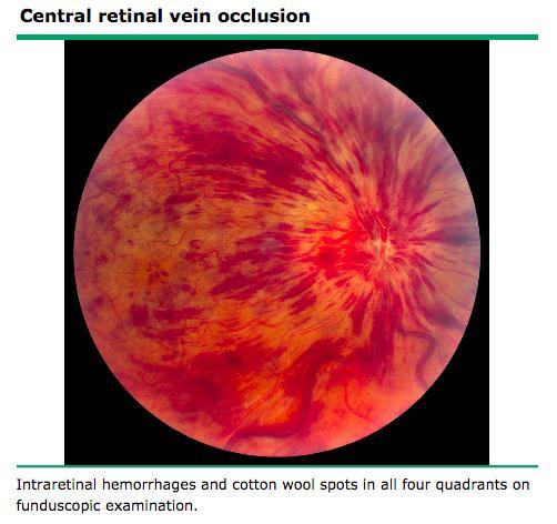
17 Findings in RVO Retinal hemorrhage Edema Dilated retinal veins Cotton wool spots CRVO Blood and Thunder


18 RVO Branch RVO Hemi-RVO Central RVO

19 Fluorescein Angiography in CRVO Retinal Nonperfusion Predict prognosis
20 Treatment of RVO Observation & Follow Up Macular edema, Neovascularization Anti VEGF Glucorticoid Laser Photocoaggulation
21 RAO Ocular Emergency minutes Form of stroke ICA opthalmic artery retinal artery Risk Factors: arteriosclerosis Carotid atherosclerotic disease, Temporal arteritis, Fibromuscular dysplasia, Moyamoya, Sickle cell Cardiogenic embolus
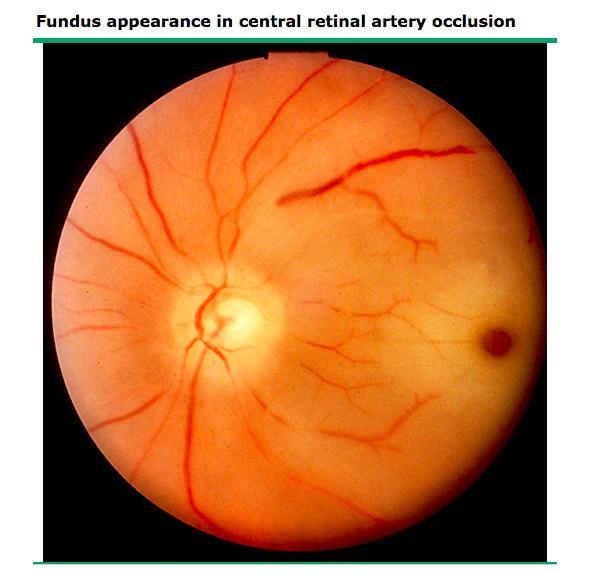
22 Presentation in RAO Preceded by fluctuating, transient course Profound vision loss Pupillary Afferent defect Findings: Ischemic retinal whitening Cherry Red Spot
23

24 Fluorescein Angiography in CRAO
25 Work Up & Treatment of RAO >50 y/o: ESR, CRP Full stroke work up Conservative Treatments Digital massage Anterior chamber paracentesis Pharmacologic reduction IOP Mannitol, Acetozolamide NTG, Pentoxyfylline Intraarterial Thrombolytics (phase II trial)
26 Back to Our Patient...
27 Admission A1c 11.1 Lipid Profile Cholestrol 267 HDL 38 LDL 171 TG 489 Endocrine, Diabetic Educator, Dietician
28 Discharge Diagnoses: DM HTN Dyslipidemia CRVO Home Medications ASA Statin Lisinopril Metformin
29 Medical Course To Date 12/30/14 Initial ED visit 1/6/15 PCP follow up 1/29/15 Eye follow up, BP 171/91 20/200 New inferior hemi-field defect, concerned for progressive end-organ damage 2/11/15 PCP: Chlorthalidone added 2/27/15 PCP: BS 288, started on glyburide 3/10/15 Next eye appointment
30 In Summary
31 Need Opthalmology NOW! CRAO/BRAO IOP>40 Temporal Arteritis Retinal Detachment Hyphema Endopthalmitis Infectious Keratitis
32 References Leveque, T. Approach to the adult with acute persistent visual loss. In: UptoDate, Post TW (Ed). (Accessed March, 2015) Douglas, J. Retinal Vein Occlusion: Epidemiology, clinical manifestations and diagnosis. In: UptoDate, Post TW (Ed). (Accessed March, 2015) Thomas, R. Central and branch retinal artery occlusion. In: UptoDate, Post TW (Ed). (Accessed March, 2015) Yuzurihara, D. Visual outcome in central retinal and branch retinal artery occlusion. Jpn J Ophthalmol 2004;48:490 Rogers, S. The prevalence of retinal vein occlusion: pooled data from population studies from the United States, Asia, and Australia. Opthalmology 2010;117:313
OCCLUSIVE VASCULAR DISORDERS OF THE RETINA
 OCCLUSIVE VASCULAR DISORDERS OF THE RETINA Learning outcomes By the end of this lecture the students would be able to Classify occlusive vascular disorders (OVD) of the retina. Correlate the clinical features
LECTURE # 7 EYECARE REVIEW: PART III
 LECTURE # 7 EYECARE REVIEW: PART III HOW TO TRIAGE EYE EMERGENCIES STEVE BUTZON, O.D. EYECARE REVIEW: HOW TO TRIAGE EYE EMERGENCIES FOR PRIMARY CARE PHYSICIANS Steve Butzon, O.D. Member Director IDOC President
EYE TRAUMA: INCIDENCE
 Introduction EYE TRAUMA: INCIDENCE 2.5 million eye injuries per year in U.S. 40,000 60,000 of eye injuries lead to visual loss Introduction Final visual outcome of many ocular emergencies depends on prompt,
Neovascular Glaucoma Associated with Cilioretinal Artery Occlusion Combined with Perfused Central Retinal Vein Occlusion
 Neovascular Glaucoma Associated with Cilioretinal Artery Occlusion Combined with Perfused Central Retinal Vein Occlusion Man-Seong Seo,* Jae-Moon Woo* and Jeong-Jin Seo *Department of Ophthalmology, Chonnam
Diabesity A Public Health Crisis: AOA Evidence Based Translation to Care Series
 Diabesity A Public Health Crisis: AOA Evidence Based Translation to Care Series Joseph J. Pizzimenti, OD, FAAO Associate Professor Nova Southeastern University The Eye Care Institute pizzimen@nova.edu
The Common Clinical Competency Framework for Non-medical Ophthalmic Healthcare Professionals in Secondary Care
 The Common Clinical Competency Framework for Non-medical Ophthalmic Healthcare Professionals in Secondary Care Medical Retina November 2016 Association of Health Professions in Ophthalmology General basic
Sahand Ensafi PA, CCPA, B.H.Sc.,Department of Emergency Medicine, University Health Network
 Sahand Ensafi PA, CCPA, B.H.Sc.,Department of Emergency Medicine, University Health Network No Disclosures Definitions Ophthalmologic Blindness Practical definition? WHO V/A less than 3/60 (snellen)
EFFICACY OF ANTI-VASCULAR ENDOTHELIAL GROWTH FACTOR AGENTS IN RETINAL DISORDER FOR BETTER VISUAL ACUITY
 EFFICACY OF ANTI-VASCULAR ENDOTHELIAL GROWTH FACTOR AGENTS IN RETINAL DISORDER FOR BETTER VISUAL ACUITY Diwakar chaudhary *1, 2, Hu shuqiong, Long Yuan and Xiong kun 1 Yangtze University, 1 Nanhuan Road
UC SF. g h. Eye Trauma. Martha Neighbor, MD Emergency Services San Francisco General Hospital University of California
 UC SF Eye Trauma sf g h Martha Neighbor, MD Emergency Services San Francisco General Hospital University of California Goals Recognize vision threatening eye emergencies Treat them when we can Know when
Objectives. Unexplained Vision Loss: Where Do I Go From Here. History. History. Drug Induced Vision Loss
 Objectives Unexplained Vision Loss: Where Do I Go From Here Denise Goodwin, OD, FAAO Coordinator, Neuro-ophthalmic Disease Clinic Pacific University College of Optometry goodwin@pacificu.edu Know the importance
OCULAR MANIFESTATIONS OF SYSTEMIC DISEASES THUCANH MULTERER, MD
 OCULAR MANIFESTATIONS OF SYSTEMIC DISEASES THUCANH MULTERER, MD UNDERGRADUATE: Philadelphia College of Pharmacy and Science 1996 MEDICAL SCHOOL: MCP Hahnemann School of Medicine, Philadelphia PA 2000 RESIDENCY:
Grand Rounds. Eddie Apenbrinck M.D. University of Louisville School of Medicine Department of Ophthalmology & Visual Sciences 6/20/2014
 Grand Rounds Eddie Apenbrinck M.D. University of Louisville School of Medicine Department of Ophthalmology & Visual Sciences 6/20/2014 Subjective CC: sudden painless loss of vision OD HPI: 75 year old
Mild NPDR. Moderate NPDR. Severe NPDR
 Diabetic retinopathy Diabetic retinopathy is the most common cause of blindness in adults aged 35-65 years-old. Hyperglycaemia is thought to cause increased retinal blood flow and abnormal metabolism in
Chris Brown, M.D. Eye Specialty Group, PLC Continuing Education Series
 Chris Brown, M.D. Eye Specialty Group, PLC 2018 Continuing Education Series Disclaimer I have no financial interests in this lecture or any information discussed therein Objectives Fluorescein Angiogram
Local Intra-arterial Fibrinolysis in Treatment of Incomplete Ophthalmic Artery Occlusion A Case Report
 CASE REPORT Local Intra-arterial Fibrinolysis in Treatment of Incomplete Ophthalmic Artery Occlusion A Case Report Shih-Ting Fang, Pao-Sheng Yen 1, Chien-Chung Chen, Yuan-Chieh Lee Department of Ophthalmology,
Neuro-Ocular Grand Rounds
 Neuro-Ocular Grand Rounds Anthony B. Litwak,OD, FAAO VA Medical Center Baltimore, Maryland Dr. Litwak is on the speaker and advisory boards for Alcon and Zeiss Meditek COMMON OPTIC NEUROPATHIES THAT CAN
Diagnosis and treatment of diabetic retinopathy. Blake Cooper MD Ophthalmologist Vitreoretinal Surgeon Retina Associates Kansas City
 Diagnosis and treatment of diabetic retinopathy Blake Cooper MD Ophthalmologist Vitreoretinal Surgeon Retina Associates Kansas City Disclosures Consulted for Novo Nordisk 2017,2018. Will be discussing
The Common Clinical Competency Framework for Non-medical Ophthalmic Healthcare Professionals in Secondary Care
 The Common Clinical Competency Framework for Non-medical Ophthalmic Healthcare Professionals in Secondary Care Cataract November 2016 Association of Health Professions in Ophthalmology General basic competences
Vascular Disease Ocular Manifestations of Systemic Hypertension
 Vascular Disease Ocular Manifestations of Systemic Hypertension Maynard L. Pohl, OD, FAAO Pacific Cataract & Laser Institute 10500 NE 8 th Street, Suite 1650 Bellevue, WA 98004 USA 425-462-7664 Cerebrovascular
 measure of your overall performance. An isolated glucose test is helpful to let you know what your sugar level is at one moment, but it doesn t tell you whether or not your diabetes is under adequate control
Professor Helen Danesh-Meyer. Eye Institute Auckland
 Professor Helen Danesh-Meyer Eye Institute Auckland Bitten by Ophthalmology Emergencies Helen Danesh-Meyer, MBChB, MD, FRANZCO Sir William and Lady Stevenson Professor of Ophthalmology Head of Glaucoma
The Common Clinical Competency Framework for Non-medical Ophthalmic Healthcare Professionals in Secondary Care
 The Common Clinical Competency Framework for Non-medical Ophthalmic Healthcare Professionals in Secondary Care Acute & Emergency Care November 2016 Association of Health Professions in Ophthalmology General
Recurrent intraocular hemorrhage secondary to cataract wound neovascularization (Swan Syndrome)
") Recurrent intraocular hemorrhage secondary to cataract wound neovascularization (Swan Syndrome) John J. Chen MD, PhD; Young H. Kwon MD, PhD August 6, 2012 Chief complaint: Recurrent vitreous hemorrhage,
Recurrent intraocular hemorrhage secondary to cataract wound neovascularization (Swan Syndrome) John J. Chen MD, PhD; Young H. Kwon MD, PhD August 6, 2012 Chief complaint: Recurrent vitreous hemorrhage,
The Human Eye. Cornea Iris. Pupil. Lens. Retina
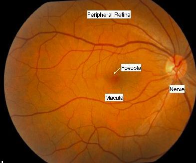
 The Retina Thin layer of light-sensitive tissue at the back of the eye (the film of the camera). Light rays are focused on the retina then transmitted to the brain. The macula is the very small area in
A Case of Carotid-Cavernous Fistula
 A Case of Carotid-Cavernous Fistula By : Mohamed Elkhawaga 2 nd Year Resident of Ophthalmology Alexandria University A 19 year old male patient came to our outpatient clinic, complaining of : -Severe conjunctival
Neuro-Ocular Grand Rounds Anthony B. Litwak,OD, FAAO VA Medical Center Baltimore, Maryland
 Neuro-Ocular Grand Rounds Anthony B. Litwak,OD, FAAO VA Medical Center Baltimore, Maryland Dr. Litwak is on the speaker and advisory boards for Alcon and Zeiss Meditek COMMON OPTIC NEUROPATHIES THAT CAN
Non-arteritic anterior ischemic optic neuropathy (NAION) with segmental optic disc edema. Jonathan A. Micieli, MD Valérie Biousse, MD
 with segmental optic disc edema. Jonathan A. Micieli, MD Valérie Biousse, MD") Non-arteritic anterior ischemic optic neuropathy (NAION) with segmental optic disc edema Jonathan A. Micieli, MD Valérie Biousse, MD A 75 year old white woman lost vision in the inferior part of her visual
Non-arteritic anterior ischemic optic neuropathy (NAION) with segmental optic disc edema Jonathan A. Micieli, MD Valérie Biousse, MD A 75 year old white woman lost vision in the inferior part of her visual
FRANZCO, MD, MBBS. Royal Darwin Hospital
 Diabetes and Eye By Dr. Nishantha Wijesinghe FRANZCO, MD, MBBS Consultant Ophthalmologist Royal Darwin Hospital 98% of Diabetics do not need to suffer from severe visual loss Yet Diabetic eye disease is
For more information about how to cite these materials visit
 Author: Jonathan Trobe, MD, 2009 License: Unless otherwise noted, this material is made available under the terms of the Creative Commons Attribution 3.0 License: http://creativecommons.org/licenses/by/3.0/
Speaker Disclosure Statement. " Dr. Tim Maillet and Dr. Vladimir Kozousek have no conflicts of interest to disclose.
 Speaker Disclosure Statement Dr. Tim Maillet and Dr. Vladimir Kozousek have no conflicts of interest to disclose. Diabetes Morbidity Diabetes doubles the risk of stroke. Diabetes quadruples the risk of
Venous Occlusive Diseases
 Venous Occlusive Diseases Bruce R. Saran, MD Adjunct Assistant Clinical Professor of Medicine Scheie Eye Institute University of Pennsylvania School of Medicine Philadelphia, PA -a division of: RVO Demographics
Grand Rounds Presentation August 25, David Mostafavi MD PL2 SUNY Downstate Ophthalmology
 Grand Rounds Presentation August 25, 2011 David Mostafavi MD PL2 SUNY Downstate Ophthalmology History of Present Illness 70 y/o black male with HTN c/o sudden painless vision loss right eye 9 hours prior
Course # Flashes and Floaters and Curtains, Oh My!
 Course # 132 Flashes and Floaters and Curtains, Oh My! FLASHES and FLOATERS and CURTAINS, OH MY!!! FLASHES OF LIGHT Vitreous is the villain Retinal traction Retinal hole Retinal tear Migraine Classic migraine
Course # Flashes and Floaters and Curtains, Oh My!
 Course # 132 Flashes and Floaters and Curtains, Oh My! FLASHES and FLOATERS and CURTAINS, OH MY!!! FLASHES OF LIGHT Vitreous is the villain Retinal traction Retinal hole Retinal tear Migraine Classic migraine
Preventing Avoidable Vision loss from Diabetic Retinopathy in Indian Country
 Diabetes in Indian Country- 2017 Preventing Avoidable Vision loss from Diabetic Retinopathy in Indian Country Albuquerque, NM 20 September2017 Mark B. Horton, OD, MD Director, IHS/JVN Teleophthalmology
5/2/2016 EYE EMERGENCIES. Nathaniel Pelsor, O.D., FAAO Talley Medical-Surgical Eye Care Associates. Anatomy. Tools
 EYE EMERGENCIES Nathaniel Pelsor, O.D., FAAO Talley Medical-Surgical Eye Care Associates Anatomy Tools 1 Contact dermatitis Blepharitis HSV Preseptal Cellulitis Anterior Chamber Subconjunctival hemorrhage
Interferon-Associated Retinopathy: Communicating with Internal Medicine Ari Wes, Esther S. Hong, MD, and Thomas A. Oetting, MS, MD
 Interferon-Associated Retinopathy: Communicating with Internal Medicine Ari Wes, Esther S. Hong, MD, and Thomas A. Oetting, MS, MD July 26, 2010 Chief Complaint: New floaters in both eyes. History of Present
8/30/2018. Eye Disorders. Patrick Sarte. Anatomy of the Eye Uveitis Scleritis vs. Episcleritis Glaucoma Retinal Findings Eyelids
 Eye Disorders Patrick Sarte Anatomy of the Eye Uveitis Scleritis vs. Episcleritis Glaucoma Retinal Findings Eyelids 1 Anatomy of the Eye Anatomy of the Eye 2 Anatomy of the Eye 3 4 A 26 year old woman
Learn Connect Succeed. JCAHPO Regional Meetings 2015
 Learn Connect Succeed JCAHPO Regional Meetings 2015 OPTIC NEUROPATHY AS EASY AS 1,2,3,4 OPTIC NERVE ANATOMY M. Tariq Bhatti, MD Departments of Ophthalmology and Neurology Duke Eye Center and Duke University
Clinically Significant Macular Edema (CSME)
") Clinically Significant Macular Edema (CSME) 1 Clinically Significant Macular Edema (CSME) Sadrina T. Shaw OMT I Student July 26, 2014 Advisor: Dr. Uwaydat Clinically Significant Macular Edema (CSME) 2
Clinically Significant Macular Edema (CSME) 1 Clinically Significant Macular Edema (CSME) Sadrina T. Shaw OMT I Student July 26, 2014 Advisor: Dr. Uwaydat Clinically Significant Macular Edema (CSME) 2
A Patient s Guide to Diabetic Retinopathy
 Diabetic Retinopathy A Patient s Guide to Diabetic Retinopathy 840 Walnut Street, Philadelphia PA 19107 www.willseye.org Diabetic Retinopathy 1. Definition Diabetic retinopathy is a complication of diabetes
Unexplained visual loss in seven easy steps
 Unexplained visual loss in seven easy steps Andrew G. Lee, MD Chair Ophthalmology, Houston Methodist Hospital, Professor, Weill Cornell MC; Adjunct Professor, Baylor COM, U Iowa, UTMB Galveston, UT MD
Bleeding in the anterior chamber, obstructing vision Caused by surgery, injury, coagulopathy, sickle cell or idiopathic Needs urgent care to prevent
 Bleeding in the anterior chamber, obstructing vision Caused by surgery, injury, coagulopathy, sickle cell or idiopathic Needs urgent care to prevent long-term vision loss TX by elevating head of bed, reducing
Retinal Vein Occlusion
 Retinal Update 2018 Retinal Vein Occlusion Case Presentations to Myself Branch Vein Occlusion What medical evaluation do you recommend for this 72 year old patient? Is there anything you ask of your medical
OPHTHALMOLOGY REFERRAL GUIDE FOR GPS
 OPHTHALMOLOGY REFERRAL GUIDE FOR GPS A guidebook to support general practitioners in the management and referral of a range of common eye problems. Contents 3 Introduction 4 Ophthalmic Workup 6 Acute Visual
Ophthalmology. Juliette Stenz, MD
 Ophthalmology Juliette Stenz, MD Required Slide Disclosures NO SIGNIFICANT FINANCIAL, GENERAL, OR OBLIGATION INTERESTS TO REPORT Required Slide At the end of this session, students will be able to: 1.
Themes for conferences No 42
 Themes for conferences No 42 Systemic Arterial Hypertension Venous Occlusive Disease Arterial Occlusive Disease Ocular Ischemic Syndrome Med. pract. Anton R. Xavier Dr. med. Claudia Zawinka Dr. med. Stephan
RVO RETINAL VEIN OCCLUSION
 RVO RETINAL VEIN OCCLUSION A guide to understanding RVO Take some time to learn about RVO - it may help you hold on to your vision Retinal vein occlusion is a common disorder of the retina and a leading
OCT Angiography The Next Frontier
 Choroid Retina avascular 5/13/2017 OCT Angiography The Next Frontier Pierce Kenworthy OD, FAAO June 9, 2017 OCT Angiography (OCTA) 2016 Non-invasive, motion contrast imaging Represents erythrocyte movement
Year 2 MBChB Clinical Skills Session Ophthalmoscopy. Reviewed & ratified by: Mr M Batterbury Consultant Ophthalmologist
 Year 2 MBChB Clinical Skills Session Ophthalmoscopy Reviewed & ratified by: o Mr M Batterbury Consultant Ophthalmologist Learning objectives o To understand the anatomy and physiology of the external and
OCT : retinal layers. Extraocular muscles. History. Central vs Peripheral vision. History: Temporal course. Optical Coherence Tomography (OCT)
") Optical Coherence Tomography (OCT) OCT : retinal layers 7 Central vs Peripheral vision Extraocular muscles RPE E Peripheral Vision: Rods (95 million) 30% Ganglion cells Central Vision: Cones (5 million)
Optical Coherence Tomography (OCT) OCT : retinal layers 7 Central vs Peripheral vision Extraocular muscles RPE E Peripheral Vision: Rods (95 million) 30% Ganglion cells Central Vision: Cones (5 million)
Diabetic Management beyond traditional risk factors and LDL-C control: Can we improve macro and microvascular risks?
 Retinopathy Diabetes has a negative effect on eyes in many ways, increasing the risk of cataracts for example, but the most common and serious ocular complication of diabetes is retinopathy. Diabetic retinopathy
ZEISS AngioPlex OCT Angiography. Clinical Case Reports
 Clinical Case Reports Proliferative Diabetic Retinopathy (PDR) Case Report 969 PROLIFERATIVE DIABETIC RETINOPATHY 1 1-year-old diabetic female presents for follow-up of proliferative diabetic retinopathy
Syllabus-Ophthalmology Rotation Course: Objectives & Goals LOYOLA UNIVERSITY CHICAGO STRITCH SCHOOL OF MEDICINE
 Syllabus-Ophthalmology Rotation Course: Objectives & Goals LOYOLA UNIVERSITY CHICAGO STRITCH SCHOOL OF MEDICINE Department of Ophthalmology Course Objectives: By Core Competencies GENERAL INFORMATION:
Retinal Plaques. Prevalence RISK FACTORS. Prevalence. Retinal Plaques 1/16/19
 Re(nal Manifesta(ons of Systemic Disease Steven Ferrucci, OD, FAAO Chief, Optometry Sepulveda VA Professor, SCCO/MBKU Several different types of plaques can often be visualized in the retinal vasculature
10/4/2013. Bruce K.Williams, MSN, RN,ACNP-BC Sisters of Charity Providence Hospitals. What is the worst thing that can go wrong with an eye?
 Red Eyes, Red Alert! Bruce K.Williams, MSN, RN,ACNP-BC Sisters of Charity Providence Hospitals Red Eyes, Red Alert! Red Eyes, Red Alert! What is the worst thing that can go wrong with an eye? 1 Red Eyes,
PART 1: GENERAL RETINAL ANATOMY
 PART 1: GENERAL RETINAL ANATOMY General Anatomy At Ora Serrata At Optic Nerve Head Fundoscopic View Of Normal Retina What Is So Special About Diabetic Retinopathy? The WHO definition of blindness is
Rapid Visual Loss. Dr Michael Johnson PhD FCOptom DipOrth DipGlauc DipTp(IP) Independent Prescribing Optometrist
 Independent Prescribing Optometrist") Rapid Visual Loss Dr Michael Johnson PhD FCOptom DipOrth DipGlauc DipTp(IP) Independent Prescribing Optometrist Outline Pathophysiology Differential diagnosis. Patient scenarios in community practice:
Rapid Visual Loss Dr Michael Johnson PhD FCOptom DipOrth DipGlauc DipTp(IP) Independent Prescribing Optometrist Outline Pathophysiology Differential diagnosis. Patient scenarios in community practice:
Supplementary Online Content
 Supplementary Online Content Park KH, Kim YK, Woo SJ, et al. Iatrogenic occlusion of the ophthalmic artery after cosmetic facial filler injections: a national survey by the Korean Retina Society. JAMA
For details on measurement and recording of visual acuity, refer to Annex 1. VISION INTERPRETING RESULTS ABSTRACT
 management update on functional decline in older adults 2012 Unit No. 5 VISION Dr Au Eong Kah Guan, Ms Yulianti, Ms Fifiana ABSTRACT Among Singaporean adults of Chinese origin aged 40 to 79 years old,
Neuro-Ophthalmic Masqueraders
 Neuro-Ophthalmic Masqueraders Leonid Skorin, Jr., OD, DO, MS, FAAO, FAOCO Mayo Clinic Health System in Albert Lea Denise Goodwin, OD, FAAO Pacific University College of Optometry Please silence all mobile
Diabetic Retinopathy. Barry Emara MD FRCS(C) Giovanni Caboto Club October 3, 2012
 Giovanni Caboto Club October 3, 2012") Diabetic Retinopathy Barry Emara MD FRCS(C) Giovanni Caboto Club October 3, 2012 Outline Statistics Anatomy Categories Assessment Management Risk factors What do you need to do? Objectives Summarize the
Diabetic Retinopathy Barry Emara MD FRCS(C) Giovanni Caboto Club October 3, 2012 Outline Statistics Anatomy Categories Assessment Management Risk factors What do you need to do? Objectives Summarize the
American Board of Optometry Board Certification Examination DETAILED OUTLINE
 American Board of Optometry Board Certification Examination DETAILED OUTLINE General Practice (160 items) The core of the examination is based in the following ten areas of general practice. 1. Ametropia/Ophthalmic
THE 35 GOLDEN EYE RULES
 THE 35 GOLDEN EYE RULES The Sense of Sight, from La Dame a la Licorne, The Lady and the Unicorn Tapestries, Late 15th Century Flemish Tapestry in wool and silk, Musée Nationale du Moyen Age, Paris. 1.
Convergence in. Introduction. Case Report: Dr. Piyali SenM.B.B.S, Dr. Abhipsha Saha M.B.B.S, Dr. Anuradha Chandra M.S,FAICO
 Convergence in Dr. Piyali SenM.B.B.S, Dr. Abhipsha Saha M.B.B.S, Dr. Anuradha Chandra M.S,FAICO Introduction non-progressive ophthalmoplegia with or without ptosis affecting part or all of the occulomotor
The Common Clinical Competency Framework for Non-medical Ophthalmic Healthcare Professionals in Secondary Care
 The Common Clinical Competency Framework for Non-medical Ophthalmic Healthcare Professionals in Secondary Care Glaucoma November 2016 Association of Health Professions in Ophthalmology General basic competences
10/6/2016. HYPERTENSIVE RETINOPATHY Amiee Ho, O.D.
 1 2 3 4 5 6 HYPERTENSIVE RETINOPATHY Amiee Ho, O.D. COURSE DESCRIPTION This course focuses on the clinical features, diagnosis and management of hypertensive retinopathy. Additionally, some background
The Prevalence of diabetic optic neuropathy in type 2 diabetes mellitus
 The Prevalence of diabetic optic neuropathy in type 2 diabetes mellitus Received: 25/4/2016 Accepted: 8/12/2016 Introduction Diabetic papillopathy is an atypical form of non-arteritic anterior ischemic
Eyes on Diabetics: How to Avoid Blindness in Diabetic Patient
 Eyes on Diabetics: How to Avoid Blindness in Diabetic Patient Rova Virgana FK Unpad Pusat Mata Nasional RS Mata Cicendo Bandung Eye Center (Hospital and Clinic) PIT IDI Jabar 2018 Keys Facts from WHO
Sudden Vision Loss. Brendan Girschek, MD, FRCSC, FACS Vitreoretinal Surgery Cedar Valley Medical Specialists
 Sudden Vision Loss Brendan Girschek, MD, FRCSC, FACS Vitreoretinal Surgery Cedar Valley Medical Specialists My Credentials -Residency in Ophthalmology at the LSU Eye Center in New Orleans, LA -Fellowship
Ophthalmology Unit Referral Guidelines
 Ophthalmology Unit Referral Guidelines Austin Health Ophthalmology Unit holds sub-specialty sessions to discuss and plan the treatment of patients with specific ocular conditions. General including cataract
Rare Presentation of Ocular Toxoplasmosis
 Case Report Rare Presentation of Ocular Toxoplasmosis Rakhshandeh Alipanahi MD From Department of Ophthalmology, Nikookari Eye Hospital, Tabriz University of Medical Sciences, Tabriz, Iran. Correspondence:
Diabetes & Your Eyes
 Diabetes & Your Eyes Diabetes is a disease that occurs when the pancreas does not secrete enough insulin or the body is unable to process it properly. Insulin is the hormone that regulates the level of
EyePACS Grading System (Part 2): Detecting Presence and Severity of Background (Non-Proliferative) Diabetic Retinopathy Lesion
: Detecting Presence and Severity of Background (Non-Proliferative) Diabetic Retinopathy Lesion") EyePACS Grading System (Part 2): Detecting Presence and Severity of Background (Non-Proliferative) Diabetic Retinopathy Lesion George Bresnick MD MPA Jorge Cuadros OD PhD Anatomy of the eye: 3 Normal Retina
EyePACS Grading System (Part 2): Detecting Presence and Severity of Background (Non-Proliferative) Diabetic Retinopathy Lesion George Bresnick MD MPA Jorge Cuadros OD PhD Anatomy of the eye: 3 Normal Retina
Outline. Preventing & Treating Diabetes Related Blindness. Eye Care Center Doctors. Justin Kanoff, MD. Eye Care Center of Northern Colorado
 Outline Preventing & Treating Diabetes Related Blindness Justin Kanoff, MD Eye Care Center of Northern Colorado 303 974 4302 Introduction to Eye Care Center of Northern Colorado How the eye works Eye problems
10 EYE EMERGENCIES. Who goes, who you better not send! Brant Slomovic, MD, FRCPC University Health Network
 10 EYE EMERGENCIES Who goes, who you better not send! Brant Slomovic, MD, FRCPC University Health Network DISCLOSURES I have none PVD CASE 1 WHAT IS A PVD? a process of aging (45-55) liquefaction of vitreous
SUPPLEMENTARY INFORMATION
 SUPPLEMENTARY INFORMATION Contents METHODS... 2 Inclusion and exclusion criteria... 2 Supplementary table S1... 2 Assessment of abnormal ocular signs and symptoms... 3 Supplementary table S2... 3 Ocular
Diabetes and Eye Health more than meets the eye Vision Initiative - in association with PSA
 Diabetes and Eye Health more than meets the eye Vision Initiative - in association with PSA Vision 2020 Australia Vision Initiative RANZCO & OAA (Vic) Proud members of Vision 2020 Australia Outline Vision
3/16/2018. Optic Nerve Examination. Hassan Eisa Swify FRCS Ed (Ophthalmology) Air Force Hospital
 Air Force Hospital") Optic Nerve Examination Hassan Eisa Swify FRCS Ed (Ophthalmology) Air Force Hospital 1 Examination Structure ( optic disc) Function Examination of the optic disc The only cranial nerve (brain tract) which
Optic Nerve Examination Hassan Eisa Swify FRCS Ed (Ophthalmology) Air Force Hospital 1 Examination Structure ( optic disc) Function Examination of the optic disc The only cranial nerve (brain tract) which
Perspectives on Screening for Diabetic Retinopathy. Dr. Dan Samaha, Optometrist, MSc Clinical Lecturer School of Optometry, Université de Montréal
 Perspectives on Screening for Diabetic Retinopathy 1 Dr. Dan Samaha, Optometrist, MSc Clinical Lecturer School of Optometry, Université de Montréal Current standards 2 According to the Canadian Diabetes
Ocular Lecture. Sue Bednar NP Ali Atwater PA-C
 Ocular Lecture Sue Bednar NP Ali Atwater PA-C Triaging Ocular Complaints Painful Eye/Red eye +/-blurry vision +/-visual loss +/-floaters +/-fevers If any of the above findings exist, pt is likely to have
CENTRAL MERSEY LOCAL OPTICAL COMMITTEE
 CENTRAL MERSEY LOCAL OPTICAL COMMITTEE OPTOMETRIC REFERRAL GUIDELINES The ocular conditions listed in this document are intended to reflect those that might be encountered in optometric practice and this
Index. Note: Page numbers of article titles are in boldface type.
 Index Note: Page numbers of article titles are in boldface type. A Acetazolamide, in idiopathic intracranial hypertension, 49 52, 60 Angiography, computed tomography, in cranial nerve palsy, 103 107 digital
Dr/ Marwa Abdellah EOS /16/2018. Dr/ Marwa Abdellah EOS When do you ask Fluorescein angiography for optic disc diseases???
 When do you ask Fluorescein angiography for optic disc diseases??? 1 NORMAL OPTIC DISC The normal optic disc on fluorescein angiography is fluorescent due to filling of vessels arising from the posterior
Faculty Financial Disclosure. Learning Objectives: Office Ophthalmology. Basic Eye Exam: What s in your pocket/office? Office Ophthalmology
 Faculty Financial Disclosure Office Ophthalmology Lynn K. Gordon, MD, PhD, has no financial relationships to disclose. Lynn K. Gordon, MD, PhD Professor and Vernon O Underwood Family Chair Department of
Goals/Objectives. Disclosures. Risk Factors RAO and RVO. Risk Factors. Retinal Artery Occlusions Branch and Central
 Jeffrey D. Perotti, OD, MS Indiana University School of Optometry Goals/Objectives RETINAL VASCULAR OCCLUSIONS FOR THE PRIMARY CARE CLINICIAN Using cases as a framework, review current evaluation and management
Sequential non-arteritic anterior ischemic optic neuropathy (NAION) Jonathan A. Micieli, MD Valérie Biousse, MD
 Jonathan A. Micieli, MD Valérie Biousse, MD") Sequential non-arteritic anterior ischemic optic neuropathy (NAION) Jonathan A. Micieli, MD Valérie Biousse, MD A 68 year old white woman had a new onset of floaters in her right eye and was found to have
Sequential non-arteritic anterior ischemic optic neuropathy (NAION) Jonathan A. Micieli, MD Valérie Biousse, MD A 68 year old white woman had a new onset of floaters in her right eye and was found to have
Preliminary report on effect of retinal panphotocoagulation on rubeosis iridis and
 British Journal of Ophthalmology, 1977, 61, 278-284 Preliminary report on effect of retinal panphotocoagulation on rubeosis iridis and neovascular glaucoma LEILA LAATIKAINEN From Moorfields Eye Hospital,
Neurology Case Presentation. Rawan Albadareen, MD 12/20/13
 Neurology Case Presentation Rawan Albadareen, MD 12/20/13 Case presentation A 49 y.o. female presented to the ED after an episode of zigzagging w a jagged bright light crossing through her Rt visual field
Incidence and Clinical Features of Neovascularization of the Iris following Acute Central Retinal Artery Occlusion
 pissn: 1011-8942 eissn: 2092-9382 Korean J Ophthalmol 2016;30(5):352-359 http://dx.doi.org/10.3341/kjo.2016.30.5.352 Original Article Incidence and Clinical Features of Neovascularization of the Iris following
Red Eyes, Red Spots, and Red Flags
 Red Eyes, Red Spots, and Red Flags Essential Knowledge of Eye Disease Andrew F. Calman, MD, PhD Associate Clinical Professor of Ophthalmology and Family & Community Medicine, UCSF Seeing Red Red Eyes Common
What is Age-Related Macular Degeneration?
 Intravitreal Injections Eylea / Lucentis Patient Information What is Age-Related Macular Degeneration? Age-related macular degeneration (AMD) is an eye condition found in older people, which may lead to
Diabetic Retinopathy WHAT IS DIABETIC RETINOPATHY? WHAT CAUSES DIABETIC RETINOPATHY? WHAT ARE THE STAGES OF DIABETIC RETINOPATHY?
 Diabetic Retinopathy WHAT IS DIABETIC RETINOPATHY? Diabetic retinopathy affects 8 million Americans with diabetes. A leading cause of blindness in American adults, it is caused by damage to the small blood
Grand Rounds Clinical Cases from Alex D. Gibberman, O.D. Harpers Point Eye Associates
 Grand Rounds Clinical Cases from 2016 Alex D. Gibberman, O.D. Harpers Point Eye Associates Relevant Financial Interests -none Case 1: 54 year old African American Female CC: Noticed a green line in
NEOVASCULAR GLAUCOMA IN A NIGERIAN AFRICAN POPULATION
 October 2006 E AST AFRICAN MEDICAL JOURNAL 559 East African Medical Journal Vol. 83 No. 10 October 2006 NEOVASCULAR GLAUCOMA IN A NIGERIAN AFRICAN POPULATION: A.O. Ashaye, FWACS, MSc, Department of Ophthalmology,
What you can expect with OZURDEX
 Important Information About Macular Edema Following Branch or Central Retinal Vein Occlusion (RVO) and Treatment For patients with RVO What you can expect with OZURDEX Approved Use OZURDEX (dexamethasone
Retinal Complications of Obstructive Sleep Apnea A Growing Concern!
 Retinal Complications of Obstructive Sleep Apnea A Growing Concern! Jay M. Haynie, OD, FAAO Financial Disclosure I have received honoraria or am on the advisory board for the following companies: Carl
NEPTUNE RED BANK BRICK
 NEPTUNE RED BANK BRICK Diabetes & The Eye Diabetics are more likely to develop Cataracts at a younger age. Diabetics are twice as likely to develop Glaucoma when compared to non-diabetics. The primary
Identify the choice that best completes the statement or answers the question.
 Chapter 5. The Eye Multiple Choice Identify the choice that best completes the statement or answers the question. 1. The most common type of eye disorder is: A. Refractive errors B. Macular conditions
Grand Rounds: Interesting and Exemplary Cases From Guanajuato and Djibouti
 Learning Community: January 25, 2015 Grand Rounds: Interesting and Exemplary Cases From Guanajuato and Djibouti JORGE CUADROS, OD, PHD EyePACS In Guanajuato Program started in 2007 Cameras go from clinic