The Asymmetric Mamillary Body: Association with Medial Temporal Lobe Disease Demonstrated with MR
|
|
|
- Harold Paul
- 5 years ago
- Views:
Transcription
1 The Mamillary Body: Association with Medial Temporal Lobe Disease Demonstrated with MR Alexander C. Mamourian, Lawrence Rodichok, and Javad Towfighi PURPOSE: To determine whether mamillary body atrophy is caused by deafferentation of the mamillary body in patients with mesial temporal sclerosis. METHODS: We studied 36 patients with thin-section MR to assess mamillary body symmetry. These patients included 10 control subjects without seizures and 26 patients with a history of seizures. Thin-section T1 scans were available for all cases. The patients with epilepsy underwent axial and coronal T2 scans as well. RESULTS: In five of eight cases with prior medial temporal lobe resection for intractable epilepsy, there was evidence of unilateral mamillary body atrophy ipsilateral to the resection. Similar findings were evident in three of six patients with MR findings of mesial temporal sclerosis without. Two patients with medial temporal stroke or tumor also had ipsilateral mamillary body atrophy. CONCLUSION: These findings provide support for the proposed mechanism of mamillary body atrophy caused by prior medial temporal lobe injury. Index terms: Brain, asymmetry/disymmetry; Brain, atrophy; Sclerosis, mesial temporal; Seizures AJNR Am J Neuroradiol 16: , March 1995 Mamillary body atrophy has been reported in autopsy cases in association with epilepsy (1). A report by Lindboe et al suggested that this atrophy is caused by deafferentation of the mamillary body in patients with mesial temporal sclerosis (2). We evaluated mamillary body symmetry in patients with medial temporal lobe abnormalities to test this proposed pathogenesis. We also studied 10 patients without seizures to determine the normal appearance of the mamillary bodies using a thin-section technique. Received October 28, 1993; accepted after revision August 24, Presented at the 31st Annual Meeting of the American Society of Neuroradiology, May 16 20, 1993, Vancouver, Canada. From the Department of Radiology, Dartmouth-Hitchcock Medical Center, Lebanon, NH (A.C.M.); and the Departments of Neurology (L.R.) and Pathology (J.T.), Milton S. Hershey-Penn State Medical Center, Hershey, Pa. Address reprint requests to Alexander C. Mamourian, MD, Department of Radiology, Dartmouth-Hitchcock Medical Center, Lebanon, NH AJNR 16: , Mar /95/ American Society of Neuroradiology 517 Materials and Methods Mamillary body size and symmetry were observed in 10 patients without epilepsy and without focal brain abnormalities by MR. These patients all underwent multiplanar 1-mm reconstruction of an MP-RAGE (magnetization-prepared rapid-acquisition gradient-echo) sequence obtained through the anterior temporal lobe region, in addition to their routine MR examination. This included an axial T1 (600/15/1 [repetition time/echo time/excitations]), axial proton-density and T2 (3000/30,90/1), and sagittal T1 (600/15/1) scan through the entire brain. In 26 patients with a history of seizures, we obtained either a 3-mm T1 scan (500/15/2) (17 cases) or threedimensional MP-RAGE sequence (18/7/1; 15 flip angle; inversion time, 500) with 1-mm reconstructed sections through the mamillary bodies (9 cases) (Fig 1). All 26 also had axial and coronal proton-density T2 scans (3000/ 30,90/1), as well as axial T1 (600/15/1). The 26 patients with seizures included 8 patients with prior temporal lobe resection for intractable epilepsy, 5 with seizures and imaging evidence of infarct or tumor, and 13 consecutive patients who were studied as part of their evaluation for epilepsy. The eight patients with temporal lobe included five men and three women with an average age of 34 years. All patients had a seizure history of more than 10 years and scans were obtained 6 months to a year after. The five with tumor or infarct included three men and two women with an average age of 35 years. The 13 with intractable epilepsy included eight women and five men with an average age of 34 years. These patients all had a modified temporal lobe resection. In this the first 4 cm (6 cm on the right) of temporal neocortex is removed, sparing the superior and often the middle tem-
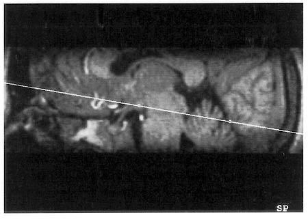
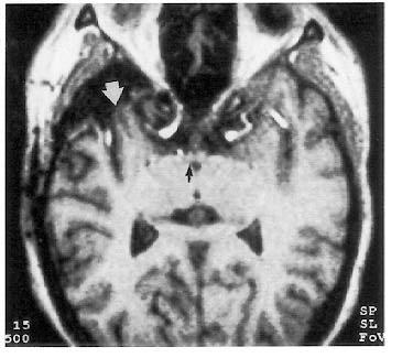
2 518 MAMOURIAN AJNR: 16, March 1995 Fig 1. T1-weighted sagittal image from an MP-RAGE sequence (18/7/1) demonstrates the preferred angled axial plane of reconstruction for seeing the mamillary bodies. Fig 2. One-millimeter axial section (18/7/1) demonstrates the prior right temporal resection (arrows). The right mamillary body (small arrow) is smaller than the left. poral gyri. The amygdala is removed, and up to 4 cm of hippocampus is resected (Table 1). Results In all 10 healthy patients the mamillary bodies were symmetric on visual inspection. Of the eight patients with prior temporal lobe for intractable epilepsy, five had asymmetric mamillary bodies. The smaller mamillary body was ipsilateral to the resection in all cases. In several cases the asymmetry was quite striking (Fig 2). Of the five patients with seizures and MR evidence of previous infarct or tumor, two had asymmetric mamillary bodies. In both cases the ipsilateral medial temporal lobe structures were absent or atrophic (Fig 3). In the other three cases with symmetric mamillary bodies, the parenchymal lesion was extratemporal. Of the 13 patients evaluated for intractable epilepsy (Table 2), 5 (38%) had asymmetric TABLE 1: Eight patients with prior temporal lobe Patient Location Basis for Location Type of Surgery Postoperative Status Pathology Mamillary Bodies 1 Temporal Clinical; response to 2 Temporal Clinical; response to 3 Temporal Clinical; response to 4 Temporal Clinical; depths; response to 5 Temporal Clinical; response to 6 Temporal Clinical; response to 7 Temporal Clinical; response to 8 Temporal Depths; response to (clinically atypical) L-modified temporal lobectomy R-modified temporal lobectomy 10/24/91 R-modified temporal lobectomy 11/15/91 L-modified temporal lobectomy 12/5/91 L-modified temporal lobectomy 3/26/92 L-modified temporal lobectomy 9/12/91 L-modified temporal lobectomy 10/3/91 R-modified temporal lobectomy 4/23/92 90% reduction gliosis gliosis gliosis No HC submitted; no change in neocortex gliosis; neocortex normal gliosis; neocortex normal gliosis; neocortex normal gliosis; neocortex normal Symmetric Symmetric Symmetric Note. Depths indicates depth electrodes and HC, hippocampus.
3 AJNR: 16, March 1995 ASYMMETRIC MAMILLARY 519 Fig 3. Coronal scan (500/15/1) demonstrates enhancement of a large right middle fossa meningioma (A). Postoperative (18/ 7/1) 1-mm axial scan (B) reveals a smaller right mamillary body than left (arrow). TABLE 2: Thirteen patients with intractable epilepsy Patient Location Basis for Location Type of Surgery 1 Temporal Clinical; depths L modified temporal lobectomy 4/22/93 Postoperative Status No change Pathology Mild nonspecific gliosis Mamillary Bodies Hippocampus Symmetric 2 R-parietal Electroencephalogram None Symmetric 3 Frontal Clinical; depths L orbital frontal resection 6/5/93 and 6/16/93 No change Normal L inferior frontal; neuronal loss and gliosis from R frontal lesion 4 Temporal Clinical; response to L modified temporal lobectomy Neuronal loss and gliosis in HC; no change in neocortex Symmetric 5 Extratemporal Clinical scalp None Symmetric monitoring 6 Unknown Clinical scalp None Symmetric monitoring 7 Nonepileptic None Symmetric Symmetric 8 Frontal Clinical; depths; L orbital frontal 90% No abnormalities Symmetric Symmetric response to response 4/14/93 decrease 9 Nonepileptic None Symmetric Symmetric 10 Multifocal Clinical; response to Callosotomy 90% Symmetric Symmetric 7/23/92 decrease 11 Temporal Clinical; depths None Symmetric Symmetric 12 Temporal Clinical; depths; response to 13 Temporal Clinical; depths; response to Note. Depths indicates depth electrode. R modified temporal lobectomy 1/13/92 L modified temporal lobectomy 9/16/93 Hippocampus; neuronal loss and astrocytosis; changes in neocortex caused by electrodes only Amygdala and HC neuronal loss and astrocytosis
4 520 MAMOURIAN AJNR: 16, March 1995 A B C Fig 4. The coronal T2-weighted image (2500/90/1) (A) shows a smaller and hyperintense left hippocampus (arrow) compared with the right. The axial (B) and coronal (C) reformatted 1-mm sections (18/7/1) demonstrate a smaller left mamillary body (small arrow) than right. The coronal scan also demonstrates the small left hippocampus (open arrow). mamillary bodies. Three of the 5 also had ipsilateral mesial temporal sclerosis by MR criteria as well as depth electrode location (Fig 4). The diagnosis of mesial temporal sclerosis was based on the presence on coronal scans of both a relatively small and hyperintense hippocampal formation (3). Although we did not do a volumetric analysis of the hippocampal volumes, visual inspection alone has a comparable sensitivity (4). Two of the 13 patients with seizures had asymmetric mamillary bodies with a normal hippocampal formation. In one of these patients (Fig 5) depth electrodes located the seizures in the temporal lobe ipsilateral to the small mamillary body. The patient subsequently underwent a temporal lobe resection but the seizure frequency was not diminished after. The other patient underwent only surface electroencephalography, which located the seizures in the ipsilateral parietal occipital region. This patient responded to medication and invasive location was not performed. The remaining 8 cases had symmetric mamillary bodies. Discussion Previous reports have documented a symmetric reduction in mamillary body size (using volumetric techniques) in patients with Wernicke encephalopathy and Korsakoff syndrome. The authors of these papers described the problem of volume averaging this small structure with surrounding cerebrospinal fluid (5, 6). In these previous studies, in which a 3-mm T1-weighted technique only was used, there were no cases among their 34 collective healthy control subjects with mamillary body Fig 5. Coronal T2-weighted image (2500/90/1) (A) demonstrates symmetric hippocampal formations. An axial T1- weighted image (600/16/1) (B) shows a marked asymmetry of the mamillary bodies. The right appears normal (arrow) but the left is virtually absent. Depth electrodes located the seizures in the left medial temporal lobe. This patient s seizure did not change after left temporal lobe.
5 AJNR: 16, March 1995 ASYMMETRIC MAMILLARY 521 asymmetry. This would support our observation of mamillary body symmetry among our control patients without seizures. All of the control patients had 1-mm sections through the region of the mamillary bodies. We used an independent console to evaluate the MP-RAGE volume in multiple planes in these cases to optimize visibility of the mamillary bodies. Artifactual asymmetry is a potential pitfall in cases with asymmetric positioning of the head or dolichoectasia of the basilar or posterior cerebral arteries. A confident assessment of mamillary symmetry usually requires an interactive selection of the optimum plane for reconstruction of the MP-RAGE scan. In five of eight patients with previous temporal lobe resection, the mamillary body was visibly smaller on the side of the resection. We did not have comparable preoperative thin-section scans of these patients, and therefore we could not determine whether there was asymmetry before. There were two among the patients with seizures who had encephalomalacia of the medial temporal lobe. In one patient this was caused by a previous infarct, and in another this was secondary to the mass effect of a middle fossa meningioma. In both, the mamillary body was smaller on the involved side. These findings suggest that mamillary body asymmetry is not unique to patients with mesial temporal sclerosis. The patient with the meningioma resection had only postoperative seizures, which responded to medication; therefore a longstanding seizure disorder could not be invoked as a potential mechanism. In contrast, one of the patients had a temporal lobe tumor (ganglioglioma) that did not involve the hippocampal formation. In that individual the mamillary bodies were symmetric. In three of the four cases with MR and clinical findings of mesial temporal sclerosis, the ipsilateral mamillary body was small. There were no cases in which the small mamillary body was contralateral to the abnormal temporal lobe. The proposed explanation for these findings is suggested by the discrete anatomic relationships of these limbic structures. Although the hippocampus proper has been implicated as the seizure focus in some patients with partial seizures (7), experimental evidence suggests that the subiculum alone provides the primary input to the mamillary body (8, 9). Thus, it is reasonable to consider that the hippocampus alone might be abnormal without significant involvement of the subiculum in some patients with mesial temporal sclerosis. In one study of non-korsakoff amnestic patients, a reduction in hippocampal volume was identified without diminished mamillary body size in some cases, and the authors proposed a similar explanation (4). There is some evidence that the subiculum is not involved in cases of mesial temporal sclerosis, however. In a study of 45 temporal lobes resected for epilepsy, there was no evidence of cell loss in subiculum (10). This observation may explain the infrequent association of mamillary body atrophy in cases of mesial temporal sclerosis. It was surprising that two cases with mamillary body atrophy had a normal appearance of the hippocampal formations on magnetic resonance. It would seem unlikely that by coincidence a patient might have a small mamillary body and seizures, particularly because mamillary body asymmetry was not seen or described as a normal variant. However, we could not exclude that possibility based on the relatively small sample of patients examined. Another explanation suggests a primary role for the mamillary body in epilepsy (11). Although there is some animal work that supports this contention, more evidence is necessary to confirm this hypothesis in humans. In our one patient with asymmetric mamillary bodies and normal hippocampal formations, did not reduce the frequency of seizures. This experience provides additional support for the previously proposed pathogenesis of mamillary body atrophy. However, our cases with asymmetric mamillary bodies and normal appearing hippocampal formations suggest the possibility of another mechanism for this atrophy in some patients with seizures. Acknowledgment We thank Sandra Billings for her considerable assistance in the preparation of this manuscript. References 1. Meldrum BS, Bruton DJ. Epilepsy. In: Adams JH, Duchen LW, eds. Greenfield s Neuropathology. 5th ed. New York: Oxford University Press, 1992: Lindboe CF, Erichsen AA, Strom EH. Atrophy and sponginess of the mamillary bodies with neuronal sparing: not only inactive Wernicke s encephalopathy. APMIS 1989;97:
6 522 MAMOURIAN AJNR: 16, March Bronen RA, Cheungh G, Charles JT, et al. Imaging findings in hippocampal sclerosis: correlation with pathology. AJNR Am J Neuroradiol 1991;12: Bronen RA, Anderson AW, Spencer DD. Quantitative MR for epilepsy: a clinical and research tool? AJNR Am J Neuroradiol 1994;15: Charness ME, DeLaPaz RL. Mammillary body atrophy in Wernicke s encephalopathy: antemortem identification using magnetic resonance imaging. Ann Neurol 1987;22: Squire LR, Amaral DG, Press GA. Magnetic resonance imaging of the hippocampal formation and mamillary nuclei distinguish medial temporal lobe and diencephalic amnesia. J Neurosci 1990; 10(9): Bronen RA. Epilepsy: the role of MR imaging. AJR Am J Roentgenol 1992;159: Meibach RC, Siegel A. The origin of fornix fibers which project to the mamillary bodies in the rat: a horseradish peroxidase study. Brain Res 1975;88: Swanson LW, Cowan WM. Hippocampo-hypothalamic connections: origins in subicular cortex, not Ammon s horn. Science 1975;189: Babb TL, Brown WJ, Pretorius J, Daverport C, Lieb JP, Crandal PH. Temporal lobe volumetric cell densities in temporal lobe epilepsy. Epilepsia 1984;25(6): Mirski M. Unraveling the neuroanatomy of epilepsy. AJNR Am J Neuroradiol 1993;14:
7
8

9

10

11
Association between Size of the Lateral Ventricle and Asymmetry of the Fornix in Patients with Temporal Lobe Epilepsy
 AJNR Am J Neuroradiol 19:9 13, January 1998 Association between Size of the Lateral Ventricle and Asymmetry of the Fornix in Patients with Temporal Lobe Epilepsy Alexander C. Mamourian, Charles H. Cho,
SWI including phase and magnitude images
 On-line Table: MRI imaging recommendation and summary of key features Sequence Pathologies Visible Key Features T1 volumetric high-resolution whole-brain reformatted in axial, coronal, and sagittal planes
An MR Protocol for Presurgical Evaluation of Patients with Complex Partial Seizures of Temporal Lobe Origin
 An MR Protocol for Presurgical Evaluation of Patients with Complex Partial Seizures of Temporal Lobe Origin Eric Achten, Paul Boon, John De Poorter, Luc Calliauw, Tom Van De Kerckhove, Jacques De Reuck,
Cerebral MRI as an important diagnostic tool in temporal lobe epilepsy
 Cerebral MRI as an important diagnostic tool in temporal lobe epilepsy Poster No.: C-2190 Congress: ECR 2014 Type: Educational Exhibit Authors: A. Puiu, D. Negru; Iasi/RO Keywords: Neuroradiology brain,
Successful Treatment of Mesial Temporal Lobe Epilepsy with Bilateral Hippocampal Atrophy and False Temporal Scalp Ictal Onset: A case report
 Hiroshima J. Med. Sci. Vol. 61, No. 2, 37~41, June, 2012 HIJM 61 7 37 Successful Treatment of Mesial Temporal Lobe Epilepsy with Bilateral Hippocampal Atrophy and False Temporal Scalp Ictal Onset: A case
MR Detection of Hippocampal Disease in Epilepsy: Factors Influencing T2 Relaxation Time
 MR Detection of Hippocampal Disease in Epilepsy: Factors Influencing T2 Relaxation Time R. A. Grunewald, G. D. Jackson, A. Connelly, and J. S. Duncan PURPOSE: To assess the reproducibility and stability
We are IntechOpen, the world s leading publisher of Open Access books Built by scientists, for scientists. International authors and editors
 We are IntechOpen, the world s leading publisher of Open Access books Built by scientists, for scientists 4,100 116,000 120M Open access books available International authors and editors Downloads Our
PRESERVE: How intensively should we treat blood pressure in established cerebral small vessel disease? Guide to assessing MRI scans
 PRESERVE: How intensively should we treat blood pressure in established cerebral small vessel disease? Guide to assessing MRI scans Inclusion Criteria Clinical syndrome Patients must have clinical evidence
Magnetic Resonance Imaging. Basics of MRI in practice. Generation of MR signal. Generation of MR signal. Spin echo imaging. Generation of MR signal
 Magnetic Resonance Imaging Protons aligned with B0 magnetic filed Longitudinal magnetization - T1 relaxation Transverse magnetization - T2 relaxation Signal measured in the transverse plane Basics of MRI
Hamartomas and epilepsy: clinical and imaging characteristics
 Seizure 2003; 12: 307 311 doi:10.1016/s1059 1311(02)00272-8 Hamartomas and epilepsy: clinical and imaging characteristics B. DIEHL, R. PRAYSON, I. NAJM & P. RUGGIERI Departments of Neurology, Pathology
MR and Positron Emission Tomography in the Diagnosis of Surgically Correctable Temporal Lobe Epilepsy
 MR and Positron Emission Tomography in the Diagnosis of Surgically Correctable Temporal Lobe Epilepsy R. Heinz, N. Ferris, E. K. Lee, R. Radtke, B. Crain, J. M. Hoffman, M. Hanson, S. Paine, and A. Friedman
Usefulness of Single Voxel Proton MR Spectroscopy in the Evaluation of Hippocampal Sclerosis
 Usefulness of Single Voxel Proton MR Spectroscopy in the Evaluation of Hippocampal Sclerosis 1, 2, 3 Kee-Hyun Chang, MD Hong Dae Kim, MD 1 Sun-Won Park, MD 1 In Chan Song, PhD 2 In Kyu Yu, MD 1 1, 2, 3
Lateralizing Ability of Single-voxel Proton MR Spectroscopy in Hippocampal Sclerosis: Comparison with MR Imaging and Positron Emission Tomography
 AJNR Am J Neuroradiol 22:625 631, April 2001 Lateralizing Ability of Single-voxel Proton MR Spectroscopy in Hippocampal Sclerosis: Comparison with MR Imaging and Positron Emission Tomography Sun-Won Park,
The Requirement for Ictal EEG Recordings Prior to Temporal Lobe Epilepsy Surgery
 Page 1 of 7 Archives of Neurology Issue: Volume 58(4), April 2001, pp 678-680 Copyright: Copyright 2001 by the American Medical Association. All Rights Reserved. Applicable FARS/DFARS Restrictions Apply
Magnetic Resonance Imaging of Mesial Temporal Sclerosis (MTS): What radiologists ought to know?
: What radiologists ought to know?") Magnetic Resonance Imaging of Mesial Temporal Sclerosis (MTS): What radiologists ought to know? Poster No.: C-0856 Congress: ECR 2012 Type: Educational Exhibit Authors: P. Singh, G. Mittal, R. Kaur, K.
Magnetic Resonance Imaging of Mesial Temporal Sclerosis (MTS): What radiologists ought to know? Poster No.: C-0856 Congress: ECR 2012 Type: Educational Exhibit Authors: P. Singh, G. Mittal, R. Kaur, K.
MR Signal Intensity of the Optic Radiation
 MR Signal Intensity of the Optic Radiation Mika Kitajima, Yukunori Korogi, Mutsumasa Takahashi, and Komyo Eto PURPOSE: To determine whether a hyperintense layer adjacent to the lateral ventricle on T2-
Visual Rating Scale Reference Material. Lorna Harper Dementia Research Centre University College London
 Visual Rating Scale Reference Material Lorna Harper Dementia Research Centre University College London Background The reference materials included in this document were compiled and used in relation to
Imaging for Epilepsy Diagnosis December 2, 2011
 Imaging for Epilepsy Diagnosis December 2, 2011 Samuel Wiebe, MD University of Calgary Canada American Epilepsy Society Annual Meeting Disclosure University of Calgary Hopewell Professorship of Clinical
Methods for Normalization of Hippocampal Volumes Measured with MR
 Methods for Normalization of Hippocampal Volumes Measured with MR S. L. Free, P. S. Bergin, D. R. Fish, M. J. Cook, S. D. Shorvon, and J. M. Stevens PURPOSE: To investigate the use of six cerebral measures
Morphometric MRI Analysis of the Parahippocampal Region in Temporal Lobe Epilepsy
 Morphometric MRI Analysis of the Parahippocampal Region in Temporal Lobe Epilepsy NEDA BERNASCONI, a ANDREA BERNASCONI, ZOGRAFOS CARAMANOS, FREDERICK ANDERMANN, FRANÇOIS DUBEAU, AND DOUGLAS L. ARNOLD Department
Role of magnetic resonance imaging for preoperative evaluation of patients with refractory epilepsy
 ACF Hui JMK Lam YL Chan KM Au-Yeung KS Wong R Kay WS Poon Key words: Epilepsy; Magnetic resonance imaging; Surgery "# Hong Kong Med J 2003;9:20-4 The Chinese University of Hong Kong, Prince of Wales Hospital,
and MR Imaging Findings in Rasmussen Encephalitis
 AJNR Am J Neuroradiol 22:1291 1299, August 2001 18 F-Fluorodeoxyglucose Positron Emission Tomography and MR Imaging Findings in Rasmussen Encephalitis David J. Fiorella, James M. Provenzale, R. Edward
Diffusion-Weighted and Conventional MR Imaging Findings of Neuroaxonal Dystrophy
 AJNR Am J Neuroradiol 25:1269 1273, August 2004 Diffusion-Weighted and Conventional MR Imaging Findings of Neuroaxonal Dystrophy R. Nuri Sener BACKGROUND AND PURPOSE: Neuroaxonal dystrophy is a rare progressive
Electro-clinical manifestations of the epilepsy associated to the different anatomical variants of hypothalamic hamartomas
 Electro-clinical manifestations of the epilepsy associated to the different anatomical variants of hypothalamic hamartomas Alberto JR Leal Hospital Fernando Fonseca, Dep. Neurology Lisbon. Abstract Objective
Discovering the hippocampus with cranial-ct.
 Discovering the hippocampus with cranial-ct. Poster No.: C-0378 Congress: ECR 2018 Type: Educational Exhibit Authors: F. Pozo Piñon, A. B. Barba Arce, E. herrera romero, V. 1 2 3 1 3 3 Fernández Lobo,
Is DTI Increasing the Connectivity Between the Magnet Suite and the Clinic?
 Current Literature In Clinical Science Is DTI Increasing the Connectivity Between the Magnet Suite and the Clinic? Spatial Patterns of Water Diffusion Along White Matter Tracts in Temporal Lobe Epilepsy.
Multimodal Imaging in Extratemporal Epilepsy Surgery
 Open Access Case Report DOI: 10.7759/cureus.2338 Multimodal Imaging in Extratemporal Epilepsy Surgery Christian Vollmar 1, Aurelia Peraud 2, Soheyl Noachtar 1 1. Epilepsy Center, Dept. of Neurology, University
Early seizure propagation from the occipital lobe to medial temporal structures and its surgical implication
 Original article Epileptic Disord 2008; 10 (4): 260-5 Early seizure propagation from the occipital lobe to medial temporal structures and its surgical implication Naotaka Usui, Tadahiro Mihara, Koichi
The Low Sensitivity of Fluid-Attenuated Inversion-Recovery MR in the Detection of Multiple Sclerosis of the Spinal Cord
 The Low Sensitivity of Fluid-Attenuated Inversion-Recovery MR in the Detection of Multiple Sclerosis of the Spinal Cord Mark D. Keiper, Robert I. Grossman, John C. Brunson, and Mitchell D. Schnall PURPOSE:
Fig. 1. Localized single voxel proton MR spectroscopy was performed along the long axis of right hippocampus after extension of patient s head to
 125 A B C Fig. 1. Localized single voxel proton MR spectroscopy was performed along the long axis of right hippocampus after extension of patient s head to obtain entire dimension of the hippocampal body.
Imaging in Epilepsy. Nucharin Supakul, MD Ramathibodi Hospital, Mahidol University August 22, 2015
 Imaging in Epilepsy Nucharin Supakul, MD Ramathibodi Hospital, Mahidol University August 22, 2015 Nothing to disclose Outline Role of Imaging and pitfalls Imaging protocol Case scenarios Clinical & Electrophysiologic
Pharmacoresistant temporal lobe epilepsy - a diagnostic performance of standardized MRI protocol in detection of epileptogenic lesion
 Pharmacoresistant temporal lobe epilepsy - a diagnostic performance of standardized MRI protocol in detection of epileptogenic lesion Poster No.: C-2226 Congress: ECR 2013 Type: Scientific Exhibit Authors:
Scalp EEG Findings in Temporal Lobe Epilepsy
 Scalp EEG Findings in Temporal Lobe Epilepsy Seyed M Mirsattari M.D., Ph.D., F.R.C.P.(C) Assistant Professor Depts. of CNS, Medical Biophysics, Medical Imaging, and Psychology University of Western Ontario
Biological Bases of Behavior. 3: Structure of the Nervous System
 Biological Bases of Behavior 3: Structure of the Nervous System Neuroanatomy Terms The neuraxis is an imaginary line drawn through the spinal cord up to the front of the brain Anatomical directions are
Epilepsy surgery in the elderly
 Clinical commentary Epileptic Disord 2009; 11 (1): 1-6 Epilepsy surgery in the elderly An unusual case of a 75-year-old man with recurrent status epilepticus Jose F. Tellez-Zenteno 1, Venkatraman Sadanand
Surgical outcome in patients with epilepsy and dual pathology
 Brain (1999), 122, 799 805 Surgical outcome in patients with epilepsy and dual pathology L. M. Li, 1 F. Cendes, 1 F. Andermann, 1 C. Watson, 2 D. R. Fish, 3 M. J. Cook, 4 F. Dubeau, 1 J. S. Duncan, 3 S.
Half-Fourier Acquisition Single-Shot Turbo Spin-Echo (HASTE) MR: Comparison with Fast Spin-Echo MR in Diseases of the Brain
 MR: Comparison with Fast Spin-Echo MR in Diseases of the Brain") Half-Fourier Acquisition Single-Shot Turbo Spin-Echo (HASTE) MR: Comparison with Fast Spin-Echo MR in Diseases of the Brain Mahesh R. Patel, Roman A. Klufas, Ronald A. Alberico, and Robert R. Edelman PURPOSE:
Half-Fourier Acquisition Single-Shot Turbo Spin-Echo (HASTE) MR: Comparison with Fast Spin-Echo MR in Diseases of the Brain Mahesh R. Patel, Roman A. Klufas, Ronald A. Alberico, and Robert R. Edelman PURPOSE:
The relevance of somatosensory auras in refractory temporal lobe epilepsies
 BRIEF COMMUNICATION The relevance of somatosensory auras in refractory temporal lobe epilepsies Ghazala Perven, Ruta Yardi, Juan Bulacio, Imad Najm, William Bingaman, Jorge Gonzalez-Martinez, and Lara
Amyotrophic lateral sclerosis (ALS) is a progressive neurodegenerative
 is a progressive neurodegenerative") ORIGINAL RESEARCH E. Matsusue S. Sugihara S. Fujii T. Kinoshita T. Nakano E. Ohama T. Ogawa Cerebral Cortical and White Matter Lesions in Amyotrophic Lateral Sclerosis with Dementia: Correlation with MR
ORIGINAL RESEARCH E. Matsusue S. Sugihara S. Fujii T. Kinoshita T. Nakano E. Ohama T. Ogawa Cerebral Cortical and White Matter Lesions in Amyotrophic Lateral Sclerosis with Dementia: Correlation with MR
CISC 3250 Systems Neuroscience
 CISC 3250 Systems Neuroscience Levels of organization Central Nervous System 1m 10 11 neurons Neural systems and neuroanatomy Systems 10cm Networks 1mm Neurons 100μm 10 8 neurons Professor Daniel Leeds
Subject Index. Band of Giacomini 22 Benton Visual Retention Test 66 68
 Subject Index Adams, R.D. 4 Addenbrooke s Cognitive Examination 101 Alzheimer s disease clinical assessment histological imaging 104 neuroimaging 101 104 neuropsychological assessment 101 clinical presentation
Utility of double inversion recovery MRI in paediatric epilepsy
 Utility of double inversion recovery MRI in paediatric epilepsy Bruno Soares, Emory University Samuel G Porter, Emory University Amit Saindane, Emory University Seena Dehkharghani, Emory University Nilesh
Computational Medical Imaging Analysis Chapter 7: Biomedical Applications
 Computational Medical Imaging Analysis Chapter 7: Biomedical Applications Jun Zhang Laboratory for Computational Medical Imaging & Data Analysis Department of Computer Science University of Kentucky Lexington,
Blurring the Lines Between Lesional and Nonlesional MRI
 Current Literature In Clinical Science Blurring the Lines Between Lesional and Nonlesional MRI Blurring in Patients With Temporal Lobe Epilepsy: Clinical, High-field Imaging and Ultrastructural Study.
Diagnosing Complicated Epilepsy: Mapping of the Epileptic Circuitry. Michael R. Sperling, M.D. Thomas Jefferson University Philadelphia, PA
 Diagnosing Complicated Epilepsy: Mapping of the Epileptic Circuitry Michael R. Sperling, M.D. Thomas Jefferson University Philadelphia, PA Overview Definition of epileptic circuitry Methods of mapping
SEIZURE OUTCOME AFTER EPILEPSY SURGERY
 SEIZURE OUTCOME AFTER EPILEPSY SURGERY Prakash Kotagal, M.D. Head, Pediatric Epilepsy Cleveland Clinic Epilepsy Center LEFT TEMPORAL LOBE ASTROCYTOMA SEIZURE OUTCOME 1 YEAR AFTER EPILEPSY SURGERY IN ADULTS
Early detection of abnormalities in partial epilepsy
 104 Institute of Child Health and Hospital for Sick Children, London, Neurosciences Unit J H Cross G D Jackson B G R Neville F J Kirkham Radiology and Physics Unit A Connelly D G Gadian Department of Clinical
ORIGINAL CONTRIBUTION. Ictal Fear in Temporal Lobe Epilepsy
 ORIGINAL CONTRIBUTION Ictal Fear in Temporal Lobe Epilepsy Surgical Outcome and Focal Hippocampal Changes Revealed by Proton Magnetic Resonance Spectroscopy Imaging Michael Feichtinger, MD; Elisabeth Pauli,
MR Volumetry of the Entorhinal, Perirhinal, and Temporopolar Cortices in Drug-Refractory Temporal Lobe Epilepsy
 AJNR Am J Neuroradiol 22:1490 1501, September 2001 MR Volumetry of the Entorhinal, Perirhinal, and Temporopolar Cortices in Drug-Refractory Temporal Lobe Epilepsy Leena Jutila, Aarne Ylinen, Kaarina Partanen,
9/30/2016. Advances in Epilepsy Surgery. Epidemiology. Epidemiology
 Advances in Epilepsy Surgery George Jallo, M.D. Director, Institute for Brain Protection Sciences Johns Hopkins All Children s Hospital St Petersburg, Florida Epidemiology WHO lists it as the second most
DIAGNOSTIC NEURORADIOLOGY. sequences
 Neuroradiology (1999) 41: 471±479 Ó Springer-Verlag 1999 DIAGNOSTIC NEURORADIOLOGY C. Oppenheim D. Dormont D. Hasboun B. Bazin S. Samson S. LehØricy M. Baulac C. Marsault Bilateral mesial temporal sclerosis:
Quantitative MRI volumetry of the entorhinal cortex in temporal lobe epilepsy
 Seizure 2000; 9: 208 215 doi: 10.1053/seiz.1999.0373, available online at http://www.idealibrary.com on Quantitative MRI volumetry of the entorhinal cortex in temporal lobe epilepsy TUULI SALMENPERÄ, REETTA
Standard magnetic resonance imaging is inadequate for patients with refractory focal epilepsy
 PAPER Standard magnetic resonance imaging is inadequate for patients with refractory focal epilepsy J von Oertzen, H Urbach, S Jungbluth, M Kurthen, M Reuber, G Fernández, C E Elger... See Editorial Commentary
Increased Anterior Temporal Lobe T2 Times in Cases of Hippocampal Sclerosis: A Multi-Echo T2 Relaxometry Study At 3 T
 AJNR Am J Neuroradiol 25:389 394, March 2004 Increased Anterior Temporal Lobe T2 Times in Cases of Hippocampal Sclerosis: A Multi-Echo T2 Relaxometry Study At 3 T Regula S. Briellmann, Ari Syngeniotis,
Methods. E. Leon Kier, Lawrence H. Staib, Lawrence M. Davis, and Richard A. Bronen. AJNR Am J Neuroradiol 25: , May 2004
 AJNR Am J Neuroradiol 25:677 691, May 2004 MR Imaging of the Temporal Stem: Anatomic Dissection Tractography of the Uncinate Fasciculus, Inferior Occipitofrontal Fasciculus, and Meyer s Loop of the Optic
José A Mendes-Ribeiro, Raquel Soares, Fernanda Simões-Ribeiro, M Luiza Guimarães
 58 Neurophysiology Unit J A Mendes-Ribeiro M L Guimarães Department of Neurology and Neurosurgery, Hospital S João, Porto, Portugal F Simões-Ribeiro Magnetic Resonance Unit, IPO, Porto, Portugal R Soares
Correlation of Apparent Diffusion Coefficient with Neuropsychological Testing in Temporal Lobe Epilepsy
 AJNR Am J Neuroradiol 26:1832 1839, August 2005 Correlation of Apparent Diffusion Coefficient with Neuropsychological Testing in Temporal Lobe Epilepsy Yvonne W. Lui, Annette O. Nusbaum, William B. Barr,
Memory. Psychology 3910 Guest Lecture by Steve Smith
 Memory Psychology 3910 Guest Lecture by Steve Smith Note: Due to copyright restrictions, I had to remove the images from the Weschler Memory Scales from the slides I posted online. Wechsler Memory Scales
Intracranial Studies Of Human Epilepsy In A Surgical Setting
 Intracranial Studies Of Human Epilepsy In A Surgical Setting Department of Neurology David Geffen School of Medicine at UCLA Presentation Goals Epilepsy and seizures Basics of the electroencephalogram
Location of the Sensorimotor Cortex: Functional and Conventional MR Compared
 Location of the Sensorimotor Cortex: Functional and Conventional MR Compared F. Zerrin Yetkin, R. Anne Papke, Leighton P. Mark, David L. Daniels, Wade M. Mueller, and Victor M. Haughton PURPOSE: To determine
Department of Cognitive Science UCSD
 Department of Cognitive Science UCSD Verse 1: Neocortex, frontal lobe, Brain stem, brain stem, Hippocampus, neural node, Right hemisphere, Pons and cortex visual, Brain stem, brain stem, Sylvian fissure,
LIMBIC SYSTEM. Dr. Amani A. Elfaki Associate Professor Department of Anatomy
 LIMBIC SYSTEM Dr. Amani A. Elfaki Associate Professor Department of Anatomy Learning Objectives Define the limbic system Identify the parts of the limbic system Describe the circulation of the limbic system
A Hippocampal Lesion Detected by High-Field 3 Tesla Magnetic Resonance Imaging in a Patient with Temporal Lobe Epilepsy
 Tohoku J. Exp. Med., 2005, A Hippocampal 205, 287-291 Lesion Detected by High Field 3 T MRI 287 A Hippocampal Lesion Detected by High-Field 3 Tesla Magnetic Resonance Imaging in a Patient with Temporal
Brain Mapping of Episodic Memory in Patients with Medial Temporal Lobe Epilepsy Using Activation Positron Emission Tomography
 Brain Mapping of Episodic Memory in Patients with Medial Temporal Lobe Epilepsy Using Activation Positron Emission Tomography Hyunwoo Nam, M.D., Sang-Kun Lee, M.D., Dong Soo Lee, M.D.*, Jae Sung Lee, M.S.*,
PRESURGICAL EVALUATION. ISLAND OF COS Hippocrates: On the Sacred Disease. Disclosure Research-Educational Grants. Patients with seizure disorders
 PRESURGICAL EVALUATION Patients with seizure disorders Gregory D. Cascino, MD Mayo Clinic Disclosure Research-Educational Grants Mayo Foundation Neuro Pace, Inc. American Epilepsy Society American Academy
multiple sclerosis by magnetic resonance imaging
 Index terms: Computed tomography Magnetic resonance sequence optimization Multiple sclerosis The evaluation of multiple sclerosis by magnetic resonance imaging Val M. Runge, M.D.*1 4, Ann C. Price, M.D.*
Single-Voxel Proton MR Spectroscopy and Positron Emission Tomography for Lateralization of Refractory Temporal Lobe Epilepsy
 AJNR Am J Neuroradiol 19:1 8, January 1998 Single-Voxel Proton MR Spectroscopy and Positron Emission Tomography for Lateralization of Refractory Temporal Lobe Epilepsy Eric Achten, Patrick Santens, Paul
Reduced Caliber of the Internal Carotid Artery: A Normal Finding with Ipsilateral Absence or Hypoplasia of the A1 Segment
 Reduced Caliber of the Internal Carotid Artery: A Normal Finding with Ipsilateral Absence or Hypoplasia of the A1 Segment Arthur G. Kane, William P. Dillon, A. James Barkovich, David Norman, Christopher
Refractory focal epilepsy: findings by MRI.
 Refractory focal epilepsy: findings by MRI. Doctors Nicolás Sgarbi, Osmar Telis Clinical Radiology Department Hospital de Clínicas Montevideo- Uruguay ABSTRACT Epilepsy is one of the most frequent neurological
Pathological reaction to disease
 Chapter1 Pathological reaction to disease Normal anatomy Figures 1.1 1.6 2 4 Brain swelling and internal herniation Figures 1.7 1.15 5 9 Epilepsy Figures 1.16 1.18 9 10 Cerebellar atrophy Figures 1.19
Analysis between clinical and MRI findings of childhood and teenages with epilepsy after hypoxic-ischemic encephalopathy in neonates periods
 Analysis between clinical and MRI findings of childhood and teenages with epilepsy after hypoxic-ischemic encephalopathy in neonates periods Poster No.: C-0401 Congress: ECR 2015 Type: Scientific Exhibit
brain MRI for neuropsychiatrists: what do you need to know
 brain MRI for neuropsychiatrists: what do you need to know Christoforos Stoupis, MD, PhD Department of Radiology, Spital Maennedorf, Zurich & Inselspital, University of Bern, Switzerland c.stoupis@spitalmaennedorf.ch
Medical Neuroscience Tutorial Notes
 Medical Neuroscience Tutorial Notes Blood Supply to the Brain MAP TO NEUROSCIENCE CORE CONCEPTS 1 NCC1. The brain is the body's most complex organ. LEARNING OBJECTIVES After study of the assigned learning
High Signal Intensity of the Infundibular Stalk on Fluid-Attenuated Inversion Recovery MR
 High Signal Intensity of the Infundibular Stalk on Fluid-Attenuated Inversion Recovery MR Yutaka Araki, Ryuichirou Ashikaga, Satoru Takahashi, Jun Ueda, and Osamu Ishida PURPOSE: To determine the MR imaging
Lecture 35 Association Cortices and Hemispheric Asymmetries -- M. Goldberg
 Lecture 35 Association Cortices and Hemispheric Asymmetries -- M. Goldberg The concept that different parts of the brain did different things started with Spurzheim and Gall, whose phrenology became quite
Auditory and Vestibular Systems
 Auditory and Vestibular Systems Objective To learn the functional organization of the auditory and vestibular systems To understand how one can use changes in auditory function following injury to localize
High Resolution Ictal SPECT: Enhanced Epileptic Source Targeting?
 High Resolution Ictal SPECT: Enhanced Epileptic Source Targeting? Marvin A Rossi MD, PhD RUSH Epilepsy Center Research Lab http://www.synapticom.net Chicago, IL USA Medically-Refractory Epilepsy 500,000-800,000
Comparative Analysis of MR Imaging, Positron Emission Tomography, and Ictal Single-photon Emission CT in Patients with Neocortical Epilepsy
 AJNR Am J Neuroradiol 22:937 946, May 2001 Comparative Analysis of MR Imaging, Positron Emission Tomography, and Ictal Single-photon Emission CT in Patients with Neocortical Epilepsy Sung-Il Hwang, Jae
Approximately 70% of childhood SURGICAL TREATMENTS FOR PEDIATRIC EPILEPSY PROCEEDINGS. Ronald P. Lesser, MD KEY POINTS
 ASIM May p153-158 5/14/01 9:19 AM Page 153 SURGICAL TREATMENTS FOR PEDIATRIC EPILEPSY Ronald P. Lesser, MD KEY POINTS Most children with epilepsy refractory to drugs can improve with surgery Temporal lobe
Investigation of hippocampal substructures in focal temporal lobe epilepsy with and without hippocampal sclerosis at 7T.
 Western University Scholarship@Western Robarts Imaging Publications Robarts Research Institute 2016 Investigation of hippocampal substructures in focal temporal lobe epilepsy with and without hippocampal
The American Approach to Depth Electrode Insertion December 4, 2012
 The American Approach to Depth Electrode Insertion December 4, 2012 Jonathan Miller, MD Director, Epilepsy Surgery University Hospitals Case Medical Center/Case Western Reserve University Cleveland, Ohio
Frontal Contributions to Memory Encoding Before and After Unilateral Medial Temporal Lobectomy
 Frontal Contributions to Memory Encoding Before and After Unilateral Medial Temporal Lobectomy Jeff Ojemann, MD Department of Neurological Surgery University of Washington Children s Hospital & Regional
Brain Structure and Epilepsy: The Impact of Modern Imaging
 Commentary Brain Structure and Epilepsy: The Impact of Modern Imaging Frederick Andermann, Professor of Neurology and Paediatrics, Department of Neurology and Neurosurgery, McGill University, Montreal,
Est-ce que l'eeg a toujours sa place en 2019?
 Est-ce que l'eeg a toujours sa place en 2019? Thomas Bast Epilepsy Center Kork, Germany Does EEG still play a role in 2019? What a question 7T-MRI, fmri, DTI, MEG, SISCOM, Of ieeg course! /HFO, Genetics
MR imaging spectrum of bilateral symmetric hippocampal lesions
 MR imaging spectrum of bilateral symmetric hippocampal lesions Poster No.: C-2510 Congress: ECR 2010 Type: Educational Exhibit Topic: Neuro Authors: P. S. Naphade, M. D. Agrawal, S. S. Sankhe, K. M. Siva,
Different Electroclinical Manifestations of the Epilepsy Associated with Hamartomas Connecting to the Middle or Posterior Hypothalamus
 Epilepsia, 44(9):1191 1195, 2003 Blackwell Publishing, Inc. C 2003 International League Against Epilepsy Different Electroclinical Manifestations of the Epilepsy Associated with Hamartomas Connecting to
The case for a relationship between human memory, hippocampus, and corpus callosum
 1 Biological Research 28: 51-57, 1995 The case for a relationship between human memory, hippocampus, and corpus callosum DAHLIA W. ZAIDEL, Ph.D. Department of Psychology, University of California, Los
OBSERVATION. Identifying Subtle Cortical Gyral Abnormalities as a Predictor of Focal Cortical Dysplasia and a Cure for Epilepsy
 OSERVATION Identifying Subtle Cortical Gyral Abnormalities as a Predictor of Focal Cortical Dysplasia and a Cure for Epilepsy Joel M. Oster, MD; Eme Igbokwe, MD; G. Rees Cosgrove, MD, FRCS(C); Andrew J.
Gross Organization I The Brain. Reading: BCP Chapter 7
 Gross Organization I The Brain Reading: BCP Chapter 7 Layout of the Nervous System Central Nervous System (CNS) Located inside of bone Includes the brain (in the skull) and the spinal cord (in the backbone)
Magnetic Resonance Imaging of the Brain in Adults Presenting With New Onset Seizures Pannag Desai K N 1, Ravi N 2
 Magnetic Resonance Imaging of the Brain in Adults Presenting With New Onset Seizures Pannag Desai K N 1, Ravi N 2 1 (Department of Radiodiagnosis, BMC&RI, India) 2 (Department of Radiodiagnosis, BMC&RI,
Imaging of Pediatric Epilepsy MRI. Epilepsy: Nonacute Situation
 Imaging of Pediatric Epilepsy Epilepsy: Nonacute Situation MR is the study of choice Tailor MR study to suspected epileptogenic zone Temporal lobe Extratemporal A. James Barkovich, MD University of California
Anatomic Evaluation of the Circle of Willis: MR Angiography versus Intraarterial Digital Subtraction Angiography
 Anatomic Evaluation of the Circle of Willis: MR Angiography versus Intraarterial Digital Subtraction Angiography K. W. Stock, S. Wetzel, E. Kirsch, G. Bongartz, W. Steinbrich, and E. W. Radue PURPOSE:
Slide 1. Slide 2. Slide 3. Tomography vs Topography. Computed Tomography (CT): A simplified Topographical review of the Brain. Learning Objective
: A simplified Topographical review of the Brain. Learning Objective") Slide 1 Computed Tomography (CT): A simplified Topographical review of the Brain Jon Wheiler, ACNP-BC Slide 2 Tomography vs Topography Tomography: A technique for displaying a representation of a cross
Slide 1 Computed Tomography (CT): A simplified Topographical review of the Brain Jon Wheiler, ACNP-BC Slide 2 Tomography vs Topography Tomography: A technique for displaying a representation of a cross
Methods. Yahya Paksoy, Bülent Oğuz Genç, and Emine Genç. AJNR Am J Neuroradiol 24: , August 2003
 AJNR Am J Neuroradiol 24:1364 1368, August 2003 Retrograde Flow in the Left Inferior Petrosal Sinus and Blood Steal of the Cavernous Sinus Associated with Central Vein Stenosis: MR Angiographic Findings
Pearls and Pitfalls in Neuroradiology of Cerebrovascular Disease The Essentials with MR and CT
 Pearls and Pitfalls in Neuroradiology of Cerebrovascular Disease The Essentials with MR and CT Val M. Runge, MD Wendy R. K. Smoker, MD Anton Valavanis, MD Control # 823 Purpose The focus of this educational
PET and SPECT in Epilepsy
 PET and SPECT in Epilepsy 12.6.2013 William H Theodore MD Chief, Clinical Epilepsy Section NINDS NIH Bethesda MD American Epilepsy Society Annual Meeting Disclosures Entity DIR NINDS NIH Elsevier Individual
Outline. Neuroradiology. Diffusion Imaging in. Clinical Applications of. Basics of Diffusion Imaging. Basics of Diffusion Imaging
 Clinical Applications of Diffusion Imaging in Neuroradiology No disclosures Stephen F. Kralik Assistant Professor of Radiology Indiana University School of Medicine Department of Radiology and Imaging
Chapter 3. Structure and Function of the Nervous System. Copyright (c) Allyn and Bacon 2004
 Allyn and Bacon 2004") Chapter 3 Structure and Function of the Nervous System 1 Basic Features of the Nervous System Neuraxis: An imaginary line drawn through the center of the length of the central nervous system, from the
Chapter 3 Structure and Function of the Nervous System 1 Basic Features of the Nervous System Neuraxis: An imaginary line drawn through the center of the length of the central nervous system, from the
Seizure Semiology and Neuroimaging Findings in Patients with Midline Spikes
 Epilepsia, 42(12):1563 1568, 2001 Blackwell Science, Inc. International League Against Epilepsy Seizure Semiology and Neuroimaging Findings in Patients with Midline Spikes *Ekrem Kutluay, *Erasmo A. Passaro,
The EEG in focal epilepsy. Bassel Abou-Khalil, M.D. Vanderbilt University Medical Center
 The EEG in focal epilepsy Bassel Abou-Khalil, M.D. Vanderbilt University Medical Center I have no financial relationships to disclose that are relative to the content of my presentation Learning Objectives