Stephanie Christine Nowak. Master s Thesis
|
|
|
- Beatrix Merritt
- 5 years ago
- Views:
Transcription
1 The Effect of Gluteus Medius Muscle Activation on Lower Limb Three-Dimensional Kinematics And Kinetics in Male and Female Athletes during three Drop Jump heights. Stephanie Christine Nowak Master s Thesis Submitted to the Faculty of Graduate and Postdoctoral Studies in partial fulfillment of the requirement for the degree of Master s of Science in Human Kinetics University of Ottawa October 2012 Stephanie Christine Nowak, Ottawa, Canada, 2012
2 Table of Contents List of Figures... i List of Tables... i Acknowledgements... ii Abstract...iii Introduction... 1 Significance... 2 Objectives... 3 Hypothesis Review of Literature... 5 The Mechanics and Incidence of ACL Injuries... 5 Possible Factors Affecting ACL Injuries... 8 Hormonal Effects... 8 Biomechanics and ACL Injuries Gluteus Medius Muscle and ACL Injuries Drop Jumps Methodology Study Design Participants Materials and Equipment Motion Recordings Ground Reaction Force Electromyography...23 Procedure
3 Data Analysis Statistical Analysis Article The effect of gender and drop jump height on lower limb kinematics, kinetics, and gluteus medius muscle activation Abstract Introduction Methods Results...38 Discussion...47 References General Discussion References Appendix A: University of Ottawa Motion Analysis Model (UOMAM)& marker set....66
4 i List of Figures Figure 1: Hip frontal plane angle and moment of force at 40DJ A. Knee frontal plane angle and ment of force at 40DJ B. IC: initial foot contact. Landing cycle (200ms before IC to 250ms after IC). * Significant difference between male and female athletes Figure 2: Hip frontal plane angle and moment of force at maxdj A. Knee frontal plane angle and moment of force at maxdj B. IC: initial foot contact. Landing cycle (200ms before IC to 250ms after IC). * Significant difference between male and female athletes Figure 3: Average sagittal plane knee angle at each drop jump height for female athletes (A) and male athletes (B). 40DJ: 40cm drop jump, maxdj: maximum countermovement drop jump, kneedj: tibial length drop jump. IC: initial foot contact.landing cycle (200ms before IC to 250ms after IC). * Significant difference between drop jump heights List of Tables Table 1. Participants average age, weight, height, BMI, day in menstrual cycle, total years of participating in high risk sport, type of sport soccer (S) and volleyball (V), Max drop jump height (MaxDJ), and Knee Drop jump height (KneeDJ). Mean (SD) Table 2. Kinematics and kinetics during the 40cm drop jump landing (40DJ), the knee height drop jump landing (KneeDJ), and the maximum CMJ drop jump landing (MaxDJ). Mean (SD) Table 3. Integrated EMG (iemg), peak EMG, and onset of the gluteus medius (GMed), gluteus maximus (GMax), vastus lateralis (VL), rectus femoris (RF), and biceps femoris (BF)during the 40DJ, kneedj, and the maxdj comparing male and female athletes. Mean (SD) Table 4. Sagittal plane kinematics and kinetics comparing the 40cm drop jump landing (40DJ), the knee height drop jump landing (KneeDJ), and the maximum CMJ drop jump landing (MaxDJ) for competitive athletes. Mean (SD) Table 5. Electromyography, normalized by MVC, comparing the 40cm drop jump landing (40DJ), the knee height drop jump landing (KneeDJ), and the maximum CMJ drop jump landing (MaxDJ) for competitive athletes. Mean (SD)
5 ii Acknowlegdements I would like to thank my committee members, Dr. Martin Bilodeau for his insight and support on the study, especially the electromyographical aspects, and Dr. Jing Xian Li for her contribution and recommendations in the development of this project. I would also like to thank my labmates for their help problem solving bumps along the road and for their endless support over the course of this study. A special thank you to the registered nurse, Ashley Mologhney, for her time, commitment, and skill this study would not have been done without her. Also to Giulia Mantovani for developing the software used in data analysis. An essential thank you goes out to my superviser, Dr. Mario Lamontagne, for giving me the opportunity to be involved in this field of research, and for his support and guidance through my Masters study. Lastly, I would like to thank my family and friends for their support and encouragment throughout these past few years.
6 iii Abstract Women have a four to eight times higher risk of injuring their anterior cruciate ligament (ACL) compared to men. It is most commonly injured by a non-contact mechanism, such as jumping, decelerating, or cutting manoeuvres, in a game time situation. The main function of the gluteus medius (GMed) muscle is to abduct and externally rotate the thigh. A less active GMed may not be able to control the internal rotation of the thigh during a jumping manouver, causing an increase in the knee joint abduction angle. The increase of knee abduction angle has been shown to augment the risk of ACL injury (Hewett et al., 2005). The purpose of this study was to determine the difference between sexes in muscle activation of the dominant lower limb, specifically the gluteus medius, and the effect on the 3D kinematics and kinetics of the lower-limb during drop jump landings from three heights. This study looked at 3D kinematics, kinetics, and muscle activation of 12 male and 12 female soccer and volleyball athletes while performing trials of two-legged drop jump tests from three heights; maximum vertical jump height, tibial length, and a commonly used height of 40cm. Results showed that, overall, females had greater hip adduction and knee abduction angles compared to male athletes. The gluteus medius muscle showed no significant difference between genders at each drop jump height.
7 Introduction In sport participation, muscles, tendons, and ligaments are most commonly injured, with the knee being extremely vulnerable having a high risk of anterior cruciate ligament (ACL) injury (Arendt& Dick, 1995). Specifically, females are four to eight times more likely to injure their ACL compared to males (Arendt& Dick, 1995; Hewett et al, 1999; Pollard et al, 2007). Although these injuries are not normally life threatening, they can lead to physical, emotional, and financial life-long consequences (Hewett et al, 1999). Therefore, it is important to try and prevent these injuries by studying different strategies and ways to limit the risk of injury in sporting activities. There are many speculations as to factors that contribute to the difference in injury incidence between genders, however no reason is completely understood, and more research is needed to identify the gender differences that affect sport performance and risk of injury. Studies have been done from many perspectives to determine the constant factors that lead to a higher risk of ACL injuries (Hewett et al., 2005; Malinzak et al., 2001; Wojtys et al., 2002). Over the years, there have remained four groups of intrinsic factors that are of interest anatomical, hormonal, neuromuscular, and biomechanical factors (Hewett et al, 2006). It has also been determined that the most common way to injure the ACL is through a non-contact mechanism, which accounts for approximately 70% of all ACL injuries, and includes decelerating maneuvers such as landing from a jump or cutting tasks. (Agel et al, 2005; Arendt & Dick, 1995; Boden et al., 2000; Decker et al., 2003; Hewett et al., 2006; Myklebust et al., 1998; Noyes et al., 1983). Furthermore, once an athlete s ACL has been torn, they have a 20% higher risk of suffering a secondary meniscal tear, as well as developing knee degenerative changes (Papadonikolakis et al., 2003). Females have also been shown to have a greater risk of increased
8 2 knee laxity and graft rupture after surgical ACL reconstruction, and generally have less successful outcomes than their male counterparts (Wolman, 2009). Gender differences have shown that women have greater hip adduction during sporting maneuvers, which leads to a higher knee abduction and greater loading on the ACL (Decker et al., 2003; Hewett et al., 2006). The gluteus medius (GMed) muscle has been determined as the primary hip abductor and pelvic stabilizer (Kendall et al., 1993), however, since women have demonstrated a greater hip adduction when compared to men during a variety of maneuvers, less activity in the GMed muscle may lead to a decrease in neuromuscular control when participating in physical activity and sport. The drop jump is a common tool used to analyze landing, since drop jumping has the ability to maximally create eccentric loading on the lower extremity (Ortiz et al., 2008). In ACL reasearch, the drop jump task is a common way to study gender differences (Carcia& Martin, 2007; Russell, et al., 2006; Zazulak, et al., 2005). However, studies have used a variety of `standard` drop heights, with no explanation as to why the specific height was chosen. Significance Physical activity and sport have been accepted as a way to create a healthy lifestyle, outweighing the risks of injury that may accompany these activities. However, some injuries, such as ACL injuries, have the potential to leave lasting negative effects, including reconstructive surgery, strenuous rehabilitation, high financial costs, and the risk of never regaining complete functionality, ending sport activities and making everyday tasks more difficult. An ACL injury also has no guarantee that treatment will be successful or that complications or further injuries will not be experienced. Therefore, it is important to prevent this injury all together. This study is a valued addition to literature since it focused on the effect of the gluteus medius muscle activity
9 3 on lower limb biomechanics between sexes using intramuscular electrodes to detect gluteus medius muscle activity during landing tasks. Along with other research on different factors influencing the risk of ACL injury, this current study will further determine the differences between men and women that may lead to a greater risk in ACL injury. Also, there is limited literature that compares the effect of drop jump height on kinematics and kinetics between genders and there is no specific height that is recommended for studying these variables. This study looked at lower limb kinematics, kinetics, and muscle activity from different drop heights, to determined if height has an effect on biomechanical measures for injury prevention between genders. Objectives This study focused specifically on the effect of gluteus medius muscle activation patterns on lower limb three-dimensional kinematics and kinetics between male and female sports players performing manouvers that lead to a higher risk of ACL injury. The relationship between gluteus medius activation using intramuscular electrodes and lower limb 3D kinematics and kinetics during three drop jump tests were assessed and compared between sexes. Studying drop jumps is believed to provide a general indicator of an athlete s ability to control lower limb kinematics and kinetics, mainly in the coronal plane (Noyes et al., 2005). A secondary objective consisted of assessing the effect of drop jump height on lower limb biomechanics to determine if there was a better height to study landing patterns from between sexes. Hypothesis It was hypothesized that women would show a different peak magnitude of gluteus medius muscle activity compared to men during the three drop jump test heights. Also it was hypothesized that there would be a negative linear relationship between peak gluteus medius
10 4 magnitude and peak angular displacement of knee abduction. Hip adduction angular displacement has been shown to increase similarly to peak angular displacement of the knee (Landry et al., 2007). With regards to effect of drop jump heights, it was hypothesized that kinematics and kinetics from higher drop jumps would be significantly different from drop jumps at lower height. Also, as drop jump height increased, sagittal plane kinematics and muscle activity would increase to help the body absorb the larger ground reaction force. Lastly, a greater difference between male and female kinematics and kinetics would be seen at higher drop jump heights due to female athletes possible lack of experience landing from these heights.
11 5 Review of Literature A large number of sport injuries occur at the knee, particularly the anterior cruciate ligament (ACL), in both males and females (Arendt& Dick, 2005). The majority of ACL injuries occur in a game/competitive setting during non-contact mechanisms including jumping or cutting tasks (Agel et al., 2005; Arendt & Dick, 1995; Boden et al., 2000; Ferretti, et al., 1992; Hewett et al., 2006; Myklebust et al., 1998; Zelisko et al., 1982). Compared to the posterior cruciate ligament, the ACL is slightly weaker and originates from the intercondylar area of the tibia. It extends superiorly, posteriorly, and laterally connecting to the posterior part of the medial side of the lateral condyle of the femur. The ACL limits anterior translation of the tibia and hyperextension of the knee joint, as well as it limits medial rotation of the femur when the foot is on the ground, and the leg is flexed (Moore& Dalley, 2006, pg. 690). The Mechanisms and Incidence of ACL Injuries Women are four to eight times more likely to injury their ACL compared to men (Arendt & Dick, 1995; Ferretti et al., 1992, Hewett et al., 1999; Hollman et al., 2003; Zelisko et al., 1982). This has been determined through a number of studies that compare incident rates relative to player exposure, game play versus practice play, level of play, and a number of other conditions. The first study to compare sport injuries between sexes where playing conditions, rules, and physical conditioning were said to be very similar was done in 1982 by Zelisko et al. Although these three conditions were similar, player exposure, number of games versus practices, and length of season differed between males and females. Females had a much shorter season with less game time and more practice time compared to males (Zelisko et al., 1982). However, through this study it can be concluded that a gender difference does exist in regards to injury incidence. The study was conducted over two seasons in two professional basketball teams, one
12 6 male and one female team. Results showed that females obtained 60% more injuries compared to males with the ankle being the most frequently injured. Knee and thigh injuries also occurred more frequently in females (16%) compared to males (12%)(Zelisko et al., 1982). A study by Ferretti et al. (1992) explored anterior cruciate ligament injuries in volleyball for a decade, starting in Results showed that 52 volleyball players were surgically treated for ACL injuries, 42 of these players being female, however player exposure was not taken into account. This study also concluded that there was a higher injury incidence during game time situations, where 32 injuries occurred, compared to practice where 20 injuries were sustained, even though practice exposure was almost 4 times as much as game exposure (Ferretti et al., 1992). It was suggested that this may be due to maximal effort put into games compared to practice or player s time at risk is greater during games. Ferretti et al. (1992) also concluded that the major mechanism for ACL injury during volleyball occurred during jumping, where 48 out of 52 injuries were sustained with this mechanism. Therefore, non-contact injury mechanisms during game time situations leads to a higher risk of ACL injury. In 2000, Boden et al. identified that the majority of ACL injuries occurred through a noncontact mechanism. This study was conducted using a questionnaire, which identified the mechanism of injury. The results showed that 72% of the ACL injuries occurred during deceleration and landing manoeuvres (Boden et al., 2000). These results, however, did not take into account gender difference and relied on participants recall ability to describe the mechanism of their injury. A few years later, a magnetic resonance imaging study was conducted where it looked at females and males with a torn ACL to determine if there was a difference in tear patterns between genders (Fayad et al., 2003). Proximal injuries to the ACL were most common, where
13 7 78% of ACL tears were sustained through a non-contact mechanism, which frequently results in a proximal injury (Fayad et al., 2003). ACL injury incidence was studied using the NCAA injury report database over a five year period. Female soccer athletes had an ACL injury rate at least two times higher compared to male soccer athletes, while female basketball players had an ACL injury rate at least three times greater than male basketball athletes (Arendt& Dick, 1995). This study further identified that male and female athletes had a nine times greater incidence of ACL injuries during a soccer game and a three times greater injury rate for basketball games, compared to practice sessions (Arendt& Dick, 1995). Ten years later, a follow-up study was published using the NCAA basketball and soccer ACL injury incidence data, evaluating reports over 13 years to determine if changes had occurred over time (Agel et al., 2005). Results showed that male athletes averaged 58% noncontact ACL injuries, while female athletes averaged 67% non-contact ACL injuries over the 13- year period. However a large percentage of this sample had no known mechanism of injury. As for injury rates between genders, female athletes had significantly higher rates of ACL injuries compared to males (Agel et al., 2005), which reflected conclusions drawn from Arendt & Dick (1995). ACL injury incidence was also noted to be higher in both male and female soccer players, compared to basketball players, as well as soccer athletes consistently sustained more ACL injuries than basketball players when comparing genders (Agel et al., 2005). Agel et al. (2005) concluded that there had been no significant change in injury incidence or nature of the injury over the 13 years of study within the sample. However, training effects and fitness of these athletes was not taken into account, therefore may have had an affect on levels of ACL injury incidence.
14 8 Over the years, literature has shown that women are much more susceptible to ACL injuries compared to males and the most common mechanism of injury is a non-contact mechanism, during game situations, compared to practice, in a variety of sports that include deceleration and landing manoeuvres. The factors leading to the cause of this injury in both males and females is still unknown, however as research furthers, it may lead to a line of attack to help prevent ACL injuries. Possible Factors Affecting ACL Injuries There are many factors that are hypothesized to increase the risk of ACL injuries. These factors are categorized into intrinsic factors, which occur within the body, and extrinsic factors, which occur outside the body. Literature has agreed that it is most likely a combination of these factors that lead to a greater risk of non-contact ACL injuries in sports involving deceleration manoeuvres, such as cutting and landing from a jump. Although the specific factors that cause non-contact ACL injuries have not been agreed on, steps have been taken to begin predicting what type of athlete is at an increased risk of obtaining an ACL injury (Huston et al., 2000). Hormonal Effects Sex hormones play a large role in appearance, structure, and functional differences in males and females. Therefore, it is suggested that they may also cause men and women to have different injury risk levels, potentially explaining why females have a higher ACL injury rate compared to males. By using the menstrual cycle as a mean of comparison between males and females, investigators are able to study the sex hormone fluctuations in gender difference. The menstrual cycle is mainly separated into three phases: follicular, ovulatory, and luteal (Wojtys et al., 2002).
15 9 The follicular phase begins on the first day of menstruation and last a mean of 9 days, but is most variable in length. The ovulatory phase last approximately 5 days, where a rapid increase of luteinizing hormone begins 24 hours before ovulation, correlated with a sharp peak in estrogen output. The luteal phase lasts approximately 14 days, where the follicle collapses and the corpus luteum is formed (Wojtys et al., 2002). The prompt stop of progesterone release causes the start of menstruation. Wojtys et al., (2002) performed a study on the relationship between menstrual cycle phase and ACL injury, where 65 women who had torn their ACL were included. Within 24 hours of the injury, a urine analysis was done to determine the hormone levels and the phase of the menstrual cycle in which the injury occurred. Results showed that there was a significantly greater number of ACL injuries during the ovulatory phase compared to the follicular and luteal phases in women not taking contraceptives. In women taking oral contraceptives, which included 14 females of the sample population, a lower risk of ACL injury was seen during the ovulatory phase suggesting that the synthetic hormones may be affecting the ACL injury distribution (Wojtys et al., 2002). Temperature is another method used to determine menstrual cycle phase using waking temperature charts. Belanger et al. (2004) used this method when testing the effects of sex hormones on knee laxity before and after exercise in a group of 27 females. Results showed no significant difference in knee joint laxity throughout the different menstrual cycle phases, before or after exercise over the 10-week study period (Belanger et al., 2004). Another study looking at the effect of estrogen on Ovine ACL fibroblasts concluded that it is unlikely that short durations of cyclic change in estrogen levels would result in rapid, clinically significant alterations in the material properties of the ACL (Seneviratne et al., 2004).
16 10 A third method of determining hormone levels and phase of menstrual cycle is using saliva samples. Slauterbeck et al. (2002) used this technique in their study, where results showed of the 31 females not using oral contraceptives, a significantly greater number of ACL injuries occurred in the follicular phase. Another study by Moller-Nielson & Hammar (1991) reported that female soccer players demonstrated a higher incidence of serious injury during the luteal phase of the menstrual cycle. The literature suggests that menstrual cycle may influence the rate of ACL injuries in women, however future research is needed to determine the relationship between ACL injury incidence and the phase of the menstrual cycle. Hewett et al. (2006) noted that it is inconclusive as to which phase of the menstrual cycle more ACL injuries occur, and should maybe be considered pre-ovulatory and post-ovulatory since ovulation is only a moment in time and not a phase. This may lead to more concise reports. Not only the study of estrogen levels, but the effects of testosterone on the ACL has been investigated (Lovering& Romani, 2005). Lovering and Romani (2005) found this hormone to be highest near ovulation and that there was a significant relationship between testosterone levels and increased ACL stiffness. This relationship was not independent but related to the concentrations of other sex hormones (Lovering& Romani, 2005). Also, it was noted that this study was conducted on reconstructed ACLs, therefore, unclear whether androgen-responsive tissue in the ACL is influenced by the inflammatory responses and remodelling that follow the injury (Lovering& Romani, 2005). The literature brings forward conflicting results pertaining to ACL injury incidence and the effects of sex hormones, with no consensus that these sex hormones play a role in the increased incidence of ACL injuries (Ireland, 2002). However, it can also be seen that there is a possible relationship between ACL injury incidence in women and menstrual cycle phase, therefore keeping hormonal influence an important factor when it comes to determining the
17 11 cause of ACL injuries. The use of oral contraceptives is also important to consider when looking at ACL injuries, since they have been shown to decrease the risk of ACL injury by controlling the cycle fluctuations (Hewett et al., 2007). Neuromuscular imbalances due to hormonal influences is another theory that may lead to higher rates of ACL injury in female athletes. Estrogen has been shown to have effects on neuromuscular function, where Sarwar et al. (1996), showed that quadriceps strength significantly increases and that there is a significant reduction of muscle relaxation during the ovulatory phase of the menstrual cycle. With serum estrogen levels constantly fluctuating during the menstrual cycle, and estrogen having significant effects on muscle function, this may be an important factor to consider when looking at ACL injury risk in females. Another study showed that estrogen had significant effects on the central nervous system, where differences were reported in physical performance in females during different phases of the menstrual cycle. Specifically, a significant decrease was seen in maximal oxygen uptake (VO 2max ) during the luteal phase of the cycle (Lebrun et al., 1994). Posthuma et al. (1987) demonstrated a decrease in motor control in the luteal phase in females with premenstrual syndrome. These studies indicate that estrogen may effect neuromuscular function, possibly effecting females in a negative manner leading them to a higher risk of ACL injury. Biomechanics and ACL Injury Biomechanical and neuromuscular factors are considered a primary reason for ACL injuries (Agel et al., 2005; Arendt & Dick, 1995; Zelisko et al., 1982). It has been stated in literature that the main mechanism for injury involves decelerating manoeuvres, such as landing from a jump and side cutting (Agel et al., 2005; Arendt & Dick, 1995; Hewett et al., 2006). Studies have been done over time to narrow the cause of ACL injury within these factors.
18 12 In a gender comparison study three dimensional knee joint motions and electromyography of the quadriceps and hamstring muscles were quantified to show that males and females had different knee motion patterns during running, cross-cutting, and side cutting (Malinzak et al., 2001). The study included 11 men and 9 women recreational athletes who played basketball, soccer, or volleyball. Results showed that females had decreased knee flexion angle, increased knee valgus angle, increased quadriceps muscle activation, and decreased hamstring muscle activation in all three movements (Malinzak et al., 2001). By firing the quadriceps without full activation of the hamstrings, this may translate the tibia anteriorly increasing the risk of ACL failure (Ireland, 2002). These found differences in females may place them at higher risk of ACL failure, but further research is needed to confirm this. In 2007, Landry et al. also looked at the kinematics, kinetics, and electromyography of the above movements in male and female adolescent soccer players. They found that not only was there a quadricepshamstring muscle imbalance, but a medial-lateral gastrocnemii imbalance, where females had greater lateral gastrocnemius activity. The authors suggested that this may place females at greater risk for ACL injury or may be a compensatory mechanism aimed at increasing joint stability (Laundry et al., 2007). To support the findings of Malinzak et al. (2001), they also found that males land with greater hip extension moments and have larger hip flexion angles. Laundry et al. (2007) stated the hip to be the most prominent kinematic difference between genders for the three joints, where female athletes had a smaller hip flexion angle when completing these movements. This was confirmed by Pollard et al. (2007) who stated that increased injury may be related to poor proximal control, where females demonstrate a greater reliance on the frontal and transverse planes at the hip when performing dynamic activities. Decker et al. (2003) verified that males and females have different movement patterns when landing from a jump. Females landed in a more erect posture with greater range of
19 13 motion at the hip and ankle and increased energy absorption at the knee, compared to men. Due to the erect landing position, the authors suggested that it might provide a mechanical disadvantage to the hamstrings and allow the quadriceps to pull the tibia anteriorly, straining the ACL. On the other hand, males did not use the full capacity of the ankle plantar flexors, and demonstrated greater knee flexions and less ankle planter-flexion. This may show that the male knee is more prepared to transfer energy up to the larger muscles such as the hip extensors (Decker et al., 2003). The authors concluded that this difference in strategy demonstrated females lack of muscular control, possibly putting more strain on the ACL (Decker et al., 2003). The effect of muscle activation on ACL injury rates has been a large focus in research. Females have demonstrated countless times that, compared to males, they have a high quadriceps to hamstring ratio (Hewett et al., 2005b; Ireland, 2002; Yu & Garrett, 2007). This may lead to strain on the ACL from full extension to of knee flexion. Decreasing the knee flexion angle increases the anterior tibial shear force at the proximal end of the tibia by increasing the patella tendon-tibia shaft angle (Hewett et al., 2005b; Yu & Garrett, 2007). Therefore, as knee flexion angle decreases, ACL loading increases. Lamontagne et al. (2005) showed that peak ACL strain occurred at the impact peak vertical ground reaction force, shortly after initial contact. This is supported by Krosshaug et al. (2007), who concluded that an ACL injury occurs between 17-50ms after initial ground contact. The quadriceps hamstring imbalance may increase this risk by decreasing the kinetic energy absorption during landing, causing the ground reaction forces and torques associated with ACL injury to increase (Hewett et al., 2005b). Females have also shown a slower response of hamstring activation, and when combined with increased quadriceps activation, increased ground reaction forces are caused, leading to ACL injury (Hewett et al., 2006).
20 14 A second muscle imbalance involves medial-lateral muscle activations. Females demonstrate a 4 times greater lateral hamstring firing pattern, as well as a low ratio of medial to lateral quadriceps recruitment (Hewett et al., 2005b). This imbalance causes a decreased medial joint compression, which may limit resistance to dynamic knee valgus, leading the female knee to medial femoral condylar lift off (Hewett et al., 2005b; Hewett et al., 2006). Alignment is another important issue when it comes to ACL injuries. A common scenario associated with ACL injury is when the tibia is externally rotated, the knee is close to full extension (Huston et al., 2001), the foot is planted, and a deceleration occurs followed by valgus collapse (Hewett et al., 2006). Compared to men, women in this situation show greater dynamic valgus, meaning hip adduction and internal rotation, decreased hip flexion (Decker et al., 2003), knee abduction, tibial external rotation, and ankle eversion. Looking at coronal plane mechanics, dynamic valgus torques build at the knee when the hip adducts and the ankle everts, which significantly increases anterior tibial translation and load on the ACL (Fukuda et al., 2003). More specifically, the hip increases the valgus torques due to muscle imbalances and decreased activation of hip musculature (White et al., 2003), indicating the difficulty women have controlling their hip during dynamic movements. Boden et al. (2000) and Olsen et al. (2004) reported that ACL injury occured when the knee was close to extension and the sagittal plane knee joint mechanisms alone could not generate enough forces to rupture the ACL (McLean et al. 2004; Pflum et al., 2004). However, decreased muscular activity at the hip, including the gluteus maximus, disallows force absorption, leading to an increased ground reaction force during landing in women (Hewett et al., 1996). Women demonstrate a greater reliance on the frontal (Kernozek et al., 2005) and transverse plane mechanisms at the hip joint during dynamic activity (Pollard et al., 2007). The most prominent kinematic difference between males and
21 15 females is seen at the hip where females generate a smaller hip flexion angle compared to males (Laundry et al., 2007). Although lower extremity static alignment has not been predictive of ACL injuries (Ireland, 2002), Hewett et al. (2005b) has used measures of dynamic valgus to predict noncontact ACL injury risk in females with 90% accuracy. In another study by Hewett et al. (2005), knee abduction moments were used, since it contributes directly to lower extremity dynamic valgus, for predicting ACL injury status with a sensitivity of 78% and a specificity of 73%. This increased knee abduction motion and moments in females before ACL injury suggest decreased neuromuscular control of the lower extremity in the coronal plane (Hewett et al., 2005). Together with valgus alignment and increased muscle imbalances, females exhibit an amplified risk of ACL injuries compared to their male counterpart. Although all three lower extremity joints have been shown to play a part in ACL injury, the hip has been noted as one of the primary factors leading to ACL injury (Laundry et al., 2007). Females show lower gluteal activity during landing, leading to misalignment and decreased load-bearing capacity of the knee joint, increasing the risk of ACL injury (Hewett et al., 2005b). Gluteus Medius Muscle and ACL Injuries The gluteus medius (GMed) muscle originates on the surface of the iliac crest and inserts into the lateral surface of the greater trochanter, where the anterior and middle portions of the gluteus medius are covered by thick fascia and the posterior portion lies deep to the gluteus maximus muscle. The main function of the gluteus medius muscle is abducting the leg. Over and over, women have shown valgus positioning during landing, where the hip is adducted compared to men. The lower limb alignment dysfunction has been associated with gluteus
22 16 medius weakness, causing contralateral pelvic drop and excessive knee valgus (Liebenson, 2006). A study was done looking at gluteus medius muscle activity in 16 male and female division-one soccer athletes to evaluate the gender difference during a forward jump (Hart et al., 2007). Surface electromyography (EMG) was collected for the gluteus medius muscle, along with hamstrings, quadriceps, and gastrocnemius muscles, where results showed that the average gluteus medius activity was significantly higher in males compared to females. However there were no significant difference between genders for any other muscles collected (Hart et al., 2007). This reduced activity may result in less resistance to hip adduction and internal rotation, which is associated with a high-risk lower extremity position that may lead to a noncontact ACL injury (Ireland, 1999). Hart et al. (2007) suggested that decreased gluteus medius activity in females may be related to gender-specific neuromuscular strategies, where males are more capable of attenuating the forces of dynamic movement patterns. On the other hand, studies have shown no difference in GMed activity between genders. A study was carried out analyzing the gluteus maximus and gluteus medius during a 30.5 cm and 45.8cm single-leg drop jump landing in male and female soccer and track athletes. Results showed that females displayed lower gluteus maximus EMG activity compared to males, however, there was no difference in gluteus medius EMG activity (Zazulak et al., 2005). Russell et al. (2006) also showed no difference in gluteus medius muscle activity between genders. Their study analyzed a 60cm single-leg drop jump with a study population of healthy subjects where activity level was unknown. Another study also looked at 20 recreationally active male and female subjects performing a bilateral drop jump from a 30cm wooden box, where subjects were instructed to land with both feet then immediately jump as high as they were able to in a vertical direction (Carcia& Martin, 2007). They found that the gluteus medius displayed similar
23 17 EMG activity between sexes before and after a drop jump. This study looked at before ground contact muscle activity to measure anticipatory muscle contraction, while post-landing muscle activity measured how a muscle responds to the imposed forces and torques as a results of the ground reaction force (Carcia& Martin, 2007). The gluteus medius muscle findings have been inconclusive with some studies showing gender differences, while others show no dissimilarity. A reason for this may be the type of electrode used to detect gluteus medius activity since it is a deep muscle. In 1970, a study done by Komi and Buskirk found that the reliability and reproducibility of the intramuscular electrodes to be weaker than surface electrodes when testing the biceps brachii. Bouisset and Maton (1972) also used the biceps brachii, however this time tests were run 55 times with electrodes placed at different locations, distances, and depths. Results were similar between tests, showing that intramuscular electrodes were more representative of the whole muscle activity while surface electrodes only measured the muscle surface. This was supported by Oberg et al. (1992), who concluded that intramuscular electrodes performed more consistently with less variance. The accuracy of intramuscular electrodes was supported in a muscle fatigue study of the rectus femoris, where both surface and intramuscular electrodes were used (Kim et al., 2007). Although muscle fatigue was seen in both electrodes, the RMS result of surface EMG was larger than that of intramuscular EMG. The authors concluded that this was due to the surface EMG representing electrical activities of many motor units, which included nearby activated muscles. On the other hand, intramuscular EMG directly measures the muscle of interest (Kim et al., 2007). In 2003, a study used surface and intramuscular electrodes to detect activity in the soleus muscle during gait analysis (Bogey et al., 2003). During the swing phase, the surface
24 18 electrodes showed activity at the soleus muscle, but the intramuscular electrodes detected no activity (Bogey et al., 2003). Furthermore, both the surface and intramuscular electrodes measured similar peak intensities, yet the duration of the signal was much longer with the surface electrodes compared to the intramuscular electrodes, showing cross-talk in the surface electrode readings (Bogey et al., 2003). This further concludes that intramuscular electrodes are better able to read the activity of specific muscle activity during human movement. Therefore, it would be difficult for surface electrodes to detect the activity of the gluteus medius muscle since it is a deep muscle covered by thick fascia and influenced by the gluteus maximus activity. With the specificity and lowered variance of intramuscular electrodes, measuring GMed activity during landings may be able to show a difference between genders, which may offer useful information to help prevent ACL injuries. Drop Jumps The majority of non-contact ACL injuries occur during a deceleration mechanism, such as landing from a jump or cutting. Therefore it is important to determine lower body strategies during these movements for future high-risk identification screening processes. The drop jump is a common tool used to analyze landing, since drop jumping has the ability to maximally create eccentric loading on the lower extremity (Ortiz et al., 2008). However, a variety of standard drop heights have been used, with no real explanation as to why the specific height was chosen. The effect of different drop heights on the biomechanics of jumps was studied, where six subjects performed a bounce drop jump from three heights; 20cm, 40cm, and 60cm (Bobbert et al., 1987). A bounce drop jump was when the downward velocity is reversed into an upward velocity as soon as possible after the landing. Results showed no significant difference between 20cm and 40cm in mechanical output about the joints during the push-off phase. There was also
25 19 no significant difference found in vertical jump achievement between drop jumps at different heights (Bobbert et al., 1987). However, peak values of moment and power output about the ankles during push-off phase were found to be smaller at a 20 cm and 60 cm drop height, compared to 40 cm (Bobbert et al., 1987). At 60 cm, the subjects could not stop their heel from contacting the ground resulting in a sharp peak of the vertical ground reaction force and the net joint reaction force. The authors concluded that dropping height should be limited to 20 or 40cm when investigating training effects of the bounce drop jump, where at 20 cm, the peak forces encountered by joint surfaces remain relativity small (Bobbert et al., 1987). Unilateral landings versus bilateral landings also lead to different effects on the biomechanics of drop landings. A study done by Pappas et al. (2007) used kinetic, kinematic, and electromyography data to compared the different types of landing and their effect on gender from a standard drop height of 40 cm. Unilateral landings showed increased knee valgus, decreased knee flexion at initial contact, decreased peak knee flexion, decreased relative hip adduction, and increased normalized electromyography of the rectus femoris, medial hamstring, lateral hamstring, and medial gastrocnemius compared to bilateral landings (Pappas et al., 2007). However, this study did not describe the drop jump design nor did it look at any data before initial ground contact. Female athletes landed with increased knee valgus and vertical ground reaction force compared to males for both types of landing. The gender and landing type interaction was insignificant. Drop jump literature focuses mainly on training effect, such as in plyometric exercises (Bobbert et al., 1987, Walsh et al., 2004). There is no specific height that is recommended for studying lower limb kinematics and kinetics during landing between genders. By studying these measurements from different drop heights, it can be determined if height has an effect on biomechanical measures for injury prevention.
26 20 From the literature it is clear that women have a significantly higher rate of ACL injuries compared to men. There are a number of factors associated with ACL injury risk that have been investigated over the years, although literature has shown inconsistencies in these factors and their relationship to ACL injury risk. It has however, been repeatedly shown that women generally perform landing and cutting manoeuvres with smaller knee flexion angles and greater knee valgus and hip adduction angles compared to men, which is linked to a high ACL injury risk position. While past studies have focused on the kinematics and kinetics of lower limb alignment, few studies have examined the contribution of the hip musculature to lower extremity control during these movements. Therefore, this study focused on the effect of the gluteus medius muscle activation patterns and its effect on lower limb alignment during landing tasks from 3 heights. As well as determine the effect of drop jump height on gender differences in lower limb kinematics and kinetics, and investigate if there is a better drop jump height to study gender differences from. The GMed plays a large role in hip abduction, therefore if women are prone to hip adduction causing knee valgus, less activity in the GMed may be seen in women performing a drop jump task, where landing is considered a movement that may lead to a greater risk of non-contact ACL injury. Along with other studies on factors affecting ACL injury risk, this current study will help further determine the differences between men and women that may lead to a greater risk of ACL injury, and help prevention programs develop a training regimen to reduce the rate of non-contact ACL injuries amoung women.
27 21 Methodology Study design This study compared the measurements of two groups: women and men. Both groups were tested in three conditions of drop jump heights. The dependent variables of interest were integrated linear envelope EMG (iemg), peaks of EMG magnitude, and onset of EMG, as well as lower limb joint angle, moment and power. This study design enabled us to see the effect of jump heights on muscle and lower limb kinematics and kinetics between genders. Participants Twenty-four participants were recruited for this study on a volunteer basis. Participants consisted of 12 female and 12 male athletes competing in high-risk sports. These high risk sports were defined as having higher rates of non contact anterior cruciate ligament injuries, such as soccer and volleyball, in comparison to other sports (Agel et al., 2005; Arendt & Dick, 1995; Ferretti et al., 1992). Inclusion criteria stated the participants were between the ages of 18 and 35 years old, and injury free at the time of testing. The participant had no history of serious injury to the lower extremities, especially the knee, defined as being out of sport for at least a week within the last 5 years of sport participation. Each participant had to have a training regiment of at least two one-hour sessions per week, and at least two years of participation in a competitive environement of the high risk sport (soccer or volleyball), defined as a higher level of the specific organized sport within a club/school organization. Prior sports experience and menstrual cycle phase (if applicable) were recorded as reported by the participant. Preceding the start of testing, each participant was required to read, understand, and sign an informed consent form outlining the inherent risks and benefits of their participation in
28 22 this study, which they also had access to prior to arriving at the study. The participant was advised that they may withdraw from the study at any point with no consequences. The University of Ottawa Health Sciences and Science Research Ethics Board (REB) approved this study. Materials and equipment For this study three-dimensional (3D) motion data, ground reaction forces, and electromyography (EMG) signals were collected during drop jumps at three heights for both groups. Motion Recordings For 3D motion analysis an infared ten-camera Vicon*Motion software system (Vicon MX-13, Oxford Metrics, Oxford, UK) was used to collect, record, and process movement signal information during the jump procedures. The system was first dynamically calibrated using a T- shaped wand (240mm) with three reflective markers to identify the marker orientation in the recording volume. A static calibration was then completed using an L-shaped stationary frame (ErgoCal 14mm) to define the origin of the recording volume. Infrared video data was captured at 500 Hz and synchronized with forceplate and EMG data. Ground Reaction Force Forces were measured using two Bertec force platforms ( , Bertec Corporation, Columbus, OH) imbedded side-by-side in the floor, creating a level surface. Measuring ranges were collected in the F x, F y, and F z directions. A mobile kistler force platform (Kistler 9286BA, Kistler Instrumente, Winterthur, Switzerland) was placed on the height-adjustable drop jump bench to determine the exact moment when the participant left the force platform.
29 23 Electromyography The electromyography (EMG) was collected using a Delsys system (DelSys Inc., MA), with 14 surface probes and two fine wire electrodes. EMG was collected bilaterally for the gluteus medius (GMed), gluteus maximus (GMax), vastus lateralis (VL), vastus medialis (VM), rectus femoris (RF), lateral gastrocnemius (LG), biceps femoris (BF), and semitendinosus (ST) muscles. Bipolar single differential silver surface electrodes 10mm in length, 1mm in diameter, and spaced 10mm apart (SP-E04, DE 2.1, DelSys Inc., MA) were placed over the muscle bellies between the origin and insertion of the muscles under investigation, excluding the GMed, and parallel to the muscle fibres. The grounding electrode was placed on the participants clavicle. Prior to the application of the surface electrodes, the skin was washed, shaven, and abraded to reduce skin impedance. The two paired fine-wire intramuscular electrodes (40 AWG, copper wire) had a 15 cm tail and 0.5cm un-insulated termination at each end, and were inserted into the gluteus medius muscle by a registered nurse, using a 26-gauge needle 1.5inches in length, to a depth of approximately three centimetres (Wilson et al., 1976). Each pair of fine-wire electrodes were inserted into the gluteus medius muscle belly at a location five centimeters below the iliac crest and posterior to the line joining the anterior superior iliac spine and the greater trochanter (Geiringer, 1994) using a ventrogluteal injection technique. The Delsys system unit was amplified via a connection to the main amplifier unit (SP-B08, Bagnoli-16, DelSys Inc., Boston, MA). EMG was sampled at 1000 Hz, band-pass filtered at Hz using a 16 bit A/D conversion board(ni PCI 6229, National Instrument Corp., Austin, TX). Procedure Upon arrival to the laboratory, the participant read and signed the consent form. They were then asked to change into spandex running shorts, a spandex t-shirt, and their sport specific shoes. Participants were then required to warm up with 20 jumping jacks to increase the
30 24 heart rate and preform stretching of all major lower extremity muscle groups, demonstrated by the investigator. Once warmed-up and stretched, the participant was asked to preform five maximal vertical counter-movement jumps (CMJ) to determine their maximum jumping height. To do this, they were instructed to perform a vertical jump on two legs with a goal to reach their maximum height. After each jump, the maximum height was calculated using force plate data and the following equation: h = 100 where the height reached was reported to the participant. The highest height of the five trials was selected for the maximum height drop jump (maxdj). Next, anthropometric measurements were taken including mass in kilograms to the nearest 0.1 kg using a balance scale, standing height to the nearest 0.5 cm using a slide ruler apparatus, and body mass index (BMI) using the equation mass in kilograms divided by height in metres squared. Surface and fine wire electrodes were then placed on the participant. Maximum voluntary isometric contractions (MVC) were recorded for six different muscle groups of each lower limb: hip flexion, hip extension, hip abduction, knee flexion, knee extension, and ankle plantar flexion. The MVCs were executed against a fixed resistence (fixed polyester strap attached to a wooden examination bench). From the three MVC trials collected, the highest peak of EMG magnitude was selected to normalize the jumping data. Forty-five reflective markers were attached to the head, trunk, arms, hands, pelvis, legs, and feet according to the University of Ottawa Motion Anaylsis Model (UOMAM) marker set a modified Helen Hayes marker set - using a headband, wristbands, and double-sided adhesive tape. These markers were used to calculate joint centres of rotation and segment length based on their position. See appendix A for marker placement. Additional anthropometric measurements were then collected including inter-asis distance, leg length, knee height, knee width, and ankle width were taken from each individual to the nearest 0.5 cm.
31 25 The participant preformed a static calibration trial where they were asked to stand feet shoulder width apart, arms infront parallel to the ground with a slight bend in the elbows, looking straight ahead, and remain motionless for 5 seconds. Each participant was next asked to perform three drop jump tasks from different platform heights onto two force platforms imbedded in the floor. Each drop jump task was performed for 2 sets of 5 repititions at the participants own speed, while 3D kinematics and kinetics, ground reaction force, and raw electromyographic signals were collected. Between each set, the participant was given a twominute rest period that is consistent with training and conditioning principles. The first five successful trials performed were used for analysis. To preform each drop jump, participants were instructed to jump out off the drop jump platform, as to minimize their vertical height, and land with one food on each side-by-side force plate where they immediately perform a maximum vertical jump. The participant was instructed that upon dropping from the platform and before jumping vertically, they were to land on the ball of their feet, trying to minimize their heel contact with the force platform. Although the heel drop is impossible to eliminate at certain heights, this encouraged the participants to not rest or relax slightly after landing. The first drop jump test height was set to the maximum CMJ height (in centimetres) determined at the beginning of the study, referred to as maxdj. The second drop jump test height was determined by 50% of the participant s knee height, which was measured from the lateral condyle to the lateral malleolus, referred to as kneedj. This drop jump height allowed the study to compare male and female athletes landing from a lower height. The thrid drop jump height was constant across all participants at 40cm, referred to as 40DJ, which represented a commonly used height seen in literature. The three drop jump heights were randomized for each participant. Once these tasks were complete, the electrodes, reflective markers, and any other equipment were removed.
32 26 Data Analysis Three-dimensional kinematics, kinetics, and electromyography data of each participant were analyzed for 15 successful drop jump trails, the first five successful trials from each drop jump height. (Note: if a variable was unusable within a successful trial, the trial would be discarded and the next successful trial was used). The cycle of interest was defined as 200ms before initial ground contact to 250ms after contact with the ground (Carcia& Martin, 2007). Two hundred fifty milliseconds was a reasonable stop time since it has been shown that an ACL injury occurs between 17-50ms after initial ground contact (Krosshaug et al., 2007). This cycle was applied to time normalize the data. The raw kinematic data was used to generate 3D coordinates in the Vicon Nexus software program (Vicon MX-13, Oxford Metrics, Oxford, UK) from the 2D camera images for each participant for each jump trial. Marker trajectories were labelled and foot contact and the cycle parameters were determined. The trajectories were filtered using Vicon s Woltring filter with a mean square error value of 15mm 2. The lower extremities were modeled as a rigid linked body and was modified to include additional markers on the medial femoral epicondyles and medial malleoli to define the knee and ankle joint centres as the mid point between the medial and lateral markers for each joint. The UOMAM was used to calculate joint centres of rotation and segment length based on the position of the skin markers. The kinetic data analyzed consisted of moments of force and powers for lower limb joints, where the three dimensional ground reaction forces obtained from the force plates were filtered using a fourth order, low-pass Butterworth filter, with a cut-off frequency of 50Hz (Hewett et al., 2005). An inverse dynamics approach (Winter, 2005) was applied to obtain weight normalized moments of force and power for lower limb joints. This approach used known forces and moments at the distal joint of a segmment, in addition to the segments
33 27 motion and inertial properties, to calculate the forces and moments at the proximal joint of the segment. Each trial was cut and time normalized to a 100% cycle. Using a custom MATLAB (The Mathworks, Inc. USA) program, peak angles, moments of force, and powers were determined for the dominant hip, knee, and ankle in the frontal and saggital planes. Kinematic and kinetic data in the transverse plane was not included in the study because of the large variability in data due to the method used of calculating trajectories with skin markers. Raw EMG signals were baseline corrected, rectified and smoothed with a critically damped filter (double pass, zero-lag, 4 th order low pass Butterworth filter), with a cutoff frequency of 6 Hz to generate a linear envelope of the EMG signal. From the linear envelope data, the signals were normalized to the participant s MVC and time normalized. The integrated EMG value and the peak muscle activity of each muscle was determined. From the raw EMG data, onset of muscle activity was determined for each trial using a custom MATLAB (The Mathworks, Inc. USA) double-threshold detector program. The onset detector operates on the raw myoelectric signal from which a first threshold is determined using the noise-to-signal ratio, detemined uniquely for each signal (Bonato et al., 1998). The second threshold, represented by r o was set at 1. A false alarm probability was set at 0.05 (Bonato et al., 1998). The timing of the onset was reported as a percentage of the landing cycle, in relation to initial ground contact, which occured at 44.5%. Therefore, a percentage of 44.4 or less indicated that the onset occurred before ground contact, while a percentage of 44.6 or over indicated muscle onset after ground contact. Statistical Analysis Statistical tests were conducted using SPSS software (SPSS for windows, version 17.0, SPSS Inc., Chicogo, USA). A 2x3 mixed between-within analysis of variance (ANOVA) was used to
34 28 determine if any main effects were found for lower limb variables between male and female athletes at three drop jump heights. Significance was set at a level of p<0.05. The lower limb variables under analysis included joint angle, moment of force, power, and muscle onset, peak EMG, and the integrated EMG. If any main effects were found to be significant (α<0.05), adequate post hoc tests were conducted with a Bonferroni correction.
35 29 Article #1 The effect of gender and drop jump height on lower limb kinematics, kinetics, and gluteus medius muscle activation Stephanie Christine owak a & Mario Lamontagne ab a School of Human Kinetics, University of Ottawa, Canada b Department of Mechanical Engineering, University of Ottawa, Canada
36 30 ABSTRACT The objectives of this study were to determine if gender differences occur in gluteus medius activity during drop jump tasks, as well as to investigate the effect of drop jump heights on lower limb kinematics, kinetics, and muscle activity. During jump landings, women are prone to valgus collapse, leading to a higher risk of anterior cruciate ligament (ACL) injury compared to men. With a valgus collapse, women have shown greater hip adduction compared to men. This may be caused by a less active gluteus medius (GMed) muscle that is unable to control the internal rotation and adduction of the thigh causing an increase in the knee joint abduction angle during a jumping maneuver, augmenting the risk of ACL injury. These sex differences are seen when testing men and women from the same drop jump height, although, on average, men are able to jump much higher than women. In this study, twelve male and twelve female competitive soccer and volleyball athletes performed bilateral drop jumps from three heights; the participants maximum vertical jump height (maxdj), 50% of the participant s tibial length (kneedj), and a commonly used height of 40cm (40DJ). Three dimensional motion data of the hip, knee, and ankle, and lower body electromyography were recorded. Intramuscular electrodes were used to collect the activity of the GMed muscle. Results showed that female athletes had greater hip adduction and knee valgus compared to male athletes during the drop jumps. GMed activity and onset were similar between sexes during landing. Sagittal plane differences were seen when comparing drop jump heights, where males showed significant differences between the maxdj and kneedj, while females showed significant differences between the maxdj and the 40DJ. This may show that testing female and male athletes at the same height may lead to errorneous conclusions on normal landing techniques between sexes. KEYWORDS: gluteus medius, ACL injury, intramuscular electrodes, drop jumps, sex differences
37 31 INTRODUCTION An anterior cruciate ligament (ACL) injury is most commonly encountered through a non-contact mechanism, such as landing from a jump or cutting tasks (Agel et al, 2005; Arendt & Dick, 1995; Boden et al., 2000; Decker et al., 2003; Hewett et al., 2006; Myklebust et al., 1998; Noyes et al., 1983). An ACL injury is also more prevalent during game environments compared to practice sessions (Ferretti et al., 1992). It is an ongoing question in research as to why women are four to eight times more likely to injury their ACL compared to men (Arendt& Dick, 1995; Hewett et al, 1999; Pollard et al, 2007). So far, studies have determined that women have decreased knee flexion angles, increased knee valgus angles, and decreased hip flexion angles (Malinzak et al., 2001), causing them to have greater ground reaction forces, landing in a more erect posture (Decker et al., 2003). The large knee valgus angle seen in women during landing may result from the hip adducting, increasing the valgus torques at the knee due to muscle imbalances at the knee and hip joint. The hip muscle imbalance in women may reduce the joint stability during dynamic movements (White et al., 2003). Together with large knee valgus angles and increased muscle imbalances at the knee and hip, women exhibit an amplified risk of ACL injuries compared to men. Although all three lower extremity joints have been shown to play a part in ACL injury, the hip has been noted as one of the primary factors leading to ACL injury (Laundry et al., 2007). Females show lower gluteal activity during landing, leading to misalignment and decreased load-bearing capacity of the knee joint, increasing the risk of ACL injury (Hewett et al., 2005b). Sex differences have shown that women have greater hip adduction during sporting maneuvers, which leads to a higher knee abduction and greater loading on the ACL (Decker et al., 2003; Hewett et al., 2006). The gluteus medius (GMed) muscle has been determined as the primary hip abductor and pelvic stabilizer (Kendall et al., 1993), however, the lower limb
38 32 alignment dysfunction has been associated with a less active gluteus medius muscle, causing contralateral pelvic drop and excessive knee valgus, decreasing neuromuscular control when participating in physical activity and sport (Liebenson, 2006). Over recent years, research on the hip has become more popular and is now thought to affect lower limb alignment and ACL loading. As mentioned above, landing from jumps are a recognized task that may lead to an ACL injury, where these landings are typically performed from various landing heights and therefore, an understanding of landing technique is important for the prevention of injuries during intensive athletic activies, such as soccer, and volleyball. Drop jumps are a common task used to study landing patterns and are executed by jumping down from a raised platform and upon landing, performing an explosive maximal vertical jump. Drop jumping is a type of plyometric exercise that uses the stretch-shortening cycle when a muscle is stretched directly before it is explosively contracted allowing higher force production and power outputs (Walsh et al., 2004). The majority of studies using drop jump tasks select commonly used heights, such as 0.2m, 0.4m, and 0.6m, with minimal explanation as to why each height was selected (Bobbert et al, 1987; Carcia & Martin, 2007; Decker et al., 2003; Walsh et al., 2004). There is no specific height that is recommended for studying lower limb kinematics and kinetics between men and women during landing studies. Therefore, this study investigated the effect of three drop jump heights on lower limb three-dimensional kinematics and kinetics of men and women athletes to determine if there was a better height to study landing patterns from. This study also focused on the effect of gluteus medius muscle activity on lower limb three-dimensional kinematics and kinetics between men and women soccer and volleyball athletes who are frequently exposed to high-
39 33 risk maneuvers. It was hypothesized that women would show a different peak magnitude and onset of gluteus medius muscle activity compared to men during the three drop jump test heights. Also it was hypothesized that there would be a negative linear relationship between peak gluteus medius magnitude and peak angular displacement of knee abduction. The relationship between gluteus medius activation measured with intramuscular electromyography (EMG) electrodes and lower limb 3D kinematics and kinetics during three drop jump heights were compared between genders. With regards to effect of drop jump heights, it was hypothesized that kinematics and kinetics from higher drop jumps would be significantly different from drop jumps at lower height. Also, as drop jump height increases, sagittal plane kinematics and muscle activity would increase to help the body absorb the larger ground reaction force. Lastly, a greater difference between male and female kinematics and kinetics would be seen at higher drop jump heights due to female athletes possible lack of experience landing from these heights. METHODS This study used a mixed between-within design, where male and female athletes were tested in three conditions of drop jump height. The dependent variables of interest were dominant lower limb joint angle, moment of force, and power, as well as dominant lower limb integrated and peak muscle activity and muscle onset. This study design enabled us to see the effect of sex on lower limb kinematics, kinetics, and muscle activity, as well as the effect of drop jump height on the lower limb variables. Participants Twenty-four participants were included in the study, consisting of 12 female and 12 male competitive athletes competing in the high-risk sports of soccer and volleyball. These
40 34 high-risk sports are defined as having higher rates of non-contact ACL injuries, in comparison to other sports (Agel et al., 2005; Arendt & Dick, 1995; Ferretti et al., 1992). Inclusion criteria stated the participants were between the ages of 18 and 35 years old, and injury free at the time of testing. The participant had no history of serious injury to the lower extremities, especially the knee, defined as being out of sport for at least a week within the last 5 years of sport participation. Each participant had to have a training regiment of at least two one-hour sessions per week, and at least two years of participation in a competitive high risk sport, defined as a higher level of the specific organized sport within a club/school organization. Prior sports experience and menstrual cycle phase, recorded as day of cycle, (if applicable) were recorded as reported by the participant. Institutional ethics board approval was obtained, and participants provided informed consent. Participant demographics are presented in Table 1. Materials Forty-five reflective markers were fixed to the participant s body, following a modified Helen Hayes model marker set (Kadaba& Ramakrishnan, 1990), which was used to calculate joint centres of rotation and segment length based on the position of the skin markers. A tencamera infrared motion analysis system (Vicon MX-13, Oxford Metrics, Oxford, UK) was used to track the markers during drop jumps at a 500Hz sampling frequency. Two side-by-side force platforms ( , Bertec Corporation, Columbus, OH) imbedded into the floor, creating a level surface, recorded 3D ground reaction forces at 1000Hz. Bipolar single differential silver surface electrodes 10mm in length, 1mm in diameter, and spaced 10mm apart (SP-E04, DE 2.1, DelSys Inc., MA) were used on the left and right vastus lateralis (VL), vastus medialis (VM), rectus femoris (RF), biceps femoris (BF), semitendinosis (ST), lateral gastrocnemius (LG), and gluteus maximus (GMax). Two paired fine-wire intramuscular electrodes were inserted by a registered nurse, using a 26-gauge needle, into the middle fibres
41 35 of the gluteus medius (GMed) muscle on the left and right side, to a depth of approximately 3 cm, using a ventrogluteal injection technique. The EMG signal was sampled at 1000Hz and connected to the main amplifier unit (SP-B08, Bagnoli-16, DelSys Inc., Boston, MA) where the signal was band-pass filtered at Hz using a 16 bit A/D conversion board, (NI PCI 6229, National Instrument Corp., Austin, TX). Protocol Each participant underwent a two-hour motion analysis session at the Human Movement Biomechanics Laboratory of the University of Ottawa. The participant wore a tight fitting black suit and their sport specific shoes. Each participant was asked to first perform five maximum vertical countermovement jumps (CMJ). The participant s maximum jumping height was determined using the force plate data and equation (1). h h = 100. (1) Maximum voluntary isometric contractions (MVC) were then recorded for each muscle using a fixed resistance (fixed polyester strap attached to a wooden examination bench). Each participant was asked to performed three MVCs for each muscle group under analysis gluteus medius, gluteus maximus, hamstrings, quadriceps, and gastrocnemius. From the three MVC trials, the EMG signal with the highest peak was selected to normalize the jumping data. Fortyfive reflective markers were then fixed to the participants body. The participant then had to perform bilateral drop-jump tasks, while 3D kinematics and kinetics, and raw electromyography (EMG) data were collected. The first drop jump test height was set to 100% of the maximum CMJ height (maxdj). The second drop jump height was determined by 50% of the participant s tibial height lateral femoral condyle to lateral malleolus (kneedj). The third drop jump height was standard across all participants at 40cm (40DJ) to represent a commonly used drop jump height in literature. Refer to Table 1. The three drop jump heights were randomized for each
42 36 participant and 2 reps of 5 sets were recorded for each participant at their own speed. Each drop jump test consisted of the participant jumping out off the drop jump platform and onto the imbedded force plates, where they immediately performed a maximum vertical jump. The participant was instructed that upon dropping from the platform and before jumping vertically, they were to land on the ball of their feet, trying to minimize their heel contact with the force platform. Although the heel drop is impossible to eliminate, this encouraged the participant to not rest or relax slightly after landing. This paper focused on the data of the first five successful trials of the dominant leg. Data Analysis The lower limbs were modelled as a rigid linked system with three degrees of freedom, where data was time normalized, from 200ms before initial ground contact on the force plate to 250ms after ground contact (Carcia& Martin, 2007). The stop time was appropriate since it has been shown that an ACL injury occurs between 17-50ms after initial ground contact (Krosshaug et al., 2007). The resulting trajectories were filtered using a Woltering filtering routine set at a mean square error of 15mm 2 (Woltering, 1986). The lower extremities were modeled as a rigid linked body and were modified to include additional markers on the medial femoral epicondyles and medial malleoli to define the knee and ankle joint centres as the mid point between the medial and lateral markers. The ground reaction forces were filtered using a fourth order low-pass butterworth filter, cutoff frequency of 50Hz (Hewett et al., 2005). An inverse dynamic approach was then used in order to obtain lower limb 3D kinetics (moments of force and power), which were normalized by body weight, as well as time. Due to unusuable date, only 8 male athletes and 6 female athletes were compared for kinetic date. The variables analyzed included the dominant hip, knee, and ankle, determined by kicking foot, peak angles for kinematics and peak moments
43 37 of force and peak power for kinetics during landing phase. The dominant electromyography was also analyzed, including the integrated EMG (iemg), peak EMG, and onset. To determine the iemg and peakemg, the raw EMG signals were baseline corrected, rectified and smoothed with a critically damped filter (double pass, zero-lag, 4 th order low pass Butterworth filter), with a cutoff frequency of 6 Hz. A linear envelope of the EMG signal was then generated. From the linear envelope data, the signals were normalized to the participant s MVC and time normalized. To determine the onset of each muscle raw EMG signals were run through a custom MATLAB (The Mathworks, Inc. USA) double-threshold detector program was used (Bonato et al., 1998). The onset detector operated on the raw myoelectric signal from which a first threshold was determined using the noise-to-signal ratio selected for each trial. The second threshold, represented by r o was set at 1. A false alarm probability was set at 0.05 (Bonato et al., 1998). The timing of the onset was reported as a percentage of the landing cycle, in relation to initial ground contact, which occured at 44.5%. Therefore, a percentage of 44.4 or less indicates that the onset occurred before ground contact, while a percentage of 44.6 or over indicates muscle onset after ground contact. Due to unusable data, the vastus medialis, semitendinosus, and lateral gastrocnemius were not analyzed in this study.the kinematic, kinetic, and electromyography variables were extracted from each of the five successful trial, and then averaged for each participant. Statistical tests were conducted using SPSS software (SPSS for windows, version 17.0, SPSS Inc., Chicogo, USA). A 2x3 mixed between-within analysis of variance (ANOVA) was used to determine if any main effects were found for lower limb variables between male and female athletes at three drop jump heights. Significance was set at a level of p<0.05. The lower limb variables under analysis included joint angle, moment of force, power, and muscle onset, peak EMG, and the integrated EMG value. If any main effects were found to be significant (α<0.05),
44 38 adequate post hoc tests were conducted with a Bonferroni correction. Independent t-tests were conducted on patient demographics. RESULTS It was found that groups did not differ and therefore were well matched for age (p=0.504), BMI (p=0.464), total years of participation in ACL injury high risk sport (p=0.473) and average weekly physical activity (p=0.827). Table 1. Participants average age, weight, height, BMI, day in menstrual cycle, total years of participating in high risk sport, type of sport soccer (S) and volleyball (V), Max drop jump height (MaxDJ), and Knee Drop jump height (KneeDJ). Mean (SD). Group (N) Age (years) Mass (kg)* Male (12) 25.4 (3.7) 75.6 (8.9) Female (12) (3.6) (8.2) *Significant difference at p<0.05 Height (cm)* (4.5) (4.0) BMI (kg/m 2 ) 23.8 (2.3) 24.7 (3.7) Menstrual Cycle (days) Total Sport Participation (years) / 14.2 (5.4) (8.5) (4.7) Sport (N) 2V 10S 2V 10S MaxDJ (cm)* 43.0 (8.0) 27.3 (4.9) KneeDJ (cm)* In all three drop jumps, differences were seen between the men and women s landing patterns in the frontal plane (Table 2). At the 40DJ, female athletes showed a greater peak hip 21.6 (1.1) 19.8 (0.7) adduction angle compared to male athletes (p= 0.019), as well as a greater peak knee abduction angle compared to men (p=0.033). At the maxdj, females showed significantly greater peak hip adduction (p=0.021) and peak knee abduction (p=0.027) compared to male athletes. Lastly, at the kneedj, females only showed a significantly larger peak knee abduction angle compared to male athletes (p=0.028).
45 39 Table 2. Kinematics and kinetics during the 40cm drop jump landing (40DJ), the knee height drop jump landing (KneeDJ), and the maximum CMJ drop jump landing (MaxDJ). Mean (SD). Variables Male (n=12) Female (n=12) Angles ( ) 40DJ MaxDJ KneeDJ 40DJ MaxDJ KneeDJ Hip peak Flexionᴥ (16.4) (16.1) (15.9) (10.7) (10.3) (10.4) Hip peak adduction/abduction^~ (5.2) (6.5) (5.1) 3.21 (4.9) 2.90 (4.4) 2.64 (4.6) Knee peak Flexion*+ᴕᴥ (11.0) (12.0) (12.6) (7.2) (7.9) (7.6) Knee peak adduction/abduction^~" (8.8) (9.6) (7.3) (8.2) (8.0) (8.4) Ankle peak dorsiflexion+ ᴕᴥ (5.4) (4.9) (5.3) (3.1) (2.3) (3.0) Ankle peak inversion/eversion 5.39 (3.9) 5.96 (5.0) 5.25 (3.0) 2.89 (4.3) 3.86 (3.1) 4.03 (3.4) Moments (Nm/kg) (n=8) (n=6) Hip peak -1.23(0.8) -1.22(0.8) -0.88(0.6) -0.85(0.5) -0.61(0.5) -0.63(0.4) flexion/extension Hip peak -0.09(0.4) -0.41(0.7) -0.36(0.4) -0.54(0.2) -0.63(0.3) -0.68(0.3) adduction/abduction Knee peak -2.56(0.6) -2.66(0.5) -2.43(0.5) -2.37(0.6) -2.25(0.5) -2.22(0.5) flexion/extension Knee peak -0.61(0.4) -0.59(0.5) -0.46(0.3) -0.18(0.2) -0.39(0.3) -0.42(0.2) adduction/abduction Ankle peak -2.39(0.6) -2.48(0.6) -2.36(0.5) -2.18(0.5) -2.11(0.5) -2.04(0.4) plantar/dorsiflexion Ankle peak -0.07(0.1) -0.10(0.1) -0.11(0.1) -0.11(0.1) -0.07(0.1) -0.06(0.1) inversion/eversion Power (W/kg) (n=8) (n=6) Hip Peak Generation 3.37 (2.7) 1.78 (2.7) 2.31 (2.2) 1.84 (0.8) 1.53 (1.1) 1.62 (1.1) Knee Peak Generation 9.95 (4.9) 9.02 (7.5) 9.15 (4.9) 8.74 (3.6) 9.21 (2.9) 9.20 (2.9) Ankle Peak Generation (3.7) (3.7) (3.3) 8.67 (2.1) 10.1 (2.8) (3.1) ^Significant difference at p<0.05 for 40cm drop jump height between genders ~Significant difference at p<0.05 for max drop jump height between genders "Significant difference at p<0.05 for knee drop jump height between genders *Significant difference at p<0.05 with bonferroni correction between 40cm and knee drop jump height in males +Significant difference at p<0.05 with bonferroni correction between max and knee drop jump height in males ᴕSignificant difference at p<0.05 with bonferroni correction between 40cm and knee drop jump height in females ᴥSignificant difference at p<0.05 with bonferroni correction between 40cm and max drop jump height in females NOTE: negative vaules represent hip and knee extension and abduction and ankle plantar flexion and eversion. Although differences were seen between sexes in the frontal plane kinematics for the 40DJ, knee DJ, and the max DJ, the electromyography of the GMed and additional muscles collected showed no significant differences between genders at all 3 drop jump heights. See Table 3.
46 40 Table 3. Integrated EMG (iemg), peak EMG, and onset of the gluteus medius (GMed), gluteus maximus (GMax), vastus lateralis (VL), rectus femoris (RF), and biceps femoris (BF)during the 40DJ, kneedj, and the maxdj comparing male and female athletes. Mean (SD). IEMG Peak EMG Onset (%) Male Female Male Female Male Female Muscles at 40DJ GMed (23.9) (24.4) 0.98 (0.6) 1.02 (0.6) (13.1) (5.3) GMax (15.8) (9.2) 0.75 (0.5) 0.69 (0.2) (17.5) (11.0) VL (30.7) (36.1) 1.85 (0.9) 2.14 (1.1) (5.1) (6.0) RF (22.4) (32.7) 1.25 (0.5) 1.24 (0.8) (7.0) (4.7) BF (10.0) (10.3) 0.46 (0.3) 0.52 (0.2) (15.4) (16.7) Muscles at maxdj GMed (23.0) (21.7) 1.00 (0.6) 0.77 (0.5) (17.1) (8.8) GMax (13.2) (9.7) 0.75 (0.4) 0.68 (0.3) (19.1) (11.0) VL (33.3) (29.9) 1.86 (1.1) 2.10 (1.0) (5.2) (5.5) RF (25.9) (23.9) 1.22 (0.6) 1.37 (0.9) (7.7) (5.8) BF (9.5) (8.5) 0.44 (0.3) 0.49 (0.1) (16.8) (14.0) Muscles at kneedj GMed (19.8) (25.0) 0.87 (0.6) 0.86 (0.6) (7.9) (6.9) GMax (15.3) (9.6) 0.69 (0.4) 0.70 (0.3) (11.6) (8.7) VL (34.6) (34.4) 1.68 (1.0) 2.07 (1.1) (5.5) (4.0) RF (22.0) (24.8) 1.76 (1.0) 2.07 (1.1) (6.9) (2.2) BF (11.1) (9.6) 0.39 (0.2) 0.48 (0.2) (19.0) (17.6) *significant difference at p<0.05 between male and female athletes No correlation was seen between peak fontal plane knee angle and peak GMed EMG at the 40DJ, r=-0.132, p=0.568, the maxdj, r=0.055, p=0.813, and the kneedj, r=0.190, p= Frontal plane kinematics and kinetics are shown in Figure 1 for the 40DJ and Figure 2 for the maxdj. The start of the analyzed landing cycle is 0% which represents 200ms before initial ground contact. Therefore, it is possible that male and female athletes are already planning to land in their normal landing pattern.
47 Female Male Hip Angle IC Cycle (%) A Figure 1: Hip frontal plane angle and moment of force at 40DJ A. Knee frontal plane angle and ment of force at 40DJ B. IC: initial foot contact. Landing cycle (200ms before IC to 250ms after IC). * Significant difference between male and female athletes. 41 Abd Degrees Add -20 Knee Angle IC * Hip Moment * Cycle (%) Abd Nm/kg Add Abd Degrees Add * Cycle (%) B Knee Moment Female Male * Cycle (%) Abd Nm/kg Add
48 Hip Angle A Female IC * Male Cycle (%) Knee Angle B IC Female Figure 2: Hip frontal plane angle and moment of force at maxdj A. Knee frontal plane angle and moment of force at maxdj B. IC: initial foot contact. Landing cycle (200ms before IC to 250ms after IC). * Significant difference between male and female athletes. 42 Abd Degrees Add Male -20 * Cycle (%) Hip Moment 1 Female Male Abd Nm/kg Add Abd Degrees Add Cycle (%) 0.6 Knee Moment Female Male Cycle (%) Abd Nm/kg Add
49 43 To determine the maxdj height for each participant, a maximum countermovement jump was performed where males jumped significantly higher than females (p<0.001). Male athletes averaged a maximum CMJ height of ±7.98 cm, while female athletes averaged a maximum CMJ height of ±4.9 cm. For the kneedj, males had significantly longer tibial lengths compared to females (p<0.001), where males averaged a length of ±1.1 cm and females averaged a length of ±0.7 cm. When comparing the lower limb variables between the three drop jump heights for competitive athletes, significant differences were only seein in the sagittal plane kinematics (Table 4). The 40DJ compared to kneedj had significant differences for peak knee flexion (p<0.001) and peak ankle dorsiflexion (p=0.003). These differences were all significantly larger at 40DJ compared to the kneedj. Peak knee flexion (p<0.001) and peak ankle dorsiflexion (p=0.003) were also significantly greater during maxdj compared to the kneedj. Table 4. Sagittal plane kinematics and kinetics comparing the 40cm drop jump landing (40DJ), the knee height drop jump landing (KneeDJ), and the maximum CMJ drop jump landing (MaxDJ) for competitive athletes. Mean (SD). Variables 40DJ MaxDJ KneeDJ Peak Angles (n=24) (n=24) (n=14) Hip Flexion (13.6) (13.3) (13.2) Knee Flexion* (9.3) (11.0) (10.4) Ankle Dorsiflexion* (4.3) (4.1) (4.3) Peak Moments (Nm/kg) (n=14) (n=14) (n=14) Hip Extension -1.07(0.7) -0.96(0.7) -0.77(0.5) Knee Extension -2.48(0.5) -2.48(0.5) -2.34(0.5) Ankle Plantar flexion -2.30(0.5) -2.32(0.5) -2.22(0.5) *Significant difference at p<0.05 with bonferroni correction between 40cm and knee drop jump height +Significant difference at p<0.05 with bonferroni correction between max drop jump height and knee drop jump height Electromyography data showed that the gluteus medius peak electromyography (GMed peakemg) was significantly greater at the 40DJ compared to the kneedj (p=0.039). The RF peakemg was significantly greater during the kneedj compared to the maxdj (p=0.017). For onset, a significant difference was only seen in the rectus femoris muscle, where onset occurred
50 44 significantly earlier during the 40DJ compared to the kneedj (p=0.007) and the maxdj compared to the kneedj (p=0.023). See Table 5. Table 5. Electromyography, normalized by MVC, comparing the 40cm drop jump landing (40DJ), the knee height drop jump landing (KneeDJ), and the maximum CMJ drop jump landing (MaxDJ) for competitive athletes. Mean (SD). 40DJ (n=24) MaxDJ (n=24) KneeDJ (n=24) MUSCLES iemg Peak Onset iemg Peak Onset iemg Peak Onset (mv*s) (%MVC) (%) (mv*s) (%MVC) (%) (mv*s) (%MVC) (%) GMed* (peak) 36.9 (23.5) 1.00 (0.6) 48.0 (10.6) 34.0 (22.0) 0.90 (0.6) 49.5 (9.8) 33.1 (21.7) 0.87 (0.6) 50.0 (7.5) GMax 30.4 (12.6) 0.73 (0.4) 46.5 (14.4) 29.0 (11.3) 0.71 (0.4) 49.5 (15.4) 29.4 (12.4) 0.70 (0.3) 49.7 (10.1) BF 22.6 (11.0) 0.49 (0.3) 41.6 (16.3) 21.5 (9.6) 0.46 (0.2) 42.3 (14.0) 21.3 (9.0) 0.44 (0.2) 38.9 (17.7) VL (33.0) (1.0) (5.8) (31.0) (1.0) (5.3) (34.2) (1.0) RF ) * (onset)+(peak & onset) (35.2) (1.3) (5.9) (24.3) (0.8) (6.7) (23.0) (1.1) *Significant difference at p<0.05 with bonferroni correction between 40cm and knee drop jump height +Significant difference at p<0.05 with bonferroni correction between max drop jump height and knee drop jump height When looking at male and females individually in post hoc tests, male athletes showed significant differences in sagittal plane kinematics (Table 2). The peak knee flexion angle was significantly greater at the 40DJ compared to the kneedj (p=0.039), as well as the maxdj compared to the kneedj (p<0.001). See Figure 3. No significant difference was found between maxdj and 40DJ for peak knee flexion in male athletes (p=1.000). The peak ankle dorsiflexion was only significantly greater in the maxdj compared to the kneedj in male athletes (p<0.001). Male athletes also showed a significantly higher rectus femoris peakemg at the kneedj (4.9) 42.7 (5.1) compared to the maxdj (p=0.032). No significant differences were seen in any of the lower limb variables between the maxdj and 40DJ in male athletes. Female athletes also showed significant differences in the sagittal plane kinematics, as well as in gluteus medius and rectus femoris EMG data (Table 2 & Table 3). The peak hip flexion angle was significantly larger in the 40DJ compared to the maxdj (p=0.016). The peak knee
51 45 flexion angle was significantly greater in the 40DJ compared to the kneedj (p=0.015) and the maxdj (p=0.002). See figure 3. The ankle dorsiflexion angle was also significantly greater in the 40DJ compared to the kneedj (p=0.030) and the maxdj (p=0.013). For EMG data (Table 3), female athletes had significantly higher GMed iemg at 40DJ compared to the maxdj (p=0.025), as well as a significantly higher GMed peakemg at 40DJ compared to the maxdj (p=0.035). The onset of the resctus femoris was significantly earlier in the kneedj compared to the 40DJ (p=0.037). No significnat differences were seen between the kneedj and the maxdj for female athletes.
52 46 Sagittal Plane Knee Angle (degrees) Sagittal Plane Knee Angle (degrees) A 40 DJ * maxdj kneedj IC Cycle (%) B 40 DJ * maxdj kneedj IC Cycle (%) Figure 3: Average sagittal plane knee angle at each drop jump height for female athletes (A) and male athletes (B). 40DJ: 40cm drop jump, maxdj: maximum countermovement drop jump, kneedj: tibial length drop jump. IC: initial foot contact.landing cycle (200ms before IC to 250ms after IC). * Significant difference between drop jump heights.
53 47 DISCUSSION It was hypothesized that female athletes would demonstrate different landing patterns and muscle activity compared to male athletes, specifically a different pattern between sexes in GMed muscle activity. To verify this, hip and knee kinematics and kinetics, and lower limb muscle activity were studied with 3D motion analysis and electromyography for 12 males and 12 females during drop jump landing tasks. The results obtained for this study revealed differences in the frontal plane kinematics between male and female athletes at the 40DJ and maxdj heights (statistical power: hip at 40DJ = 67.5%, knee at 40DJ = 58.5%, hip at maxdj = 65.9%, knee at maxdj = 62.2%). In the most part, the results confirmed the hypotheses for the frontal plane kinematics. However, the electromyography results were not able to prove the hypotheses, most importantly this study showed no significant differences between sexes for the muscle activity and onset of the gluteus medius muscle. The gluteus medius is the main muscle responsible for hip abduction and to reduce hip internal rotation (Clark& Haynor, 1987), and although significant differences were found between male and female athletes in the hip frontal plane kinematics, the gluteus medius muscle seemed to act similar for both men and women in onset, iemg, and peak muscle activity. The lack of difference in GMed activity between sexes has been shown in previous studies where a group of 22 soccer and track athletes (13 females, 9 males) performed drop jump landings from 30.5cm-45.8cm, with surface EMG, and no significant difference was seen in GMed activity. However, females displayed lower GMax EMG activity compared to males (Zazulak et al., 2005). This was not seen in the current study where all other muscles under analysis, including the GMax, showed no significant differences between sexes. Russell et al. (2006) also showed no difference in gluteus medius muscle activity between sexes (16 males, 16 females). Their study analyzed a 60cm single-leg drop jump, using surface EMG and 3D motion
54 48 analysis, with a study population of healthy subjects where activity level was unknown. The above study did not report hip kinematics and kinetics, however did show a significant difference between sexes for the frontal plane knee angle. At initial contact, females landed in a valgus position while male athletes landed in a varus position. At maximum knee flexion Russel et al. (2006) showed that although still significantly different, both men (15.26 ± 9.41 ) and women (3.13 ± 6.84 ) had varus knee angles. This contradicts the current study results where peak frontal plane knee kinematics demonstrated female athletes have significantly greater knee valgus angles ( ± 8.2 ) compared to male athletes (-4.22 ± 8.8 ). A previous study that showed difference between sexes in GMed activity was conducted by Hart et al. (2007), where 8 male and 8 female division-one soccer athletes performed a single-leg forward jump. Surface EMG was collected for the gluteus medius, along with hamstrings, quadriceps, and gastrocnemius muscles, where results showed that the average gluteus medius activity was significantly higher in males compared to females. There was no significant difference between sexes for any other muscles collected (Hart et al., 2007). Hart et al. (2007) did not look at kinematics and kinetics during the forward jump, therefore it is unable to relate the difference in GMed activity to a possible sex difference in hip alignment. The main difference in this study was the task performed by the athletes, where a single leg forward jump may place different demands on lower extremity muscles compared to a drop jump task. However, a study of intertask comparisons compared frontal plane knee position and moment in female athletes across different tasks concluded that female athletes who demonstrate high ACL risk knee-position and loading are likely to do so across tasks (Harty et al., 2009). Also, all the above studies used surface EMG to detect GMed activity, when the gluteus medius muscle is a deep muscle, hidden by the gluteus maximus muscle and gluteal aponeurosis. Therefore, by using intramuscular electrodes to detect the activity of the GMed muscle, this would provide a
55 49 more accurate reading of the GMed activity for the measured task. However, result still coincided with the majority of studies investigating the GMed muscle during landings, where no difference in activity exists between male and females in the drop jump task. There was also no correlation found between the GMed activity and the frontal plane knee kinematics during drop jumps, suggesting that the GMed activity may not be a factor in controling lower limb alignment during landing. Despite no significant difference in GMed activity between sexes, female athletes displayed a significantly great hip adduction angle compared to male athletes, which is associated with a high-risk lower extremity position that may lead to a non-contact ACL injury (Ireland, 1999). These results are supported in literature where females are prone to a valgus collapse during deceleration movements, such as landing from a jump (Decker et al., 2003; Hewett et al., 2006; Malinzak et al., 2001). Literature also shows a sex difference in the sagittal plane where female athletes land in a more erect position (Decker et al., 2003, Laundry et al., 2007), however results show no significant differences between sexes in the sagittal plane kinematics and kinetics at the 40DJ and maxdj heights. This study also looked at the effect of drop jump height, where it was hypothesized that lower limb sagittal plane angles and moments of force would increase as drop jump height increased and that sex differences would be more prominent at greater heights. The results obtained in this study confirmed the hypotheses, revealing differences in the sagittal plane kinematics between different drop jump heights and a larger number of sex differences at greater drop jump heights. With males having a maximum jump height larger than 40cm and females preforming a maximum jump height less than 40cm, this may show the lack of control in females landing since they do not reach a height of 40cm during their jumping maneuvers in sports. Therefore, trying
56 50 to land from a height greater than their maximum jump height may increase the lack of control and improper technique during landing. On the other hand, male athletes have more experience landing from a 40cm jump height, and may show more control in their landing patterns. Bobbert et al (1987) showed that if the landing technique is altered, the chances of an off-balanced landing and high impact forces increased. Therefore, it may be unrealistic to study landing techniques of male and female athletes from the same drop jump height, since the majority of female athletes cannot perform jumps as high as male athletes and have less experience landing from greater drop jump heights. At the kneedj height, the only sex difference was seen at the peak knee abduction angle, which is a common difference seen in literature (Decker et al., 2003; Hewett et al., 2006; Malinzak et al., 2001). This landing height may be assumed common between sexes since both male and females kneedj heights were similar and less than their maximum countermovement jump height. Therefore landing from a height of approximately 20cm is frequent for both sexes in their sporting activities resulting in better controlled landings. The difference between each drop jump height showed no significant differences in the frontal plane (Table 7). However, frontal plane differences were seen when comparing sexes at each drop jump height. This may suggest that the height of the drop does not alter an individual s frontal plane kinematics and kinetics during landing. If the individual is prone to a high risk position, this may show that they are more likely to land in this high risk position at any height compared to individuals with low-risk landing positions. A study conducted by Yeow et al. (2009) also did not report any significant differences for frontal plane kinematics between a 30cm and 60cm drop jump height. However, it must be noted that when male and female athletes are combined into one group for statistical tests, there is much greater variability within the group for each landing variable, compared to a group of just male or female athletes.
57 51 Women showed significant differences in sagittal plane kinematics between the 40DJ and max DJ, while men showed significant differences between the maxdj and the kneedj. Both male and female athletes showed significant differences between 40DJ and kneedj at the peak knee flexion, as well as the peak ankle dorsiflexion for females. It has been suggested that male and female athletes have different optimum dropping heights which allow them to achieve a maximum vertical jump (Bobbert, 1990; Komi and Bosco, 1978). At the 40DJ, female athletes may have been experiencing more of an overload compared to male athletes since females more commonly land from heights much lower than 40cm. This is supported by Komi and Bosco (1978) where they report the optimal dropping height for male physical education students is 0.63m, while females is 0.48m. Although, athletes in this study were only landing from 40cm, female athletes were much closer to their optimal height compared to the male athletes. Also, the subject population in the current study was much older than the high school athletes used in Komi and Bosco s (1978) study. This study s strength include the use of intramuscular electrodes to detect the activity of the GMed muscle in different heights of drop jump, as well as the use of competitive athletes. This study is also one of the first to study to look at the effect of drop jump heights between sexes. A main limitation of this study may be the inter-subject differences in landing style. It has been shown that contact time may affect lower limb variables in landing (Walsh et al., 2004). By having the participants focus on forefoot landing with minimal heel drop, contact time was controlled to a certain degree. However this study s main objective was to focus on the effect of drop jump height on participant s lower limb kinematics and kinetics, therefore the participants were asked to perform their natural landing style. Therefore, it was assumed that the participants were landing in their natural landing style even with the invasive protocol of a tight fitting suit, wire across their body, and intramuscular electrodes in the GMed muscle. The
58 52 placement of the GMed intramuscular electrode was controlled by having the same registered nurse insert it for each participant using the same technique; however, we were unable to verify that the intramuscular electrode was in the same portion of the muscle for each participant. Another limitation is the use of stick on reflective markers to measure joint kinematics, when the participants are performing high impact landings. With these high impacts, vibrations occur and the reflective markers move over the anatomical landmarks that were originally used for marker placement. This may cause an innate degree of error in the kinematic measurements. The sex ratio is unequal for the kinetic data in the study due to unusable data; the increased number of males could have skewed results, along with the much smaller sample size for kinetics. Also, fitness strength and condition testing was not done with participants, therefore the influence of measured fitness on GMed activity could not be considered, however MVCs were collected to minimize this effect. Future work should include electromyography of a greater number of hip muscles, and ideally all three portions of the GMed muscle to further decipher if the greater hip adduction in female athletes can be attributed to a possible decrease in activity of hip musculature. This study concluded that sex differences occur in the frontal plane kinematics during drop jump tasks. It also detected no significant difference in GMed activity and onset between male and female athletes with the use of intramuscular electrodes, although hip adduction and knee valgus still occurred in female athletes. Also, with female athletes commonly achieving lower jumping heights compared to male athletes, when drop jumps are performed from greater heights, females tend to have less experience landing from these heights compared to males. Females showed a greater number of significant differences between the 40DJ and kneedj, while male athletes primarily had significant differences between the maxdj and kneedj. This suggests that studying male and female landing patterns at the same drop jump
59 53 height may not show realistic sex differences. This study also showed that different landing heights only affected sagittal plane kinematics, and no differences were seen in frontal plane kinematics between different drop jump heights. Therefore if an athlete is prone to hip adduction and knee valgus, this may be prominent at all landing heights compared to an athlete that does not display a high risk landing position.
60 54 REFERENCES Agel, J., Arendt, E. A., & Bershadsky, B. (2005). Anterior cruciate ligament injury in national collegiate athletic association basketball and soccer: A 13-year review. Am J Sports Med, 33(4): Arendt, E. & Dick, R. (1995). Knee injury patterns amoung men and women in collegiate basketball and soccer: NCAA Data and Review of Literature. The American Journal of Sports Medicine 23(6): Bobbert MF. (1990). Drop jumping as a training method for jumping ability. Sports Medicine, 9:7-22. Bobbert MF, Huijing PA, & Van Ingen Schenau GJ. (1987). Drop Jumping II: The influence of dropping height on the biomechanics of drop jumping. Medicine and Science in Sports and Exercise, 19(4): Boden, BP., Dean, GS., Feagin, JA., & Garrett, WE. (2000). Mechanisms of anterior cruciate ligament injury. Orthopedics, 23: Carcia CR, & Martin RL. (2007). The influence of gender on gluteus medius activity during a drop jump. Physical Therapy in Sport, 8: Clark, J. M., & Haynor, D. R. (1987). Anatomy of the abductor muscles of the hip as studied by computed tomography. Journal of Bone and Joint Surgery - Series A, 69(7), Decker, M.J., Torry, M.R., Wyland, D.J., Sterett, W.I., & Steadman, J.R. (2003). Gender differences in lower extremity kinematics, kinetics and energy absorption during landing. Clinical Biomechanics, 18: Ferretti, A., Papandrea, P., Conteduca, F., & Mariani, P. P. (1992). Knee ligament injuries in volleyball players. American Journal Of Sports Medicine, 20(2), Hart, JM., Garrison, JC., Kerrigan, DC., Palmieri-Smith, R., & Ingersoll, C.D. (2007). Gender differences in gluteus medius muscle activity exist in soccer players performing a forward jump. Research in Sports Medicine, 15(2): Harty, CM., DuPont, CE., Chmielewski, TL., & Mizner, RL. (2009). Intertask comparison of frontal plane knee position and moment in female athletes during three distinct movement tasks. Scandinavian Journal of Medicine & Science in Sports, 21(1): Hewett, T. E., Lindenfeld, T. N., Riccobene, J. V., & Noyes, F. R. (1999). The effect of neuromuscular training on the incidence of knee injury in female athletes. A prospective study. Am J Sports Med, 27(6): Hewett, T.E., Myer, G.D., & Ford, K.R. (2006). Anterior Cruciate Ligament Injuries in Female Athletes: Part 1, Mechanisms and Risk Factors. The American Journal of Sports Medicine, 34: Hewett, T.E., Myer, G.D., Ford, K.R., Heidt, R.S., Colosimo, A.J., McLean, S.G., van den Bogert, A.J., Paterno, M.V., & Succop, P. (2005). Biomechanical measures of neuromuscular control and valgus loading of the knee predict anterior cruciate ligament injury risk in female athletes: A prospective study. The American Journal of Sports Medicine, 33: Hewett, T.E., Zazulak, B.T., Myer, G.D., & Ford, K.R. (2005b). A review of electromyographic activation levels, timing differences, and increased anterior cruciate ligament injury incidence in female athletes. British Journal of Sports Medicine, 39: Ireland, ML. (1999). Anterior crucitate ligament injury in female athletes: Epidemiology. Journal of Athletic Training, 34(2): Kadaba, M. P., H. K. Ramakrishnan, et al. (1990). "Measurement of Lower Extremity Kinematics During Level Walking." Journal of Orthopaedic Research, 8:
61 Kendall, F. P., Kendall McCreary, E., & Geise Provance, P. (1993). Muscles, testing and function with posture and pain (4th ed.). Baltimore: Williams & Wilkins. Komi PV, & Bosco C. (1978). Utilization of stored elastic energy in leg extensor muscles by men and women. Medicine and Science in Sports and Exercise, 10: Kovács I, Tihanyi J, Devita P, Rácz L, Barrier J, & Hortobágyi T. (1999). Foot placement modifies kinematics and kinetics during drop jumping. Medicine and Science in Sports and Exercise, 31(5): Krosshaug T., Nakamae A., Boden BP., Engebretsen L., Smith G., Slauterbeck JR., Hewett TE., & Bahr R. (2007). Mechanisms of anterior cruciate ligament injury in basketball. American Journal of Sports Medicine, 35(3): Landry, S.C., McKean, K.A., Hubley-Kozey, C.L., Stanish, W.D., & Deluzio, K.J. (2007). Neuromuscular and lower limb biomechanical differences exist between male and female elite adolescent soccer players during an unanticipated run and crosscut maneuver. The American Journal of Sports Medicine, 35: Liebenson, C. (2006). Functional problems associated with the knee Part 1: Sources of biomechanical overload. Journal of Bodywork and Movement Therapies, 10: Malinzak, R.A., Colby, S.M., Kirkendall, D.T., Yu, B., & Garrett, W.E. (2001). A comparison of knee joint motion patterns between men and women in selected athletic tasks. Clinical Biomechanics, 16: Myklebust, G., Maehlum, S., Holm, I., & Bahr, R. (1998). A prospective cohort study of anterior cruciate ligament injuries in elite Norwegian team handball. Scand J Med Sci Sports, 8(3), Noyes FR, Mooar PA, Matthews DS. (1983). The symptomatic anterior cruciate-deficient knee. Part I: The long-term functional disability in athletically active individuals. Journal of Bone and Joint Surgery, 65A: Pappas E, Hagins M, Sheikhzadeh A, Nordin M, & Rose, D. (2007). Biomechanical differences between unilateral and bilateral landings from a jump: gender differences. Clinical Journal of Sport Medicine, 17(4): Pollard, C.D., Sigward, S.M., & Powers, C.M. (2007). Mechanisms of ACL injury: Current perspectives. Lower Extremity Injuries, 40(2): S254. Russell, KA., Palmieri, RM., Zinder, SM., & Ingersoll, CD. (2006). Sex differences in valgus knee angle during a single-leg drop jump. Journal of Athletic Training, 41(2): Shultz SJ, Schmitz RJ, Nguyen A, & Levine BJ. (2010). Joint laxity is related to lower extremity energetics during a drop jump landing. Medicine and Science in Sports and Exercise, 42(4): Walsh M, Arampatzis A, Schade F, & Brűggemann GP. (2004). The effect of drop jump starting height and contact time on power, work performed, and moment of force. Strength Conditioning and Research, 18(3): White, KK., Lee, SS., Cutuk, A., Hargens, AR., & Pedowitz, RA. (2003). EMG power spectra of intercollegiate athletes and anterior cruciate ligament injury risk in females. Medicine & Science in Sports & Exercise, 35: Woltring, H. J. (1986). A Fortran package for generalized, cross-validatory spline smoothing and differentiation. Advances in Engineering Software, 8(2): Yeow CH, Lee PVS, Goh JCH. (2009). Effect of landing height on frontal plane kinematics, kinetics and energy dissipation at lower extremity joints. Journal of Biomechanics, 42: Zazulak BT, Ponce PL, Straub SJ, Medvecky MJ, & Avedisian L, & Hewett TE. (2005). Gender comparison of hip muscle activity during single-leg landing. Journal of Orthopaedic and Sports Physiotherapy, 35(5):
62 56 General Discussion The hip s role in lower limb kinematics and kinetics has recently become a primary focus when studying ACL injuries, risks, and prevention techniques. It is thought that the abduction of the knee leading to a higher risk of ACL injury may be caused due to a misalignment and/or weakness of the hip during decelerating movements (Hewett et al., 2005b; Pollard et al., 2007; White et al., 2003). Literature has shown that during decelerating movements females are prone to a valgus collapse (Hewett et al., 2006). This increased hip adduction in female athletes during sporting maneuvers leads to greater knee abduction and increased loading on the ACL (Decker et al., 2003; Hewett et al., 2006). The purpose of the current study was to analyze the effect of the gluteus medius muscle activation patterns on the dominant lower limb 3D kinematics and kinetics between male and female athletes performing drop jumps from three heights. Kinematics, kinetics, and electromyography during the drop jumps were compared between sexes to determine if women demonstrated a different peak magnitude of gluteus medius muscle activity compared to men when performing this high ACL injury risk task. According to White et al. (2003), the hip increases the valgus torques due to muscle imbalances and decreased activation of hip musculature. However, there is minimal literature that specifically looks at the effect of hip musculature on lower limb alignment. Three different variables of gluteus medius activity were studied; IEMG, peakemg, and onset. For all variables, male and female athletes had statistically similar GMed muscle activity for each drop jump height (Table 3). It is interesting to note that although not significant, the onset of the GMed muscle in females occurred approximately 22-36ms after initial contact, while the onset in male athletes occurred only approximately 2-13ms after initial contact. No study has looked at GMed muscle onset during drop jump tasks, so we were unable to confirm these results. A larger sample size possibly would have shown statistically significant results. This
63 57 possible difference may help explain the reason females land with greater hip adduction and knee valgus. However, this study was not able to confirm the hypotheses, therefore, we cannot conclude that the gluteus medius activity is different between men and women and does not affect lower limb alignment during decelerating maneuvers. These results are supported by Zazulak and colleagues (2005) and Carcia and Martin (2007), who showed no significant differences in GMed activity during drop jumps between sexes. However, both studies only looked at surface electromyography data and did not perform a motion analysis. Therefore it is unknown whether the kinematics and kinetics differed between sexes. Similar to the current study, both these studies used a drop jump task to compare sexes, however Zazulak et al., (2005) used a single-leg drop jump. A single-leg drop jump may require more muscle stability to prevent falling over, and may explain why their results showed a significant difference in peak and mean GMax activity between sexes (Zazulak et al., 2005), while the current study showed no significant difference between sexes for this muscle. A third study supported the current results for GMed activity between sexes, however the knee kinematics that were also recorded showed females had a valgus angle at initial contact and moved into a varus angle at maximum knee flexion during the 60cm single-leg drop jump landing (Russell et al., 2006). The current study showed that female athletes landed in knee valgus and never corrected it throughout the landing motion (Figure 1 and Figure 2). Female athletes also demonstrated greater hip adduction compared to male athletes, however Russell et al (2006), did not report this variable, which would have been an asset in the study to see if the change in female knee position was linked with a change in hip position. The only study that contradicted the current results for gluteus medius activity was Hart et al., (2007), where female competitive soccer athletes had a much lower RMS value
64 58 (2.62±0.95) compared to male competitive soccer athletes (7.16±3.16). However, with no other variables being recorded, it is not known if lower limb kinematics or kinetics were different between the athletes in this study, therefore there is no possible explanation for the difference in gluteus medius activity. While these studies record gluteus medius activity during decelerating movements, there is no study, to our knowledge, that provide complete lower limb analysis during these ACL injury high risk mechanisms. Also, all the studies used surface electrodes to detect the GMed activity even though this muscle is considered a deep muscle being covered by the gluteus maximus and a thick fascia, therefore the use of intramuscular electrodes may detect activity more accurately (Bouisset& Maton, 1972; Oberg et al., 1992). Although this study supports previous work that there is no difference in gluteus medius activity between sexes (Carcia& Martin, 2007; Russell et al., 2006; Zazulak et al., 2005), these studies look at the task of landing from a drop jump, while a sex difference was seen during a forward jump task (Hart et al., 2007). The current results did not verify the main hypothesis and it cannot be concluded that a less active gluteus medius muscle in female athletes may be a reason for their high risk positioning leading to ACL injury. Therefore, future research should focus on hip muscle activity, including all three portions of the GMed, between sexes during a number of different tasks, such as forward jumps, drop jumps, the whole jump cycle, side cutes, etc. This study also compared the three drop jump heights to determine if height had an effect on dominant lower limb three-dimensional kinematics, kinetics, and electromyography. It was hypothesized that as drop jump height increases, sagittal plane kinematics and muscle activity would increase to help the body absorb the larger ground reaction force.
65 59 Results showed no significant differences in the frontal plane variables between the three drop jump heights, however, significant differences were seen in the sagittal plane for both male and female athletes. Males showed significant differences between the maxdj and the kneedj, while females showed significant differences between the 40DJ and the maxdj. This may show that investigating landing patterns between sexes at the same drop jump height can lead to different results due to the inexperience or lack of overload in one group compared to the other. For example, in this study, the male athletes had a maxdj higher than 40cm, while the female athletes had a maxdj much lower than 40cm. Therefore, having the female athletes perform a normal landing pattern from a 40cm dropping height may accentuate their lack of control and experience in landing from jumps at this height, while male athletes can execute their normal landing pattern since they can land from higher heights. Komi and Bosco et al., (1978) have determined that the optimal dropping height for male physical education students was 0.63m, while the height for female students was 0.48m. These heights are higher than the maxdj height for each sex, therefore they are subjected to muscle overloading and landing from heights that they do not normally experience. Results also showed no significant differences in the frontal plane between drop jump heights, even though they differ between sexes at each drop jump height. Therefore, if a high risk position is seen at one height, it may be seen at all heights. Similar to these results, Harty et al., (2009) showed that high ACL risk frontal plane knee positioning and moment in female athletes are seen across different tasks. Limitations in this study should be considered when referring to the results. A main limitation is due to the use of intramuscular electrodes being uncomfortable for the participant while preforming MVCs or drop jumps. They may have limited the participant from performing a true MVC or normal landing pattern because of pain or the knowledge of a wire being in their
66 60 hip muscle. Although most participants said that after moving around for a while, the intramuscular electrode was not painful, it sometimes lead to cramping during MVC performance. This mainly happened in male participants, and even though these MVC trials were discarded, if they did not give their full effort during the hip abduction MVC, then this may have heightened their normalized data, possibly leading to closer male and female athlete muscle activity results. However, results from a bilateral landing study supported our results, showing no significant difference in GMed activity between males and females (Carcia& Martin, 2007). Also, the invasive techniques and amount of equipment each participant had to wear to perform the drop jumps may have caused the participant to not land in their normal landing pattern. Although each participant was told to land as normal as possible and each participant had the same amount of equipment on, some participants felt more restrained compared to others. Another limitation is the sample size used in this study. Twenty-four participants is not a small number, however, additional subjects may have revealed statistically significant results for the onset of gluteus medius activity, demonstrating females have a late onset compared to male athletes. Also, due to unusable data, the kinetics analyzed in this study had a much smaller sample size, possibly affecting significance. This study, furthermore, only looked at competitive soccer and volleyball players, therefore the results are not able to be generalized to athletes playing in other sports than the ones included in the study. Since this study focused on gluteus medius activity, a limitation may include that fitness strength and condition testing was not done with participants, therefore the influence of measured fitness on GMed activity was not considered. This effect was minimized by collecting MVC data. Some inherent errors that are present in this biomechanical analysis include the skin based marker system used for 3D motion analysis. The location of the centre of rotation is
67 61 determined by the placement of these markers, however during movement, the skin moves over the bony landmarks used to place the markers, not predicting definite movement of the bones and centre of rotations. Electromyography data is also difficult to quantitatively interpret, even though standard procedure of normalizing with MVC data is used. The high impact landing of drop jumps can cause large movement artifacts, and surface electrodes may pick up crosstalk. This was minimized by the use of filters and checking each signal before the drop jump task was performed. In conclusion, this study confirmed women and men show different landing patterns, mainly in the frontal plane, however gluteus medius activity was similar between sexes. Having the participants drop from three heights allowed us to see if height had an effect on lower limb alignment, where there was no significant difference in the lower limb frontal plane biomechanics. Although there was no statistical difference in gluteus medius activity between sexes for the bilateral drop jump, a difference has been seen in forward jump tasks (Hart et al., 2007). This shows that future research should compare hip muscle activity while incorporating a variety of decelerating movements that are associated with a higher risk of ACL injury. This study is also limited to the peak, integrated, and onset values of electromyography data, where future studies should consider other measures of EMG, such as time to peak amplitude or relation of peak amplitude to peak kinematics. Also, several muscles associated with hip biomechanics should be analyzed to see if sex difference exists during decelerating maneuvers, including the three portions of the gluteus medius muscle. The ultimate goal of this topic in research is trying to reduce the large number of non-contact ACL injuries in athletes, especially female athletes.
68 62 References Agel, J., Arendt, E. A., & Bershadsky, B. (2005). Anterior cruciate ligament injury in national collegiate athletic association basketball and soccer: A 13-year review. Am J Sports Med, 33(4): Arendt, E. & Dick, R. (1995). Knee injury patterns amoung men and women in collegiate basketball and soccer: NCAA Data and Review of Literature. The American Journal of Sports Medicine 23(6): Belanger, M.J., Moore, D.C., Crisco III, J.J., Fadale, P.D., Hulstyn, M.J., & Ehrlich, M.G. (2004). Knee laxity does not vary with the menstrual cycle, before or after exercise. American Journal of Sports Medicine, 32: Bobbert MF, Huijing PA, & Van Ingen Schenau GJ. (1987). Drop Jumping II: The influence of dropping height on the biomechanics of drop jumping. Medicine and Science in Sports and Exercise, 19(4): Boden, BP., Dean, GS., Feagin, JA., & Garrett, WE. (2000). Mechanisms of anterior cruciate ligament injury. Orthopedics, 23: Bogey, R., Cerny, K., & Mohammed, O. (2003). Repeatability of wire and surface electrodes in gait. Am J Phys Med Rehabil, 82(5), Bonato, P., D Alessio, T., Knaflitz, M. (1998). A statistical method for the measurement of muscle activation intervals from surface myoelectric signal during gait. IEEE Transactions on Biomechical Engineering, 45(3): Bouisset, S., & Maton, B. (1972). Quantitative relationship between surface emg and intramuscular electromyographic activity in voluntary movement. Am J Phys Med, 51(6), Carcia CR, & Martin RL. (2007). The influence of gender on gluteus medius activity during a drop jump. Physical Therapy in Sport, 8: Clark, J. M., & Haynor, D. R. (1987). Anatomy of the abductor muscles of the hip as studied by computed tomography. Journal of Bone and Joint Surgery - Series A, 69(7): Decker, M.J., Torry, M.R., Wyland, D.J., Sterett, W.I., & Steadman, J.R. (2003). Gender differences in lower extremity kinematics, kinetics and energy absorption during landing. Clinical Biomechanics, 18: Fayad, L.M., Parellada, J.A., Parker, L., & Schweitzer, M.E. (2003). MR imaging of anterior cruciate ligament tears: is there a gender gap? Skeletal Radiology, 32: Ferretti, A., Papandrea, P., Conteduca, F., & Mariani, P. P. (1992). Knee ligament injuries in volleyball players. American Journal Of Sports Medicine, 20(2), Fukuda, Y., Woo, S.L., Loh, J.C., Tsuda, E., Tang, P., McMahon, P.J., & Debski, R.E. (2003). A wuantitative analysis of valgus torque on the ACL: a human cadaveric study. Journal of Orthopaedic Research, 21(6): Geiringer, S. R. (1994). Anatomic localization for needle electromyography. Philadelphia: Hanley & Belfus, Inc. Hart, JM., Garrison, JC., Kerrigan, DC., Palmieri-Smith, R., & Ingersoll, C.D. (2007). Gender differences in gluteus medius muscle activity exist in soccer players performing a forward jump. Research in Sports Medicine, 15(2): Hewett, T. E., Lindenfeld, T. N., Riccobene, J. V., & Noyes, F. R. (1999). The effect of neuromuscular training on the incidence of knee injury in female athletes. A prospective study. Am J Sports Med, 27(6):
69 Hewett, T.E., Myer, G.D., & Ford, K.R. (2006). Anterior Cruciate Ligament Injuries in Female Athletes: Part 1, Mechanisms and Risk Factors. The American Journal of Sports Medicine, 34: Hewett, T.E., Myer, G.D., Ford, K.R., Heidt, R.S., Colosimo, A.J., McLean, S.G., van den Bogert, A.J., Paterno, M.V., & Succop, P. (2005). Biomechanical measures of neuromuscular control and valgus loading of the knee predict anterior cruciate ligament injury risk in female athletes: A prospective study. The American Journal of Sports Medicine, 33: Hewett, TE., Stroupe, AL., Nance, TA., & Noyes, FR. (1996). Plyometric training in female athletes: Decreased impact forces and increased hamstring torques. American Journal of Sports Medicine, 24(6): Hewett, T.E., Zazulak, B.T., Myer, G.D., & Ford, K.R. (2005b). A review of electromyographic activation levels, timing differences, and increased anterior cruciate ligament injury incidence in female athletes. British Journal of Sports Medicine, 39: Hewett, T.E., Zazulak, B.T., & Myer, G.D. (2007). Effects of the menstrual cycle on anterior cruciate ligament injury risk: A systematic review. The American Journal of Sports Medicine, 35: Hollman, J.H., Deusinger, R.H., Van Dillen, L.R., & Matava, M.J. (2003). Gender difference in surface rolling and gliding kinematics of the knee. Clinical Orthopaedics and Related Research, 413: Huston, LJ., Greenfield, MLV., & Wojtys, EM. (2000). Anterior cruciate ligament injuries in he female athlete: Potential Risk Factors. Clinical Orthopaedics & Related Research, 372: Huston, LJ., Vibert, B., Ashton-Miller, JA., & Wojtys, EM. (2001). Gender differences in knee angle when landing from a drop-jump. The American Journal of Knee Surgery, 14(4): Ireland, ML. (1999). Anterior crucitate ligament injury in female athletes: Epidemiology. Journal of Athletic Training, 34(2): Ireland, M.L. (2002). The female ACL: why is it more prone to injury? Orthopedic clinics of North America, 33: Kendall, F. P., Kendall McCreary, E., & Geise Provance, P. (1993). Muscles, testing and function with posture and pain (4th ed.). Baltimore: Williams & Wilkins. Kernozek, TW., Torry, MR., Van Hoof, H., Cowley, H., & Tanner, S. (2005). Gender difference in frontal and sagittal plane biomechanics during drop landings. Medicine and Science in Sports and Exercise, 37(6): Kim G, Ahad MA, Ferdjallah M, & Harris GF. (2007). Correlation of muscle fatigue indices between intramuscular and surface EMG signals. SoutheastCon, March 22-25: pg Komi, P. V., & Buskirk, E. R. (1970). Reproducibility of electromyographic measurements with inserted wire electrodes and surface electrodes. Electromyography, 10(4), Krosshaug T., Nakamae A., Boden BP., Engebretsen L., Smith G., Slauterbeck JR., Hewett TE., & Bahr R. (2007). Mechanisms of anterior cruciate ligament injury in basketball. American Journal of Sports Medicine, 35(3): Lamontagne, M., Benoit, D.L., Ramsey, D.K. (2005). What can we learn from in vivo biomechanical investigation of lower extremity? Proceedings of XXIII International Symposium of Biomechanics in Sports. Beijing: The China Institute of Sports Science, pg Landry, S.C., McKean, K.A., Hubley-Kozey, C.L., Stanish, W.D., & Deluzio, K.J. (2007). Neuromuscular and lower limb biomechanical differences exist between male and female elite adolescent soccer players during an unanticipated run and crosscut maneuver. The American Journal of Sports Medicine, 35:
70 Lebrun, CM. (1994). The effect of the phase of the menstrual cycle and the birth control pill in athletic performance. Clinics in Sports Medicine, 13(2): Liebenson, C. (2006). Functional problems associated with the knee Part 1: Sources of biomechanical overload. Journal of Bodywork and Movement Therapies, 10: Lovering, R.M, & Romani, W.A. (2005). Effect of testosterone on the female anterior cruciate ligament. The American Journal of Physiology Regulatory, Integrative and Comparative Physiology, 289: Malinzak, R.A., Colby, S.M., Kirkendall, D.T., Yu, B., & Garrett, W.E. (2001). A comparison of knee joint motion patterns between men and women in selected athletic tasks. Clinical Biomechanics, 16: McLean, SG., Huang, X., Su, A., Van Den Bogert, AJ. (2004). Effect of gender and defensive opponent on the biomechanics of sidestep cutting. Clinical Biomechanics, 19: Moller-Nielson, J. & Hammar, M. (1991). Sports injuries and oral contraceptive use: is there a relationship? Sports Medicine, 12: Moore, K.L. & Dalley, A.F. (2005). Clinically Oriented Anatomy, Fifth Edition. Philadelphia: Lippincott Willams & Wilkins. Myklebust, G., Maehlum, S., Holm, I., & Bahr, R. (1998). A prospective cohort study of anterior cruciate ligament injuries in elite Norwegian team handball. Scand J Med Sci Sports, 8(3), Noyes FR, Mooar PA, Matthews DS. (1983). The symptomatic anterior cruciate-deficient knee. Part I: The long-term functional disability in athletically active individuals. Journal of Bone and Joint Surgery, 65A: Oberg, T., Sandsjo, L., & Kadefors, R. (1992). Arm movement and EMG mean power frequency in the trapezius muscle: A comparison between surface and intramuscular recording techniques. Electromyogr Clin Neurophysiol, 32(1-2), Olsen, OE., Myklebust, G., Engebretsen, L., Bahr, R. (2004). Injury Mechanisms for anterior crucitate ligament injuries in team handball: a systematic video analysis. The American Journal of Sports Medicine, 32: Ortiz, A., Olson, S., Libby, C.L., Trudelle-Jackson, E., Kwon, Y., Etnyre, B., & Bartlett, W. (2008). Landing mechanics between noninjured women and women with anterior cruciate ligament reconstruction during 2 jump tasks. The American Journal of Sports Medicine, 36: Papadonikolakis, A., Cooper, L., Stergior, N., Georgoulis, A.D., & Soucacos, P.N. (2003). Compensatory mechanisms in anterior cruciate ligament deficiency. Knee Surg Sports Traumatol Arthrosc, 11: Pappas E, Hagins M, Sheikhzadeh A, Nordin M, & Rose, D. (2007). Biomechanical differences between unilateral and bilateral landings from a jump: gender differences. Clinical Journal of Sport Medicine, 17(4): Pflum, MA., Shelburne, KB., Torry, MR., Decker, MJ., & Pandy, MG. (2004). Model predicition of anterior cruciate ligament force during drop-landings. Medicine and Science in Sports and Exercise, 36: Pollard, C.D., Sigward, S.M., & Powers, C.M. (2007). Mechanisms of ACL injury: Current perspectives. Lower Extremity Injuries, 40(2): S254. Posthuma, BW., Bass, MJ., Bull, SB., & Nisker, JA. (1987). Detecting changes in functional ability in women with premenstrual syndrome. American Journal of Obstetrics and Gynecology, 156(2): Russell, KA., Palmieri, RM., Zinder, SM., & Ingersoll, CD. (2006). Sex differences in valgus knee angle during a single-leg drop jump. Journal of Athletic Training, 41(2):
71 Sarwar, R., Beltran, NB., & Rutherford, OM. (1996). Changes in muscle strength, relaxation rate and fatiguability during the human menstrual cycle. Journal of Physiology, 493(1): Seneviratne, A., Attia, E., Williams, R.J., Rodeo, S.A., & Hannafin, J.A. (2004). The effect of estrogen on ovine anterior cruciate ligament fibroblasts: Cell proliferation and collagen synthesis. American Journal of Sports Medicine, 32: Slauterbeck, J.R., Fuzie, S.F., & Smith, M.P. (2002). The menstrual cycle, sex hormones, and anterior cruciate ligament injury. Journal of Athletic Training, 37: Walsh, M., Arampatzis, A., Schade, F., & Brűggemann, GP. (2004). The effect of drop jump starting height and contact time on power, work performed, and moment of force. Strength Conditioning and Research, 18(3): White, KK., Lee, SS., Cutuk, A., Hargens, AR., & Pedowitz, RA. (2003). EMG power spectra of intercollegiate athletes and anterior cruciate ligament injury risk in females. Medicine & Science in Sports & Exercise, 35: Wilson, G.L., Capen, E. K., & Stubbs, N. B. (1976). A fine-wire electromyographic investigation of the gluteus minimus and gluteus medius muscles. Research Quarterly, 47(4), Wojtys, E.M., Huston, L.J., Boynton, M.D., Spindler, K.P., & Lindenfeld, T.N. (2002). The effect of the menstrual cycle on anterior cruciate ligament injuries in women as determined by hormone levels. American Journal of Sports Medicine, 30(2): Wolman, R. (2009). Epidemiology: Knee injuries in female athletes. Nature Reviews Rheumatology, 5: Yu, B. & Garrett, W.E. (2007). Mechanisms of non-contact ACL injuries. British Journal of Sports Medicine, 41: i47-i51. Zazulak BT, Ponce PL, Straub SJ, Medvecky MJ, & Avedisian L, & Hewett TE. (2005). Gender comparison of hip muscle activity during single-leg landing. Journal of Orthopaedic and Sports Physiotherapy, 35(5): Zelisko, J.A., Noble, H. B., & Porter, M. (1982). A comparison of men's and women's professional basketball injuries. American Journal of Sports Medicine, 10(5),

72 Appendix A 66
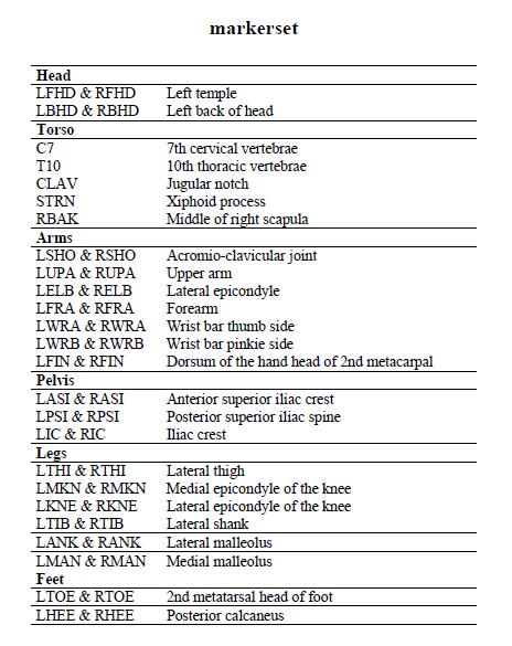
73 67
5/13/2016. ACL I Risk Factors AAP Position Statement. Anterior Cruciate Ligament Injuries: Diagnosis, Treatment and Prevention.
 ACL I Risk Factors AAP Position Statement Timothy E. Hewett, PhD 2016 Chicago Sports Medicine Symposium Chicago, Illinois August 5-7, 2016 2015 MFMER slide-1 Anterior Cruciate Ligament Injuries: Diagnosis,
CHAPTER 8: THE BIOMECHANICS OF THE HUMAN LOWER EXTREMITY
 CHAPTER 8: THE BIOMECHANICS OF THE HUMAN LOWER EXTREMITY _ 1. The hip joint is the articulation between the and the. A. femur, acetabulum B. femur, spine C. femur, tibia _ 2. Which of the following is
Re training Movement Behavior for ACL Injury Prevention and Rehabilitation: A Matter of Strength or Motor Control?
 Re training Movement Behavior for ACL Injury Prevention and Rehabilitation: A Matter of Strength or Motor Control? Christopher M. Powers, PT, PhD, FACSM, FAPTA Beth Fisher, PT, PhD, FAPTA Division of Biokinesiology
CHANGES IN LOWER-LIMB MUSCLE FORCES WITH PROPHYLACTIC KNEE BRACING DURING LANDING AND STOP-JUMP TASKS
 CHANGES IN LOWER-LIMB MUSCLE FORCES WITH PROPHYLACTIC KNEE BRACING DURING LANDING AND STOP-JUMP TASKS Katie Ewing 1, Rezaul Begg 2, Peter Lee 1 Department of Mechanical Engineering, University of Melbourne,
DIFFERENCES IN THE MECHANICS BETWEEN THE DOMINANT AND NON-DOMINANT PLANT LIMB DURING INSTEP SOCCER KICKING. Cassidy M. Berlin.
 DIFFERENCES IN THE MECHANICS BETWEEN THE DOMINANT AND NON-DOMINANT PLANT LIMB DURING INSTEP SOCCER KICKING by Cassidy M. Berlin A thesis submitted in partial fulfillment of the requirements for the degree
Mechanisms of ACL Injury: Implications for Rehabilitation, Injury Prevention & Return to Sport Decisions. Overarching research theme:
 Mechanisms of ACL Injury: Implications for Rehabilitation, Injury Prevention & Return to Sport Decisions Associate Professor Co Director, Musculoskeletal Biomechanics Research Laboratory University of
Anatomy and Biomechanics
 Introduction Increased participation= increased injury rates Females were found to be 5.4 times more likely to sustain injury than males. And females injured their ACL ad a rate of 7.8 times more than
The Female Athlete: Train Like a Girl. Sarah DoBroka Wilson, PT, SCS Ron Weathers, PT, DPT, ATC, LAT
 The Female Athlete: Train Like a Girl Sarah DoBroka Wilson, PT, SCS Ron Weathers, PT, DPT, ATC, LAT Page 1 of 6 The Female Athlete: Train Like a Girl Sarah DoBroka Wilson PT, SCS Ron Weathers PT, DPT,
Research Theme. Cal PT Fund Research Symposium 2015 Christopher Powers. Patellofemoral Pain to Pathology Continuum. Applied Movement System Research
 Evaluation and Treatment of Movement Dysfunction: A Biomechanical Approach Research Theme Christopher M. Powers, PhD, PT, FAPTA Understanding injury mechanisms will lead to the development of more effective
ABSTRACT THE EFFECTS OF A 6-WEEK NEUROMUSCULAR TRAINING PROGRAM ON KNEE JOINT MOTOR CONTROL DURING SIDECUTTING IN HIGH-SCHOOL FEMALE ATHLETES
 ABSTRACT THE EFFECTS OF A 6-WEEK NEUROMUSCULAR TRAINING PROGRAM ON KNEE JOINT MOTOR CONTROL DURING SIDECUTTING IN HIGH-SCHOOL FEMALE ATHLETES by Justin Phillip Waxman The purpose of this study was to implement
Commonality of ACL Injuries and Prevention Methods in Women. Anterior cruciate ligament or ACL injuries have become more common in
 4/6/12 Period 2 Commonality of ACL Injuries and Prevention Methods in Women Anterior cruciate ligament or ACL injuries have become more common in the past decade than at any other time in our history.
ACL Injury Prevention: Considerations for Children and Adolescents
 ACL Injury Prevention: Considerations for Children and Adolescents Susan Sigward PhD, PT, ATC Human Performance Laboratory University of Southern California 1 ACL Injury Season ending/ Career ending 82%
Effects of Altered Surface Inclinations on Knee Kinematics During Drop Landing
 Pursuit - The Journal of Undergraduate Research at the University of Tennessee Volume 7 Issue 1 Article 14 April 2016 Effects of Altered Surface Inclinations on Knee Kinematics During Drop Landing Cicily
BIOMECHANICAL INFLUENCES ON THE SOCCER PLAYER. Planes of Lumbar Pelvic Femoral (Back, Pelvic, Hip) Muscle Function
 Muscle Function") BIOMECHANICAL INFLUENCES ON THE SOCCER PLAYER Functional performance of the soccer player reflects functional capability of certain specific muscle and muscle groups of the back, pelvis and hip to work
BIOMECHANICAL INFLUENCES ON THE SOCCER PLAYER Functional performance of the soccer player reflects functional capability of certain specific muscle and muscle groups of the back, pelvis and hip to work
Female Athlete Knee Injury
 Female Athlete Knee Injury Kelly C. McInnis, DO Physical Medicine and Rehabilitation Massachusetts General Hospital Sports Medicine Center Outline Historical Perspective Gender-specific movement patterns
Evaluating the Athlete Questionnaire
 Evaluating the Athlete Questionnaire Prior to developing the strength and conditioning training plan the coach should first evaluate factors from the athlete s questionnaire that may impact the strength
ACL and Knee Injury Prevention. Presented by: Zach Kirkpatrick, PT, MPT, SCS
 ACL and Knee Injury Prevention Presented by: Zach Kirkpatrick, PT, MPT, SCS ACL Anatomy ACL Mechanism of Injury Contact ACL Tear Noncontact ACL Tear ACL MOI and Pathology Common in young individual who
EMG Analysis of Lower Extremity Muscles in Three Different Squat Exercises
 REPORT EMG Analysis of Lower Extremity Muscles in Three Different Squat Exercises Gaston Ariel NISHIWAKI 1, Yukio URABE 1 and Kosuke TANAKA 1 1 Department of Sport Rehabilitation, Graduate School of Health
Muscles of the Hip 1. Tensor Fasciae Latae O: iliac crest I: lateral femoral condyle Action: abducts the thigh Nerve: gluteal nerve
 Muscles of the Hip 1. Tensor Fasciae Latae O: iliac crest I: lateral femoral condyle Action: abducts the thigh Nerve: gluteal nerve 2. Gluteus Maximus O: ilium I: femur Action: abduct the thigh Nerve:
Main Menu. Joint and Pelvic Girdle click here. The Power is in Your Hands
 1 Hip Joint and Pelvic Girdle click here Main Menu K.6 http://www.handsonlineeducation.com/classes//k6entry.htm[3/23/18, 2:01:12 PM] Hip Joint (acetabular femoral) Relatively stable due to : Bony architecture
Sports Medicine 15. Unit I: Anatomy. The knee, Thigh, Hip and Groin. Part 4 Anatomies of the Lower Limbs
 Sports Medicine 15 Unit I: Anatomy Part 4 Anatomies of the Lower Limbs The knee, Thigh, Hip and Groin Anatomy of the lower limbs In Part 3 of this section we focused upon 11 of the 12 extrinsic muscles
Functional Movement Screen (Cook, 2001)
") Functional Movement Screen (Cook, 2001) TEST 1 DEEP SQUAT Purpose - The Deep Squat is used to assess bilateral, symmetrical, mobility of the hips, knees, and ankles. The dowel held overhead assesses bilateral,
Functional Movement Screen (Cook, 2001) TEST 1 DEEP SQUAT Purpose - The Deep Squat is used to assess bilateral, symmetrical, mobility of the hips, knees, and ankles. The dowel held overhead assesses bilateral,
Balanced Body Movement Principles
 Balanced Body Movement Principles How the Body Works and How to Train it. Module 3: Lower Body Strength and Power Developing Strength, Endurance and Power The lower body is our primary source of strength,
THE EFFECTS OF HIP STRENGTH ON GLUTEAL MUSCLE ACTIVATION AMPLITUDES AND HOW THESE FACTORS PREDICT LOWER EXTREMITY KINEMATICS. Katie J.
 THE EFFECTS OF HIP STRENGTH ON GLUTEAL MUSCLE ACTIVATION AMPLITUDES AND HOW THESE FACTORS PREDICT LOWER EXTREMITY KINEMATICS Katie J. Homan A thesis submitted to the faculty of the University of North
Primary Movements. Which one? Rational - OHS. Assessment. Rational - OHS 1/1/2013. Two Primary Movement Assessment: Dynamic Assessment (other)
") Primary Movements Practical Application for Athletic Trainers Two Primary Movement Assessment: NASM-CES Overhead Squat Single-leg Squat Dynamic Assessment (other) Single-leg Step Off Functional Movement
Primary Movements Practical Application for Athletic Trainers Two Primary Movement Assessment: NASM-CES Overhead Squat Single-leg Squat Dynamic Assessment (other) Single-leg Step Off Functional Movement
Myology of the Knee. PTA 105 Kinesiology
 Myology of the Knee PTA 105 Kinesiology Objectives Describe the planes of motion and axes of rotation of the knee joint Visualize the origins and insertions of the muscles about the knee List the innervations
BIOMECHANICS AND CONTEXT OF ACUTE KNEE INJURIES. Uwe Kersting MiniModule Idræt Biomekanik 2. Objectives
 BIOMECHANICS AND CONTEXT OF ACUTE KNEE INJURIES Uwe Kersting MiniModule 06 2011 Idræt Biomekanik 2 1 Objectives Know about the static and dynamic organisation of the knee joint (anatomy & function) Be
BIOMECHANICAL RISK FACTORS ASSOCIATED WITH NON-CONTACT ANTERIOR CRUCIATE LIGAMENT INJURIES DURING LANDING PERFORMANCE
 BIOMECHANICAL RISK FACTORS ASSOCIATED WITH NON-CONTACT ANTERIOR CRUCIATE LIGAMENT INJURIES DURING LANDING PERFORMANCE Thomas W. Kernozek University of Wisconsin-La Crosse, La Crosse, WI, USA Anterior cruciate
Dynamic Trunk Control Influence on Run-to-Cut Maneuver: A Risk Factor for ACL Rupture
 Dynamic Trunk Control Influence on Run-to-Cut Maneuver: A Risk Factor for ACL Rupture By: Steve Jamison 1 INTRODUCTION Anterior cruciate ligament (ACL) rupture is one of the most common knee injuries for
2. Iliotibial Band syndrome
 2. Iliotibial Band syndrome Iliotibial band (ITB) syndrome (so called runners knee although often seen in other sports e.g. cyclists and hill walkers). It is usually an overuse injury with pain felt on
Uranie P. Browne. Chapel Hill 2007
 A Comparison of Risk Factors Between a Cutting Task and a Stop-Jump as it Relates to the Non Contact Anterior Cruciate Ligament Injury Uranie P. Browne A thesis submitted to the faculty of the University
HAMSTRING NEUROMECHANICAL PROPERTIES AND BIOMECHANICS IN AN ACL INJURED POPULATION. David Robert Bell
 HAMSTRING NEUROMECHANICAL PROPERTIES AND BIOMECHANICS IN AN ACL INJURED POPULATION David Robert Bell A dissertation submitted to the faculty of the University of North Carolina at Chapel Hill in partial
Obesity is associated with reduced joint range of motion (Park, 2010), which has been partially
, which has been partially") INTRODUCTION Obesity is associated with reduced joint range of motion (Park, 2010), which has been partially attributed to adipose tissues around joints limiting inter-segmental rotations (Gilleard, 2007).
INTRODUCTION Obesity is associated with reduced joint range of motion (Park, 2010), which has been partially attributed to adipose tissues around joints limiting inter-segmental rotations (Gilleard, 2007).
Rehabilitation Guidelines for Anterior Cruciate Ligament (ACL) Reconstruction
 Reconstruction") Rehabilitation Guidelines for Anterior Cruciate Ligament (ACL) Reconstruction The knee is the body's largest joint, and the place where the femur, tibia, and patella meet to form a hinge-like joint. These
Rehabilitation Guidelines for Anterior Cruciate Ligament (ACL) Reconstruction The knee is the body's largest joint, and the place where the femur, tibia, and patella meet to form a hinge-like joint. These
The Lower Limb II. Anatomy RHS 241 Lecture 3 Dr. Einas Al-Eisa
 The Lower Limb II Anatomy RHS 241 Lecture 3 Dr. Einas Al-Eisa Tibia The larger & medial bone of the leg Functions: Attachment of muscles Transfer of weight from femur to skeleton of the foot Articulations
Knee valgus in self-initiated vertical jump landings: developmental and gender comparisons
 Bridgewater State University Virtual Commons - Bridgewater State University Movement Arts, Health Promotion and Leisure Studies Faculty Publications Movement Arts, Health Promotion and Leisure Studies
Muscle Testing of Knee Extensors. Yasser Moh. Aneis, PhD, MSc., PT. Lecturer of Physical Therapy Basic Sciences Department
 Muscle Testing of Knee Extensors Yasser Moh. Aneis, PhD, MSc., PT. Lecturer of Physical Therapy Basic Sciences Department Muscle Testing of Knee Extensors othe Primary muscle Quadriceps Femoris -Rectus
Certified Personal Trainer Re-Certification Manual
 Certified Personal Trainer Re-Certification Manual Section II 1 Anatomy & Physiology Terms Anatomy and physiology are closely related fields of study: anatomy is the study of form, and physiology is the
Disclosures. Objectives. Overview. Patellofemoral Syndrome. Etiology. Management of Patellofemoral Pain
 Management of Patellofemoral Pain Implications of Top Down Mechanics Disclosures I have no actual or potential conflict of interest in relation to this presentation David Nolan, PT, DPT, MS, OCS, SCS,
When are athletes ready for return to sports??? Functional Testing for Return to Sports. Important Factors Involved in Return to Sport
 Functional Testing for Return to Sports Meg Jacobs PT Momentum Physical Therapy and Sports Rehab Mjacobs@wegetyouhealthy.com When are athletes ready for return to sports??? Post ACL reconstruction, average
CHAPTER 1: 1.1 Muscular skeletal system. Question - text book page 16. Question - text book page 20 QUESTIONS AND ANSWERS. Answers
 QUESTIONS AND ANSWERS CHAPTER 1: 1.1 Muscular skeletal system Question - text book page 16 Using the information on pages 12 to 14 above, complete the table below. joint joint type articulating bones associated
The effect of patellofemoral pain syndrome on the hip and knee neuromuscular control on dynamic postural control task
 The University of Toledo The University of Toledo Digital Repository Theses and Dissertations 2009 The effect of patellofemoral pain syndrome on the hip and knee neuromuscular control on dynamic postural
ANTICIPATORY EFFECTS ON LOWER EXTREMITY KINETICS AND KINEMATICS DURING A LAND AND CROSS STEP MANEUVER IN FEMALE VOLLEYBALL PLAYERS
 ANTICIPATORY EFFECTS ON LOWER EXTREMITY KINETICS AND KINEMATICS DURING A LAND AND CROSS STEP MANEUVER IN FEMALE VOLLEYBALL PLAYERS A THESIS SUBMITTED TO THE GRADUATE SCHOOL IN PARTIAL FULFILLMENT OF THE
Influence of Fatigue on Jump and Land Movement Patterns
 St. Catherine University SOPHIA Doctor of Physical Therapy Research Papers Physical Therapy 4-2016 Influence of Fatigue on Jump and Land Movement Patterns Sarah Bard St. Catherine University Beth Anne
Development of an ergonomic musculoskeletal model to estimate muscle forces during vertical jumping
 Available online at www.sciencedirect.com Procedia Engineering 13 (2011) 338 343 5 th Asia-Pacific Congress on Sports Technology (APCST) Development of an ergonomic musculoskeletal model to estimate muscle
Lifting your toes up towards your tibia would be an example of what movement around the ankle joint?
 NAME: TEST 1 ANATOMY IN SPORT SCIENCE: SEMESTER 1, 2017 TOTAL MARKS = 58 Total: / 58 marks Percentage: Grade: TERMINOLOGY: The structures that connect bone to bone are called: The ankle joint is to the
Effect of frontal plane foot position on lower extremity muscle activation and limb positioning in a single leg squat
 University of North Dakota UND Scholarly Commons Physical Therapy Scholarly Projects Department of Physical Therapy 2012 Effect of frontal plane foot position on lower extremity muscle activation and limb
Practical 1 Worksheet
 Practical 1 Worksheet ANATOMICAL TERMS 1. Use the word bank to fill in the missing words. reference side stand body arms palms anatomical forward All anatomical terms have a(n) point which is called the
Nicholas Robert Heebner. B.S. Kinesiology, The Pennsylvania State University, M.S. Sports Medicine, University of Pittsburgh, 2012
 THE EFFECT OF JUMP DISTANCE ON BIOMECHANICAL RISK FACTORS FOR ACL INJURY DURING LANDING AND THEIR RELATIONSHIP WITH SENSORIMOTOR CHARACTERISTICS AT THE KNEE by Nicholas Robert Heebner B.S. Kinesiology,
Muscles of the Thigh. 6.1 Identify, describe the attachments of and deduce the actions of the muscles of the thigh: Anterior group
 Muscles of the Thigh 6.1 Identify, describe the attachments of and deduce the actions of the muscles of the thigh: Anterior group Sartorius: This is a long strap like muscle with flattened tendons at each
PROPHYLACTIC ANKLE STABILIZERS AND THEIR EFFECT ON LOWER EXTREMITY LANDING MECHANICS DURING DROP JUMP LANDINGS TO FATIGUE MASTER OF SCIENCE
 PROPHYLACTIC ANKLE STABILIZERS AND THEIR EFFECT ON LOWER EXTREMITY LANDING MECHANICS DURING DROP JUMP LANDINGS TO FATIGUE A THESIS SUBMITTED TO THE GRADUATE SCHOOL IN PARTIAL FULFILLMENT OF THE REQUIREMENTS
GFM Platform Exercise Manual
 GFM Platform Exercise Manual STEPHEN NEWHART, CSCS*D What is Whole Body Vibration? Body vibration is delivered through a variety of machines, including the most recent inclusion of whole body vibration
Muscles to know. Lab 21. Muscles of the Pelvis and Lower Limbs. Muscles that Position the Lower Limbs. Generally. Muscles that Move the Thigh
 Muscles to know Lab 21 Muscles of the Pelvis, Leg and Foot psoas major iliacus gluteus maximus gluteus medius sartorius quadriceps femoris (4) gracilus adductor longus biceps femoris semitendinosis semimembranosus
ANTICIPATORY EFFECTS ON LOWER EXTREMITY KINEMATICS AND KINETICS DURING CUTTING MOVEMENTS
 ANTICIPATORY EFFECTS ON LOWER EXTREMITY KINEMATICS AND KINETICS DURING CUTTING MOVEMENTS By RYAN ASHLEY MIZELL A THESIS PRESENTED TO THE GRADUATE SCHOOL OF THE UNIVERSITY OF FLORIDA IN PARTIAL FULFILLMENT
Biomechanics of Skeletal Muscle and the Musculoskeletal System
 Biomechanics of Skeletal Muscle and the Musculoskeletal System Hamill & Knutzen (Ch 3) Nordin & Frankel (Ch 5), or Hall (Ch. 6) Muscle Properties Ø Irritability Ø Muscle has the capability of receiving
An estimated anterior cruciate ligament. Mechanisms of Noncontact Anterior Cruciate Ligament Injury
 Journal of Athletic Training 2008;43(4):396 408 g by the National Athletic Trainers Association, Inc www.nata.org/jat systematic review Mechanisms of Noncontact Anterior Cruciate Ligament Injury Yohei
RN(EC) ENC(C) GNC(C) MN ACNP *** MECHANISM OF INJURY.. MOST IMPORTANT *** - Useful in determining mechanism of injury / overuse
 ENC(C) GNC(C) MN ACNP *** MECHANISM OF INJURY.. MOST IMPORTANT *** - Useful in determining mechanism of injury / overuse") HISTORY *** MECHANISM OF INJURY.. MOST IMPORTANT *** Age of patient Sport / Occupation - Certain conditions are more prevalent in particular age groups (Osgood Schlaters in youth / Degenerative Joint Disease
HISTORY *** MECHANISM OF INJURY.. MOST IMPORTANT *** Age of patient Sport / Occupation - Certain conditions are more prevalent in particular age groups (Osgood Schlaters in youth / Degenerative Joint Disease
Stronger Legs & Lower Body
 Stronger Legs & Lower Body CORRESPONDENCE EDUCATION PROGRAM # 139. Check your receipt for course expiration date. After that date no credit will be awarded for this program. 2012 by Exercise ETC Inc. All
Active-Assisted Stretches
 1 Active-Assisted Stretches Adequate flexibility is fundamental to a functional musculoskeletal system which represents the foundation of movement efficiency. Therefore a commitment toward appropriate
Retrieved from DalSpace, the institutional repository of Dalhousie University
 Retrieved from DalSpace, the institutional repository of Dalhousie University https://dalspace.library.dal.ca/handle/10222/72293 Version: Post-print Publisher s version: Wilson, Janie L. Astephen, Michael
A Principal Component Analysis Investigation of Drop Landings for Defining Anterior Cruciate Ligament Injury Risk Factors
 Marquette University e-publications@marquette Master's Theses (2009 -) Dissertations, Theses, and Professional Projects A Principal Component Analysis Investigation of Drop Landings for Defining Anterior
EVALUATION OF THE ANKLE ROLL GUARD S EFFECTIVENESS TO IMPROVE ITS CLINICAL BENEFIT PROGRESS REPORT. Prepared By:
 EVALUATION OF THE ANKLE ROLL GUARD S EFFECTIVENESS TO IMPROVE ITS CLINICAL BENEFIT PROGRESS REPORT Prepared By: Dr. Tyler Brown, Principal Investigator Wyatt Ihmels, Graduate Research Assistant Research
The Influence of Static Stretching of Knee Flexors on Knee Biomechanics
 Wright State University CORE Scholar Browse all Theses and Dissertations Theses and Dissertations 2018 The Influence of Static Stretching of Knee Flexors on Knee Biomechanics Joshua David Perrin Wright
BIOMECHANICAL ANALYSIS OF THE DEADLIFT DURING THE 1999 SPECIAL OLYMPICS WORLD GAMES
 63 Biomechanics Symposia 2001 / University of San Francisco BIOMECHANICAL ANALYSIS OF THE DEADLIFT DURING THE 1999 SPECIAL OLYMPICS WORLD GAMES Rafael F. Escamilla, Tracy M. Lowry, Daryl C. Osbahr, and
Hamstring Strain. 43 Thames Street, St Albans, Christchurch 8013 Phone: (03) Website: philip-bayliss.com.
 Website: philip-bayliss.com.") 43 Thames Street, St Albans, Christchurch 8013 Phone: (03) 356 1353. Website: philip-bayliss.com Hamstring Strain The hamstring muscles are very susceptible to tears, strains and other common sporting
43 Thames Street, St Albans, Christchurch 8013 Phone: (03) 356 1353. Website: philip-bayliss.com Hamstring Strain The hamstring muscles are very susceptible to tears, strains and other common sporting
Gender differences in knee kinematics during landing from. Gerwyn Hughes James Watkins, Nick Owen and Mike Lewis.
 Title: Gender differences in knee kinematics during landing from volleyball block jumps. Authors: Gerwyn Hughes James Watkins, Nick Owen and Mike Lewis. Institution: Department of Sports Science, Swansea
Anterior Cruciate Ligament Surgery
 Anatomy Anterior Cruciate Ligament Surgery Roger Ostrander, MD Andrews Institute Anatomy Anatomy Function Primary restraint to anterior tibial translation Secondary restraint to internal tibial rotation
Jennifer L. Cook, MD
 Jennifer L. Cook, MD Florida Joint Replacement and Sports Medicine Center 5243 Hanff Lane New Port Richey, FL 34652 Phone: (727)848-4249 Fax: (727) 841-8934 ANTERIOR CRUCIATE LIGAMENT RECONSTRUCTION POST-OPERATIVE
APTA Intro to Identity. The Movement System The Kinesiopathologic Model Movement System Impairment Syndromes of the Knee THE HUMAN MOVEMENT SYSTEM
 The Movement System The Kinesiopathologic Model Movement System Impairment Syndromes of the Knee Shirley Sahrmann, PT, PhD, FAPTA Professor Emerita Statement of Privacy To protect the privacy of the subjects
Diane Vives, MS, CSCS, *D Owner, Fit4Austin, Austin, TX Member of the Board of Directors, NSCA
 Diane Vives, MS, CSCS, *D Owner, Fit4Austin, Austin, TX Member of the Board of Directors, NSCA 1. Introduction a. Through my experience as a strength coach over the last 11 years, I have often been faced
BARBELL HIP THRUST. Eckert, RM 1 and Snarr, RL 1,2
 Eckert, RM and Snarr, RL. Barbell hip thrust. J Sport Human Perf 2014;2(2):1-9. DOI: 10.12922/jshp.0037.2014 1 SHORT REPORT BARBELL HIP THRUST OPEN ACCESS Eckert, RM 1 and Snarr, RL 1,2 1 School of Nutrition
RN(EC) ENC(C) GNC(C) MN ACNP *** MECHANISM OF INJURY.. MOST IMPORTANT ***
 ENC(C) GNC(C) MN ACNP *** MECHANISM OF INJURY.. MOST IMPORTANT ***") HISTORY *** MECHANISM OF INJURY.. MOST IMPORTANT *** Age of patient - Certain conditions are more prevalent in particular age groups (Hip pain in children may refer to the knee from Legg-Calve-Perthes
HISTORY *** MECHANISM OF INJURY.. MOST IMPORTANT *** Age of patient - Certain conditions are more prevalent in particular age groups (Hip pain in children may refer to the knee from Legg-Calve-Perthes
JAT Vol. 49 #3 CEU Questions
 JAT Vol. 49 #3 CEU Questions Aronson et al 1. According to Aronson et al, which of the following age groups demonstrated less medial knee stiffness than the other age groups? a. 8 10 year olds b. 18 40
Discrepancies in Knee Joint Moments Using Common Anatomical Frames Defined by Different Palpable Landmarks
 Journal of Applied Biomechanics, 2008, 24, 185-190 2008 Human Kinetics, Inc. Discrepancies in Knee Joint Moments Using Common Anatomical Frames Defined by Different Palpable Landmarks Dominic Thewlis,
Compiled and Designed by: Sport Dimensions - 2 -
 SOCCER TRAINING While all reasonable care has been taken during the preparation of this edition, neither the publisher, nor the authors can accept responsibility for any consequences arising from the use
The influence of forefoot binding force change on vertical jump kinematics variation
 Available online www.jocpr.com Journal of Chemical and Pharmaceutical Research, 2014, 6(2):554-558 Research Article ISSN : 0975-7384 CODEN(USA) : JCPRC5 The influence of forefoot binding force change on
A STOCHASTIC BIOMECHANICAL MODEL FOR RISK AND RISK FOCTORS OF SUSTAINING NON-CONTACT ACL INJURIES. Cheng-Feng Lin, MS
 A STOCHASTIC BIOMECHANICAL MODEL FOR RISK AND RISK FOCTORS OF SUSTAINING NON-CONTACT ACL INJURIES Cheng-Feng Lin, MS A dissertation submitted to the faculty of the University of North Carolina at Chapel
The Effects of Augmented Feedback on Knee Valgus Angles and Muscle Activity During a Jump Landing Task. Michelle M. Bensman BS, LAT, ATC
 The Effects of Augmented Feedback on Knee Valgus Angles and Muscle Activity During a Jump Landing Task Michelle M. Bensman BS, LAT, ATC A thesis submitted to the faculty of The University of North Carolina
Military Movement Training Program Improves Jump-Landing Mechanics Associated With Anterior Cruciate Ligament Injury Risk
 Military Movement Training Program Improves Jump-Landing Mechanics Associated With Anterior Cruciate Ligament Injury Risk Brett D. Owens, MD; Kenneth L. Cameron, PhD, ATC; Michele L. Duffey, MS; Donna
EXCERPT ADAPTED FROM CARL PETERSEN S NEW BOOK FIT 2 SKI
 ACL THE ABC S OF PROTECTION EXCERPT ADAPTED FROM CARL PETERSEN S NEW BOOK FIT 2 SKI by CARL PETERSEN BPE, BSc (PT) Injuries to the knee are a regular occurrence for skiing professionals and anterior cruciate
Knee Conditioning Program
 Knee Conditioning Program Purpose of Program After an injury or surgery, an exercise conditioning program will help you return to daily activities and enjoy a more active, healthy lifestyle. Following
Literature Review of Female Anterior Cruciate Ligament Injuries
 Eastern Michigan University DigitalCommons@EMU Senior Honors Theses Honors College 2004 Literature Review of Female Anterior Cruciate Ligament Injuries Allison M. Nichol Follow this and additional works
Recognizing common injuries to the lower extremity
 Recognizing common injuries to the lower extremity Bones Femur Patella Tibia Tibial Tuberosity Medial Malleolus Fibula Lateral Malleolus Bones Tarsals Talus Calcaneus Metatarsals Phalanges Joints - Knee
Anatomy & Physiology. Muscles of the Lower Limbs.
 Anatomy & Physiology Muscles of the Lower Limbs http://www.ishapeup.com/musclecharts.html Muscles of the Lower Limbs Among the strongest muscles in the body. Because pelvic girdle is composed of heavy,
Human anatomy reference:
 Human anatomy reference: Weak Glut Activation Weak gluteal activation comes from poor biomechanics, poor awareness when training or prolonged exposure in deactivated positions such as sitting. Weak Glut
What is Kinesiology? Basic Biomechanics. Mechanics
 What is Kinesiology? The study of movement, but this definition is too broad Brings together anatomy, physiology, physics, geometry and relates them to human movement Lippert pg 3 Basic Biomechanics the
MUSCLE ACTIVATION AND RANGE OF MOTION PATTERNS OF INDIVIDUALS WHO DISPLAY A LATERAL HIP SHIFT DURING AN OVERHEAD SQUAT. Kerry J.
 MUSCLE ACTIVATION AND RANGE OF MOTION PATTERNS OF INDIVIDUALS WHO DISPLAY A LATERAL HIP SHIFT DURING AN OVERHEAD SQUAT Kerry J. Peterson A thesis submitted to the faculty of the University of North Carolina
Anterior Cruciate Ligament (ACL) Injuries
 Injuries") Anterior Cruciate Ligament (ACL) Injuries Mark L. Wood, MD The anterior cruciate ligament (ACL) is one of the most commonly injured ligaments of the knee. The incidence of ACL injuries is currently estimated
Anterior Cruciate Ligament (ACL) Injuries Mark L. Wood, MD The anterior cruciate ligament (ACL) is one of the most commonly injured ligaments of the knee. The incidence of ACL injuries is currently estimated
Knee Conditioning Program
 Prepared for: Prepared by: Purpose of Program After an injury or surgery, an exercise conditioning program will help you return to daily activities and enjoy a more active, healthy lifestyle. Following
SKELETAL KINEMATICS OF THE ANTERIOR CRUCIATE LIGAMENT DEFICIENT KNEE WITH AND WITHOUT FUNCTIONAL BRACES
 SKELETAL KINEMATICS OF THE ANTERIOR CRUCIATE LIGAMENT DEFICIENT KNEE WITH AND WITHOUT FUNCTIONAL BRACES Dan K. Ramsey 1, Mario Lamontagne, Per F.Wretenberg 3 and Gunnar Németh 1 School of Human Kinetics,
Fatigue Induced Hip Abductor Strength and Single Leg Drop- Jumps in Recreationally Active Females
 International Journal of Science and Engineering Investigations vol. 7, issue 78, July 2018 ISSN: 2251-8843 Fatigue Induced Hip Abductor Strength and Single Leg Drop- Jumps in Recreationally Active Females
ACL Rehabilitation and Return To Play
 ACL Rehabilitation and Return To Play Seth Gasser, MD Director of Sports Medicine Florida Orthopaedic Institute Introduction Return to Play: the point in recovery from an injury when a person is safely
Current trends in ACL Rehab. James Kelley, MDS, PT
 Current trends in ACL Rehab James Kelley, MDS, PT Objectives Provide etiological information Discuss the criteria for having an ACL reconstruction Review the basic rehabilitation principles behind ACL
Muscles of the Gluteal Region
 Muscles of the Gluteal Region 1 Some of the most powerful in the body Extend the thigh during forceful extension Stabilize the iliotibial band and thoracolumbar fascia Related to shoulders and arms because
Examining the Impact of Adding Gluteal Strengthening Exercises to the FIFA 11+ Warm-Up Program on High School GIrls' Basketball Reported Injuries.
 University of Montana ScholarWorks at University of Montana Graduate Student Theses, Dissertations, & Professional Papers Graduate School 2018 Examining the Impact of Adding Gluteal Strengthening Exercises
A controlled intervention study assessing the relation between hip abductor strength and knee valgus
 Are Grytdal A controlled intervention study assessing the relation between hip abductor strength and knee valgus Master s thesis in Human Movement Science Trondheim, June 2015 Norwegian university of science
5/14/2013. Acute vs Chronic Mechanism of Injury:
 Third Annual Young Athlete Conference: The Lower Extremity February 22, 2013 Audrey Lewis, DPT Acute vs Chronic Mechanism of Injury: I. Direct: blow to the patella II. Indirect: planted foot with a valgus
PRELIMINARY HSC PDHPE. CQ1 How do the musculoskeletal and cardiorespiratory systems of the body influence and respond to movement?
 PRELIMINARY HSC PDHPE CQ1 How do the musculoskeletal and cardiorespiratory systems of the body influence and respond to movement? How do the musculoskeletal and cardiorespiratory systems of the body influence
The Muscular System. Chapter 10 Part D. PowerPoint Lecture Slides prepared by Karen Dunbar Kareiva Ivy Tech Community College
 Chapter 10 Part D The Muscular System Annie Leibovitz/Contact Press Images PowerPoint Lecture Slides prepared by Karen Dunbar Kareiva Ivy Tech Community College Table 10.14: Muscles Crossing the Hip and
ACE s Essentials of Exercise Science for Fitness Professionals TRUNK
 ACE s Essentials of Exercise Science for Fitness Professionals TRUNK Posture and Balance Posture refers to the biomechanical alignment of the individual body parts and the orientation of the body to the