Chiari Malformations. Google. Objectives Seventh Annual NKY TBI Conference 3/22/13. Kerry R. Crone, M.D.
|
|
|
- Isabella Jefferson
- 5 years ago
- Views:
Transcription
1 Chiari Malformations Kerry R. Crone, M.D. Professor of Neurosurgery and Pediatrics University of Cincinnati College of Medicine University of Cincinnati Medical Center Cincinnati Children s Hospital Medical Center Cincinnati, Ohio. USA kerry.crone@cchmc.org Objectives Define Chiari Malformations Explain the differences among the various types of malformations. Review the presenting symptoms for Chiari malformations. Discuss the options for treatment. Recall what you learn today. Google About 35% of U.S. adults say they have used the Internet to figure out what medical condition they or someone else might have. A new survey from the Pew Research Center in Washington, D.C. reports that: Among adults who use the Internet to get any kind of health information, 59% admit to diagnostic sleuthing. 1
2 From the Internet Chiari malformations are named for Hans Chiari, an Austrian pathologist, who first identified types I-III in Julius Arnold further expanded the definition of Chiari malformation type II. Summary Information Some medical sources began using the name Arnold-Chiari malformation. Nowadays, some medical sources, use Arnold-Chiari malformation as a broad term for all forms. Summary Information Chiari malformations have also been known as: Congenital tonsillar herniation Tonsillar ectopia Tonsillar descent 2

 that covered the fourth ventricle and extended into the")
3 The Chiari Description 1891-Dr. Hans Chiari Austrian pathologist 17yo female with hydrocephalus who died from typhoid fever and at autopsy her brain displayed an elongation of the tonsils and medial parts of the inferior lobes of the cerebellum The Arnold Description 1894-Dr. Julius Arnold German Anatomist Portrayed an infant with spina bifida and described an elongated inferior portion of the cerebellum (vermis) that covered the fourth ventricle and extended into the spinal canal. Synthesized Terminology 1907 Schwalbe and Gredig first applied the Arnold-Chiari eponym to patients previously characterized as having the Chiari type 2 malformation. Their differentiation was an improper modification of Chiari s original description. This eponym has continued into modern times to define rhombencephalon deformities. 3


4 Why the Difference is Important! The Cerebellum Chiari Malformations Chiari I Malformation (Most common) Chiari II (Arnold-Chiari) Malformation Chiari III Malformation Chiari IV Malformation Acquired Chiari Malformation Chiari 1.5 Malformation 4


5 Anatomic Findings in Chiari I Tonsillar herniation Hydrocephalus Related Anomalies Anatomic Metabolic Genetic Anatomic Findings in Chiari II Herniation of vermis, brainstem and fourth ventricle Myelomenigocele Syringomyelia Hydrocephalus Chiari III Malformation Posterior fossa encephalocele that contains cerebellar and brainstem tissue with herniation through an upper cervical spina bifida. Grave prognosis. Must distinguish this malformation from high cervical myelomeningoceles which may have a favorable prognosis. 5
6 Chiari IV Malformation Absent hindbrain herniation Cerebellar hypoplasia or aplasia Minimal function Acquired Chiari Malformation Spinal Fluid Diversion Development of craniocerebral disproportion related to chronic cranial diversion of cerebrospinal fluid. Essentially The skull becomes to small for the brain. Herniation of cerebellar tonsils either from chronic lumbar spinal fluid drainage related to lumboperitoneal shunts or acutely related to lumbar external drains Chiari 1.5 Malformation Transitional form of Chiari malformation where both tonsils and brainstem are caudally descended into the cervical spine. Myelomeningocele is absent. 6
 pain Clumsiness Dysphagia (difficulty swallowing) Dysarthria (difficulty speaking) Dys means bad, painful or disordered Chiari I Clinical Findings Oscillopsia Esotropia")
7 MR Findings in Chiari I Herniation of cerebellar tonsils Absent intracranial mass lesion Tonsillar tip configuration Cine MR Pain Chiari I Symptoms Occipital-cervical (lower head and neck) pain Back, shoulder, and limb (arm) pain Clumsiness Dysphagia (difficulty swallowing) Dysarthria (difficulty speaking) Dys means bad, painful or disordered Chiari I Clinical Findings Oscillopsia Esotropia Crossed Eyes Bradycardia Slow Heart Rate Apnea Central Hoarseness Choking Gagging Hiccoughs 7


8 The Three Chiari I Syndromes Brainstem Syndrome Spinal Cord Syndrome Cerebellar Syndrome Brainstem Syndrome Usually from birth through early years Respiratory Irregularities Nystagmus Lower cranial nerve dysfunction Recurrent aspiration Pneumonia Reactive airway disease Brainstem Syndrome 8


9 Spinal Cord Syndrome Late childhood through second decade Headache Scoliosis (spinal curvature) Motor/sensory losses Hyporeflexia Hyperreflexia Spinal Cord Syndrome Cerebellar Syndrome Usually from 18 months through adult life Truncal and appendicular ataxia Sensory motor disturbances 9

10 Radiographic Studies Magnetic Resonance Imaging Pathological hindbrain herniation defined as a distance greater than two standard deviations beyond the range of normal. 6mm First Decade 5mm Second and third decade 4mm Fourth through eighth decade Dysphagia Dysphagia is disordered or impaired swallowing Neuromotor dysfunction Developmental disabilities Phases of Swallowing Oral Phase Pharyngeal Phase Esophageal Phase 10


, pharynx, or")
11 Anatomy of the Medulla The Gag Reflex and Vomiting The gag reflex is a normal defense mechanism that prevents foreign bodies from entering the trachea (airway), pharynx, or larynx. Physiology of Gagging 11
")
12 History Physical Exam Diagnosis Radiological Studies MR Scan Head Spine Sleep Studies (apnea) Swallowing Studies (aspiration) Treatment There are only two options in the treatment of Chiari malformations Observation Most common in asymptomatic patients Limited published data Surgery Non uniform approaches Surgical Treatment Bony decompression Foramen magnum (Boney opening skull base) Lamina of first cervical ring Dural augmentation Tonsillar coagulation Fourth ventricle to subarachnoid stent 12


13 Surgical Treatment Posterior Fossa Anatomy Bony Decompression 13

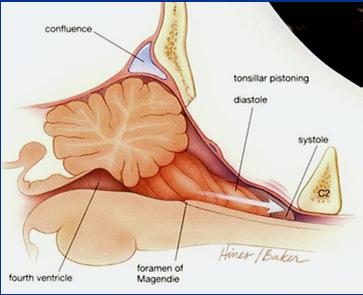
14 Dural Band Incision Dural Band Decompression Tonsillar Pistoning 14
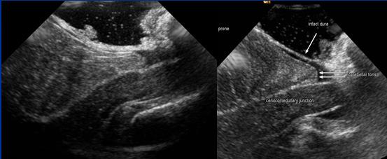
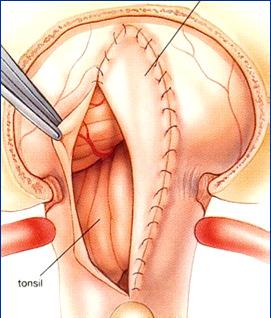
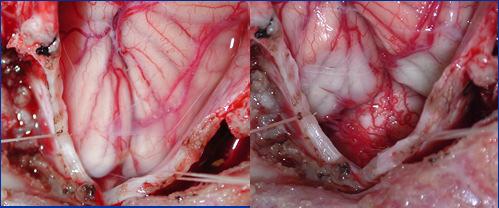
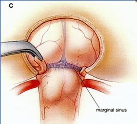
15 Intraoperative Ultrasound Dural Augmentation Intraoperative Appearance 15
16 CCHMC Chiari Database Over 1200 patients referred since patients have undergone surgical treatment Temporary Complications < 2.0% 90% Patients demonstrated Improvement 5% Presenting symptoms stabilized 5% No Change Summary Defined the Chiari Malformation Described the differences in malformation types. Related presenting signs and symptoms. Discussed options for treatment. Provided an overview of surgery. My gratitude for attention today. 16
Chiari malformations. A fact sheet for patients and carers
 A fact sheet for patients and carers Chiari malformations This fact sheet provides information on Chiari malformations. It focuses on Chiari malformations in adults. Our fact sheets are designed as general
Djamila Kafoufi Al Galaa Military Hospital Cairo
 Djamila Kafoufi Al Galaa Military Hospital Cairo Herniation cerebellar tonsils below the foramen magnum, Hans Chiari 4 types Chiari I less than 5mm,HDC rare,syringomyelia often present. Chiari II,protrusion
Chiari FAQ's. 1. What is a Chiari Malformation?
 Chiari FAQ's These FAQ's are for informational purposes only and in no way represent an attempt to provide medical advice. This information may or may not apply to your case and anyone with a question
Arnold Chiari Malformation - A hospital based autopsy study
 Rapotra Megha et al / International Journal of Biomedical Research 2017; 8(05): 250-254. 250 International Journal of Biomedical Research ISSN: 0976-9633 (Online); 2455-0566 (Print) Journal DOI: https://dx.doi.org/10.7439/ijbr
Morphometric Analysis of Left & Right Tonsils in Adult Symptomatic Type 1 Chiari Patients and Healthy Controls
 The University of Akron IdeaExchange@UAkron Honors Research Projects The Dr. Gary B. and Pamela S. Williams Honors College Spring 2015 Morphometric Analysis of Left & Right Tonsils in Adult Symptomatic
Chiari bridges review Chiari Treatments & Potential Pitfalls
 Chiari bridges review Chiari Treatments & Potential Pitfalls Once diagnosed, you will usually be referred to a specialist (not a Chiari Specialist, but an everyday, run-of-the-mill neurologist or neurosurgeon).
Role of MRI in Selection of Patients for Surgery and Assessing the Post Operative Outcome in Chiari 1 Malformation
 DOI: 10.7860/IJARS/2017/13599:2222 Radiology Section Original Article Role of MRI in Selection of Patients for Surgery and Assessing the Post Operative Outcome in Chiari 1 Malformation Rajesh Kumar V,
Suboccipital decompression for Chiari malformation associated scoliosis: risk factors and time course of deformity progression
 J Neurosurg Pediatrics 1:456 460, 2008 Suboccipital decompression for Chiari malformation associated scoliosis: risk factors and time course of deformity progression FRANK J. ATTENELLO, M.S., MATTHEW J.
What Every Spine Surgeon Should Know About Neurosurgical Issues
 What Every Spine Surgeon Should Know About Neurosurgical Issues Amer Samdani, MD Chief of Surgery Shriners Hospitals for Children Philadelphia, PA Objectives Main intraspinal lesions Chiari malformation
Disclosures None. Common Neurosurgical Problems Seen in Office Encounters. Macrocephaly Low Back Pain Sacral Dimple Concussion Chiari Malformation
 Common Neurosurgical Problems Seen in Office Encounters When to Manage, When to Refer Andrew Jea MD FAAP Professor and Chief of Pediatric Neurosurgery Riley Hospital for Children Indiana University School
Brain Imaging. Bearbeitet von Klaus Sartor, Stefan Hähnel, Bodo Kress
 Brain Imaging Bearbeitet von Klaus Sartor, Stefan Hähnel, Bodo Kress 1. Auflage 2007. Taschenbuch. 312 S. Paperback ISBN 978 3 13 143961 1 Format (B x L): 12,5 x 19 cm Weitere Fachgebiete > Medizin > Sonstige
five minutes to change a patient s life?
 Will you spend five minutes to change a patient s life? That s all it takes to learn what you need to know about the disorder that claimed our daughter www.shanno 7300 Bur Austin, Tex (512)90 Shannon s
Presented by : Shashwat Mishra
 Presented by : Shashwat Mishra Named after Hans Chiari (1851 1916). Professor of Pathology in Prague, Czechoslovakia, Paper entitled Concerning alterations in the cerebellum resulting from cerebral hydrocephalus
Significance of Cerebellar Tonsillar Position on MR
 795 Significance of Cerebellar Tonsillar Position on MR A. J. Barkovich 1. 3 F. J. Wippold 2. 3 J. L. Sherman 3. 4 C. M. Citrin 4. 5 It has been noted that a low degree of ectopia of the cerebellar tonsils
CNS Embryology 5th Menstrual Week (Dorsal View)
") Imaging of the Fetal Brain; Normal & Abnormal Alfred Abuhamad, M.D. Eastern Virginia Medical School CNS Embryology 5th Menstrual Week (Dorsal View) Day 20 from fertilization Neural plate formed in ectoderm
Imaging of the Fetal Brain; Normal & Abnormal Alfred Abuhamad, M.D. Eastern Virginia Medical School CNS Embryology 5th Menstrual Week (Dorsal View) Day 20 from fertilization Neural plate formed in ectoderm
Introduction to Neurosurgical Subspecialties:
 Introduction to Neurosurgical Subspecialties: Pediatric Neurosurgery Brian L. Hoh, MD 1 and Gregory J. Zipfel, MD 2 1 University of Florida, 2 Washington University Pediatric Neurosurgery Pediatric neurosurgeons
Complex Hydrocephalus
 2012 Hydrocephalus Association Conference Washington, DC - June 27-July1, 2012 Complex Hydrocephalus Marion L. Walker, MD Professor of Neurosurgery & Pediatrics Primary Children s Medical Center University
Abstract !"# $% &%'(% )* ( % +$$ '% % % Presentation Notes
* ( % +$$ '% % % Presentation Notes") Presenter Name: John Oro, MD Topic: Chiari & Syringomyelia 101 A Brief Look at Neuroanatomy The brain is enclosed and protected by a rounded skull made of rigid bone. The bottom of the skull contains multiple
Presenter Name: John Oro, MD Topic: Chiari & Syringomyelia 101 A Brief Look at Neuroanatomy The brain is enclosed and protected by a rounded skull made of rigid bone. The bottom of the skull contains multiple
Idiopathic cervical syringomyelia can be associated. Pediatric Chiari malformation Type 0: a 12-year institutional experience.
 J Neurosurg J Neurosurg Pediatrics Pediatrics 8:000 000, 8:1 5, 2011 Pediatric Chiari malformation Type 0: a 12-year institutional experience Clinical article Joshua J. Chern, M.D., Ph.D., Amber J. Gordon,
The Walton Centre. NHS Foundation Trust CHIARI MALFORMATION PATIENT INFORMATION
 The Walton Centre NHS Foundation Trust CHIARI MALFORMATION PATIENT INFORMATION What is a Chiari malformation? A Chiari Malformation (Hindbrain hernia) is a developmental abnormality which affects the
Intracranial hypotension secondary to spinal CSF leak: diagnosis
 Intracranial hypotension secondary to spinal CSF leak: diagnosis Spinal cerebrospinal fluid (CSF) leak is an important and underdiagnosed cause of new onset headache that is treatable. Cerebrospinal fluid
Neuroanatomy. Assistant Professor of Anatomy Faculty of Medicine The University of Jordan Dr Maha ELBeltagy
 Neuroanatomy Dr. Maha ELBeltagy Assistant Professor of Anatomy Faculty of Medicine The University of Jordan 2018 Development of the Central Nervous System Development of the nervous system Development
Fetal Medicine. Case Presentations. Dr Ermos Nicolaou Fetal Medicine Unit Chris Hani Baragwanath Hospital. October 2003
 Case Presentations Dr Ermos Nicolaou Fetal Medicine Unit Chris Hani Baragwanath Hospital October 2003 Case 1 Ms A M 22year old P0 G1 Referred from Sebokeng Hospital at 36w for polyhydramnios On Ultrasound:
Multimodal Evaluation Of Cerebrospinal Fluid (csf ) Dynamics Following Extradural Decompression For Chiari I Malformation
 Dynamics Following Extradural Decompression For Chiari I Malformation") Yale University EliScholar A Digital Platform for Scholarly Publishing at Yale Yale Medicine Thesis Digital Library School of Medicine January 2015 Multimodal Evaluation Of Cerebrospinal Fluid (csf ) Dynamics
Yale University EliScholar A Digital Platform for Scholarly Publishing at Yale Yale Medicine Thesis Digital Library School of Medicine January 2015 Multimodal Evaluation Of Cerebrospinal Fluid (csf ) Dynamics
Pediatric and adult Chiari malformation Type I surgical series : a review of demographics, operative treatment, and outcomes
 PEDIATRICS literature review Pediatric and adult Chiari malformation Type I surgical series 1965 2013: a review of demographics, operative treatment, and outcomes aska arnautovic, bsc, 1 bruno splavski,
Spinal Imaging. Bearbeitet von Herwig Imhof. 1. Auflage Taschenbuch. 312 S. Paperback ISBN Format (B x L): 12,5 x 19 cm
: 12,5 x 19 cm") Spinal Imaging Bearbeitet von Herwig Imhof 1. Auflage 2007. Taschenbuch. 312 S. Paperback ISBN 978 3 13 144071 6 Format (B x L): 12,5 x 19 cm Weitere Fachgebiete > Medizin > Sonstige Medizinische Fachgebiete
Spinal Imaging Bearbeitet von Herwig Imhof 1. Auflage 2007. Taschenbuch. 312 S. Paperback ISBN 978 3 13 144071 6 Format (B x L): 12,5 x 19 cm Weitere Fachgebiete > Medizin > Sonstige Medizinische Fachgebiete
Stroke School for Internists Part 1
 Stroke School for Internists Part 1 November 4, 2017 Dr. Albert Jin Dr. Gurpreet Jaswal Disclosures I receive a stipend for my role as Medical Director of the Stroke Network of SEO I have no commercial
NEUROSURGERY WORKING GROUP
 NEUROSURGERY WORKING GROUP OUTCOMES Primary Outcome Protect neurocognitive development by optimizing CSF dynamics throughout the life span. Optimize metrics for the management of CSF anomalies to protect/optimize
Neurosurgery. Neurosurgery
 Neurosurgery Neurosurgery Neurosurgery Telephone Numbers: Appointment: 202-476-3020 Fax: 202-476-3091 Administration: 202-476-3020 Evenings and Weekends: 202-476-5000 Robert Keating, MD, Chief The Division
Prior Authorization Review Panel MCO Policy Submission
 Prior Authorization Review Panel MCO Policy Submission A separate copy of this form must accompany each policy submitted for review. Policies submitted without this form will not be considered for review.
A review of the disagreements in the prevalence and treatment of the tethered cord syndromes with chiari 1 malformations
 SNI: Spine OPEN ACCESS For entire Editorial Board visit : http://www.surgicalneurologyint.com Editor: Nancy E. Epstein, MD Winthrop Hospital, Mineola, NY, USA Review Article A review of the disagreements
Cranio-cervical decompression. Information for patients Neurosurgery
 Cranio-cervical decompression Information for patients Neurosurgery page 2 of 12 What is a cranio-cervical decompression? A cranio-cervical decompression is an operation involving the back of the head
Brain ميهاربا لض اف دمح ا د The Meninges 1- Dura Mater of the Brain endosteal layer does not extend meningeal layer falx cerebri tentorium cerebelli
 .احمد د فاضل ابراهيم Lecture 15 Brain The Meninges Three protective membranes or meninges surround the brain in the skull: the dura mater, the arachnoid mater, and the pia mater 1- Dura Mater of the Brain
River North Pain Management Consultants, S.C., Axel Vargas, M.D., Regional Anesthesiology and Interventional Pain Management.
 River North Pain Management Consultants, S.C., Axel Vargas, M.D., Regional Anesthesiology and Interventional Pain Management. Chicago, Illinois, 60611 Phone: (888) 951-6471 Fax: (888) 961-6471 Clinical
Decompression of the spinal subarachnoid space as a solution for syringomyelia without Chiari malformation
 (2002) 40, 501 ± 506 ã 2002 International Society All rights reserved 1362 ± 4393/02 $25.00 www.nature.com/sc Original Article Decompression of the spinal subarachnoid space as a solution for syringomyelia
Brain Meninges, Ventricles and CSF
 Brain Meninges, Ventricles and CSF Lecture Objectives Describe the arrangement of the meninges and their relationship to brain and spinal cord. Explain the occurrence of epidural, subdural and subarachnoid
MR Imaging of Chiari II Malformation
 1037 MR Imaging of Chiari II Malformation Taher EI Gammal 1 Edward K. Mark2 Betty S. Brooks 1 High-field-strength MR imaging was performed in one patient with Chiari III and 19 patients with Chiari II
INCREASED INTRACRANIAL PRESSURE
 INCREASED INTRACRANIAL PRESSURE Sheba Medical Center, Acute Medicine Department Irene Frantzis P-Year student SGUL 2013 Normal Values Normal intracranial volume: 1700 ml Volume of brain: 1200-1400 ml CSF:
The NIHSS score is 4 (considering 2 pts for the ataxia involving upper and lower limbs.
 Neuroscience case 5 1. Speech comprehension, ability to speak, and word use were normal in Mr. Washburn, indicating that aphasia (cortical language problem) was not involved. However, he did have a problem
Malformations of the Nervous System November 10, Dr. Peter Ostrow
 Malformations of the Nervous System November 10, 2016 Dr. Peter Ostrow Malformations of the Nervous System 1. Abnormal closure of the neural tube 1. Disorders of forebrain formation 1. Cortical anomalies
BRITISH BIOMEDICAL BULLETIN
 Journal Home Page www.bbbulletin.org BRITISH BIOMEDICAL BULLETIN Case Report Neurofibromatosis Type 1 with Chiari Malformation Type 1 in Child: A Case Report Mallesh Kariyappa*, Febna A. Rahiman and Jayanthi
Pediatric Sleep-Disordered Breathing: More than OSA
 Pediatric Sleep-Disordered Breathing: More than OSA Carolyn M. D Ambrosio Associate Professor of Medicine Harvard Medical School Brigham and Women s Hospital Boston, MA Disclosures 1. Section Editor, Dynamed,
Supplementary Online Content
 Supplementary Online Content Honein MA, Dawson AL, Petersen E, et al; US Zika Pregnancy Registry Collaboration. Birth Defects Among Fetuses and Infants of US Women With Laboratory Evidence of Possible
Fifteen-minute consultation: incidental findings on brain and spine imaging
 BEST PRACTICE Editor s choice Scan to access more free content Department of Neurosurgery, Birmingham Children s Hospital, Birmingham, UK Correspondence to Chirag Patel, Department of Neurosurgery, Birmingham
J. Neurol. Neurosurg. Psychiat., 1953, 16, 227.
 J. Neurol. Neurosurg. Psychiat., 1953, 16, 227. THE ARNOLD-CHIARI MALFORMATION RADIOLOGICAL EXAMINATION WITH THE " ZIEDSES DES PLANTES " PROCEDURE BY Flom the Neurosurgical Department of the Neurological
Enhancement of Cranial US: Utility of Supplementary Acoustic Windows and Doppler Harriet J. Paltiel, MD
 Enhancement of Cranial US: Utility of Supplementary Acoustic Windows and Doppler Harriet J. Paltiel, MD Boston Children s Hospital Harvard Medical School None Disclosures Conventional US Anterior fontanelle
CHAPTER 11 Tumors Originating in the Brain Medulloblastomas, PNETs and Ependymomas
 Tumors Originating in the Brain Medulloblastomas, PNETs and Ependymomas Foolishly, I waited 7 months before I joined this (or any) group. By that time, my son had radiation, chemo, and a recurrence of
Sample page. Radiology. Cross Coder. Essential links from CPT codes to ICD-10-CM and HCPCS
 Cross Coder 2018 Radiology Essential links from CPT codes to ICD-10-CM and HCPCS POWER UP YOUR CODING with Optum360, your trusted coding partner for 32 years. Visit optum360coding.com. Contents Introduction...
Primary Outcomes. Outcomes
 Neurosurgery Jeffrey Blount, MD, Chair Robin Bowman, MD Michael Partington, MD Brandon Roque, MD Elias Rizk, MD Mark Dias, MD, FAAP, FAANS Betsy Hopson, MS Outcomes Primary Outcomes Protect neurocognitive
Microsurgery of Arnold-Chiari malformation in adults with and without hydromyelia
 Microsurgery of Arnold-Chiari malformation in adults with and without hydromyelia ALBERT L. RHOTON, JR., M.D. Division of Neurological Surgery, University of Florida Health Center, Gainesville, Florida
1 Normal Anatomy and Variants
 1 Normal Anatomy and Variants 1.1 Normal Anatomy MR Technique. e standard MR protocol for a routine evaluation of the spine always comprises imaging in sagittal and axial planes, while coronal images are
The Chiari Malformation: What Does It Mean?
 The Chiari Malformation: What Does It Mean? (also, see PowerPoint presentation The Chiari Malformation: Incidental Finding or Pathological Process? ) Joseph H Piatt, Jr, MD, FAAP Chief, Section of Neurosurgery
Anatomy Lab (1) Theoretical Part. Page (2 A) Page (2B)
 Theoretical Part. Page (2 A) Page (2B)") Anatomy Lab (1) This sheet only includes the extra notes for the lab handout regarding the theoretical part, as for the practical part it includes everything the doctor mentioned. Theoretical Part Page
Anatomy Lab (1) This sheet only includes the extra notes for the lab handout regarding the theoretical part, as for the practical part it includes everything the doctor mentioned. Theoretical Part Page
Anatomy, Terminology and Treatment in Pediatric Neurosurgery Part I
 Anatomy, Terminology and Treatment in Pediatric Neurosurgery Part I John Ragheb, MD, FACS, FAAP Professor of Neurosurgery and Pediatrics, Affiliated Faculty of University of Miami, Miller School of Medicine
Moderators: Dr Manmohan Singh Dr Pankaj Singh Presentor : Dr Kanwaljeet Garg
 Moderators: Dr Manmohan Singh Dr Pankaj Singh Presentor : Dr Kanwaljeet Garg History! Hans Chiari (1851 1916) was born in Vienna, Austria! He was Professor of pathology at Prague, Czechoslovakia! His initial
Lecture 4 The BRAINSTEM Medulla Oblongata
 Lecture 4 The BRAINSTEM Medulla Oblongata Introduction to brainstem 1- Medulla oblongata 2- Pons 3- Midbrain - - - occupies the posterior cranial fossa of the skull. connects the narrow spinal cord
Ligaments of the vertebral column:
 In the last lecture we started talking about the joints in the vertebral column, and we said that there are two types of joints between adjacent vertebrae: 1. Between the bodies of the vertebrae; which
Cranial Nerves VII to XII
 Cranial Nerves VII to XII MSTN121 - Neurophysiology Session 13 Department of Myotherapy Cranial Nerve VIII: Vestibulocochlear Sensory nerve with two distinct branches. Vestibular branch transmits information
MR Imaging of Hindbrain Deformity in Chiari II Patients with and Without Symptoms of Brainstem Compression
 293 MR Imaging of Hindbrain Deformity in Chiari II Patients with and Without Symptoms of Brainstem Compression John T. Curnes 2 W. Jerry Oakes 3 Orest B. Boyko We examined the MR appearance of the hindbrain
TUMOURS IN THE REGION OF FORAMEN MAGNUM
 TUMOURS IN THE REGION OF FORAMEN MAGNUM Abstract Pages with reference to book, From 119 To 122 Naim-ur-Rahman ( Department of Neurosurgery, Rawalpindi Medical College, Rawalpindi. ) A very unusual case
Cerebellar tonsil herniation: Its diverse pathogenesis
 Cerebellar tonsil herniation: Its diverse pathogenesis Poster No.: C-0959 Congress: ECR 2014 Type: Educational Exhibit Authors: H. Mukai, H. Yokota, T. Horikoshi, K. Motoori, T. Uno; Chiba/JP Keywords:
Surgical Privileges Form: "Neurosurgery" Clinical Privileges Request. Requested (To be completed by the applicant) Not Recommended (For committee use)
 Not Recommended (For committee use)") Surgical Form: Clinical Request "Neurosurgery" Applicant s Name:. License No. (If Any):... Date:... Scope of Practice:. Facility:.. Place of Work:. the applicant) CATEGORY I: Core : 1. Interpretation of
Surgical Form: Clinical Request "Neurosurgery" Applicant s Name:. License No. (If Any):... Date:... Scope of Practice:. Facility:.. Place of Work:. the applicant) CATEGORY I: Core : 1. Interpretation of
Central nervous system
 Central nervous system By Dr. Mohsen Dashti Clinical Medicine & Pathology 316 7 th Lecture Lecture outline Review of structure & function. Symptoms, signs & tests. Specific diseases. Review of structure
CASE OF THE WEEK PROFESSOR YASSER METWALLY
 CLINICAL PICTURE CLINICAL PICTURE A 13 years old male patient presented clinically with atrophy of the small muscles of the hands, bulbar cranial nerve manifestations and cerebellar manifestations. Physical
Chapter 7 The Skeletal System:The Axial Skeleton
 Chapter 7 The Skeletal System:The Axial Skeleton Axial Skeleton 80 bones lie along longitudinal axis skull, hyoid, vertebrae, ribs, sternum, ear ossicles Appendicular Skeleton 126 bones upper & lower limbs
A case report on result of posterior fossa decompression on syringomyelia in a case of chiary type I malformation
 Romanian Neurosurgery Volume XXXII Number 1 2018 January-March Article A case report on result of posterior fossa decompression on syringomyelia in a case of chiary type I malformation Asheesh Kumar Gupta,
Meninges and Ventricles
 Meninges and Ventricles Irene Yu, class of 2019 LEARNING OBJECTIVES Describe the meningeal layers, the dural infolds, and the spaces they create. Name the contents of the subarachnoid space. Describe the
Brainstem. By Dr. Bhushan R. Kavimandan
 Brainstem By Dr. Bhushan R. Kavimandan Development Ventricles in brainstem Mesencephalon cerebral aqueduct Metencephalon 4 th ventricle Mylencephalon 4 th ventricle Corpus callosum Posterior commissure
Preoperative evaluation and surgical management of the Arnold-Chiari II malformation
 J Neurosurg 64:363-370, 1986 Preoperative evaluation and surgical management of the Arnold-Chiari II malformation JOAN L. VENES, M.D., KEITH L. BLACK, M.D., AND JOSEPH T. LATACK, M.D. Section of Neurosurgery,
W IDENING of an intervertebral foramen
 Agenesis of a Pedicle in the Cervical Spine LESLIE i. ZATZ, M.D., PETER W. BURGESS, M.D. AND John W. HANBERY, M.D. Departments of Radiology and of Surgery, Neurosurgical Division, Stanford University School
Chiari malformation. A guide for patients and carers
 Chiari malformation A guide for patients and carers 1 The Brain & Spine Foundation provides support and information on all aspects of neurological conditions. Our publications are designed as guides for
Slide 1. Slide 2. Slide 3. Tomography vs Topography. Computed Tomography (CT): A simplified Topographical review of the Brain. Learning Objective
: A simplified Topographical review of the Brain. Learning Objective") Slide 1 Computed Tomography (CT): A simplified Topographical review of the Brain Jon Wheiler, ACNP-BC Slide 2 Tomography vs Topography Tomography: A technique for displaying a representation of a cross
Slide 1 Computed Tomography (CT): A simplified Topographical review of the Brain Jon Wheiler, ACNP-BC Slide 2 Tomography vs Topography Tomography: A technique for displaying a representation of a cross
The neurvous system senses, interprets, and responds to changes in the environment. Two types of cells makes this possible:
 NERVOUS SYSTEM The neurvous system senses, interprets, and responds to changes in the environment. Two types of cells makes this possible: the neuron and the supporting cells ("glial cells"). Neuron Neurons
Training the Clinical Anatomy Trainer Level 2 Teaching Objectives
 Training the Clinical Anatomy Trainer Level 2 Presentations o Large group teaching o Small group teaching Prosection-based teaching Osteology teaching Surface anatomy Innovative teaching Conceptual teaching
Han-Sung Kwon M.D. Department of Obstetrics and Gynecology Konkuk University School of Medicine Seoul, Korea
 Han-Sung Kwon M.D. Department of Obstetrics and Gynecology Konkuk University School of Medicine Seoul, Korea Embryologic features of the developing hindbrain Embryologic features of the developing hindbrain
The Chiari malformation I (CM I) is a disorder that has been
 is a disorder that has been") DOI: 10.5137/1019-5149.JTN.18349-16.1 Received: 31.05.2016 / Accepted: 01.07.2016 Published Online: 22.08.2016 Original Investigation Are Herniated Cerebellar Tonsils the Main Culprit of Chiari Malformation
DOI: 10.5137/1019-5149.JTN.18349-16.1 Received: 31.05.2016 / Accepted: 01.07.2016 Published Online: 22.08.2016 Original Investigation Are Herniated Cerebellar Tonsils the Main Culprit of Chiari Malformation
Nsci 2100: Human Neuroanatomy Examination 1
 Name KEY Lab Section Nsci 2100: Human Neuroanatomy Examination 1 On this page, write your name and lab section. On your scantron answer sheet, enter your name (last name, space, first name), internet ID
Respiratory/Sleep Disorder Breathing (SDB) SDB is highly prevalent, under recognized, under reported and under treated
 SDB is highly prevalent, under recognized, under reported and under treated") Respiratory/Sleep Disorder Breathing (SDB) Definitions SDB is highly prevalent, under recognized, under reported and under treated Central 1. Central sleep apnea (CSA) is defined by the cessation of air
Respiratory/Sleep Disorder Breathing (SDB) Definitions SDB is highly prevalent, under recognized, under reported and under treated Central 1. Central sleep apnea (CSA) is defined by the cessation of air
Malformations of the cranio-cervical junction: basilar impression Gonzalo Bertullo 1, Viviana Cabrera 2
 Malformations of the cranio-cervical junction: basilar impression Gonzalo Bertullo 1, Viviana Cabrera 2 Abstract Cranio-cervical junction abnormalities are a rare combination of congenital or acquired
Chapter 8. Pediatric Surgery
 Chapter 8 Pediatric Surgery 8.1 Hydrocephalus Hydrocephalus is a congenital disorder. There may be difficulties during normal vaginal delivery due large size of the head. In 1970s, when these pictures
Skull base growth in children with Chiari malformation Type I
 See the corresponding editorial in this issue, pp 187. J Neurosurg (3 Suppl Pediatrics) 107:188 192, 2007 Skull base growth in children with Chiari malformation Type I SPYROS SGOUROS, M.D., F.R.C.S.(SN),
HEAD AND NECK IMAGING. James Chen (MS IV)
") HEAD AND NECK IMAGING James Chen (MS IV) Anatomy Course Johns Hopkins School of Medicine Sept. 27, 2011 OBJECTIVES Introduce cross sectional imaging of head and neck Computed tomography (CT) Review head
HEAD AND NECK IMAGING James Chen (MS IV) Anatomy Course Johns Hopkins School of Medicine Sept. 27, 2011 OBJECTIVES Introduce cross sectional imaging of head and neck Computed tomography (CT) Review head
Clinical Anatomy, Embryology and Imaging BMS 6115C. Summer Semester 2009 Lynn J. Romrell, Ph.D. Course Director. Course Schedule
 Anatomy, Embryology and Imaging BMS 6115C Summer Semester 2009 Lynn J. Romrell, Ph.D. Course Director Course Schedule Color codes for course activities: Anatomy Sessions Anatomic Radiology Sessions Embryology
NMH happens when there is an abnormal reflex interaction between the heart and the brain, although both are structurally normal.
 Neurally mediated hypotension: is also known as: the fainting reflex, neurocardiogenic syncope, vasodepressor syncope, the vaso-vagal reflex, and autonomic dysfunction. (Hypotension= low blood pressure,
Stroke: Every Minute Counts! Primary Stroke Center, Ingalls Memorial Hospital
 Stroke: Every Minute Counts! Primary Stroke Center, Ingalls Memorial Hospital Objectives Describe the A & P of the nervous system Outline pathophysiological changes in the nervous system that may alter
Minimally invasive subpial tonsillectomy for Chiari I decompression
 Acta Neurochir (2016) 158:1807 1811 DOI 10.1007/s00701-016-2877-2 HOW I DO IT - NEUROSURGICAL TECHNIQUES Minimally invasive subpial tonsillectomy for Chiari I decompression Jeffrey S. Beecher 1 Yong Liu
CAYUGA COMMUNITY COLLEGE Division of Computer Science, Mechanical Technology, Electrical Technology, GIS, Math, Nursing, Science
 CAYUGA COMMUNITY COLLEGE Division of Computer Science, Mechanical Technology, Electrical Technology, GIS, Math, Nursing, Science Anatomy and Physiology I - Biology 203 4 Credit Hours CATALOG DESCRIPTION
Arterial Blood Supply
 Arterial Blood Supply Brain is supplied by pairs of internal carotid artery and vertebral artery. The four arteries lie within the subarachnoid space Their branches anastomose on the inferior surface of
Abstract Objective: Multiple surgical strategies exist for the management of the symptomatic Chiari II malformation. To date, no comprehensive
 Childs Nerv Syst (2004) 20:375 381 DOI 10.1007/s00381-004-0969-4 R E V I E W P A P E R R. Shane Tubbs Treatment and management of the Chiari II W. Jerry Oakes malformation: an evidence-based review of
Clinical Policy: Evoked Potential Testing
 Clinical Policy: Evoked Potential Testing Reference Number: PA.CP.MP.134 Last Review Date: 09/18 Effective Date: 09/18 Coding Implications Revision Log Description Evoked potentials evaluate electrical
DIANE MUELLER, N.D., R.N., C-F.N.P., AND JOHN J. ORO, M.D.
 Neurosurg Focus 18 (2):ECP2, 2005 Prospective analysis of self-perceived quality of life before and after posterior fossa decompression in 112 patients with Chiari malformation with or without syringomyelia
Pediatric Spinal Anomalies
 Department of Radiology University of California San Diego Pediatric Spinal Anomalies John R. Hesselink, M.D. Spine Embryogenesis 1. Primitive streak 2. Proliferation of cells at primitive pit (Hensen's
Subspecialty Rotation: Otolaryngology
 Subspecialty Rotation: Otolaryngology Faculty: Evelyn Kluka, M.D. GOAL: Hearing Loss. Understand the morbidity of hearing loss, intervention strategies, and the pediatrician's and other specialists' roles
The MRI in Arnold-Chiari Syndrome I and Idiopathic Syringomyelia
 The MRI in Arnold-Chiari Syndrome I and Idiopathic Syringomyelia Author: Miguel B. Royo Salvador, MD, PhD Introduction Magnetic resonance imaging (MRI) is a crucial diagnostic tool for men, women, and
May have excessive movement in the unfused segment to compensate. Flexion extension better preserved than lateral bend or rotation
 IV CONGENITAL SPINE KLIPPEL FLAIL SYNDROME Prevalence 0.60% Mainly around upper 3 vertebrae [75%] Commonest: C2 3 Lower Cervical spine fusion may be associated with syndromes: Fetal alcohol syndrome Goldenhar
CT - Brain Examination
 CT - Brain Examination Submitted by: Felemban 1 CT - Brain Examination The clinical indication of CT brain are: a) Chronic cases (e.g. headache - tumor - abscess) b) ER cases (e.g. trauma - RTA - child
Meningioma tumor. Meningiomas are named according to their location (Fig. 1) and cause various symptoms: > 1
 and cause various symptoms: > 1") Meningioma tumor Overview A meningioma is a type of tumor that grows from the protective membranes, called meninges, which surround the brain and spinal cord. Most meningiomas are benign (not cancer) and
Meningioma tumor Overview A meningioma is a type of tumor that grows from the protective membranes, called meninges, which surround the brain and spinal cord. Most meningiomas are benign (not cancer) and
The "Keyhole": A Sign of
 473 The "Keyhole": A Sign of Herniation of a Trapped Fourth Ventricle and Other Posterior Fossa Cysts Barbara J. Wolfson' Eric N. Faerber' Raymond C. Truex, Jr. 2 When a cystic structure in the posterior
Anatomy of the Nervous System. Brain Components
 Anatomy of the Nervous System Brain Components NERVOUS SYSTEM INTRODUCTION Is the master system of human body, controlling the functions of rest of the body systems Nervous System CLASSIFICATION A. Anatomical