CASE OF THE WEEK PROFESSOR YASSER METWALLY
|
|
|
- Lee Rose
- 5 years ago
- Views:
Transcription

. Plain x ray image (B) and Precontrast MRI T1 image (C).")
1 CLINICAL PICTURE CLINICAL PICTURE A 13 years old male patient presented clinically with atrophy of the small muscles of the hands, bulbar cranial nerve manifestations and cerebellar manifestations. Physical examination revealed a short neck, low hairline, painful torticollis, restricted neck motion, and a loss of the normal cervical spine lordosis. Neurologic examination showed downbeat nystagmus, bulbar cranial nerve impairment, bilateral corticospinal tract signs, ataxia, and weakness and sensory loss in the hands. RADIOLOGICAL FINDINGS RADIOLOGICAL FINDINGS CASE OF THE WEEK PROFESSOR YASSER METWALLY Figure 1. Sagittal CT scan reconstructed image (A). Plain x ray image (B) and Precontrast MRI T1 image (C). Notice that the odontoid process is abnormally large, subluxated, and posteriorly located. Also notice basilar invagination, The tip of the odontoid process is in touch of the lower pons. A syringomyelic cavity is seen opposite the second vertebra and extends for only one spinal segment. The cerebellar tonsils are herniated below the level of the foramen magnum. The brain stem is abnormally elongated with herniation of the medullar below the level of the foramen magnum (Type II Chiari malformation). Notice malalignment of the cervical vertebrae.
 with complete fusion of the atlas with the occipital bone (atlanto-occipital fusion or assimilation")

2 Figure 2. The atlas is abnormally high, almost completely invaginated intracranially (basilar invagination) with complete fusion of the atlas with the occipital bone (atlanto-occipital fusion or assimilation of atlas) Figure 3. Precontrast MRI T1 images. Notice that the odontoid process is abnormally large, subluxated, and posteriorly located. Also notice basilar invagination, The tip of the odontoid process is in touch of the lower pons. A syringomyelic cavity is seen opposite the second vertebra and extends for only one spinal segment. The cerebellar tonsils are herniated below the level of the foramen magnum. The brain stem is abnormally elongated with herniation of the medullar below the level of the foramen magnum (Type II Chiari malformation). Interestingly the foramen of magendi (which is situated at the caudal end of the 4th ventricle and can easily be appreciated on the T1 MRI sagittal images) is appreciated as being markedly diminished in volume and transformed into a long slit that opened below the level of the foramen magnum. Notice the extradural space occupying lesion compressing and anteriorly displacing the upper cervical sinal cord.

.")


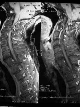
3 Figure 4. Precontrast MRI T1 images. Notice that the odontoid process is abnormally large, subluxated, and posteriorly located. Also notice basilar invagination, The tip of the odontoid process is in touch of the lower pons. A syringomyelic cavity is seen opposite the second vertebra and extends for only one spinal segment. The cerebellar tonsils are herniated below the level of the foramen magnum. The brain stem is abnormally elongated with herniation of the medullar below the level of the foramen magnum (Type II Chiari malformation). Interestingly the foramen of magendi (which is situated at the caudal end of the 4th ventricle and can easily be appreciated on the T1 MRI sagittal images) is appreciated as being markedly diminished in volume and transformed into a long slit that opened below the level of the foramen magnum. Notice the extradural space occupying lesion compressing and anteriorly displacing the upper cervical sinal cord. Figure 5. MRI T2,T1 images. Notice that the odontoid process is abnormally large, subluxated, and posteriorly located. Also notice basilar invagination, The tip of the odontoid process is in touch of the lower pons. A syringomyelic cavity is seen opposite the second vertebra and extends for only one spinal segment. The cerebellar tonsils are herniated below the level of the foramen magnum. The brain stem is abnormally elongated with herniation of the medullar below the level of the foramen magnum (Type II Chiari malformation). Interestingly the foramen of magendi (which is situated at the caudal end of the 4th ventricle and can easily be appreciated on the T1 MRI sagittal images) is appreciated as being markedly diminished in volume and transformed into a long slit that opened below the level of the foramen magnum. Notice the extradural space occupying lesion compressing and


.")
 is appreciated as being markedly diminished in volume and transformed into a long slit that opened below the level of the foramen magnum.")
4 anteriorly displacing the upper cervical sinal cord. Figure 6. MRI T2 images. Notice that the odontoid process is abnormally large, subluxated, and posteriorly located. Also notice basilar invagination, The tip of the odontoid process is in touch of the lower pons. A syringomyelic cavity is seen opposite the second vertebra and extends for only one spinal segment. The cerebellar tonsils are herniated below the level of the foramen magnum. The brain stem is abnormally elongated with herniation of the medullar below the level of the foramen magnum (Type II Chiari malformation). Interestingly the foramen of magendi (which is situated at the caudal end of the 4th ventricle and can easily be appreciated on the T1 MRI sagittal images) is appreciated as being markedly diminished in volume and transformed into a long slit that opened below the level of the foramen magnum. Notice the extradural space occupying lesion compressing and anteriorly displacing the upper cervical sinal cord. Figure 7. Post contrast MRI T1 image (A) Showing a dumb-bell cystic neurofibroma (Confirmed surgically) at C 2 vertebra compressing and displacing the spinal cord. Notice malalignment of the cervical vertebrae and the high odontoid process.
5 In the above report case we have a primary pathology, a secondery pathology and a tertiary pathology The primary pathology is basilar invagination (intracranial invagination of atlas and axis) Basilar impression/invagination is a congenital skeletal malformation that combines deformity of the osseous structures of the base of the skull with invagination of the foramen magnum and upper cervical spine into the posterior fossa. Basilar invagination is almost invariably associated with assimilation of atlas (atlantooccipital fusion). In most of the cases the odontoid precess is fixed and not subluxated. In the author experience, hydrosyringomyelia and syringobulbia are more likely to occur when the odontoid process is fixed. Metwally 12 regarded basilar invagination as a risk factor that increases the probability of a cervical syringomyelic cavity breaking upward into the medulla and forming syringobulbia. Probably the craniocervical junction is more crowded when the odontoid process is fixed than when it is mobile and subluxated, thus increasing the probably of the emergence of the secondary and the tertiary pathology as explained later. Complete intracranial invagination of the atlas and axis (basilar impression or invagination) is demonstrated in about 25% of cases 12 with congenital syringomyelia. Basilar impression/invagination acts by reducing the volume capacity of the posterior fossa and crowds the cerebellum thus producing cerebellar tonsillar herniation, thereby sitting up the substrate for the funneling of the CSF pressure waves into the central canal of the spinal cord thus creating hydrosyringomyelia. 12 Secondary pathology is cerebellar tonsillar herniation (Arnold Chiari malformation) Basilar impression/invagination acts by reducing the volume capacity of the posterior fossa and crowds the cerebellum thus producing cerebellar tonsillar herniation, thereby sitting up the substrate for the funneling of the CSF pressure waves into the central canal of the spinal cord thus creating hydrosyringomyelia. 12 Tertiary Pathology is hydrosyringomyelia Funneling of the CSF pressure waves into the central canal of the spinal cord thus creating hydrosyringomyelia. DIAGNOSIS: DIAGNOSIS: MULTIPLE CONGENITAL BONY AND SOFT TISSUE CRANIOCERVICAL ANOMALIES (BASILAR INVAGINATION, ASSIMILATION OF ATLAS, SUBLUXATION OF THE ODONTOID PROCESS, TYPE II CHIARI MALFORMATION) ASSOCIATED WITH EXTRADURAL CYSTIC NEUROFIBROMA AND A SINGLE SPINAL SEGMENT SYRINGOMYELIC CAVITY AT THE LEVEL OF THE NEUROFIBROMA (C 2 VERTEBRA) DISCUSSION DISCUSSION Three main points must be commented upon while discussing this case. The Basilar impression/invagination, The syringomyelia and the epidural neurofibroma Basilar impression/invagination Basilar impression is a congenital skeletal malformation that combines deformity of the osseous structures of the base of the skull with invagination of the foramen magnum and upper cervical spine into the posterior fossa. The foramen magnum is small and deformed. The floor of the posterior fossa and the odontoid process are displaced upward, further narrowing the foramen magnum and compressing the medulla and upper cervical spinal cord. Metabolic bone disease, such as osteomalacia, osteogenesis imperfecta, congenital hypothyroidism, rickets, and Paget's disease, may result in acquired basilar impression. Basilar impression is also seen in association with mucopolysaccharidoses II (Hurler syndrome) and Down's syndrome. The congenital condition is inherited as an


6 autosomal dominant trait with variable expression. Figure 8. Normal craniocervical junction


7 Figure 9. Normal atlas and axis, A, Axis & Atlas Articulated Posterior View (1-Dens, 2- Foramen transversarium or transverse foramen, 3- Spinous process or spine, 5- Anterior arch of atlas, 6- Posterior arch of atlas, 7- Transverse process, 8- Articular facet for base of skull). B, C1, 1st Cervical Vertebra or Atlas Superior View (1- Anterior tubercle, 2- Anterior arch, 3- Facet for dens, 4- Transverse process, 5- Foramen transversarium or transverse foramen, 6- Superior articular facet on lateral mass, 7- Posterior arch, 8- Posterior tubercle, 9- Vertebral foramen) C, C2, 2nd Cervical Vertebra or Axis Superior View (1- Dens, 2- Superior articular surface, 3- Body, 4- Pedicle, 5- Lamina, 6- Inferior articular process, 7- Transverse process, 8- Spinous process or spine) D, C2, 2nd Cervical Vertebra or Axis Posterior View ( 1- Dens, 2- Superior articular surface, 3- Body, 4- Pedicle, 5- Transverse process, 6- Lamina, 7- Bifid spinous process or spine, 8- Inferior articular surface) Basilar impression (or invagination) is the deformity of the bones of the base of the skull at the margin of the foramen magnum. The floor of the skull appears to be indented by the upper cervical spine; therefore, the tip of the odontoid is more cephalad. This increases the risk of neurologic damage from injury, circulatory embarrassment, or impairment of cerebrospinal fluid (CSF) flow. Two types of basilar impression exist: (1) primary, a congenital abnormality often associated with other anomalies such as atlanto-occipital fusion, hypoplasia of the atlas, bifid posterior arch of atlas, odontoid abnormalities, Klippel-Feil syndrome, and Goldenhar syndrome; and (2) secondary, a developmental condition usually attributed to softening of the bone, in which the deformity develops later in life. Table 1. Characteristics of basilar invagination Elevation of the floor of the posterior fossa (the floor the posterior fossa is convex upward) The upper cervical spine (atlas and axis) encroaches on the brainstem and spinal cord (the tip of the odontoid is more cephalad) as the base of the skull is displaced toward the cranial vault Partial to complete atlanto-occipital fusion (assimilation of atlas) The odontoid process might be partially of completely subluxated Compression of the cervico-medullary junction by the odontoid process, fibrous band, or tight dura at the level of foramen magnum Chiari malformation, and congenital syringomyelia might be associated with basilar invagination With basilar impression, the upper cervical spine encroaches on the brainstem and spinal cord as the base of the
.")
.")

8 skull is displaced toward the cranial vault. Motor and sensory disturbances are noted in 85% of individuals who are symptomatic. However, most affected patients remain asymptomatic until the second or third decade of life, when they may present with headache, neck ache, and neurologic compromise (prevalence of symptoms, 15%). 12 Figure 10. Basilar invagination. The base of the skull is elevated and convex upward with high odontoid process. Figure 11. A,B Plain X ray of the cervical spine in two cases with congenital syringomyelia. Notice widening of the cervical canal (A,B). Also notice the associated Klippel-Feil anomaly and basilar impression with high odontoid proceess (B) Complete intracranial invagination of the atlas and axis (basilar impression or invagination) is demonstrated in about 25% of cases with congenital syringomyelia. Basilar impression acts by reducing the volume capacity of the posterior fossa and crowds the cerebellum thus producing cerebellar tonsillar herniation, thereby sitting up the substrate for the funneling of the CSF pressure waves into the central canal of the spinal cord thus creating hydrosyringomyelia. In most cases with basilar impression the odontoid process was fixed with no evidence of
.")
 occur in about 5% of cases of congenital syringomyelia, however it might be present as an isolated phenomenon.")


9 subluxation. Congenital syringomyelia, when associated with basilar invagination, is more likely to be associated with extension of the cervical intramedullary cavitation into the lower brain stem (syringobulbia). 12 Metwally 12 regarded basilar invagination as a risk factor that increases the probability of a cervical syringomyelic cavity breaking upward into the medulla and forming syringobulbia. Assimilation of the atlas (complete fusion between the atlas and the occipital condyles) occur in about 5% of cases of congenital syringomyelia, however it might be present as an isolated phenomenon. In atlanto-occipital fusion, the odontoid process is abnormally high, subluxated and compressing the cervico-medullary junction posteriorly. The most significant finding in assimilation of the atlas with neurological symptoms is an odontoid process with abnormal size, in abnormal position and with abnormal mobility. Assimilation of the atlas is invariably present with basilar invagination and some degree of atlanto-occipital fusion is invariably present in basilar invagiantion. 10,11,12,15 When the atlas is fused with the occiput, flexion of the head results in partial forward subluxation of the fused atlas on the axis. Posterior displacement of the odontoid process then occurs, resulting in compression of the cervicomedullary junction. As the posterior luxation of the odontoid process is intermittent (only during head flexion), so intermittent compression of the cervico-medullary junction might result, initially, in intermittent neurological manifestations. 10 Figure 12. CT myelography showing basilar invagination with atlanto-occipital fusion. The odontoid process is more


10 cephalad than normal and subluxated and compressing the cervicomedullary junction posteriorly upon head flexion. Atlanto-occipital fusion, also termed "occipitalization of the atlas," is the most commonly recognized anomaly involving the craniovertebral junction, encountered in 0.14% to 0.25% of the population. 7 It is characterized by complete or partial fusion of the atlas to the occiput and likely results from failure of segmentation of the last occipital and first cervical sclerotomes and the hypochordal bow. Affected patients often have low hairline, short neck, and restricted neck movements, similar to the Klippel-Feil syndrome. 2 Associated congenital anomalies, present in 20% of patients, include incomplete clefting of the nasal cartilage, cleft palate, congenital external ear deformities, cervical ribs, hypospadias, and urinary tract anomalies. 2 Atlanto-occipital fusion invariably results in basilar invagination, less severe when only the anterior arch is fused to the basion and most pronounced when there is complete assimilation, including fusion of the lateral masses to the occipital condyles which, themselves, may be hypoplastic. Seventy percent of patients additionally have associated congenital nonsegmentation of C2-3 (partial or complete). 3 This places greater demands on the atlantoaxial joint leading to atlantoaxial instability in 50% of patients. 7,15 Sudden death in patients with atlanto-occipital assimilation has been reported. 1,6 Figure 13. Atlanto-occipital assimilation with atlantoaxial subluxation. A, Reconstructed midsagittal CT scan reveals complete assimilation of the anterior and posterior atlas arches to the occipital bone. The odontoid process (0) lies at the level of the foramen magnum. A = anterior arch; P = posterior arch; B = basion. B, Midsagittal Tl -weighted MR image reveals elongation and a "comma" configuration to the anterior lip of the foramen magnum produced by incorporation of the anterior atlas arch to the basion (dot). A similar finding is identified at the posterior margin of the foramen magnum. There is an increase in the width of the anterior atlantodental interval (dotted line), signifying incompetence of the transverse ligament, such that the displaced odontoid process (0) is producing marked compression of the cervicomedullary junction. Also note congenital incomplete segmentation of C2 and C3. As opposed to atlanto-occipital fusions, atlantoaxial fusions are rare. 5 Fusion of the anterior atlas arch to the odontoid process is frequently accompanied by hypoplasia of the posterior atlas arch and congenital block vertebrae in the lower cervical spine. The odontoid process itself may be relatively hypoplastic. 9 This fusion anomaly does not predispose to any instability. There is an increased incidence of atlantoaxial fusion anomalies associated with the Chiari I malformation, in addition to other malformations of the skull base and cervical vertebrae. 5 An unusual case of anterior atlas arch fusion to the basion and posterior atlas arch fusion to the posterior arch of C2, associated with insufficiency of the transverse ligament, has been reported. 8,15


11 Figure 14. Chiari I malformation with basilar invagination. A, Midsagittal Tl -weighted MR scan demonstrates caudal displacement of the cerebellar tonsils (solid arrow), almost to the C3-4 level. Marked basiocciput hypoplasia is present resulting in shortening of the clivus and basilar imagination with all of the anterior Cl arch, the odontoid process, and much of the axis body, positioned above Chamberlain's line (dotted line). Note how the basion (open arrow) is located at the midpons level such that, essentially, all of the medulla lies within the upper cervical spinal canal. Congenital nonsegmentation of C2-3 is also identified. B, Midsagittal Tl -weighted MR image of the thoracic spine demonstrates associated hydromyelia with multiple loculations. Clinical Characteristics Neurologic symptoms most often begin after age 10 years. Neck stiffness. progressive spasticity and weakness of the legs, occipital headaches, and difficulty walking are common complaints. Basilar impression should be considered in cases of progressive cerebellar ataxia or spastic paraparesis and brain- stem or cervical cord syndromes, which might be mistaken for multiple sclerosis, spinal cord tumors, Arnold-Chiari malformation, or syringomyelia. Basilar impression may be associated with the Arnold-Chiari malformation and aqueductal stenosis; therefore, hydrocephalus may occur. The progressive symptoms are attributed to pressure on the medulla and upper cervical spinal cord, similar to that of other craniocervical junction defects, such as Arnold-Chiari malformation. Interference with the local blood supply, as well as the direct compression of neural structures, has been postulated as a pathogenic mechanism. 10,11,12 Physical examination reveals a short neck, low hairline, painful torticollis, restricted neck motion, and a loss of the normal cervical spine lordosis. Neurologic findings may include downbeat or periodic alternating nystagmus, fixed or intermittent cranial nerve impairment, bilateral corticospinal tract signs, ataxia, and weakness or sensory loss in the hands. 12 Figure 15. A, Posterior photo of a patient with basilar invagination syndrome and an anomaly of the atlantooccipital fusion. The image shows an elevated left shoulder, a short webbed neck, and a low hairline. B, This patient has basilar invagination and an anomaly of the occipitocervical junction. The patient's flexion and extension after the oatlanto-occipital fusion is demonstrated. His rotation was very limited. C, basilar invagination. Flexion of the
.")


. The odontoid process is more cephalad than normal. The diagnosis is confirmed by a lateral roentgenogram of the skull.")
12 cervical spine in a patient who had an atlanto-occipital fusion. Figure 16. CT myelography showing basilar invagination associated with Arnold Chiari malformation and syringobulbia in (B). Notice the partial to complete atlanto-occipital fusion with no evidence of subluxation of the odontoid process. The odontoid process is more cephalad than normal. Figure 17. MRI T1 precontrast image (A) and CT bone window (B) showing basilar invagination associated with Arnold Chiari malformation and syringomyelia. The hyperintense odontoid process is seen indenting the brain stem (the high MRI T1 contract of the odontoid process is due to increased fat content). Notice the partial to complete atlanto-occipital fusion with no evidence of subluxation of the odontoid process. The odontoid process is more cephalad than normal. The diagnosis is confirmed by a lateral roentgenogram of the skull. The odontoid process extends above a line drawn from the hard palate to the posterior edge of the foramen magnum (Chamberlain's line 13 ). MR imaging and CT clearly define the existence of neural compression.


13 Figure 18. CT myelography showing basilar invagination associated with Arnold Chiari malformation. Notice the partial to complete atlanto-occipital fusion with no evidence of subluxation of the odontoid process. The odontoid process is more cephalad than normal. Management. Surgical decompression of the posterior fossa and the upper cervical cord is the treatment for symptomatic patients with congenital basilar impression. Decompressive surgery should be avoided in children with acquired basilar impression resulting from a metabolic bone disease because surgery may worsen the deformity. 12 Figure 19. A, Showing incomplete fusion of posterior arch of atlas and complete fusion of anterior arch with the occipital bone on the left side. B, Showing fusion of posterior and anterior arch of atlas with the occipital bone. The associated syringomyelic cavity The intramedullary syringomyelic cavity demonstrated in this patient is a very shot one (one spinal segment). There are two explanation for the associated syringomyelic cavity. 1-The cavity is secondary to the cerebellar tonsilar herniation and represent the starting point that will ultimately extends to involve the cervico-dorsal spinal cord. Cerebellar tonsilar herniation produces the following anatomical variations as follows Stenosis of the foramen magnum and the foramen of Magendie. This is produced either mechanically when the foramen magnum is crowded by the descent of the cerebellar tonsils through it or by the adhesions that were demonstrated between the cerebellar tonsils and the posterior aspect of the cervico-medullary regions. The foramen of Magendie is transformed into an abnormally elongated slit-like channel that opened below the foramen magnum in all patients.


14 The foramen of Magendie now lies below the level of the calamus scriptorious (the outflow channel of the central canal of the spinal cord). This occurs due to tonsillar ectopia as the foramen of Magendie opens between the two cerebellar tonsils. Normally the ventricular CSF arterial pulse wave escapes through the foramen of Magendie into the subarachnoid spaces and is progressively damped as it passes down behind the spinal cord through the foramen magnum. In this way the central canal of the spinal cord is bypassed and is not subjected to the ventricular fluid pulse wave. The central canal of the spinal cord is normally a potentially distensible vestigial structure. When the foramen of Magendie lies below the foramen magnum, the CSF pulse waves are funneled into the central canal of the spinal cord, distending it and finally creating hydrosyringomyelia. 11,12,15 Figure 20. The anatomic substrate of congenital syringomyelia and/or hydromyelia is based upon cerebellar tonsillar ectopia in fetal life. Blockade of the foramen of Magendie and stenosis of the foramen magnum will funnel the CSF arterial pulse waves into the spinal canal, distending it and eventually creating hydrosyringomyelia The anatomical aberrations induced by hindbrain herniation will block the normal pathway for the escape of the ventricular fluid arterial pulse wave and redirect it towards the calamus scriptorious. The CSF arterial pulse wave will then be funneled through the calamus scriptorious into the central canal of the spinal cord dilating it and eventually creating hydrosyringomyelia. Accordingly a direct cause-effect relationship could be established between hindbrain herniation and the development of hydrosyringomyelia. 11,12,15 Accordingly hindbrain herniation should constitute the anatomical substrate of hydrosyringomyelia and the cause of syringomyelia should be sought not in the spinal cord but in the herniation of the hindbrain. The diagnosis of congenital syringomyelia is not possible unless this the aetiological factor (hindbrain herniation) is demonstrated radiologically. 11,12,15 However the demonstrated syringomyelic cavity is a very short one and this is not the case for congenital hydrosyringomyelia. In congenital hydrosyringomyelia the syringomyelic cavity is usually a long multisegmental one that involves most of the cervico-dorsal spinal cord. However the demonstrated intramedullary cavity could be just the beginning, the starting point, that will ultimately extends further upward and downwards as the disease progresses. The pathological process, in this patient, was probably caught early enough to demonstrate such a small syringomyelic cavity. 2- The second explanation for the syringomyelia cavity is that it is secondary to the epidural neurofibromas (neoplastic syringomyelia) The cavity is demonstrated anatomically opposite to an epidural neurofibroma. Spinal cord cavitations are known to coexist with primary spinal cord neoplasms. They could be secondary to CSF flow obstructions and in this way they resemble obstructive hydrocephalus. The dumbbell neurofibroma Neurofibromas are known to coexist with craniocervical anomalies and hydrosyringomyelia 16,17 Some even regarded the craniocervical anomalies and the associated syringomyelia cavity (when associated with optic nerve gliomas or neurofibromas for example) as a part of neurofibromatosis type 1. 18
15 SUMMARY SUMMARY Occipitalization of the atlas or atlanto-occipital fusion is one of the most common osseous anomalies of the craniovertebral junction. Occipitalization represents the most cephalic blocked vertebra encountered in the spine. It is characterized by complete or partial fusion of the bony ring of the atlas to the base of the occipital bone. The patients with craniovertebral joint anomalies exhibit the first neurological signs and symptoms usually no sooner than the second decade of life. In patients with the atlanto-occipital fusion, the clinical findings suggest that the major neurological compression is due to the odontoid projection into the foramen magnum. The signs and symptoms of pyramidal tract, anterior bulbar and cranial nerve involvement may be present. Assimilation of atlas is an osseous abnormality, which occurs in the base of skull in the region of foramen magnum. The union of the atlas with the occipital bone constitutes the anomaly. There may be partial or complete union. The occipital bone is derived from basioccipital, exoccipital and supraoccipital portions, all of which surround the foramen magnum. The basiocciput goes on to develop into four occipital somites. The caudal portion of the fourth occipital somite goes onto fuse with the cranial portion of the first cervical somite to form the proatlas; the proatlas is assimilated into the occiput to form the articular condyles and the tip of the odontoid process. The caudal half of the first cervical somite along with the cranial part of the second cervical somite goes on to form the atlas and the odontoid process of the axis. A paracondylar process represents vestiges of the cranial half of the first cervical sclerotome. This formation is referred to as a caudal shifting (a vertebra taking on the characteristics of its caudal neighbor) where the occipital vertebra separates from the occiput. The symptoms and signs of pyramidal tract, anterior bulbar and cranial nerves involvement may be present. Less commonly, if the compression occurred posteriorly by the posterior lip of the foramen magnum, are the symptoms and signs related to the involvement of the posterior column of spinal cord. Patients with occipitalization of the atlas have short neck and restricted neck movements. Symptoms referable to the vertebral artery compression, such as dizziness, seizures, mental deterioration, and syncope may occur alone or in combination with those of the spinal cord compression. Any morphological and structural alteration of the cervical spine may lead to stenosis or substenosis of the vertebral arterial circulation and hence to brain stem anoxia. Fusion between occiput and atlas occurs anteriorly between the arch and the rim of the foramen with some segment of the posterior arch of atlas present in some instances. This fragment can frequently constrict the spinal canal causing intermittent symptoms depending on the position of the head. Although atlanto-occipital fusion is a congenital condition, many patients do not develop the symptoms until the second decade of life. This may be due to a gradual increasing degree of ligamentous laxity and instability with aging. The onset of clinical symptoms can be sudden and precipitated by relatively minor trauma, the most common course is a progressive, but sudden onset or instant death has also been reported. Addendum A new version of this PDF file (with a new case) is uploaded in my web site every week (every Saturday and remains available till Friday.) To download the current version follow the link " You can also download the current version from my web site at " To download the software version of the publication (crow.exe) follow the link: The case is also presented as a short case in PDF format, to download the short case follow the link:
16 At the end of each year, all the publications are compiled on a single CD-ROM, please contact the author to know more details. Screen resolution is better set at 1024*768 pixel screen area for optimum display REFERENCES References 1. Bezi L: Assimilation of the atlas and compression of medulla. Arch Pathol 12: , HensingerRN: Anomalies of the atlas. In Cervical Spine Research Society Editorial Committee (eds): The Cervical Spine, ed 2. Philadelphia, JB Lippincott, 1989, p McRae DL: Bony abnormalities in the region of the foramen magnum: Correlation of the anatomic and neurologic findings. Acta Radiol 40: , Naidich TP, McLone DC, Harwood-Nash DC: Malformations of the craniocervical junction. In Newton TH, Potts DG (eds): Computed Tomography of the Spine and Spinal Cord. San Anselmo, CA, Clavedel Press, 1983, p Olbrantz K, Bohrer SP: Fusion of the anterior arch of the atlas and dens. Skeletal Radiol 12:21-22, Vakili ST, Aguilar JC, Muller J: Sudden unexpected death associated with atlanto-occipital fusion. Am j Forensic Med Pathol 6:39-43, von Tbrklus D, Gehle W: The Upper Cervical Spine. New York, Grune and Stratton, Wackenheim A: Occipitalization of the ventral part and vertebralization of the dorsal part of the atlas with insufficiency of the transverse ligament. Neuroradiology 24:45-47, Wackenheim A: Cervico-occipital Joint. Berlin, Springer-Verlag, Metwally MYM: Imaging of syringomyelia, a comparative study. Read at the Scientific Meeting of the Egyptian Society of neurology, Psychiatry and Neurosurgery October Metwally MYM: The congenital craniocervical anomalies and their role in the pathogenesis of congenital hydrosyringomyelia. Ain Shams medical journal, Vol. 50, number 4,5,6 pp Metwally MYM: Congenital syringobulbia, a radiological study with clinical correlation. Ain Shams medical journal,vol 51, No 1,2,3 pp Chamberlain WE: Basilar impression (platybasia): A bizarre developmental anomaly of the occipital bone and upper cervical spine with striking and misleading neurologic manifestations. Yale J Biol Med 1939; 11: Charnes LR, Marini JC: Communicating hydrocephalus, basilar invagination, and other neurologic features in osteogenesis imperfecta. Neurology 43: , Metwally, MYM: Textbook of neurimaging, A CD-ROM publication, (Metwally, MYM editor) WEB-CD agency for electronic publishing, version 9.1a January Fernandez JA, Calleja PB, Paseual CI : Syringomyelia, Chiari's malformation and scoliosis in a patient with type 1 neurofibromatosis. An Esp Paeditr 1998; 48 : Shenoy SN, Raja A. Cystic cervical intramedullary schwannoma with syringomyelia. Neurol India 2005;53: Chakravarty A, Bhargava A, Nandy S. A patient with optic pathway glioma, scoliosis, Chiari type I
17 malformation and syringomyelia : is it Neurofibromatosis type 1?. Neurol India 2002;50:520
Cranial Vertebral Junction Anatomy and Pathology
 Cranial Vertebral Junction Anatomy and Pathology October 2016 Mary Scanlon MD FACR Goals and Objective Goal-Understand CVJ anatomy & pathology Objective-After attending this lecture you will be able to
Brain Imaging. Bearbeitet von Klaus Sartor, Stefan Hähnel, Bodo Kress
 Brain Imaging Bearbeitet von Klaus Sartor, Stefan Hähnel, Bodo Kress 1. Auflage 2007. Taschenbuch. 312 S. Paperback ISBN 978 3 13 143961 1 Format (B x L): 12,5 x 19 cm Weitere Fachgebiete > Medizin > Sonstige
Ligaments of the vertebral column:
 In the last lecture we started talking about the joints in the vertebral column, and we said that there are two types of joints between adjacent vertebrae: 1. Between the bodies of the vertebrae; which
Malformations of the cranio-cervical junction: basilar impression Gonzalo Bertullo 1, Viviana Cabrera 2
 Malformations of the cranio-cervical junction: basilar impression Gonzalo Bertullo 1, Viviana Cabrera 2 Abstract Cranio-cervical junction abnormalities are a rare combination of congenital or acquired
1 Normal Anatomy and Variants
 1 Normal Anatomy and Variants 1.1 Normal Anatomy MR Technique. e standard MR protocol for a routine evaluation of the spine always comprises imaging in sagittal and axial planes, while coronal images are
May have excessive movement in the unfused segment to compensate. Flexion extension better preserved than lateral bend or rotation
 IV CONGENITAL SPINE KLIPPEL FLAIL SYNDROME Prevalence 0.60% Mainly around upper 3 vertebrae [75%] Commonest: C2 3 Lower Cervical spine fusion may be associated with syndromes: Fetal alcohol syndrome Goldenhar
Chiari Malformations. Google. Objectives Seventh Annual NKY TBI Conference 3/22/13. Kerry R. Crone, M.D.
 Chiari Malformations Kerry R. Crone, M.D. Professor of Neurosurgery and Pediatrics University of Cincinnati College of Medicine University of Cincinnati Medical Center Cincinnati Children s Hospital Medical
Cervical Spine Anatomy and Biomechanics. Typical Cervical Vertebra C3 6. Typical Cervical Vertebra Anterior 10/5/2017
 Cervical Spine Anatomy and Biomechanics Typical Cervical Vertebra C3 6 Small, relatively broad body Bifid SpinousProcess Long and narrow laminae Spinal Canal: large, triangular; remarkably consistent dimensions
Involvement of the spine is common in rheumatoid. Incidence been reported to be 85% radiologically but only 30% have neurological signs and symptoms.
 RHEUMATOID SPINE Involvement of the spine is common in rheumatoid. Incidence been reported to be 85% radiologically but only 30% have neurological signs and symptoms. When neurology is present it may manifest
The craniocervical junction
 Anver Jameel, MD The craniocervical junction A biomechanical and anatomical unit that extends from the skull base to C2 Includes the clivus, foramen magnum and contiguous occipital bone, the occipital
THE VERTEBRAL COLUMN. Average adult length: In male: about 70 cms. In female: about 65 cms.
 THE VERTEBRAL COLUMN Average adult length: In male: about 70 cms. In female: about 65 cms. 1 Vertebral Column (Regions and Curvatures) Curvatures of the vertebral column: A. Primary curvature: C-shaped;
Imaging of Cervical Spine Trauma Tudor H Hughes, M.D.
 Imaging of Cervical Spine Trauma Tudor H Hughes, M.D. General Considerations Most spinal fractures are due to a single episode of major trauma. Fatigue fractures of the spine are unusual except in the
Vertebral Column. Backbone consists of 26 vertebrae. Five vertebral regions. Cervical
 Vertebral Column Backbone consists of 26 vertebrae. Five vertebral regions Cervical vertebrae (7) in the neck. Thoracic vertebrae (12) in the thorax. Lumbar vertebrae (5) in the lower back. Sacrum (5,
Cervical Spine Disorder in Children
 Cervical Spine Disorder in Children Embryology of Vertebra Para axial Mesoderm 42~44, 42~44 somite 4 occipital somite 8 cervical somite 12 thoracic somite 5 lumbar somite 10 sacral somite 5 coccyx somite
River North Pain Management Consultants, S.C., Axel Vargas, M.D., Regional Anesthesiology and Interventional Pain Management.
 River North Pain Management Consultants, S.C., Axel Vargas, M.D., Regional Anesthesiology and Interventional Pain Management. Chicago, Illinois, 60611 Phone: (888) 951-6471 Fax: (888) 961-6471 Clinical
2. The vertebral arch is composed of pedicles (projecting from the body) and laminae (uniting arch posteriorly).
 and laminae (uniting arch posteriorly).") VERTEBRAL COLUMN 2018zillmusom I. VERTEBRAL COLUMN - functions to support weight of body and protect spinal cord while permitting movements of trunk and providing for muscle attachments. A. Typical vertebra
VERTEBRAL COLUMN 2018zillmusom I. VERTEBRAL COLUMN - functions to support weight of body and protect spinal cord while permitting movements of trunk and providing for muscle attachments. A. Typical vertebra
VERTEBRAL COLUMN VERTEBRAL COLUMN
 VERTEBRAL COLUMN FUNCTIONS: 1) Support weight - transmits weight to pelvis and lower limbs 2) Houses and protects spinal cord - spinal nerves leave cord between vertebrae 3) Permits movements - *clinical
AXIAL SKELETON FORM THE VERTICAL AXIS OF THE BODY CONSISTS OF 80 BONES INCLUDES BONES OF HEAD, VERTEBRAL COLUMN, RIBS,STERNUM
 AXIAL SKELETON FORM THE VERTICAL AXIS OF THE BODY CONSISTS OF 80 BONES INCLUDES BONES OF HEAD, VERTEBRAL COLUMN, RIBS,STERNUM APPENDICULAR SKELETON BONES OF THE FREE APPENDAGES & THEIR POINTS OF ATTACHMENTS
Inferior view of the skull showing foramina (Atlas of Human Anatomy, 5th edition, Plate 12)
") Section 1 Head and Neck Skull, Basal View Incisive foramen Choanae Foramen ovale Foramen lacerum Foramen spinosum Carotid canal Jugular fossa Mastoid process Inferior view of the skull showing foramina
Section 1 Head and Neck Skull, Basal View Incisive foramen Choanae Foramen ovale Foramen lacerum Foramen spinosum Carotid canal Jugular fossa Mastoid process Inferior view of the skull showing foramina
Copyright 2010 Pearson Education, Inc.
 E. VERTEBRAL COLUMN 1. The vertebral column extends from the skull to the pelvis and forms the vertical axis of the skeleton. 2. The vertebral column is composed of vertebrae that are separated by intervertebral
You have 24 vertebrae in your spinal column. Two are special enough to be individually named.
 You have 24 vertebrae in your spinal column. Two are special enough to be individually named. Your atlas (C01) and axis (C02) are very important vertebrae. Without them, head and neck movement would be
Axial Skeleton: Vertebrae and Thorax
 Axial Skeleton: Vertebrae and Thorax Function of the vertebral column (spine or backbone): 1) 2) 3) Composition of Vertebral column The vertebral column is formed by 33 individual vertebrae (some of which
Musculoskeletal Development and Sports Injuries in Pediatric Patients
 Dynamic Chiropractic October 21, 2010, Vol. 28, Issue 22 Musculoskeletal Development and Sports Injuries in Pediatric Patients By Deborah Pate, DC, DACBR Physical activity is extremely important for everyone,
Any of the vertebra in the cervical (neck) region of the spinal column. The cervical vertebra are the smallest vertebra in the spine, reflective of th
 region of the spinal column. The cervical vertebra are the smallest vertebra in the spine, reflective of th") Any of the vertebra in the cervical (neck) region of the spinal column. The cervical vertebra are the smallest vertebra in the spine, reflective of the fact that they support the least load. In humans,
Any of the vertebra in the cervical (neck) region of the spinal column. The cervical vertebra are the smallest vertebra in the spine, reflective of the fact that they support the least load. In humans,
Spinal Cord Injuries: The Basics. Kadre Sneddon POS Rounds October 1, 2003
 Spinal Cord Injuries: The Basics Kadre Sneddon POS Rounds October 1, 2003 Anatomy Dorsal columntouch, vibration Corticospinal tract- UMN Anterior horn-lmn Spinothalamic tractpain, temperature (contralateral)
INDEPENDENT LEARNING: DISC HERNIATION IN THE NATIONAL FOOTBALL LEAGUE: ANATOMICAL FACTORS TO CONSIDER IN REVIEW
 INDEPENDENT LEARNING: DISC HERNIATION IN THE NATIONAL FOOTBALL LEAGUE: ANATOMICAL FACTORS TO CONSIDER IN REVIEW CDC REPORT - CAUSES OF DISABILITY, 2005 REVIEW QUESTIONS ABOUT DISC HERNIATION IN THE NATIONAL
Human Anatomy and Physiology - Problem Drill 07: The Skeletal System Axial Skeleton
 Human Anatomy and Physiology - Problem Drill 07: The Skeletal System Axial Skeleton Question No. 1 of 10 Which of the following statements about the axial skeleton is correct? Question #01 A. The axial
Role of MRI in Selection of Patients for Surgery and Assessing the Post Operative Outcome in Chiari 1 Malformation
 DOI: 10.7860/IJARS/2017/13599:2222 Radiology Section Original Article Role of MRI in Selection of Patients for Surgery and Assessing the Post Operative Outcome in Chiari 1 Malformation Rajesh Kumar V,
Chapter 7. Skeletal System
 Chapter 7 Skeletal System 1 Skull A. The skull is made up of 22 bones: 8 cranial bones, 13 facial bones, and the mandible. B. The Cranium encloses and protects the brain, provides attachments for muscles,
Spinal Imaging. Bearbeitet von Herwig Imhof. 1. Auflage Taschenbuch. 312 S. Paperback ISBN Format (B x L): 12,5 x 19 cm
: 12,5 x 19 cm") Spinal Imaging Bearbeitet von Herwig Imhof 1. Auflage 2007. Taschenbuch. 312 S. Paperback ISBN 978 3 13 144071 6 Format (B x L): 12,5 x 19 cm Weitere Fachgebiete > Medizin > Sonstige Medizinische Fachgebiete
Spinal Imaging Bearbeitet von Herwig Imhof 1. Auflage 2007. Taschenbuch. 312 S. Paperback ISBN 978 3 13 144071 6 Format (B x L): 12,5 x 19 cm Weitere Fachgebiete > Medizin > Sonstige Medizinische Fachgebiete
Chiari III Joseph Junewick, MD FACR
 Chiari III Joseph Junewick, MD FACR 07/02/2010 History Newborn with suboccipital mass. Diagnosis Chiari III Additional Clinical Surgery-Skin covered suboccipital cystic mass confined by the dura. Pathology-Leptomeningeal
Clarification of Terms
 Clarification of Terms The Spine, Spinal Column, and Vertebral Column are synonymous terms referring to the bony components housing the spinal cord Spinal Cord = made of nervous tissue Facet = a small,
Clarification of Terms
 Clarification of Terms The Spine, Spinal Column, and Vertebral Column are synonymous terms referring to the bony components housing the spinal cord Spinal Cord = made of nervous tissue Facet = a small,
Presented by : Shashwat Mishra
 Presented by : Shashwat Mishra Named after Hans Chiari (1851 1916). Professor of Pathology in Prague, Czechoslovakia, Paper entitled Concerning alterations in the cerebellum resulting from cerebral hydrocephalus
SUBAXIAL CERVICAL SPINE TRAUMA- DIAGNOSIS AND MANAGEMENT
 SUBAXIAL CERVICAL SPINE TRAUMA- DIAGNOSIS AND MANAGEMENT 1 Anatomy 3 columns- Anterior, middle and Posterior Anterior- ALL, Anterior 2/3 rd body & disc. Middle- Posterior 1/3 rd of body & disc, PLL Posterior-
Idiopathic cervical syringomyelia can be associated. Pediatric Chiari malformation Type 0: a 12-year institutional experience.
 J Neurosurg J Neurosurg Pediatrics Pediatrics 8:000 000, 8:1 5, 2011 Pediatric Chiari malformation Type 0: a 12-year institutional experience Clinical article Joshua J. Chern, M.D., Ph.D., Amber J. Gordon,
ANATOMY & PHYSIOLOGY I Laboratory Version B Name Section. REVIEW SHEET Exercise 10 Axial Skeleton
 ANATOMY & PHYSIOLOGY I Laboratory Version B Name Section REVIEW SHEET Exercise 10 Axial Skeleton 1 POINT EACH. THE SKULL MULTIPLE CHOICE 1. The major components of the axial skeleton include the 7. The
Clarification of Terms
 Clarification of Terms The Spine, Spinal Column, and Vertebral Column are synonymous terms referring to the bony components housing the spinal cord Spinal Cord = made of nervous tissue Facet = a small,
Skeletal System. Axial Division
 Skeletal System Axial Division The Axial Skeleton You will see that each bone has special features (overviewed in section I below) that provide Sites of Attachment (for muscles, ligaments, tendons, etc.)
Atlantoaxial joint distraction as a treatment for basilar invagination: A report of an experience with 11 cases
 Original Article Atlantoaxial joint distraction as a treatment for basilar invagination: A report of an experience with 11 cases Atul Goel, Abhidha Shah Department of Neurosurgery, King Edward 7 th Memorial
Human Anatomy - Problem Drill 06: The Skeletal System Axial Skeleton & Articualtions
 Human Anatomy - Problem Drill 06: The Skeletal System Axial Skeleton & Articualtions Question No. 1 of 10 Instructions: (1) Read the problem and answer choices carefully, (2) Work the problems on paper
Caudal Occipital Malformation Syndrome
 Caudal Occipital Malformation Syndrome Robert L Bergman, DVM, MS, Dip ACVIM Carolina Veterinary Specialists With the increasing availability of MRI, as well as improved education of pet owners, caudal
VERTEBRAL COLUMN ANATOMY IN CNS COURSE
 VERTEBRAL COLUMN ANATOMY IN CNS COURSE Vertebral body Sections of the spine Atlas (C1) Axis (C2) What type of joint is formed between atlas and axis? Pivot joint What name is given to a fracture of both
PEDIATRIC CERVICAL SPINE DEFORMITY.
 PEDIATRIC CERVICAL SPINE DEFORMITY www.fisiokinesiterapia.biz DEVELOPMENT EACH VERTEBRAE DEVELOP FROM THE CAUDAL AND CRANIAL ½ OF 2 SCLEROTOMES C1 and C2 primitive centrum fuse to form odontoid process
Chapter 7 Craniocervical Developmental Anatomy and Its Implications
 Chapter 7 Craniocervical Developmental Anatomy and Its Implications Arnold H. Menezes, M.D. INTRODUCTION Congenital and developmental osseous anomalies and abnormalities that affect the craniovertebral
International Journal of Pharma and Bio Sciences
 Original Research Article Anatomy and Allied sciences International Journal of Pharma and Bio Sciences ISSN 0975-6299 SCREENING FOR ANOMALIES IN OCCIPITO-CERVICAL JUNCTION USING CRANIOMETRY IN COMPUTED
Torticollis in children - differential diagnosis approach
 Torticollis in children - differential diagnosis approach Poster No.: C-2101 Congress: ECR 2015 Type: Educational Exhibit Authors: A. Eran, A. Ilivitzki; Haifa/IL Keywords: Neoplasia, Infection, Education,
Common fracture & dislocation of the cervical spine. Theerachai Apivatthakakul Department of Orthopaedic Chiangmai University
 Common fracture & dislocation of the cervical spine Theerachai Apivatthakakul Department of Orthopaedic Chiangmai University Objective Anatomy Mechanism and type of injury PE.and radiographic evaluation
human anatomy 2015 lecture four Dr meethak ali ahmed neurosurgeon
 The Vertebral Column the vertebral columnis central pillar of the body.it serve to protect the spinal cord and support the weight of the head trunk, which it transmits to the hip bones & the lower limbs.
The MRI in Arnold-Chiari Syndrome I and Idiopathic Syringomyelia
 The MRI in Arnold-Chiari Syndrome I and Idiopathic Syringomyelia Author: Miguel B. Royo Salvador, MD, PhD Introduction Magnetic resonance imaging (MRI) is a crucial diagnostic tool for men, women, and
Anatomy of the Nervous System. Brain Components
 Anatomy of the Nervous System Brain Components NERVOUS SYSTEM INTRODUCTION Is the master system of human body, controlling the functions of rest of the body systems Nervous System CLASSIFICATION A. Anatomical
Chapter 7 Part B The Skeleton
 Chapter 7 Part B The Skeleton 7.2 The Vertebral Column General Characteristics Extends from skull to pelvis Also called spine or spinal column Functions to transmit weight of trunk to lower limbs, surround
Arnold Chiari Malformation - A hospital based autopsy study
 Rapotra Megha et al / International Journal of Biomedical Research 2017; 8(05): 250-254. 250 International Journal of Biomedical Research ISSN: 0976-9633 (Online); 2455-0566 (Print) Journal DOI: https://dx.doi.org/10.7439/ijbr
Structure and Function of the Vertebral Column
 Structure and Function of the Vertebral Column Posture Vertebral Alignment Does it really matter? Yes it does! Postural Curves The vertebral column has a series of counterbalancing curves posterior anterior
HEAD AND NECK IMAGING. James Chen (MS IV)
") HEAD AND NECK IMAGING James Chen (MS IV) Anatomy Course Johns Hopkins School of Medicine Sept. 27, 2011 OBJECTIVES Introduce cross sectional imaging of head and neck Computed tomography (CT) Review head
HEAD AND NECK IMAGING James Chen (MS IV) Anatomy Course Johns Hopkins School of Medicine Sept. 27, 2011 OBJECTIVES Introduce cross sectional imaging of head and neck Computed tomography (CT) Review head
Chiari bridges review Chiari Treatments & Potential Pitfalls
 Chiari bridges review Chiari Treatments & Potential Pitfalls Once diagnosed, you will usually be referred to a specialist (not a Chiari Specialist, but an everyday, run-of-the-mill neurologist or neurosurgeon).
MDCT and MRI evaluation of cervical spine trauma
 Insights Imaging (2014) 5:67 75 DOI 10.1007/s13244-013-0304-2 PICTORIAL REVIEW MDCT and MRI evaluation of cervical spine trauma Michael Utz & Shadab Khan & Daniel O Connor & Stephen Meyers Received: 10
W IDENING of an intervertebral foramen
 Agenesis of a Pedicle in the Cervical Spine LESLIE i. ZATZ, M.D., PETER W. BURGESS, M.D. AND John W. HANBERY, M.D. Departments of Radiology and of Surgery, Neurosurgical Division, Stanford University School
The vault bones Frontal Parietals Occiput Temporals Sphenoid Ethmoid
 The Vertebral Column Head, Neck and Spine Bones of the head Some consider the bones of the head in terms of the vault bones and the facial bones hanging off the front of them The vault bones Frontal Parietals
S YRINGOMYELIA and syringobulbia are
 Syringomyelia: A Look at Surgical Therapy J. GRAFTON LOVE, M.D. AND RICHARD A. OLAFSON, M.D. Mayo Clinic and Mayo Foundation, Section of Neurologic Surgery, and Mayo Graduate School of Medicine, University
o Diaphysis o Area where red marrow is found o Area where yellow marrow is found o Epiphyseal plate AXIAL SKELETON Skull
 64 Anatomy & Physiology Coloring Workbook 7. Figure 5-2A is a midlevel, cross-sectional view of the diaphysis of the femur. Label the membrane that lines the cavity and the membrane that covers the outside
THEME 2. VERTEBRAE (GENERAL DATA). CERVICAL, THORACIC AND LUMBAR VERTEBRAE. SACRUM. COCCYX. THE VERTEBRAL COLUMN AS A WHOLE
. CERVICAL, THORACIC AND LUMBAR VERTEBRAE. SACRUM. COCCYX. THE VERTEBRAL COLUMN AS A WHOLE") THEME 2. VERTEBRAE (GENERAL DATA). CERVICAL, THORACIC AND LUMBAR VERTEBRAE. SACRUM. COCCYX. THE VERTEBRAL COLUMN AS A WHOLE Osteology of the Vertebral Column Bone Description vertebra Notes a vertebra
THEME 2. VERTEBRAE (GENERAL DATA). CERVICAL, THORACIC AND LUMBAR VERTEBRAE. SACRUM. COCCYX. THE VERTEBRAL COLUMN AS A WHOLE Osteology of the Vertebral Column Bone Description vertebra Notes a vertebra
Significance of Cerebellar Tonsillar Position on MR
 795 Significance of Cerebellar Tonsillar Position on MR A. J. Barkovich 1. 3 F. J. Wippold 2. 3 J. L. Sherman 3. 4 C. M. Citrin 4. 5 It has been noted that a low degree of ectopia of the cerebellar tonsils
Copyright 2010 Pearson Education, Inc. Copyright 2010 Pearson Education, Inc. Figure Sectioned spinous process. Interspinous.
 PowerPoint Lecture Slides prepared by Janice Meeking, Mount Royal College C H A P T E R 7 The Skeleton: Part B Vertebral Column Transmits weight of trunk to lower limbs Surrounds and protects spinal cord
Spinal Dynamics I: The Axio-atlanto-occipital Assemblage
 Spinal Dynamics I: The Axio-atlanto-occipital Assemblage Bones interact through joints. The relative placements of bones across joints determine how they move in space. In this section we will consider
Spine. Neuroradiology. Spine. Spine Pathology. Distribution of fractures. Radiological algorithm. Role of radiology 18/11/2015
 Spine Neuroradiology Spine Prof.Dr.Nail Bulakbaşı X Ray: AP/L/Oblique Vertebra & disc spaces CT & CTA Vertebra, discs, vessels MRI & MRA Vertebra, disc, vessels, meninges Spinal cord & nerves Myelography
LUMBAR SPINAL STENOSIS
 LUMBAR SPINAL STENOSIS Always occurs in the mobile segment. Factors play role in Stenosis Pre existing congenital or developmental narrowing of the lumbar spinal canal Translation of one anatomic segment
Fractures of the thoracic and lumbar spine and thoracolumbar transition
 Most spinal column injuries occur in the thoracolumbar transition, the area between the lower thoracic spine and the upper lumbar spine; over half of all vertebral fractures involve the 12 th thoracic
Anatomy of the Spine. Figure 1. (left) The spine has three natural curves that form an S-shape; strong muscles keep our spine in alignment.
 The spine has three natural curves that form an S-shape; strong muscles keep our spine in alignment.") 1 2 Anatomy of the Spine Overview The spine is made of 33 individual bony vertebrae stacked one on top of the other. This spinal column provides the main support for your body, allowing you to stand upright,
1 2 Anatomy of the Spine Overview The spine is made of 33 individual bony vertebrae stacked one on top of the other. This spinal column provides the main support for your body, allowing you to stand upright,
Chiari malformations. A fact sheet for patients and carers
 A fact sheet for patients and carers Chiari malformations This fact sheet provides information on Chiari malformations. It focuses on Chiari malformations in adults. Our fact sheets are designed as general
The Trunk and Spinal Column Kinesiology Cuneyt Mirzanli Istanbul Gelisim University
 The Trunk and Spinal Column Kinesiology Cuneyt Mirzanli Istanbul Gelisim University The Trunk and Spinal Column Vertebral column 24 articulating vertebrae 31 pairs of spinal nerves Abdominal muscles some
Journal of Biological and Chemical Research An International Peer Reviewed / Refereed Journal of Life Sciences and Chemistry
 Atlanto-Occipital Synostosis: Embryological Basis By Deepshikha Kori, Archana Rani, R.K. Diwan and Ganpat Prasad ISSN 0970-4973 Print ISSN 2319-3077 Online/Electronic Journal Impact Factor: 4.275 Global
TUMOURS IN THE REGION OF FORAMEN MAGNUM
 TUMOURS IN THE REGION OF FORAMEN MAGNUM Abstract Pages with reference to book, From 119 To 122 Naim-ur-Rahman ( Department of Neurosurgery, Rawalpindi Medical College, Rawalpindi. ) A very unusual case
Anatomy and Physiology II. Spine
 Anatomy and Physiology II Spine Bones and Other Structures Vertibrae Contains Cervical, Thoracic, Lumbar, Sacral and Coccygeal regions We use Capital letters to refer to these (C, T, L, S, and Co) and
Craniovertebral Junction Realignment for the Treatment of Basilar Invagination With Syringomyelia: Preliminary Report of 12 Cases
 Neurol Med Chir (Tokyo) 45, 512 518, 2005 Craniovertebral Junction Realignment for the Treatment of Basilar Invagination With Syringomyelia: Preliminary Report of 12 Cases Atul GOEL and Praveen SHARMA
Spinal canal stenosis Degenerative diseases F 06
 What is spinal canal stenosis? The condition known as spinal canal stenosis is a narrowing (stenosis) of the spinal canal that in most cases develops due to the degenerative (wear-induced) deformation
Normal Value of Skull Base Angle. Using the Modified Magnetic Resonance Imaging Technique in Thai Population
 Open Access Journal of Oral Health and Craniofacial Science Research Article Normal Value of Skull Base Angle ISSN 2573-6191 Using the Modified Magnetic Resonance Imaging Technique in Thai Population Siriporn
Biology 218 Human Anatomy. Adapted from Martini Human Anatomy 7th ed. Chapter 6 The Skeletal System: Axial Division
 Adapted from Martini Human Anatomy 7th ed. Chapter 6 The Skeletal System: Axial Division Introduction The axial skeleton: Composed of bones along the central axis of the body Divided into three regions:
Djamila Kafoufi Al Galaa Military Hospital Cairo
 Djamila Kafoufi Al Galaa Military Hospital Cairo Herniation cerebellar tonsils below the foramen magnum, Hans Chiari 4 types Chiari I less than 5mm,HDC rare,syringomyelia often present. Chiari II,protrusion
Pathogenesis of Chiari malformation: a morphometric study of the posterior cranial fossa
 Pathogenesis of Chiari malformation: a morphometric study of the posterior cranial fossa Misao Nishikawa, M.D., Hiroaki Sakamoto, M.D., Akira Hakuba, M.D., Naruhiko Nakanishi, M.D., and Yuichi Inoue, M.D.
Cervical spondylarthrotic myelopathy with early onset in Down's syndrome: five cases and a review of the literature
 283 Journal of Intellectual Disability Research VOLUME 43 PART 4 pp 283±288 AUGUST 1999 Cervical spondylarthrotic myelopathy with early onset in Down's syndrome: five cases and a review of the literature
Neuroanatomy. Assistant Professor of Anatomy Faculty of Medicine The University of Jordan Dr Maha ELBeltagy
 Neuroanatomy Dr. Maha ELBeltagy Assistant Professor of Anatomy Faculty of Medicine The University of Jordan 2018 Development of the Central Nervous System Development of the nervous system Development
Cervical Spine Exercise and Manual Therapy for the Autonomous Practitioner
 Cervical Spine Exercise and Manual Therapy for the Autonomous Practitioner Eric Chaconas PT, PhD, DPT, FAAOMPT Assistant Professor and Assistant Program Director Doctor of Physical Therapy Program Eric
The dura is sensitive to stretching, which produces the sensation of headache.
 Dural Nerve Supply Branches of the trigeminal, vagus, and first three cervical nerves and branches from the sympathetic system pass to the dura. Numerous sensory endings are in the dura. The dura is sensitive
Abstract !"# $% &%'(% )* ( % +$$ '% % % Presentation Notes
* ( % +$$ '% % % Presentation Notes") Presenter Name: John Oro, MD Topic: Chiari & Syringomyelia 101 A Brief Look at Neuroanatomy The brain is enclosed and protected by a rounded skull made of rigid bone. The bottom of the skull contains multiple
Presenter Name: John Oro, MD Topic: Chiari & Syringomyelia 101 A Brief Look at Neuroanatomy The brain is enclosed and protected by a rounded skull made of rigid bone. The bottom of the skull contains multiple
Introduction to Neuroimaging spine. John J. McCormick MD
 Introduction to Neuroimaging spine John J. McCormick MD Neuroanatomy Netter drawings Radiographic Anatomy Cervical Spine Cervical Spine Oblique View Cervical Spine Dens View Thoracic Spine Lumbar Spine
Intracranial hypotension secondary to spinal CSF leak: diagnosis
 Intracranial hypotension secondary to spinal CSF leak: diagnosis Spinal cerebrospinal fluid (CSF) leak is an important and underdiagnosed cause of new onset headache that is treatable. Cerebrospinal fluid
Overview of the Skeleton: Bone Markings
 Name Overview of the Skeleton: Bone Markings Match the terms in column B with the appropriate description in column A. Column A 1. sharp, slender process* 2. small rounded projection* 3. narrow ridge of
Lecture 4 The BRAINSTEM Medulla Oblongata
 Lecture 4 The BRAINSTEM Medulla Oblongata Introduction to brainstem 1- Medulla oblongata 2- Pons 3- Midbrain - - - occupies the posterior cranial fossa of the skull. connects the narrow spinal cord
APPENDICULAR SKELETON 126 AXIAL SKELETON SKELETAL SYSTEM. Cranium. Skull. Face. Skull and associated bones. Auditory ossicles. Associated bones.
 SKELETAL SYSTEM 206 AXIAL SKELETON 80 APPENDICULAR SKELETON 26 Skull Skull and associated s 29 Cranium Face Auditory ossicles 8 4 6 Associated s Hyoid Thoracic cage 25 Sternum Ribs 24 Vertebrae 24 column
OCCIPITO-CERVICAL SYNOSTOSIS: ITS OCCURANCE AND EMBRYOLOGICAL BASIS
 Original Research Article OCCIPITO-CERVICAL SYNOSTOSIS: ITS OCCURANCE AND EMBRYOLOGICAL BASIS Rakesh Kumar Diwan *, Deepshikha Kori, R.K Verma, A.K Pankaj, Garima Sehgal, Sushma Tomar. ABSTRACT Introduction:
Bony framework of the vertebral column Structure of the vertebral column
 5.1: Vertebral column & back. Overview. Bones o vertebral column. o typical vertebra. o vertebral canal. o spinal nerves. Joints o Intervertebral disc. o Zygapophyseal (facet) joint. Muscles o 2 compartments:
Morphometric Analysis of Left & Right Tonsils in Adult Symptomatic Type 1 Chiari Patients and Healthy Controls
 The University of Akron IdeaExchange@UAkron Honors Research Projects The Dr. Gary B. and Pamela S. Williams Honors College Spring 2015 Morphometric Analysis of Left & Right Tonsils in Adult Symptomatic
DEVELOPMENT OF BRAIN
 Ahmed Fathalla OBJECTIVES At the end of the lecture, students should: List the components of brain stem. Describe the site of brain stem. Describe the relations between components of brain stem & their
8/4/2012. Causes and Cures. Nucleus pulposus. Annulus fibrosis. Vertebral end plate % water. Deforms under pressure
 Causes and Cures Intervertebral discs Facet (zygopophyseal) joints Inter body joints Spinal nerve roots Nerve compression Pathological conditions Video Causes of back pain Nucleus pulposus Annulus fibrosis
CNS Embryology 5th Menstrual Week (Dorsal View)
") Imaging of the Fetal Brain; Normal & Abnormal Alfred Abuhamad, M.D. Eastern Virginia Medical School CNS Embryology 5th Menstrual Week (Dorsal View) Day 20 from fertilization Neural plate formed in ectoderm
Imaging of the Fetal Brain; Normal & Abnormal Alfred Abuhamad, M.D. Eastern Virginia Medical School CNS Embryology 5th Menstrual Week (Dorsal View) Day 20 from fertilization Neural plate formed in ectoderm
Anatomy and Physiology II. Review Spine and Neck
 Anatomy and Physiology II Review Spine and Neck Spine regions How many cervical vertibrae are there? 7 The curvature is the cervical region posterior? Concave posterior How many thoracic? And curvature?
CLINICAL EXAM IMAGING AND TREATMENT OF THE NECK
 Arbeitsgruppe Pferd Advanced applications of ultrasound imaging in horses Bonn Germany sept.2010 CLINICAL EXAM IMAGING AND TREATMENT OF THE NECK Philippe HEILES DVM MSc Les Bréviaires France philippeheiles@equineclinic.fr
Basilar Invagination: cranio-cervical kyphosis rather than prolapse from the upper cervical spine
 Botelho RV, Melo Diniz J. J Neurol Neuromedicine (2017) 2(3): 15-19 www.jneurology.com Neuromedicine www.jneurology.com Mini Review Open Access Basilar Invagination: cranio-cervical kyphosis rather than
Subaxial Cervical Spine Trauma
 Subaxial Cervical Spine Trauma Pooria Salari, MD Assistant Professor Of Orthopaedics Department of Orthopaedic Surgery St. Louis University School of Medicine St. Louis, Missouri, USA Initial Evaluation