Neurology Case Presentation. Rawan Albadareen, MD 12/20/13
|
|
|
- Vernon Brooks
- 6 years ago
- Views:
Transcription
1 Neurology Case Presentation Rawan Albadareen, MD 12/20/13
2 Case presentation A 49 y.o. female presented to the ED after an episode of zigzagging w a jagged bright light crossing through her Rt visual field early that morning that lasted 15 min then resolved. Later that afternoon she heard a sudden loud noise in her ears associated with Rt LE weakness (transient) along with Rt visual field deficit(persisted). She complained of a throbbing HA earlier that resolved by the time of presentation. Pt has Hx of classic migraines with visual aura and field defect that can last up to 24 hours. Her migraines in the past were related to birth control pill use that she stopped 5 years ago followed by resolution of her HA. This summer she began using estrogen injections to control her hot flashes, her most recent injection was day prior to presentation
3 PMHx: SHx Ocular migraines (classic migraines with visual aura) Never smoked Medications: HRT, clonazepam
4 Neurological Exam Mental status: alert, oriented to person/place/time Speech: fluent with no dysarthria Cranial Nerves: II-XII intact except for Rt homonomous hemianopsia (mainly upper quadrantopia) Muscle/motor: 5/5 all over Sensation: intact to LT/PP/ vibration Coordination: intact FTN and HTS Reflexes: 2/4 all over with down going toes
5 Ophthalmological Exam Near Visual Acuity: 20/20 OU Pupils: PERRL, no APD CVF: right homonymous hemianopia Motility: EOMI DILATED FUNDUS EXAM: Optic Nerve: 0.2 elevated nerve with refractile drusen, no edema OU

6 Where? What?


7


8
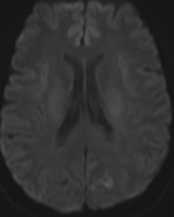
9 MRI/MRA H&N Left PCA territory acute/subacute infarct. Widely patent cervical and intracranial and vertebral arteries without evidence of narrowing or dissection.
10 Hypercoagulability work up ve ANA B2glycoprotein Cardiolpin Protein C Protein S Anti-thrombin 3 ESR CRP Factor 2 mutation LDL 88 HbA1c 4.9 Echo: Nl EF and no cardiac thrombus

11 Visual Pathway
12 Homonymous Hemianopia Lesions in the retrochiasmatic pathways (affecting the optic tract, lateral geniculate body, optic radiations, or occipital lobe). Lesions of the tract and lateral geniculate body tend to be incongruous, but the more posterior the lesion, the greater the congruity of the defect in either field. In general, tumors produce sloping field defects, whereas vascular lesions produce sharp field defects.

 in the eye with temporal field loss")
13 Optic Tract Lesions Complete unilateral optic tract lesions cause a complete macular splitting homonymous hemianopia, usually without impaired visual acuity Partial optic tract lesions are more common than complete ones and result in an incongruous field defect that may be scotomatous. Optic tract lesions are often associated with a relative afferent pupillary defect (RAPD) in the eye with temporal field loss (contralateral to the side of the lesion) This reflects the difference in light sensitivity between the intact temporal and nasal hemifield

 is caused by occlusion of the anterior choroidal artery A horizontal homonymous sector defect (wedge-shaped) is")
14 Lateral Geniculate Body Lesions Lateral geniculate body lesions may cause a complete macular splitting homonymous hemianopia Partial lesions result in an incongruous homonymous field defect. Two relatively specific patterns of congruous homonymous field defects A homonymous defect in the upper and lower quadrants with the sparing of a horizontal sector (quadruple sectoranopia) is caused by occlusion of the anterior choroidal artery A horizontal homonymous sector defect (wedge-shaped) is caused by interruption of the posterior lateral choroidal artery, which perfuses the central portion of the lateral geniculate

15 Optic Radiation Lesions Superior homonymous quadrantic defects ( pie-in-the-sky field defects) may result from a lesion in the temporal (Meyer s) loop of the optic radiations or in the inferior bank of the calcarine fissure For a quadrantic defect the lesion has to be quite extensive; small lesions result in scotomata Certain fibers from the ipsilateral eye travel more anteriorly and laterally in Meyer s loop and supports the hypothesis that visual field defects due to anterior retrogeniculate lesions are incongruous because of anatomic differences in the afferent pathway
 Such defects are usually more congruous")

16 Optic radiations in the depth of the parietal lobe gives rise to an inferior quadrantic defect ( pie-on-the-floor defect) Such defects are usually more congruous than those produced by lesions of the temporal lobe, and because the entire optic radiation passes through the parietal lobe, large lesions may produce complete homonymous hemianopia


17 Occipital Lesions Lesion of the extrastriate cortex (areas V2 and V3) has sharp horizontal edge because V2 and V3 are divided along the horizontal meridian into separate halves flanking the striate (V1) cortex Medial occipital lesions cause highly congruous homonymous hemianopias When both the upper and the lower calcarine cortices are affected, a complete homonymous hemianopia, usually with macular sparing, develops Macular sparing is common with occipital lesions, due to a combination of a large macular representation (as much as 50% 60%) and dual blood supply Anterior lesions adjacent to the parieto-occipital fissure affect the monocular temporal crescent of the contralateral visual field (temporal crescent or half-moon syndrome)
, keyhole fields.")
 may result in")
18 Bilateral occipital lobe lesions may occur from a single or from consecutive events and may cause bilateral homonymous scotomas, usually with some macular sparing ( ring scotomas), keyhole fields. Posterior lesions located in the posterior 50% to 60% of the striate cortex, including the occipital pole and operculum, and affect macular vision Bilateral homonymous hemianopia (double hemianopia) may result in cortical blindness
19 Clinical Features In patients with occipital lesions, the field defects often occurs in isolation, while other localizing signs of parietal involvement are evident in patients with parietal lesions Patients with purely occipital lesions are partially or fully aware of the hemianopia, whereas patients with larger or more anterior lesions, affecting parietal regions may be unaware of their defecit Hemianopic anosognosia occurs predominantly in right-sided lesions Spontaneous improvement of homonymous hemianopia is seen in at least 50% of patients first seen within 1 month of injury. In most cases, the improvement occurs within the first 3 months from injury.
20 Association between HRT and subsequent arterial and venous vascular events: a meta-analysis Sare, Gray, and Bath ( Eur Heart J. 2008) HRT is associated with an increased risk of stroke, stroke severity, and VTE, but not of CHD events. Although most trials studied older patients, increased risk was not related to age. Combined HRT increases the risk of VTE compared with estrogen monotherapy.

21 THANK YOU!!
Pathologies of postchiasmatic visual pathways and visual cortex
 Pathologies of postchiasmatic visual pathways and visual cortex Optic radiation: anatomy Pathologies of the postchiamsatic visual pathways and visual cortex Characterized by homonymous hemianopsia. This
LISC-322 Neuroscience. Visual Field Representation. Visual Field Representation. Visual Field Representation. Visual Field Representation
 LISC-3 Neuroscience THE VISUAL SYSTEM Central Visual Pathways Each eye sees a part of the visual space that defines its visual field. The s of both eyes overlap extensively to create a binocular. eye both
HOMONYMOUS VISUAL FIELD DEFECTS Perimetric findings and corresponding neuro-imaging results
 Homonymous visual field defects 511 HOMONYMOUS VISUAL FIELD DEFECTS Perimetric findings and corresponding neuro-imaging results JAN SCHILLER 1, TRAUGOTT J. DIETRICH 1, LIESE LORCH 1, MARTIN SKALEJ 2, CHRISTOPH
Course # Flashes and Floaters and Curtains, Oh My!
 Course # 132 Flashes and Floaters and Curtains, Oh My! FLASHES and FLOATERS and CURTAINS, OH MY!!! FLASHES OF LIGHT Vitreous is the villain Retinal traction Retinal hole Retinal tear Migraine Classic migraine
Course # Flashes and Floaters and Curtains, Oh My!
 Course # 132 Flashes and Floaters and Curtains, Oh My! FLASHES and FLOATERS and CURTAINS, OH MY!!! FLASHES OF LIGHT Vitreous is the villain Retinal traction Retinal hole Retinal tear Migraine Classic migraine
Neuroanatomy, Text and Atlas (J. H. Martin), 3 rd Edition Chapter 7, The Visual System, pp ,
, 3 rd Edition Chapter 7, The Visual System, pp ,") Normal CNS, Special Senses, Head and Neck TOPIC: FACULTY: LECTURE: READING: RETINA and CENTRAL VISUAL PATHWAYS P. Hitchcock, Ph.D. Department Cell and Developmental Biology Kellogg Eye Center Friday, 20
Normal CNS, Special Senses, Head and Neck TOPIC: FACULTY: LECTURE: READING: RETINA and CENTRAL VISUAL PATHWAYS P. Hitchcock, Ph.D. Department Cell and Developmental Biology Kellogg Eye Center Friday, 20
THE VISUAL PATHWAY FOR DENTAL STUDENTS
 Neuroanatomy Suzanne S. Stensaas, Ph.D. February 16, 2012 Objectives: THE VISUAL PATHWAY FOR DENTAL STUDENTS A. Draw the expected visual fields seen in classic lesions of the nerve, chiasm, thalamus, optic
Visual Field Screening and Analysis PART 2 A SYSTEMATIC APPROACH TO INTERPRETATION Revised 1/16/2011
 Visual Field Screening and Analysis PART 2 A SYSTEMATIC APPROACH TO INTERPRETATION Revised 1/16/2011 VF Screening Systematic Approach to Interpretation Patient info Field testing strategy and test point
C:\Documents and Settings\sstensaas\Desktop\dental visual 2010\VisualPath dental 2010.docVisualPath dental 2010.doc
 Neuroanatomy Suzanne Stensaas April 8, 2010, 10:00-12:00 p.m. Reading: Waxman Ch. 15, Computer Resources: HyperBrain Ch 7 THE VISUAL PATHWAY Objectives: 1. Describe the pathway of visual information from
Learn Connect Succeed. JCAHPO Regional Meetings 2017
 Learn Connect Succeed JCAHPO Regional Meetings 2017 Visual Field Testing Suzanne Hansen, M.Ed., COMT, OSC Why are these tests ordered? Visual field testing is ordered to help the physician diagnose and
Stroke School for Internists Part 1
 Stroke School for Internists Part 1 November 4, 2017 Dr. Albert Jin Dr. Gurpreet Jaswal Disclosures I receive a stipend for my role as Medical Director of the Stroke Network of SEO I have no commercial
Pupil Exams and Visual Fields
 Pupil Exams and Visual Fields A Closer Look at Cranial Nerves No Financial Interests Amy Jost does not have any financial interests related to this presentation AMY JOST, BS, COMT, CCRC, OSC CINCINNATI
Dr Jo-Anne Pon. Consultant Ophthalmologist and Oculoplastic Surgeon Southern Eye Specialists Christchurch
 Dr Jo-Anne Pon Consultant Ophthalmologist and Oculoplastic Surgeon Southern Eye Specialists Christchurch 12:15-12:30 Visual Migraines to be Worried About Visual Migraines To Be Worried About Jo-Anne Pon
OBJECTIVES. At the end of the lecture, students should be able to: List the cerebral arteries.
 DR JAMILA EL MEDANY OBJECTIVES At the end of the lecture, students should be able to: List the cerebral arteries. Describe the cerebral arterial supply regarding the origin, distribution and branches.
Learn Connect Succeed. JCAHPO Regional Meetings 2015
 Learn Connect Succeed JCAHPO Regional Meetings 2015 VISUAL FIELDS No financial conflicks Florida Society of Ophthalmology 2015 Gary Schemmer, MD Definition of Visual Field The area in space perceived by
Vision II. Steven McLoon Department of Neuroscience University of Minnesota
 Vision II Steven McLoon Department of Neuroscience University of Minnesota 1 Ganglion Cells The axons of the retinal ganglion cells form the optic nerve and carry visual information into the brain. 2 Optic
Disclosures. Visual Pathways. Visual Pathways. Visual Loss Understanding the Patterns. I have no financial disclosures. Tabby A.
 Visual oss Understanding the Patterns Tabby A. Kennedy, MD University of Wisconsin Department of adiology I have no financial disclosures Acknowledgements: indell Gentry Greg Avey JP Yu Judy Chen Disclosures
Parallel streams of visual processing
 Parallel streams of visual processing RETINAL GANGLION CELL AXONS: OPTIC TRACT Optic nerve Optic tract Optic chiasm Lateral geniculate nucleus Hypothalamus: regulation of circadian rhythms Pretectum: reflex
Required Slide. Session Objectives
 Vision: CNS 2018 Required Slide Session Objectives Visual system: CNS At the end of this session, students will be able to: 1. Understand how axons from the eyes travel through the optic nerves and tracts
Case Follow Up. Sepi Jooniani PGY-1
 Case Follow Up Sepi Jooniani PGY-1 Triage 54 year old M Pt presents to prelim states noticed today he had reddness to eyes, states worse in R eye. Pt denies any pain or itching. No further complaints.
/ / / / / / Hospital Abstraction: Stroke/TIA. Participant ID: Hospital Code: Multi-Ethnic Study of Atherosclerosis
 Multi-Ethnic Study of Atherosclerosis Participant ID: Hospital Code: Hospital Abstraction: Stroke/TIA History and Hospital Record 1. Was the participant hospitalized as an immediate consequence of this
OCT : retinal layers. Extraocular muscles. History. Central vs Peripheral vision. History: Temporal course. Optical Coherence Tomography (OCT)
") Optical Coherence Tomography (OCT) OCT : retinal layers 7 Central vs Peripheral vision Extraocular muscles RPE E Peripheral Vision: Rods (95 million) 30% Ganglion cells Central Vision: Cones (5 million)
Optical Coherence Tomography (OCT) OCT : retinal layers 7 Central vs Peripheral vision Extraocular muscles RPE E Peripheral Vision: Rods (95 million) 30% Ganglion cells Central Vision: Cones (5 million)
A Case of Carotid-Cavernous Fistula
 A Case of Carotid-Cavernous Fistula By : Mohamed Elkhawaga 2 nd Year Resident of Ophthalmology Alexandria University A 19 year old male patient came to our outpatient clinic, complaining of : -Severe conjunctival
Topical Diagnosis of Chiasmal and Retrochiasmal Disorders
 Topical Diagnosis of Chiasmal and Retrochiasmal Disorders Leonard A. Levin CHAPTER 12 TOPICAL DIAGNOSIS OF OPTIC CHIASMAL LESIONS Visual Field Defects Etiologies of the Optic Chiasmal Syndrome Masqueraders
1. The responses of on-center and off-center retinal ganglion cells
 1. The responses of on-center and off-center retinal ganglion cells 2. Responses of an on-center ganglion cell to different light conditions 3. Responses of an on-center ganglion cells to different light
Headache Assessment In Primary Eye Care
 Headache Assessment In Primary Eye Care Spencer Johnson, O.D., F.A.A.O. Northeastern State University Oklahoma College of Optometry johns137@nsuok.edu Course Objectives Review headache classification Understand
Visual Evoked Potentials. Outline. Visual Pathway Anatomy
 Visual Evoked Potentials Elayna Rubens, MD Assistant Professor of Neurology Weill Cornell Medical College Memorial Sloan Kettering Cancer Center Outline Visual Pathway Anatomy Basic VEP principles -VEP
Ganglion cell analysis by optical coherence tomography (OCT) Jonathan A. Micieli, MD Valérie Biousse, MD
 Jonathan A. Micieli, MD Valérie Biousse, MD") Ganglion cell analysis by optical coherence tomography (OCT) Jonathan A. Micieli, MD Valérie Biousse, MD Figure 1. Normal OCT of the macula (cross section through the line indicated on the fundus photo)
Ganglion cell analysis by optical coherence tomography (OCT) Jonathan A. Micieli, MD Valérie Biousse, MD Figure 1. Normal OCT of the macula (cross section through the line indicated on the fundus photo)
CNS 2 Physiology lab
 It should be noted that the doctor emphasized that this material is also considered as continuation of the theory material and is INCLUDED IN THE THEORY EXAM. Presbiopia: is decrease in accommodation of
Assessing the Stroke Patient. Arlene Boudreaux, MSN, RN, CCRN, CNRN
 Assessing the Stroke Patient Arlene Boudreaux, MSN, RN, CCRN, CNRN Cincinnati Pre-Hospital Stroke Scale May be done by EMS o One of many o F facial droop on one side o A arm drift (hold a pizza box, close
Carotid Cavernous Fistula
 Chief Complaint: Double vision. Carotid Cavernous Fistula Alex W. Cohen, MD, PhD; Richard Allen, MD, PhD May 14, 2010 History of Present Illness: A 46 year old female patient presented to the Oculoplastics
Unexplained visual loss in seven easy steps
 Unexplained visual loss in seven easy steps Andrew G. Lee, MD Chair Ophthalmology, Houston Methodist Hospital, Professor, Weill Cornell MC; Adjunct Professor, Baylor COM, U Iowa, UTMB Galveston, UT MD
CVA. Alison Atwater PA-C
 CVA Alison Atwater PA-C Types of CVAs Ischemic strokes 80% of strokes 2/3 are thrombotic 1/3 are embolic emboli from the heart or arteries feeding the brain such as carotids, vertebral and basilar etc
Neuroanatomy of a Stroke. Joni Clark, MD Professor of Neurology Barrow Neurologic Institute
 Neuroanatomy of a Stroke Joni Clark, MD Professor of Neurology Barrow Neurologic Institute No disclosures Stroke case presentations Review signs and symptoms Review pertinent exam findings Identify the
Pathway from the eye to the cortex
 Vision: CNS 2017 Pathway from the eye to the cortex Themes of this lecture Visual information is analyzed in more complicated ways than in the retina. One major pathway from the eye leads to the striate
Relationship between visual field defect and arterial occlusion in the posterior cerebral circulation
 Journal of Neurology, Neurosurgery, and Psychiatry, 1974, 37, 1022-1030 Relationship between visual field defect and arterial occlusion in the posterior cerebral circulation S. N. KAUL, G. H. DU BOULAY,
OCCLUSIVE VASCULAR DISORDERS OF THE RETINA
 OCCLUSIVE VASCULAR DISORDERS OF THE RETINA Learning outcomes By the end of this lecture the students would be able to Classify occlusive vascular disorders (OVD) of the retina. Correlate the clinical features
Band atrophy of the optic nerve: A report on different anatomical locations in three patients
 Saudi Journal of Ophthalmology (2013) 27, 65 69 Case Report Band atrophy of the optic nerve: A report on different anatomical locations in three patients Alberto Gálvez-Ruiz, MD a,b, ; Nawal Arishi, MD
Alan Barber. Professor of Clinical Neurology University of Auckland
 Alan Barber Professor of Clinical Neurology University of Auckland Presented with Non-fluent dysphasia R facial weakness Background Ischaemic heart disease Hypertension Hyperlipidemia L MCA branch
3/16/2018. Optic Nerve Examination. Hassan Eisa Swify FRCS Ed (Ophthalmology) Air Force Hospital
 Air Force Hospital") Optic Nerve Examination Hassan Eisa Swify FRCS Ed (Ophthalmology) Air Force Hospital 1 Examination Structure ( optic disc) Function Examination of the optic disc The only cranial nerve (brain tract) which
Optic Nerve Examination Hassan Eisa Swify FRCS Ed (Ophthalmology) Air Force Hospital 1 Examination Structure ( optic disc) Function Examination of the optic disc The only cranial nerve (brain tract) which
NEURORADIOLOGY DIL part 4
 NEURORADIOLOGY DIL part 4 Strokes and infarcts K. Agyem MD, G. Hall MD, D. Palathinkal MD, Alexandre Menard March/April 2015 OVERVIEW Introduction to Neuroimaging - DIL part 1 Basic Brain Anatomy - DIL
VISUAL REFLEXES. B. The oculomotor nucleus, Edinger-Westphal nucleus, and oculomotor nerve at level of the superior colliculus.
 Neuroanatomy Suzanne Stensaas February 24, 2011, 10:00-12:00 p.m. Reading: Waxman Ch. 15 HyperBrain: Ch 7 with quizzes and or Lab 7 videotape http://www-medlib.med.utah.edu/kw/hyperbrain/anim/reflex.html
The NIHSS score is 4 (considering 2 pts for the ataxia involving upper and lower limbs.
 Neuroscience case 5 1. Speech comprehension, ability to speak, and word use were normal in Mr. Washburn, indicating that aphasia (cortical language problem) was not involved. However, he did have a problem
Non-arteritic anterior ischemic optic neuropathy (NAION) with segmental optic disc edema. Jonathan A. Micieli, MD Valérie Biousse, MD
 with segmental optic disc edema. Jonathan A. Micieli, MD Valérie Biousse, MD") Non-arteritic anterior ischemic optic neuropathy (NAION) with segmental optic disc edema Jonathan A. Micieli, MD Valérie Biousse, MD A 75 year old white woman lost vision in the inferior part of her visual
Non-arteritic anterior ischemic optic neuropathy (NAION) with segmental optic disc edema Jonathan A. Micieli, MD Valérie Biousse, MD A 75 year old white woman lost vision in the inferior part of her visual
No Financial Interest
 Pituitary Apoplexy Michael Vaphiades, D.O. Professor Department of Ophthalmology, Neurology, Neurosurgery University of Alabama at Birmingham, Birmingham, AL No Financial Interest N E U R O L O G I C
Acute stroke. Ischaemic stroke. Characteristics. Temporal classification. Clinical features. Interpretation of Emergency Head CT
 Ischaemic stroke Characteristics Stroke is the third most common cause of death in the UK, and the leading cause of disability. 80% of strokes are ischaemic Large vessel occlusive atheromatous disease
Neuroophthalmology and otoneurology. 1. Which of the following is shown on this fundoscopy?
 EANS/UEMS European examination in neurosurgery Part I (written) Variants of questions with answers (compilation - Vyacheslav S. Botev, Department of Neurosurgery, M.Gorky Donetsk National Medical University)
Image Formation and Phototransduction. By Dr. Abdelaziz Hussein Lecturer of Physiology
 Image Formation and Phototransduction By Dr. Abdelaziz Hussein Lecturer of Physiology Vision Vision is a complex process through which an image of the external environment is formed on the photosensitive
Medical Neuroscience Tutorial Notes
 Medical Neuroscience Tutorial Notes Blood Supply to the Brain MAP TO NEUROSCIENCE CORE CONCEPTS 1 NCC1. The brain is the body's most complex organ. LEARNING OBJECTIVES After study of the assigned learning
Fundus Autofluorescence. Jonathan A. Micieli, MD Valérie Biousse, MD
 Fundus Autofluorescence Jonathan A. Micieli, MD Valérie Biousse, MD The retinal pigment epithelium (RPE) has many important functions including phagocytosis of the photoreceptor outer segments Cone Rod
Neuro-Ocular Grand Rounds
 Neuro-Ocular Grand Rounds Anthony B. Litwak,OD, FAAO VA Medical Center Baltimore, Maryland Dr. Litwak is on the speaker and advisory boards for Alcon and Zeiss Meditek COMMON OPTIC NEUROPATHIES THAT CAN
CEREBRAL ANEURYSMS PRESENTING WITH VISUAL FIELD DEFECTS*
 Brit. J. Ophthal. (1966) 50, 251 CEREBRAL ANEURYSMS PRESENTING WITH VISUAL FIELD DEFECTS* BY University Department of Ophthalmology and Royal Infirmary, Edinburgh ANEURYSMS occur more frequently within
LECTURE # 7 EYECARE REVIEW: PART III
 LECTURE # 7 EYECARE REVIEW: PART III HOW TO TRIAGE EYE EMERGENCIES STEVE BUTZON, O.D. EYECARE REVIEW: HOW TO TRIAGE EYE EMERGENCIES FOR PRIMARY CARE PHYSICIANS Steve Butzon, O.D. Member Director IDOC President
CSE511 Brain & Memory Modeling. Lect21-22: Vision Central Pathways
 CSE511 Brain & Memory Modeling CSE511 Brain & Memory Modeling Lect02: BOSS Discrete Event Simulator Lect21-22: Vision Central Pathways Chapter 12 of Purves et al., 4e Larry Wittie Computer Science, StonyBrook
Neuroscience Tutorial
 Neuroscience Tutorial Brain Organization : cortex, basal ganglia, limbic lobe : thalamus, hypothal., pituitary gland : medulla oblongata, midbrain, pons, cerebellum Cortical Organization Cortical Organization
TIA AND STROKE. Topics/Order of the day 1. Topics/Order of the day 2 01/08/2012
 Charles Ashton Medical Director TIA AND STROKE Topics/Order of the day 1 What Works? Clinical features of TIA inc the difference between Carotid and Vertebral territories When is a TIA not a TIA TIA management
LISC-322 Neuroscience Cortical Organization
 LISC-322 Neuroscience Cortical Organization THE VISUAL SYSTEM Higher Visual Processing Martin Paré Assistant Professor Physiology & Psychology Most of the cortex that covers the cerebral hemispheres is
CHAPTER 13 CLINICAL CASES INTRODUCTION
 2 CHAPTER 3 CLINICAL CASES INTRODUCTION The previous chapters of this book have systematically presented various aspects of visual field testing and is now put into a clinical context. In this chapter,
Do You See What I See!!! Shane R. Kannarr, OD
 Do You See What I See!!! Shane R. Kannarr, OD skannarr@kannarreyecare.com Define Specialty Testing Additional Test to: Prove/Disprove Diagnosis To monitor progression of a condition To document a condition
Neuro-Ophthalmic Masqueraders
 Neuro-Ophthalmic Masqueraders Leonid Skorin, Jr., OD, DO, MS, FAAO, FAOCO Mayo Clinic Health System in Albert Lea Denise Goodwin, OD, FAAO Pacific University College of Optometry Please silence all mobile
Inside Your Patient s Brain Michelle Peterson, APRN, CNP Centracare Stroke and Vascular Neurology
 Inside Your Patient s Brain Michelle Peterson, APRN, CNP Centracare Stroke and Vascular Neurology Activity Everyone stand up, raise your right hand, tell your neighbors your name 1 What part of the brain
Index. Note: Page numbers of article titles are in boldface type.
 Index Note: Page numbers of article titles are in boldface type. A Acetazolamide, in idiopathic intracranial hypertension, 49 52, 60 Angiography, computed tomography, in cranial nerve palsy, 103 107 digital
Article 4Small Spot, Big Problem: Low Vision Management of Central Neurological Visual Field Defects
 Article 4Small Spot, Big Problem: Low Vision Management of Central Neurological Visual Field Defects Noah Merhar, OD, VA Central Western Massachusetts Healthcare System, Leeds, Massachuesetts ABSTRACT
Divine Intervention Episode 36 USMLE Ophthalmology. Some PGY1
 Divine Intervention Episode 36 USMLE Ophthalmology Some PGY1 1 71 yo M with central scotomas. Straight lines appear wavy and blurred when he tries to read. Diagnosis? Types of disease? Which is more rapid
The phenomenon of unilateral loss of vision in
 554 Short Communication Bilateral Loss of Vision in Bright Light David 0. Wiebers, MD, Jerry W. Swanson, MD, Terrence L. Cascino, MD, and Jack P. Whisnant, MD We describe four patients with episodic bilateral
Anterior Ischemic Optic Neuropathy (AION)
") Anterior Ischemic Optic Neuropathy (AION) Your doctor thinks you have suffered an episode of anterior ischemic optic neuropathy (AION). This is the most common cause of sudden decreased vision in patients
Anterior Ischemic Optic Neuropathy (AION) Your doctor thinks you have suffered an episode of anterior ischemic optic neuropathy (AION). This is the most common cause of sudden decreased vision in patients
Principles Arteries & Veins of the CNS LO14
 Principles Arteries & Veins of the CNS LO14 14. Identify (on cadaver specimens, models and diagrams) and name the principal arteries and veins of the CNS: Why is it important to understand blood supply
The occipital lobe is involved in many aspects of
 728 Distribution of the Occipital Branches of the Posterior Cerebral Artery Correlation With Occipital Lobe Infarcts Slobodan V. Marinkovic", MD, Milan M. Milisavljevic", MD, Vera Lolid-Draganid, MD, and
Neurology. Hollie Wilson
 Neurology Hollie Wilson Objectives Anatomy Physiology: Functional centres of brain UMN lesion vs. LMN lesion Spinal cord Main tracts ascending and descending Nerve roots and peripheral nerves action potentials
Neuro-Ocular Grand Rounds Anthony B. Litwak,OD, FAAO VA Medical Center Baltimore, Maryland
 Neuro-Ocular Grand Rounds Anthony B. Litwak,OD, FAAO VA Medical Center Baltimore, Maryland Dr. Litwak is on the speaker and advisory boards for Alcon and Zeiss Meditek COMMON OPTIC NEUROPATHIES THAT CAN
Excellent Network Courses. Department of Neurology Affiliated hospital of Jiangsu University
 Excellent Network Courses Department of Neurology Affiliated hospital of Jiangsu University Agnosia Visual Agnosia Lissauer (1890) described 2 types: a) Apperceptive Cannot see objects b) Associative Does
Evaluation of ONH Pallor in Glaucoma Patients and Suspects. Leticia Rousso, O.D. Joseph Sowka, O.D
 Evaluation of ONH Pallor in Glaucoma Patients and Suspects Leticia Rousso, O.D Joseph Sowka, O.D I. Abstract This case report will evaluate a young glaucoma suspect with unilateral sectoral optic nerve
Essentials of Clinical MR, 2 nd edition. 14. Ischemia and Infarction II
 14. Ischemia and Infarction II Lacunar infarcts are small deep parenchymal lesions involving the basal ganglia, internal capsule, thalamus, and brainstem. The vascular supply of these areas includes the
Sudden Vision Loss. Brendan Girschek, MD, FRCSC, FACS Vitreoretinal Surgery Cedar Valley Medical Specialists
 Sudden Vision Loss Brendan Girschek, MD, FRCSC, FACS Vitreoretinal Surgery Cedar Valley Medical Specialists My Credentials -Residency in Ophthalmology at the LSU Eye Center in New Orleans, LA -Fellowship
A Curious Case of Bilateral Optic Disc Edema Brittney Dautremont, DO, MPH
 A Curious Case of Bilateral Optic Disc Edema Brittney Dautremont, DO, MPH PGY2 Ophthalmology Resident Grandview Medical Center Dayton, OH CASE PRESENTATION 51 year old white female presenting with blurred
The Ophthalmoscope, the Fundus Oculi, and Central and Peripheral Vision
 The Ophthalmoscope, the Fundus Oculi, and Central and Peripheral Vision 1 The examination of the eye consists of five parts. This chapter deals with the fundus oculi and with central and peripheral vision.
Overview INTRODUCTION 3/15/2018. Headache Emergencies. Other way to differentiate between them? Is there an easy way to differentiate between them?
 Overview Headache Emergencies Primary versus Secondary headache disorder Red flags 4 cases of unusual headache emergencies Disclaimer: we will not talk about brain bleed as patients usually go the ED.
Objectives. Unexplained Vision Loss: Where Do I Go From Here. History. History. Drug Induced Vision Loss
 Objectives Unexplained Vision Loss: Where Do I Go From Here Denise Goodwin, OD, FAAO Coordinator, Neuro-ophthalmic Disease Clinic Pacific University College of Optometry goodwin@pacificu.edu Know the importance
3/16/2018. Perimetry
 Perimetry The normal visual field extends further away from fixation temporally and inferiorly than superiorly and nasally. From the center of the retina this sensitivity decreases towards the periphery,
CEREBRO VASCULAR ACCIDENTS
 CEREBRO VASCULAR S MICHAEL OPONG-KUSI, DO MBA MORTON CLINIC, TULSA, OK, USA 8/9/2012 1 Cerebrovascular Accident Third Leading cause of deaths (USA) 750,000 strokes in USA per year. 150,000 deaths in USA
PORT WINE STAINS AND STURGE-WEBER SYNDROME
 PORT WINE STAINS AND STURGE-WEBER SYNDROME Ong Hian Tat It is important for general practitioners to recognize cutaneous port-wine stains as these could signify important association with Sturge Weber
Blood Supply. Allen Chung, class of 2013
 Blood Supply Allen Chung, class of 2013 Objectives Understand the importance of the cerebral circulation. Understand stroke and the types of vascular problems that cause it. Understand ischemic penumbra
Clinical and Experimental Neuropsychology. Lecture 3: Disorders of Perception
 Clinical and Experimental Neuropsychology Lecture 3: Disorders of Perception Sensation vs Perception Senses capture physical energy from environment that are converted into neural signals and elaborated/interpreted
Michael Horowitz, MD Pittsburgh, PA
 Michael Horowitz, MD Pittsburgh, PA Introduction Cervical Artery Dissection occurs by a rupture within the arterial wall leading to an intra-mural Hematoma. A possible consequence is an acute occlusion
Jacqueline Theis, O.D., F.A.A.O.
 Neuro-Ophthalmological Emergencies Presenting in Primary Care Optometry Describes the symptoms, signs, and management of neuro-ophthalmological emergencies. Signs/Symptoms to be Concerned about (especially
o A cushion of fat surrounds most of the eye
 Name Period SPECIAL SENSES The Senses of touch o Temperature o Pressure o Pain o Smell o Taste o Sight o Hearing o Equilibrium The Eye and Vision are in the eyes has over a o Most of the eye is enclosed
Neuroanatomy Dr. Maha ELBeltagy Assistant Professor of Anatomy Faculty of Medicine The University of Jordan 2018
 Neuroanatomy Dr. Maha ELBeltagy Assistant Professor of Anatomy Faculty of Medicine The University of Jordan 2018 Blood Supply of Brain and Spinal Cord Arterial Supply of Brain The brain receives blood
Module 4. Ischemia in Carotid Territory
 Module 4. Ischemia in Carotid Territory Introduction and Key Clinical Examples Objectives for Module 4 Knowledge! Describe two common TIAs (mini-strokes) that are seen with ischemia in carotid territory.!
Neuro Op Grand Rounds: Fields and Diplopia Utah Optometric Association June 2018
 Disclosures Neuro Op Grand Rounds: Fields and Diplopia Utah Optometric Association June 2018 I have received honorarium from the following: Glaukos CE in Italy Heidelberg Engineering Review of Optometry
Stroke/TIA. Tom Bedwell
 Stroke/TIA Tom Bedwell tab1g11@soton.ac.uk The Plan Definitions Anatomy Recap Aetiology Pathology Syndromes Brocas / Wernickes Investigations Management Prevention & Prognosis TIAs Key Definitions Transient
Disclosures. Objectives 6/2/2017
 Classification: Migraine and Trigeminal Autonomic Cephalalgias Lauren Doyle Strauss, DO, FAHS Assistant Professor, Child Neurology Assistant Director, Child Neurology Residency @StraussHeadache No disclosures
Lesions of the optic radiations mimicking lateral geniculate nucleus visual field defects
 Journal of Neurology, Neurosurgery, and Psychiatry 1985;48:982-988 Lesions of the optic radiations mimicking lateral geniculate nucleus visual field defects JOHN E CARTER,* PATRICK O'CONNOR,t DAVID SHACKLETT,t
Key Clinical Concepts
 Cerebrovascular Review and General Vascular Syndromes, Including Those That Impact Dizziness Key Clinical Concepts Basic Review of Cerebrovascular Circulation Circulation to the brain is divided into anterior
LOOKING AT BLINDNESS FROM NEUROLOGIST S PERSPECTIVE
 Vet Times The website for the veterinary profession https://www.vettimes.co.uk LOOKING AT BLINDNESS FROM NEUROLOGIST S PERSPECTIVE Author : LAURENT S GAROSI Categories : Vets Date : June 23, 2008 LAURENT
Learn Connect Succeed. JCAHPO Regional Meetings 2017
 Learn Connect Succeed JCAHPO Regional Meetings 2017 NO FINANCIAL DISCLOSURES Technician s Role in Neuro-Ophthalmology Workup Beth Koch COT, ROUB Cleveland 9/16/2017 What Tests Are You Expected To Perform?
Lab 2. we will look into several angled horizontal sections ( orbitomeatal plane ) i.e passing from the orbit into the ear
 i.e passing from the orbit into the ear") we will look into several angled horizontal sections ( orbitomeatal plane ) i.e passing from the orbit into the ear Figure I page 76 : looking at the key on the left side this section passed through the
we will look into several angled horizontal sections ( orbitomeatal plane ) i.e passing from the orbit into the ear Figure I page 76 : looking at the key on the left side this section passed through the
Gross Organization I The Brain. Reading: BCP Chapter 7
 Gross Organization I The Brain Reading: BCP Chapter 7 Layout of the Nervous System Central Nervous System (CNS) Located inside of bone Includes the brain (in the skull) and the spinal cord (in the backbone)
T HE blood supply of cerebral arteriovenous malformations is often extensive
 NOVEMBER, 1974 ROENTGENOGRAPHIC ANALYSIS OF ARTERIOVENOUS MALFORMATIONS OF THE OCCIPITAL LOBE* By B. TODD TROOST, M.D.,t and THOMAS H. NEWTON, M.D4 T HE blood supply of cerebral arteriovenous malformations