Sudden Vision Loss. Brendan Girschek, MD, FRCSC, FACS Vitreoretinal Surgery Cedar Valley Medical Specialists
|
|
|
- Barnard Hunter
- 5 years ago
- Views:
Transcription
1 Sudden Vision Loss Brendan Girschek, MD, FRCSC, FACS Vitreoretinal Surgery Cedar Valley Medical Specialists
2 My Credentials -Residency in Ophthalmology at the LSU Eye Center in New Orleans, LA -Fellowship in Retinal Surgery at the Hamilton Eye Institute/Univ of Tennessee in Memphis, TN -Board Certified in the US & Canada -Fellow of the American College of Surgeons
3 What is a Retinal Surgeon? -Uniquely-trained ophthalmologist who works behind the lens -Typically manage cases of sudden vision loss which often involve the retina and/or optic nerve -Requires additional 2-year fellowship
4 Initial Concerns -Unilateral vs. Bilateral -Altitudinal vs. Hemispheric vs. Central -Sudden vs. Progressive -Pain? Recent surgical procedure? PMHx?
5 What Are We Worried About -Things that a retinal surgeon can repair -retinal detachment surgery -Things that cannot afford to wait -giant cell/temporal arteritis -Things that don t require expensive imaging procedures (ie. when to not order a CT/MRI) -no need for CT/MRI with unilateral vision loss
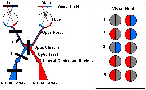
6 The Role of the Pupil -Insults posterior to the chiasm will NOT cause an afferent or efferent pupillary defect -Evaluate reaction to pupil(s) with strong light -Assess for speed and briskness of contraction to light (may have up to 1mm anisocoria) -Ask about subjective light intensity
7
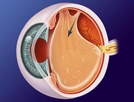
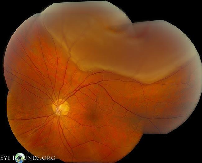
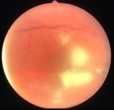
8 Retinal Detachment -Requires surgery in nearly all circumstances -Macula on vs Macula off -Timing of surgery depends on macular status -If macula on - repair immediately (<24h), often within 6 hours; these are the eyes with 20/20 vision and peripheral vision loss! -If macula off - repair within one week
9 Retinal Detachment -Visual loss does not adhere to any specific quadrant/altitudinal defect -May or may not be associated with flashes +/floaters -Be concerned if post-surgical (cataract surgery)
10 Retinal Detachment -Progressive visual field loss over hours to days -Majority of RDs begin supero-temporal causing infero-nasal visual loss (where one s nose is); insidious onset -Describe a dark curtain starting peripherally moving centripetally
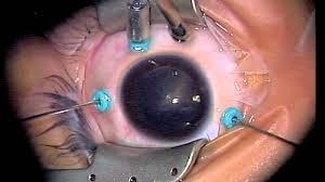
11 Retinal Detachment -Repair with Pars Plana Vitrectomy (PPV) -25g (0.5mm) sutureless incisions; soon 27g -Surgical time < 60 minutes; often < 45 minutes -Internal tamponade with gas vs oil -90+% success rate with initial repair -Local anesthesia only
12
13
14
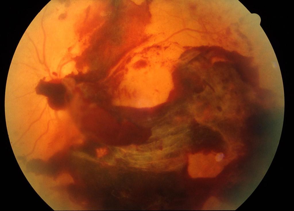
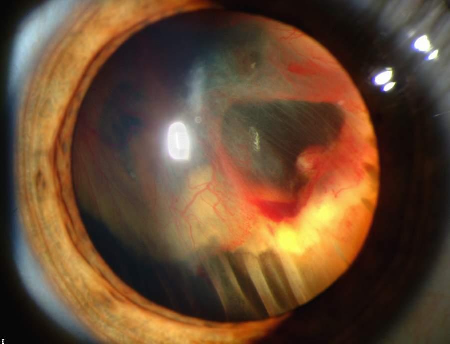
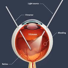
15 Vitreous Hemorrhage -Sudden-onset cloud of haze or floaters in vision to one eye -Often begins centrally, diffuses within the vitreous -Rule out retinal tear and/or detachment -May be due to proliferative retinopathy (diabetes, sickle cell disease, hypertension)
16
17
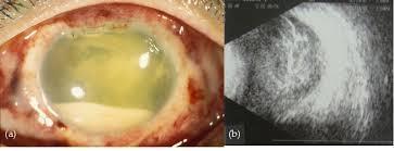
18 Endophthalmitis -Intraocular inflammation commonly due to infection -Be concerned with red/painful/light sensitive eye with vision loss within one week of intraocular surgery -Requires immediate treatment with intravitreal injection of antibiotics/steroids vs vitrectomy
19 Endophthalmitis -Uncommon presentation with sepsis -Bacteria and fungi can seed the vitreous and cause floaters and retinal necrosis -Due to significant vascularity to the choroid (vascular bed underlying the retina) -Can present uni- or bi-laterally -Be concerned in patients with hardware
20
21 Giant Cell/Temporal Arteritis -Natural history leads to bilateral irreversible blindness and possible death due to aortic aneurysm or stroke -Vague history of amaurosis fugax, temporallylocated pain, recent weight loss, proximal muscle weakness/pain, fevers, etc. -Rule out with ESR/CRP/platelet screen (check CXR, UA, UCx if elevated white count noted)
22 Giant Cell/Temporal Arteritis -Requires temporal artery biopsy if clinical suspicion is high (regardless of labs) -Nodular granulomatous inflammation of medium-to-large sized arteries -Fragmentation of internal elastic lamina with transmural cellular infiltration
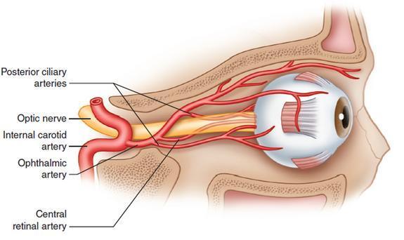
23 Giant Cell/Temporal Arteritis -Most commonly involved vessels are superficial temporal artery, ophthalmic artery, posterior ciliary artery and vertebral arteries -Start systemic corticosteroids immediately if clinical suspicion is high and biopsy within 1 wk -Taper slowly and follow symptoms +/- labs
24 Cortical Stroke -Does NOT cause unilateral altitudinal defects -Visual field loss is hemispheric (left/right) and often with obvious bilateral involvement -Homonymous loss indicates posterior infarct -Often will have macular sparing (ie. reading vision intact) -Will NOT cause an afferent pupillary defect!
25 Retinal Artery Occlusion -May be either branch or central depending on where the occlusion occurred -If occlusion is proximal to retinal artery bifurcation then CRAO, distal then BRAO -Sudden onset of altitudinal defect with BRAO, near complete loss of total visual field with CRAO (may have macular sparing)
26
27
28
29 Retinal Artery Occlusion -Evaluate the pupil! -The greater the area of infarction, the greater the pupillary defect -Evaluate with carotid doppler (MRA if clinical suspicion warrants) and transthoracic echocardiogram (transesophogeal if clinical suspicion warrants)
30 Retinal Artery Occlusion -Do not forget about giant cell/temporal arteritis! -No effective treatment available to restore visual function -Find the source of the emboli and anticoagulate if warranted
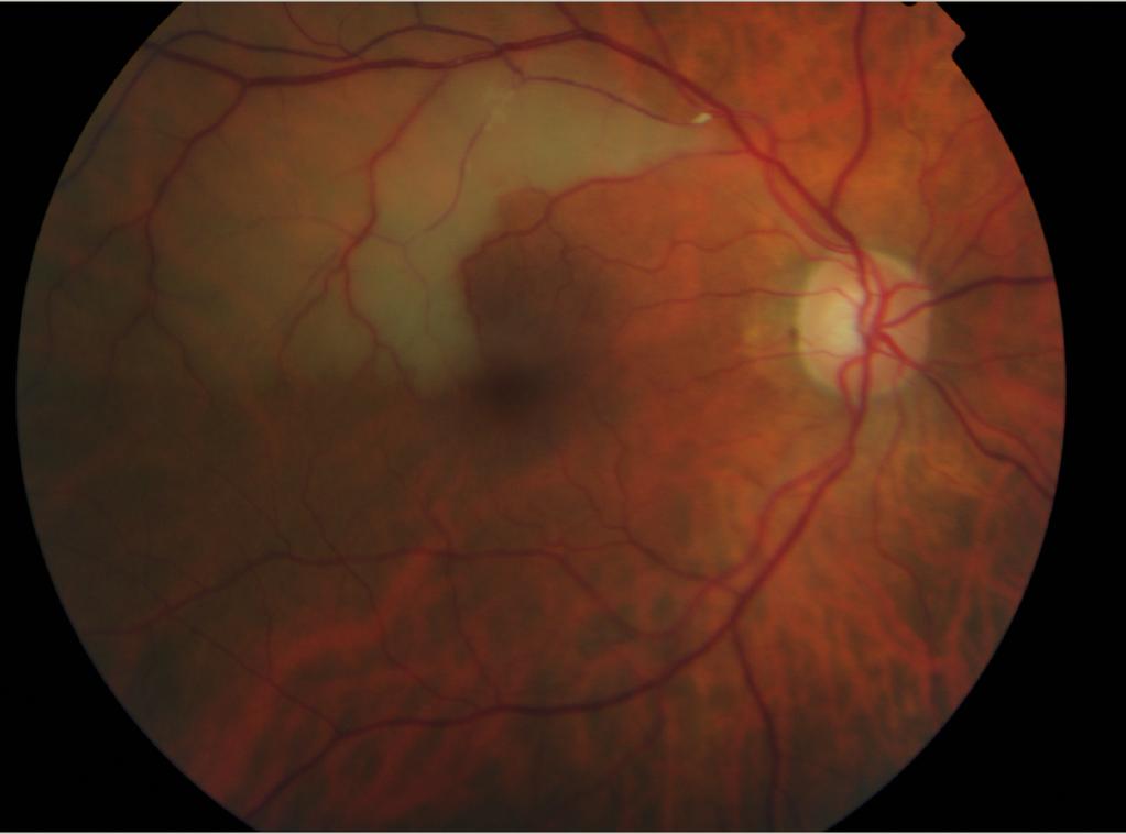
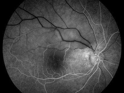
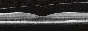
31 Central/Macular Vision Loss -Various retinal conditions such as macular degeneration, diabetic macular edema, macular hole -These conditions will not present in a simultaneous bilateral manner -Non-urgent follow-up with retinal specialist or general ophthalmologist recommended
32
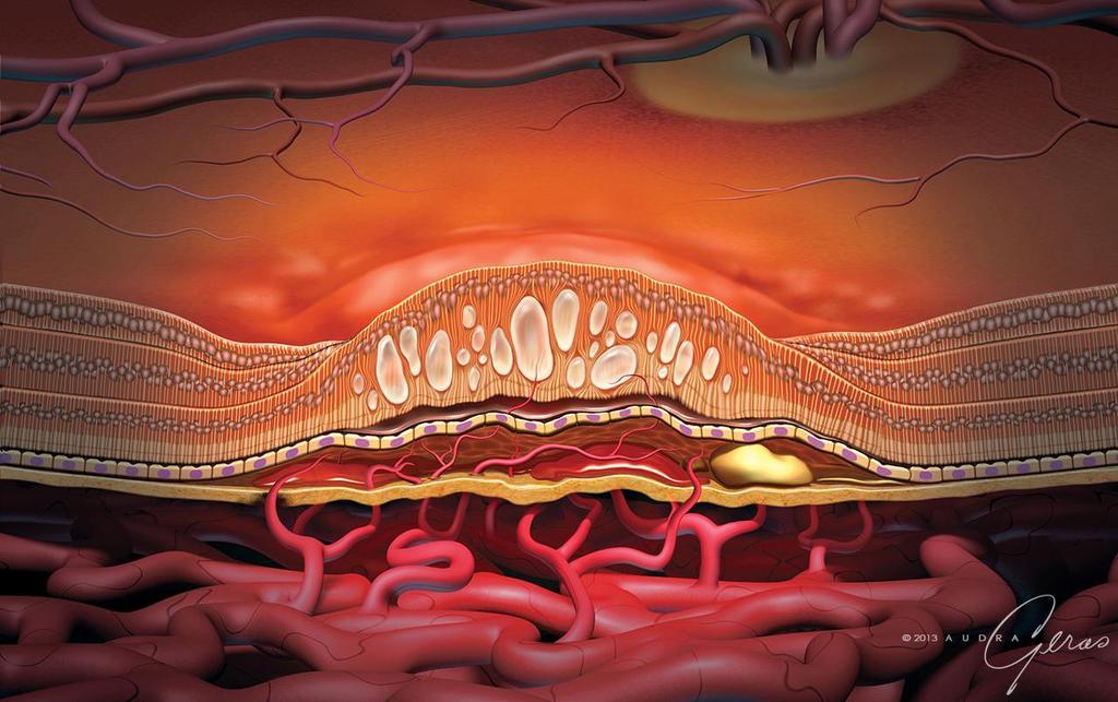
33 Macular Degeneration -No longer a blinding problem for most -Regular intravitreal injections of anti-vegf medications stabilizes vision loss in most and may improve vision in many -Various imaging modalities and treatment options including medication, laser and vitrectomy surgery available
34
35
36
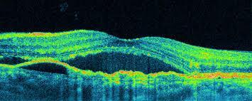
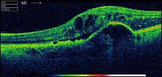
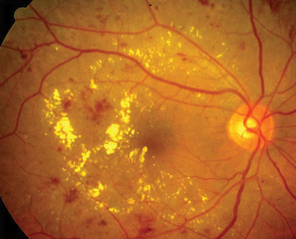
37 Diabetic Macular Edema -Most common cause of vision loss in diabetes is due to macular edema -Treated with regular intravitreal anti-vegf medications, laser surgery and/or vitrectomy surgery -Often occur concurrently with nephropathy and peripheral neuropathy
38
39
40 Diabetic Retinopathy -I ask about the patient s hemoglobin A1C at every visit -A1C extremely important in determining followup interval in those with retinopathy -Encourage patient to know the A1C value (not just good or ok )
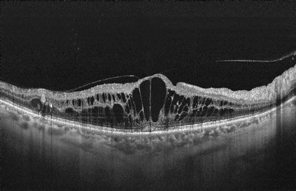
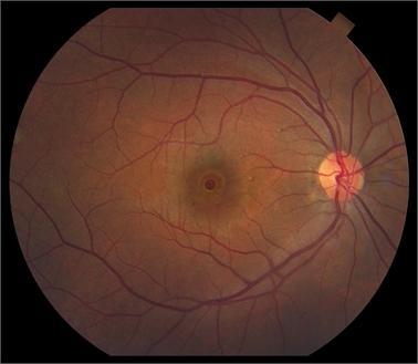
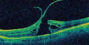
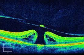
41 Macular Hole -Present as a relative sudden-onset loss of central/reading vision in one eye without associated symptoms -No pupillary abnormality noted -Due to persistent vitreo-retinal traction at the fovea -Requires vitrectomy surgery to repair
42
43 Pars Plana Vitrectomy -Pars plana is mm posterior to the limbus -Behind the ciliary muscle, ahead of the retina -Safe point for entry into the posterior segment -3 ports: 1) infusion, 2) light source, 3) cutter/forceps/aspiration
44 Vitreoretinal Surgery

45 Retinal Detachment Repair

46 Macular Hole Repair
47 Q&A
A Patient s Guide to Diabetic Retinopathy
 Diabetic Retinopathy A Patient s Guide to Diabetic Retinopathy 840 Walnut Street, Philadelphia PA 19107 www.willseye.org Diabetic Retinopathy 1. Definition Diabetic retinopathy is a complication of diabetes
Diabetes & Your Eyes
 Diabetes & Your Eyes Diabetes is a disease that occurs when the pancreas does not secrete enough insulin or the body is unable to process it properly. Insulin is the hormone that regulates the level of
Course # Flashes and Floaters and Curtains, Oh My!
 Course # 132 Flashes and Floaters and Curtains, Oh My! FLASHES and FLOATERS and CURTAINS, OH MY!!! FLASHES OF LIGHT Vitreous is the villain Retinal traction Retinal hole Retinal tear Migraine Classic migraine
Course # Flashes and Floaters and Curtains, Oh My!
 Course # 132 Flashes and Floaters and Curtains, Oh My! FLASHES and FLOATERS and CURTAINS, OH MY!!! FLASHES OF LIGHT Vitreous is the villain Retinal traction Retinal hole Retinal tear Migraine Classic migraine
Ophthalmology. Juliette Stenz, MD
 Ophthalmology Juliette Stenz, MD Required Slide Disclosures NO SIGNIFICANT FINANCIAL, GENERAL, OR OBLIGATION INTERESTS TO REPORT Required Slide At the end of this session, students will be able to: 1.
9/11/11. Temporal Arteritis. Background. Background. Richard E. Castillo, OD, DO NORTHEASTERN STATE UNIVERSITY Director, Ophthalmic Surgery Service
 Temporal Arteritis Richard E. Castillo, OD, DO NORTHEASTERN STATE UNIVERSITY Director, Ophthalmic Surgery Service 1 Background Giant Cell Arteritis Temporal Arteritis Cranial Arteritis Granulomatous Arteritis
Office Based Practice. Vitreoretinal Disease & Surgery. Coding Fiesta Vitreoretinal Disease & Surgery September 23, 2017 ADULT RETINA
 Vitreoretinal Disease & Surgery Coding Fest 2017 Vitreoretinal Surgery & Disease University of FL College of Medicine ADULT RETINA Medical Retina Surgical Retina Age Related Vascular Disease Vascular Disease
Macular Hole. Helpline
 Macular Hole The retina is a light-sensitive layer of tissue lining the back of the eye. The macula is a small area at the centre of the retina responsible for all of our central vision, most of our colour
INFORMED CONSENT FOR AVASTIN TM (BEVACIZUMAB) INTRAVITREAL INJECTION
 INTRAVITREAL INJECTION") INFORMED CONSENT FOR AVASTIN TM (BEVACIZUMAB) INTRAVITREAL INJECTION INDICATIONS: Age-related macular degeneration (AMD) is the leading cause of blindness in people over 50 years of age. It is caused by
INFORMED CONSENT FOR AVASTIN TM (BEVACIZUMAB) INTRAVITREAL INJECTION INDICATIONS: Age-related macular degeneration (AMD) is the leading cause of blindness in people over 50 years of age. It is caused by
Diagnosis and treatment of diabetic retinopathy. Blake Cooper MD Ophthalmologist Vitreoretinal Surgeon Retina Associates Kansas City
 Diagnosis and treatment of diabetic retinopathy Blake Cooper MD Ophthalmologist Vitreoretinal Surgeon Retina Associates Kansas City Disclosures Consulted for Novo Nordisk 2017,2018. Will be discussing
Royal Berkshire Hospital Dunedin Hospital. Prince Charles Eye Unit Pi Princess Margaret Hospital
 Vitreoretinal Surgery Mr Vaughan Tanner www.tanner-eyes.co.uk eyes Reading Royal Berkshire Hospital Dunedin Hospital Windsor Prince Charles Eye Unit Pi Princess Margaret Hospital Success rates VR surgery
Vanderbilt Eye Institute Clinical Trials
 April, 2010 Vanderbilt Eye Institute Clinical Trials Ophthalmology Actively Recruiting Studies For information on our clinical trials and other studies, please contact: Sandy Owings, COA, CCRP Clinic Director
FRANZCO, MD, MBBS. Royal Darwin Hospital
 Diabetes and Eye By Dr. Nishantha Wijesinghe FRANZCO, MD, MBBS Consultant Ophthalmologist Royal Darwin Hospital 98% of Diabetics do not need to suffer from severe visual loss Yet Diabetic eye disease is
OCCLUSIVE VASCULAR DISORDERS OF THE RETINA
 OCCLUSIVE VASCULAR DISORDERS OF THE RETINA Learning outcomes By the end of this lecture the students would be able to Classify occlusive vascular disorders (OVD) of the retina. Correlate the clinical features
Mild NPDR. Moderate NPDR. Severe NPDR
 Diabetic retinopathy Diabetic retinopathy is the most common cause of blindness in adults aged 35-65 years-old. Hyperglycaemia is thought to cause increased retinal blood flow and abnormal metabolism in
Moncef Khairallah, MD
 Moncef Khairallah, MD Department of Ophthalmology, Fattouma Bourguiba University Hospital Faculty of Medicine, University of Monastir Monastir, Tunisia INTRODUCTION IU: anatomic form of uveitis involving
GENERAL INFORMATION DIABETIC EYE DISEASE
 GENERAL INFORMATION DIABETIC EYE DISEASE WHAT IS DIABETIC EYE DISEASE? Diabetic eye disease is a term used to describe the common eye complications seen in people with diabetes. It includes: Diabetic retinopathy
Brampton Hurontario Street Brampton, ON L6Y 0P6
 Diabetic Retinopathy What is Diabetic Retinopathy Diabetic retinopathy is one of the leading causes of blindness world-wide. Diabetes damages blood vessels in many organs of the body including the eyes.
Repairing Macular Hole and Macular Pucker
 Transcript Details This is a transcript of an educational program accessible on the ReachMD network. Details about the program and additional media formats for the program are accessible by visiting: https://reachmd.com/programs/revealing-retina/repairing-macular-hole-and-macular-pucker/3924/
NEPTUNE RED BANK BRICK
 NEPTUNE RED BANK BRICK Diabetes & The Eye Diabetics are more likely to develop Cataracts at a younger age. Diabetics are twice as likely to develop Glaucoma when compared to non-diabetics. The primary
Rapid Visual Loss. Dr Michael Johnson PhD FCOptom DipOrth DipGlauc DipTp(IP) Independent Prescribing Optometrist
 Independent Prescribing Optometrist") Rapid Visual Loss Dr Michael Johnson PhD FCOptom DipOrth DipGlauc DipTp(IP) Independent Prescribing Optometrist Outline Pathophysiology Differential diagnosis. Patient scenarios in community practice:
Rapid Visual Loss Dr Michael Johnson PhD FCOptom DipOrth DipGlauc DipTp(IP) Independent Prescribing Optometrist Outline Pathophysiology Differential diagnosis. Patient scenarios in community practice:
Retinal Tear and Detachment
 Retinal Tear and Detachment Introduction The retina is the layer of tissue in the back of the eye that is responsible for vision. It is attached to the choroid tissue, which supplies the retina with blood.
Detached and Torn. Se Habla Español
 Detached and Torn Retina www.fleyedocs.com Se Habla Español Retinal Detachments Occur in 1 Out of 10,000 Americans Each Year A retinal detachment is not as common as other eye conditions such as glaucoma
Facts About Diabetic Eye Disease
 Facts About Diabetic Eye Disease Points to Remember 1. Diabetic eye disease comprises a group of eye conditions that affect people with diabetes. These conditions include diabetic retinopathy, diabetic
Objectives. Unexplained Vision Loss: Where Do I Go From Here. History. History. Drug Induced Vision Loss
 Objectives Unexplained Vision Loss: Where Do I Go From Here Denise Goodwin, OD, FAAO Coordinator, Neuro-ophthalmic Disease Clinic Pacific University College of Optometry goodwin@pacificu.edu Know the importance
Advanced Vitreoretinal Techniques & Technology Symposium
 14th ANNUAL Advanced Vitreoretinal Techniques & Technology Symposium JULY 25-27, 2014 SWISSÔTEL CHICAGO CHICAGO, IL CME ACCREDITATION AND CREDIT STATEMENT This activity has been planned and implemented
Ophthamology Directorate. Eye Injection for Macular Disorders Information for Patients
 Ophthamology Directorate Eye Injection for Macular Disorders Information for Patients As discussed at your appointment today, please call the Medical Retinal Services Coordinator as soon as possible (within
Diabetic Retinopathy
 Diabetic Retinopathy Introduction People with diabetes are more likely to have eye problems that can lead to blindness. Diabetic retinopathy is a disease of the eye s retina that is caused by diabetes.
The Foundation WHAT IS THE RETINA? continued next page. RETINA HEALTH SERIES Facts from the ASRS
 The Foundation American Society of Retina Specialists Committed to improving the quality of life of all people with retinal disease. Epiretinal Membranes (ERMs), also commonly known as cellophane maculopathy
 measure of your overall performance. An isolated glucose test is helpful to let you know what your sugar level is at one moment, but it doesn t tell you whether or not your diabetes is under adequate control
5/2/2016 EYE EMERGENCIES. Nathaniel Pelsor, O.D., FAAO Talley Medical-Surgical Eye Care Associates. Anatomy. Tools
 EYE EMERGENCIES Nathaniel Pelsor, O.D., FAAO Talley Medical-Surgical Eye Care Associates Anatomy Tools 1 Contact dermatitis Blepharitis HSV Preseptal Cellulitis Anterior Chamber Subconjunctival hemorrhage
X-Plain Diabetic Retinopathy Reference Summary
 X-Plain Diabetic Retinopathy Reference Summary Introduction Patients with diabetes are more likely to have eye problems that can lead to blindness. Diabetic retinopathy is a disease of the eye s retina
When to Refer to RETINA. Joseph M. Coney, MD February 17, 2017 Memphis, TN
 When to Refer to RETINA Joseph M. Coney, MD February 17, 2017 Memphis, TN Financial Disclosure Commercial Interest What was received For what role Aerpio Grant Support Contracted Research Alcon Laboratories
RETINAL SURGERY. Shifa. Shifa Ophthalmology Clinic Sector H-8/4 Islamabad - Pakistan For Appointment & Information:
 RETINAL SURGERY An Informatory Guide for Retinal Disorders and Surgeries Shifa International Hospitals Ltd. Shifa Ophthalmology Clinic Sector H-8/4 Islamabad - Pakistan For Appointment & Information: 051-8464646
The Human Eye. Cornea Iris. Pupil. Lens. Retina
 The Retina Thin layer of light-sensitive tissue at the back of the eye (the film of the camera). Light rays are focused on the retina then transmitted to the brain. The macula is the very small area in
Retinal Detachment PATIENT EDUCATION
 Retinal Detachment PATIENT EDUCATION What is Retinal Detachment (RD)? Retina is the light-sensitive layer at the back of the eye that converts light images into nerve impulses that are relayed to the brain
CASE PRESENTATION. DR.Sravani 1 st yr PG Dept of Ophthalmology
 CASE PRESENTATION DR.Sravani 1 st yr PG Dept of Ophthalmology Name : X X X X X Age : 50yrs Sex : male Occupation : Farmer Residence : Mothkur CHIEF COMPLAINTS : - Diminision of vision in Right Eye since
Identify the choice that best completes the statement or answers the question.
 Chapter 5. The Eye Multiple Choice Identify the choice that best completes the statement or answers the question. 1. The most common type of eye disorder is: A. Refractive errors B. Macular conditions
Diabetic retinopathy damage to the blood vessels in the retina. Cataract clouding of the eye s lens. Cataracts develop at an earlier age in people
 Diabetic Retinopathy What is diabetic eye disease? Diabetic eye disease refers to a group of eye problems that people with diabetes may face as a complication of diabetes. All can cause severe vision loss
Scrub In. What is the function of vitreous humor? What does the pupil do when exposed to bright light? a. Maintain eye shape and provide color vision
 Scrub In What is the function of vitreous humor? a. Maintain eye shape and provide color vision b. Maintain eye shape and refract light rays c. Provide night vision and color vision d. Provide night vision
UNDERSTAND MORE ABOUT UVEITIS UVEITIS
 UNDERSTAND MORE ABOUT UVEITIS UVEITIS Uveitis What is uveitis? Uveitis is inflammation of the uvea, the middle layer of your eye. The eye is shaped much like a tennis ball, with three different layers
Ophthalmology. Ophthalmology Services
 Ophthalmology Ophthalmology Services The Ophthalmology service offers the latest and most comprehensive eye care for patients. With a dedicated team of eye surgeons and consultants, we treat vision problems
Retinal Detachments
 Retinal Detachments What is a retinal detachment? The retina is the light sensitive layer covering the inside of the back of the eye. It is analogous to the film in a camera. The retina has many layers.
PRECISION PROGRAM. Injection Technique Quick-Reference Guide. Companion booklet for the Video Guide to Injection Technique
 Injection Technique Quick-Reference Guide PRECISION PROGRAM Companion booklet for the Video Guide to Injection Technique Available at www.ozurdexprecisionprogram.com Provides step-by-step directions with
Diabetic Retinopathy
 Diabetic Retinopathy Diabetes mellitus is one of the leading causes of irreversible blindness worldwide. In the United States, it is the most common cause of blindness in people younger than 65 years.
Blindness In An Elderly Woman
 Blindness In An Elderly Woman A 74 y/o woman with a chief complaint of: a cloud in front of my right eye and I can t t see through it Symptoms began 24 hours prior to examination. Visual loss was painless
Age-Related Macular Degeneration
 Age-Related Macular Degeneration Age-Related Macular Degeneration Age-related macular degeneration (AMD) is one of the most common causes of poor vision after age 60. AMD is a deterioration or breakdown
OPTIC DISC PIT Pathogenesis and Management OPTIC DISC PIT
 OPTIC DISC PIT Pathogenesis and Management Abdel-Latif Siam Ain Shams University Cairo Egypt OPTIC DISC PIT Congenital pit is an atypical coloboma usually located on the temporal edge of the disc, associated
Progressive Symptomatic Retinal Detachment Complicating Retinoschisis. Initial Reporting Questionnaire
 Progressive Symptomatic Retinal Detachment Complicating Retinoschisis In association with the British Ophthalmological Surveillance Unit Ethics ref: 13/NW/0037 Initial Reporting Questionnaire Case Definition:
Diabetic Eye Disease
 Manchester Royal Eye Hospital Medical Retinal Services Information for Patients Diabetic Eye Disease This leaflet sets out to answer some of your questions about diabetic eye disease. You may wish to discuss
CENTRAL MERSEY LOCAL OPTICAL COMMITTEE
 CENTRAL MERSEY LOCAL OPTICAL COMMITTEE OPTOMETRIC REFERRAL GUIDELINES The ocular conditions listed in this document are intended to reflect those that might be encountered in optometric practice and this
Champlain LHIN. Estimated that 55,563 people over age 18 live with diabetes
 Champlain LHIN Estimated that 55,563 people over age 18 live with diabetes Healthy, caring communities supported by health services of choice that achieve results- today and for the future Impact of Diabetes
PART 1: GENERAL RETINAL ANATOMY
 PART 1: GENERAL RETINAL ANATOMY General Anatomy At Ora Serrata At Optic Nerve Head Fundoscopic View Of Normal Retina What Is So Special About Diabetic Retinopathy? The WHO definition of blindness is
evaluation of vitreoretinal adhesions in exudative AMD using optical coherence tomography
 evaluation of vitreoretinal adhesions in exudative AMD using optical coherence tomography Dr. Mahmoud Alaa Abouhusssein, FRCO Lecturer of ophthalmology, Alexandria university Dr. Amir Ramadan Gomaa, MD
 www.brisbaneeyeclinic.com.au Brisbane Eye Clinic is a modern ophthalmology practice focused on the provision of excellent medical eye care. The Clinic has two convenient consulting locations, our Wickham
Choroidal Neovascularization in Sympathetic Ophthalmia
 Choroidal Neovascularization in Sympathetic Ophthalmia Lucia Sobrin, Miguel Cordero Coma, C. Stephen Foster Case Report A 49-year-old man presented after a ruptured globe repair of his left eye status
Anina Abraham, Consultant, Swarup Eye Centre, Hyderabad, India. The author has no financial interests
 Reduced Incidence of Sclerotomy Related Breaks during 23-Gauge Vitrectomy Anina Abraham, Consultant, Swarup Eye Centre, Hyderabad, India The author has no financial interests Introduction Sclerotomy related
Sahand Ensafi PA, CCPA, B.H.Sc.,Department of Emergency Medicine, University Health Network
 Sahand Ensafi PA, CCPA, B.H.Sc.,Department of Emergency Medicine, University Health Network No Disclosures Definitions Ophthalmologic Blindness Practical definition? WHO V/A less than 3/60 (snellen)
Professor Helen Danesh-Meyer. Eye Institute Auckland
 Professor Helen Danesh-Meyer Eye Institute Auckland Bitten by Ophthalmology Emergencies Helen Danesh-Meyer, MBChB, MD, FRANZCO Sir William and Lady Stevenson Professor of Ophthalmology Head of Glaucoma
 www.brisbaneeyeclinic.com.au Brisbane Eye Clinic is a modern ophthalmology practice focused on the provision of excellent medical eye care. The Clinic has two convenient consulting locations, our Wickham
Diabetic Retinopathy WHAT IS DIABETIC RETINOPATHY? WHAT CAUSES DIABETIC RETINOPATHY? WHAT ARE THE STAGES OF DIABETIC RETINOPATHY?
 Diabetic Retinopathy WHAT IS DIABETIC RETINOPATHY? Diabetic retinopathy affects 8 million Americans with diabetes. A leading cause of blindness in American adults, it is caused by damage to the small blood
Local Coverage Determination (LCD): Scanning Computerized Ophthalmic Diagnostic Imaging (SCODI) (L34431)
: Scanning Computerized Ophthalmic Diagnostic Imaging (SCODI) (L34431)") Local Coverage Determination (LCD): Scanning Computerized Ophthalmic Diagnostic Imaging (SCODI) (L34431) Links in PDF documents are not guaranteed to work. To follow a web link, please use the MCD Website.
Local Coverage Determination (LCD): Scanning Computerized Ophthalmic Diagnostic Imaging (SCODI) (L34431) Links in PDF documents are not guaranteed to work. To follow a web link, please use the MCD Website.
Anterior Ischemic Optic Neuropathy (AION)
") Anterior Ischemic Optic Neuropathy (AION) Your doctor thinks you have suffered an episode of anterior ischemic optic neuropathy (AION). This is the most common cause of sudden decreased vision in patients
Anterior Ischemic Optic Neuropathy (AION) Your doctor thinks you have suffered an episode of anterior ischemic optic neuropathy (AION). This is the most common cause of sudden decreased vision in patients
The Wilmer Eye Institute s 34 th Annual Current Concepts in Ophthalmology March 13-17, 2017 Vail Marriott * Vail, Colorado
 The Wilmer Eye Institute s 34 th Annual Current Concepts in Ophthalmology March 13-17, 2017 Vail Marriott * Vail, Colorado Tentative 1/23/17 PROGRAM MONDAY, MARCH 13, 2017 Morning Session 6:00-7:00 Registration
Speaker Disclosure Statement. " Dr. Tim Maillet and Dr. Vladimir Kozousek have no conflicts of interest to disclose.
 Speaker Disclosure Statement Dr. Tim Maillet and Dr. Vladimir Kozousek have no conflicts of interest to disclose. Diabetes Morbidity Diabetes doubles the risk of stroke. Diabetes quadruples the risk of
Diabetic Retinopathy A Presentation for the Public
 Diabetic Retinopathy A Presentation for the Public Ray M. Balyeat, MD The Eye Institute Tulsa, Oklahoma The Healthy Eye Light rays enter the eye through the cornea, pupil and lens. These light rays are
GENERAL INFORMATION BONDI JUNCTION CLINIC
 GENERAL INFORMATION BONDI JUNCTION CLINIC visioneyeinstitute.com.au Quality Management. ISO 9001 SPECIALIST EYE CARE FOR PATIENTS Vision Eye Institute Bondi Junction s team of highly regarded doctors provides
Diabetic Retinopathy
 Diabetic Retinopathy Diabetes can be classified into type 1 diabetes mellitus and type 2 diabetes mellitus, formerly known as insulin-dependent diabetes mellitus, and non-insulin diabetes mellitus, respectively.
RETINAL CONDITIONS RETINAL CONDITIONS
 GENERAL INFORMATION RETINAL CONDITIONS RETINAL CONDITIONS WHAT ARE RETINAL CONDITIONS? Retinal conditions affect the light-sensitive tissue at the back of eye known as the retina. They include diseases
LECTURE # 7 EYECARE REVIEW: PART III
 LECTURE # 7 EYECARE REVIEW: PART III HOW TO TRIAGE EYE EMERGENCIES STEVE BUTZON, O.D. EYECARE REVIEW: HOW TO TRIAGE EYE EMERGENCIES FOR PRIMARY CARE PHYSICIANS Steve Butzon, O.D. Member Director IDOC President
Comparison Between 20- Gauge And 23-Gauge Vitrectomy In Diabetic Patients
 Asok Nataraj MS Abstract Aim: - Comparison Between 20- Gauge And 23-Gauge Vitrectomy In Diabetic Patients The purpose of this study was to directly compare the outcome, safety and efficacy of the 20G and
Floaters. Information for patients Ophthalmology (Vitreal Retina) Large Print
 Large Print") Floaters Information for patients Ophthalmology (Vitreal Retina) Large Print page 2 of 8 What are floaters? Floaters are shapes that people can see drifting across their vision. Floaters are small bits
Floaters Information for patients Ophthalmology (Vitreal Retina) Large Print page 2 of 8 What are floaters? Floaters are shapes that people can see drifting across their vision. Floaters are small bits
The Foundation. RETINA HEALTH SERIES Facts from the ASRS
 Complex Retinal Detachment: Proliferative Vitreoretinopathy and Giant Retinal Tears Proliferative vitreoretinopathy (PVR) is a condition in which retinal scar tissue, or membranes form; this may occur
Sudden loss of vision
 Sudden loss of vision Abstract Du Toit N, MBChB, DipOphth(SA), FRCS(Ed), FCOphth(SA), MMed, Senior Lecturer University of Cape Town; Groote Schuur Hospital, Cape Town Correspondence to: Nagib du Toit,
Information for Patients undergoing Intravitreal Triamcinolone Acetonide (Kenalog) Injection
 Injection") Information for Patients undergoing Intravitreal Triamcinolone Acetonide (Kenalog) Injection Kenalog/SS/ST/04.2012/v1.1 review 05.2013 Page 1 Introduction Your doctor has found that you have leakage of
Information for Patients undergoing Intravitreal Triamcinolone Acetonide (Kenalog) Injection Kenalog/SS/ST/04.2012/v1.1 review 05.2013 Page 1 Introduction Your doctor has found that you have leakage of
Grand Rounds. Eddie Apenbrinck M.D. University of Louisville School of Medicine Department of Ophthalmology & Visual Sciences 6/20/2014
 Grand Rounds Eddie Apenbrinck M.D. University of Louisville School of Medicine Department of Ophthalmology & Visual Sciences 6/20/2014 Subjective CC: sudden painless loss of vision OD HPI: 75 year old
Understanding Diabetic Retinopathy
 Understanding Diabetic Retinopathy What Is Diabetic Retinopathy? Diabetes damages blood vessels in the rear of the eye. This condition is called diabetic retinopathy. It can lead to vision loss or blindness.
Flashers and Floaters
 Flashers and Floaters Introduction Sometimes people see small, moving spots or specks in their field of vision. These sensations are called floaters. About 7 out of 10 people experience floaters at some
GENERAL INFORMATION FOOTSCRAY CLINIC
 GENERAL INFORMATION FOOTSCRAY CLINIC visioneyeinstitute.com.au Quality Management. ISO 9001 SPECIALIST EYE CARE FOR PATIENTS Vision Eye Institute Footscray is a well-established private ophthalmology clinic
Optical Coherence Tomography in Diabetic Retinopathy. Mrs Samantha Mann Consultant Ophthalmologist Clinical Lead of SEL-DESP
 Optical Coherence Tomography in Diabetic Retinopathy Mrs Samantha Mann Consultant Ophthalmologist Clinical Lead of SEL-DESP Content OCT imaging Retinal layers OCT features in Diabetes Some NON DR features
Venturi versus peristaltic pumps 33 vitrectomy dynamics 34 Fluorescein, vitreous staining 120
 Subject Index Accurus 35, 83 Aflibercept, diabetic macular edema management 167, 168 Air-forced infusion, Stellaris PC 12, 13 Alcon Constellation, see Constellation system Autoclave sterilization lens
Treatment of central retinal artery occlusions
 Treatment of central retinal artery occlusions Jeffrey N. Weiss Retina Associates of South Florida, Margate, Florida, USA To the Editor: As a practicing retinal specialist for the last 28 years, I read
Chris Brown, M.D. Eye Specialty Group, PLC Continuing Education Series
 Chris Brown, M.D. Eye Specialty Group, PLC 2018 Continuing Education Series Disclaimer I have no financial interests in this lecture or any information discussed therein Objectives Fluorescein Angiogram
Marcus Gonzales, OD, FAAO Cedar Springs Eye Clinic
 Marcus Gonzales, OD, FAAO Cedar Springs Eye Clinic 25.6 million adults 11.3% of the adult population 10.9 million adults 65 years and older 26.9% of this age population 79 million people are Pre-diabetic!!
The Foundation WHAT IS THE RETINA? continued next page. RETINA HEALTH SERIES Facts from the ASRS
 The Foundation American Society of Retina Specialists Committed to improving the quality of life of all people with retinal disease. Vitreomacular Traction Syndrome The vitreous humor is a transparent,
Diabetic eye disease. Diabetic retinopathy. Sam S. Dahr, M.D. Retina Center of Oklahoma.
 Diabetic eye disease Sam S. Dahr, M.D. Retina Center of Oklahoma www.rcoklahoma.com Downloaded from: The Retina (on 28 May 2007 12:48 AM) 2007 Elsevier Diabetic retinopathy Downloaded from: The Retina
OUR EYES & HOW WE SEE
 OUR EYES & HOW WE SEE UNDERSTAND MORE ABOUT OUR EYES & HOW WE SEE Our Eyes & How We See The eye is our visual gateway to the world. Within it, an array of delicate components labour away to give us the
SURGICAL VITREORETINAL FELLOWSHIP PROGRAM. UNIVERSITY OF KENTUCKY AND RETINA ASSOCIATES OF KENTUCKY Lexington, Kentucky
 SURGICAL VITREORETINAL FELLOWSHIP PROGRAM UNIVERSITY OF KENTUCKY AND RETINA ASSOCIATES OF KENTUCKY Lexington, Kentucky UK Fellowship Director P. Andrew Pearson, M.D. UK Vitreoretinal Faculty Romulo Albuquerque,
Patient with Daily Headache NTERNATIONAL CLASSIFICATION HEADACHE DISORDERS. R. Allan Purdy, MD, FRCPC,FACP. Professor of Medicine (Neurology)
") Patient with Daily Headache NTERNATIONAL CLASSIFICATION of R. Allan Purdy, MD, FRCPC,FACP HEADACHE DISORDERS Professor of Medicine (Neurology) 2nd edition (ICHD-II) Learning Issues Headaches in the elderly
Patient with Daily Headache NTERNATIONAL CLASSIFICATION of R. Allan Purdy, MD, FRCPC,FACP HEADACHE DISORDERS Professor of Medicine (Neurology) 2nd edition (ICHD-II) Learning Issues Headaches in the elderly
GHPI0100_06_10 Contact: Ophthalmology Review due: June What is a Cataract?
 GHPI0100_06_10 Contact: Ophthalmology Review due: June 2013 What is a Cataract? Further information If you or a relative have access to the internet, you can use the following websites for further information:
Practical Care of the Cataract Patient with Retinal Disease
 Practical Care of the Cataract Patient with Retinal Disease Brooks R. Alldredge, OD, FAAO Kelly L. Cyr, OD, FAAO The Retina Center Eye Associates of New Mexico 4411 The 25 Way NE, Suite 325 Albuquerque,
Information for Patients. Vitrectomy
 Manchester Royal Eye Hospital Vitreoretinal Services Information for Patients Vitrectomy Your eye doctor has advised you that you require vitrectomy surgery. This leaflet gives you information that will
Unexplained visual loss in seven easy steps
 Unexplained visual loss in seven easy steps Andrew G. Lee, MD Chair Ophthalmology, Houston Methodist Hospital, Professor, Weill Cornell MC; Adjunct Professor, Baylor COM, U Iowa, UTMB Galveston, UT MD
10 EYE EMERGENCIES. Who goes, who you better not send! Brant Slomovic, MD, FRCPC University Health Network
 10 EYE EMERGENCIES Who goes, who you better not send! Brant Slomovic, MD, FRCPC University Health Network DISCLOSURES I have none PVD CASE 1 WHAT IS A PVD? a process of aging (45-55) liquefaction of vitreous
Posterior Segment Disease: Case Challenges
 CHRPE Posterior Segment Disease: Case Challenges Steven Ferrucci, OD, FAAO Chief, Optometry Sepulveda VA Professor, SCCO/MBKU! Lesions are almost always stable in size, but color may change. Very rare
Eyes on Diabetics: How to Avoid Blindness in Diabetic Patient
 Eyes on Diabetics: How to Avoid Blindness in Diabetic Patient Rova Virgana FK Unpad Pusat Mata Nasional RS Mata Cicendo Bandung Eye Center (Hospital and Clinic) PIT IDI Jabar 2018 Keys Facts from WHO
ZEISS AngioPlex OCT Angiography. Clinical Case Reports
 Clinical Case Reports Proliferative Diabetic Retinopathy (PDR) Case Report 969 PROLIFERATIVE DIABETIC RETINOPATHY 1 1-year-old diabetic female presents for follow-up of proliferative diabetic retinopathy
Patient Information: Macular Hole Surgery
 Mr Vaughan Tanner BSc MBBS FRCOphth Consultant Ophthalmic Surgeon Telephone: 0800 644 0900 0800 644 0700 http://www.tanner-eyes.co.uk Patient Information: Macular Hole Surgery Mr Tanner has advised you
The Outcome Of 23 Gauge Pars Plana Vitrectomy Without Scleral Buckle For Management Of Rhegmatogenous Retinal Detachment. By:
 The Outcome Of 23 Gauge Pars Plana Vitrectomy Without Scleral Buckle For Management Of Rhegmatogenous Retinal Detachment. By: Mohamed El-Deeb, MD, M.Sc, ICO, FRCS. Vitreoretinal Consultant, Magrabi Eye
Retina of Auburn & Metro-Columbus
 INFORMED CONSENT FOR VITRECTOMY SURGERY What is a vitrectomy? Vitrectomy is the surgical removal of the vitreous gel from the middle of the eye. This procedure may be done for several reasons. To remove