Ocular Lecture. Sue Bednar NP Ali Atwater PA-C
|
|
|
- Patrick O’Brien’
- 5 years ago
- Views:
Transcription
1 Ocular Lecture Sue Bednar NP Ali Atwater PA-C
2 Triaging Ocular Complaints Painful Eye/Red eye +/-blurry vision +/-visual loss +/-floaters +/-fevers If any of the above findings exist, pt is likely to have a more urgent/emergent eye problem and therefore requires increased level of acuity. Visual Acuities (MUST BE PERFORMED ON ALL PTS WITH EYE COMPLAINT) Objective baseline measurement Comparison (OD vs OS) Include with or without correction
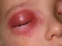
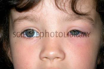
3 Triaging Ocular Complaints Periorbital swelling Surrounding eye redness i.e periorbital cellulitis Commonly seen in children Requires IV antibiotics and admission Complications: CNS infection (meningitis, epidural abscess, subdural empyemas, brain abscess), orbital involvment (cellulitis, abscess), Cavernous sinus thrombosis, Toxic shock syndrome, Eschar formation leading to scarring
4 Triaging Ocular Complaints Facial Symmetry Proptosis Facial droop Consider Bells Palsy/CVA
5 Ocular Emergencies Vision loss Closed-angle glaucoma Retinal Detachment FB Orbital Fracture Corneal Abrasion/Lacerations Chemical Burn Central Retinal Artery Occlusion Ruptured Globe Retrobulbar hematoma
6 Acute Painless Visual Loss Differential Diagnosis CVA Central Retinal Artery Occlusion Central Retinal Vein Occlusion Wet Macular Degeneration Vitreous Hemorrhage Retinal Detachment
7 Retinal Detachment True Emergency Painless vision loss Associated with Vascular disorders Congenital malformations Metabolic disarray Trauma Shrinking of the vitreous Myopia Degeneration And less commonly with diabetic retinopathy and uveitis Separation of the neurosensory layer of the retina from the underlying choroid and retinal pigment epithelium


8 Presentation Black curtain coming down over visual field Visual loss: sudden and starts peripherally. Bright flashes of light, floaters, fine dots, cobwebs Visual acuity correlates with the extent of macular involvement

9 Physical Findings Afferent pupillary defect is possible CHECK for consensual reaction The retina may appear gray or translucent or may seem out of focus

10 Treatment Early diagnosis and treatment are imperative preserve vision Consult Optho ASAP Retina needs to be replaced onto underlying nourishing layers. Laser photocoagulation or cyrotherapy Surgical repair to fix tear

11 Vitreous Hemorrhage Bleeding into preretinal space or the vitreous cavity itself. Difficult to distinguish from a retinal detachment Painless vision loss Floaters, cobwebs No afferent pupil defect This will help distinguish from a retinal detachment
12 Central Retinal Artery Occlusion Embolus from the carotid artery that lodges in the ophthalmic artery (leads to an ocular stroke) Visual complaints Caused by ischemia s/s: extremely sudden, acute unilateral PAINLESS vision loss


13 Central Retinal Artery Occlusion Ocular exam Cherry red spot on fundoscopic exam (cilioretinal artery will maintain perfusion of macula, so the macula appears pink and healthy against the ischemic retina) Listen for bruits over carotids, tenderness over the temporal arteries Examine for AFIB
14 Treatment Immediate! Visual loss is usually irreversible after 2 hours of ischemia Intermittent globe massage to dislodge clot Anterior chamber paracentesis and IV Acetazolamide Both decrease IOP & allow for better perfusion of the retinal artery Inhaled Carbogen (mixture of O2 and CO2) Dilates the vasculature, thereby increasing retinal PO2 Admit to the hospital if sudden vision loss To determine the underlying cause

 Non-ischemic (venous stasis")
15 Central Retinal Vein Occlusion Risk Factors CV disease, HTN, Glaucoma, Venous stasis, hypercoaguable conditions, collagen vascular disease, diabetes Two Types Ischemic (aka hemorrhagic retinopathy) Non-ischemic (venous stasis retinopathy)
16 Types Ischemic Acute and profound vision loss Afferent pupil defect Non-Ischemic Progressive blurry vision Worse in the morning BOTH Funduscopic exam shows Edema to optic disc and macula Dilated retinal veins Retinal hemorrhage Cotton wool spots
17 Diagnostic Tests Diagnosis of exclusion Excluding all the other processes that cause painless visual loss
18 Treatment Immediate optho consult Search for a cause In order to protect the contralateral eye No specific treatment exists Interventions (which are outside the scope of the ER) Laser photocoagulation Steroids
 Corneal Inflammation/Infection Dacryocystitis")
19 The Red Eye FB Iritis Keratoconjunctivitis Narrow-angle glaucoma Pterygium Scleritis Subconjunctival hemorrhages Differential Diagnosis Blepharitis Canaliculitis Conjunctivitis (viral, bacterial, allergic) Corneal Inflammation/Infection Dacryocystitis Episcleritis
20 ?
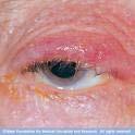
21 Blepharitis Inflammation of the eyelids usually involving the lid margins Often associated with conjunctivitis
22 ?

23 Canaliculitis Characterized by a mildly red eye Usually unilateral Slight discharge that can be expressed from the canaliculus
24 ?
25 Conjunctivitis Vascular dilation Inflammation of the mucous membranes that line the sclera and the inner eyelids Cellular infiltration Exudation
26 Types Viral Adenovirus-most common Extremely contagious Itchy, watery discharge Herpes simplex Usually unilateral, watery discharge Photophobia and FB sensation Look for dendritic pattern on slit lamp/fluorescein exam Look for facial vesicles Hutchinson s sign Seen with herpes zoster Lesion on the tip of the nose Involvement of the nasociliary branch of the 5 th nerve Much higher likelihood of ocular involvement


27 Types Bacterial Erythema, fb sensation, purulent drainage, morning crusting of the eye. Staph, Strep and Heamophilus Gonococcal Usually unilateral Seen in infants, health care workers and sexually active young adults May also have urethritis or arthritis Usually more discharge than other bacterial infections
28 Types cont d Chlamydial Sexually active young adults and neonates May have associated GC Urethral discharge and arthritis Scant seropurulent eye dishcarge Pseudomonas Immune compromised Contact lens wearers Yellow green discharge, sticky Inspect for corneal perforation which is a major concern with this organism Allergic Typically bilat. Watery discharge Might see chemosis Fungal Actinomyces, Aspergillus, candida, coccidioides and mucor Diabetic pts immunocompromised pts and pts with eye trauma with vegetable matter May have corneal infiltration May see hypopyon Chemical
29 Treatment Supportive Warm compresses Artificial tears Alleviates photophobia and keratitis Broad spectrum antibiotic drops (prevent superinfection w/ other bugs) Erythromycin For uncomplicated cases Fluoroquinolones Cover for pseudomonas Use in contact lenses wearers Topical Corticosteroids Only if consult optho NEVER USE IN PT WITH SUSPECTED HERPES Adenovirus Decongestants Lubricants Herpes Simplex Topical antivirals Vidarabine 3% Trifluridine 1% Herpes Simplex Ophthalmicus Systemic antivirals
30 Treatment Uncomplicated Bacterial Conjunctivitis Erythromycin Sulfacetamide 10% Gentamicin 0.3% Neosporin Cipro Gonococcal Emergent optho consult Topical and parenteral antibiotic therapy IM Ceftriaxone Frequent eye irrigation Prevent corneal perforation Chlamydial Oral and topical antibiotics Fungal Natamycin 5% Allergic Supportive Diphenhydramine


31 Ocular Foreign Bodies Generally superficial Photophobia, fb sensation, tearing, conjunctival injection & lid/corneal edema +/-flare ant chamber +/- visual acuities decrease Metallic FB RUST RING


32 Corneal Abrasion One of the most common ocular injuries 10% of ED visits related to eye complaints Abrasion of the corneal epithelium More common in contact lens wearers Severe injuries can involve the deeper thicker stromal layers
33 Corneal Abrasions Presentation Fb sensation, pain, watery discharge, photophobia If over the pupil, then may have decreased VA Physical exam findings Proparacaine first to control pain Evert eyelid Don t miss FB Fluoroscein exam with slit lamp Uptake-blue light Cell/Flare-white light Hyphema-white light Treatment for both FB and Corneal abrasions Update TDAP Preventative antibiotic drops If contact lens wearer, cover for pseudomonas Cycloplegic Homatropine Relief from photophobia and blepharospasm AVOID PATCHING May lead it increased infection No contact lens use while healing.
34 Corneal Ulcers Infectious in etiology Pseudomonas in contact lens wearers S/S: Fb sensation, pain, watery or purulent discharge, photophobia If over the pupil, then may have decreased VA


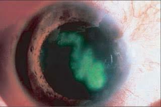
35 Slit lamp exam- Corneal Abrasion vs Corneal Ulcer
36 Treatment of Corneal Ulcer Immediate optho consult Cycloplegic agent Optho adovacate discharge with follow up the next day Homatropine for comfort Frequent topical antibiotic therapy A drop every 1-2 hours until follow up the next day
37 ?
38 Corneal Infection Decreased visual acuity Photophobia Severe pain Opacification of the cornea OPHTHALMIC EMERGENCY!!
39 ?
40 Dacrocystitis Localized pain,edema, erythema over the lacrimal sac at the medial canthus Usually unilateral Often purulent drainage
41 ?
42 Episcleritis Differentiated from the injection of themore superficial conjunctival vessels and from the deeper scleral vessels. Unlike Conjunctivitis, the inflammation tends to be limited to an ISOLATED PATCH Hx of recurrent episodes is common Mild to moderate tenderness over the area of injection
43 ?
44 Iritis Peri-limbal flush due to dilation of the radial vessels Compared to conjunctivitis, in which the intensity of the vascular engorgement decreases toward the limbus +/- decreased visual acuity Usually unilateral
 Oral anlagesics")
45 Traumatic Iritis Onset 1-4 days after trauma Presentation and PE Eye pain, photophobia Impaired vision Peirlimbal conjunctival injection Cells and flare in the anterior chamber Contricted,weakly dilated pupil Treatment Cycloplegic Homatropine 5% Topical steroid (consult optho) Oral anlagesics

46 ?

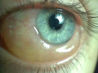
47 Subconjunctival Hemorrhage May occur spontaneously i.e. If pt is on anticoagulants Or secondary to trauma i.e. Increased pressure: singing, screaming etc May appears as a flat thin Hemorrhage or a thicker collection of blood No treatment required Absorbs on its own

48 ?
49 Hyphema Mostly due to trauma. Post-injury accumulation of blood in the anterior chamber The agent producing a hyphema is usually projectile Spontaneous hyphemas are rare. i.e secondary to neoplasms, vascular anomalies, neovascularization such as from DM, ischemia
50 Hyphemas Present with eye pain, decreased va, photophobia. Diagnosis is made by looking for blood in anterior chamber May see with your naked eye Esp if pt is upright and it has had time to layer out Slit lamp exam May see cells in anterior chamber or layered blood If large, then check IOP

51 Treatment stop damage to vision Elevate pt s head Dilate the pupil Avoid pupillary play Topical B Blockers (lower IOP) Topical carbonic anhydrase inhibitor Systemic acetazolamide or mannitol Avoid ASA/antiplatelet meds Pain control Surgery to remove clot When other therapies do not work. Complications: rebleeding
52 ?
53 Chemosis Cornea is recessed and the conjunctiva is swollen Due to allergic reaction. Treat with antihistamines
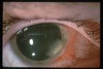
54 Narrow (Closed) Angle Glaucoma Presentation >50 years and older Unilateral severely painful red eye and head pain Nausea, vomiting common Halos around light are common IOP elevated Reduced visual acuity Immediate referral to optho

55 Diagnostic testing Visual Acuities Slit-lamp exam Depth of anterior chamber If <¼ of the corneal thickness, then chamber is narrow. Likely done by Optho and not in the ER May have a middilated pupil and corneal haziness IOP measurement with tonometer
56 Treatment Ophthalmologic emergency- Consult Optho ASAP! Therapy is geared toward Decreasing the aqueous production Increasing aqueous outflow Reducing vitreous volume to lower IOP Topical B-blockers Timolol (decrease aqueous production) Parasympathomimetic Miotic Agent Pilocarpine (improves aqueous outflow) Carbonic Anhydrase Inhibitor Acetazolamide (limits aqueous humor formation) Osmotic Diuretic Mannitol (creates an osmotic gradient b/w the vitreous and the blood to cause vitreous volume reduction).

57 Chemical Burns Presentation Eye pain, limited visual acuity Corneal cloudiness Scleral whitening Eye may be erythematous or whitened Chemosis Vascular engorgement FB sensation Physical Findings Punctate lesions on the cornea Conjunctival injection Decreased visual acuity
58 Chemical Burn A TRUE OCULAR EMERGENCY Test ph IRRIGATE, IRRIGATE, IRRIGATE 30 minutes using IV NS or LRs with morgans lens. Morgans lens alternatives Use the tubing and tape over pt s eye. Acid vs Alkali Alkali causes necrosis. Will destroy vessels and denature collagen (Lipophilic- so it absorbs into the eye more easily, causing more damage) i.e household cleaners (bleach), fertilizers Acid also causea necrosis. Less common i.e sulfuric acid (automobile batteries), industrial cleaners

59 Ruptured Globe From penetrating trauma to cornea or sclera Extravasation of the intraocular contents May lead to irreversible vision loss or Endophthalmitis
60 Ruptured Globe S/S: pain, decreased vision, hyphema, tear drop pupil, severe subconjunctival hemorrhage Management Immediately place an eye shield to protect eye from further manipulation DO NOT PERFORM TONOMETRY

61 Orbital and Periorbital Celllulitis Orbital cellulitis without treatment causes blindness and death in 20% of patients Venous drainage of the orbital region communciates with vessint in the brain via the cavernous since, infection can progress rapidly
62 Peds! Preseptal cellulitis is more common than orbital cellulitis. 2 pediatric case series 94% and 87% of cases, respectively, were dx as pre-septal cellulitis. Remainder were dx as orbital cellulitis. accordingly, most of the data regarding these infections comes from studies in children. Pub Med references

63 Orbital Septum Orbital Cellulitis Tissues within the orbit posterior to the orbital septum Periorbital Cellulitis Confined to the tissues anterior to the septum
64
65
66 Presentation Erythema, edema, warmth of the external eye tissues Unilateral usually Infection can be bilat Fever and malaise Ocular pain, ophthalmoplegia, pain with movement of EOM SUGGESTS ORBITAL INFECTION May also have decreased VA and pupillary paralysis
67 Treatment Pre-orbital infection Oral antibiotics and close f/u outpt Lower threshold for admission in children Cover for staph, strep, enterobacteriaceae Clindamycin in children and adults or Trimethoprim-sulfamethoxazole (TMP-SMX; in children and adults plus one of the following: Amoxicillin or Amoxicillin-clavulanic acid or Cefpodoxime or Cefdinir In pts with comorbid conditions, admit!
68 Treatment Orbital celllulitis Broad spectrum IV antibotics Vancomycin in children and adults plus one of the following: Ceftriaxone in children and adults or Cefotaxime in children and adults or Ampicillin-sulbactam in children and adults or Piperacillin-tazobactam in children and adults Admit I&D if imaging reveals collection
69 References Adams Up to date rbital+cellulitis+children&selectedtitle=1~14# H bital+cellulitis+children&selectedtitle=1~25&s ectionrank=1&anchor=h24#h24
Ocular Lecture. Sue Bednar NP Ali Atwater PA-C
 Ocular Lecture Sue Bednar NP Ali Atwater PA-C Triaging Ocular Complaints Painful Eye/Red eye +/-blurry vision +/-visual loss +/-floaters +/-fevers If any of the above findings exist, pt is likely to have
10/4/2013. Bruce K.Williams, MSN, RN,ACNP-BC Sisters of Charity Providence Hospitals. What is the worst thing that can go wrong with an eye?
 Red Eyes, Red Alert! Bruce K.Williams, MSN, RN,ACNP-BC Sisters of Charity Providence Hospitals Red Eyes, Red Alert! Red Eyes, Red Alert! What is the worst thing that can go wrong with an eye? 1 Red Eyes,
EYE TRAUMA: INCIDENCE
 Introduction EYE TRAUMA: INCIDENCE 2.5 million eye injuries per year in U.S. 40,000 60,000 of eye injuries lead to visual loss Introduction Final visual outcome of many ocular emergencies depends on prompt,
Acute Eyes for ED. Enis Kocak. The Alfred Ophthalmology
 Acute Eyes for ED Enis Kocak The Alfred Ophthalmology The problem with eyes Things to cover Ocular anatomy Basic assessment Common presentations Eye first aid and procedures Ophthalmic emergencies What
Ocular Urgencies and Emergencies
 Ocular Urgencies and Emergencies Pam Boyce, O.D., F.A.A.O. Boyce Family Eye Care, Ltd. 528 Devon Ave. Park Ridge, IL 60068 847-518-0303 Somebody s going to lose an eye Epidemiology 2.4 million ocular and
10 EYE EMERGENCIES. Who goes, who you better not send! Brant Slomovic, MD, FRCPC University Health Network
 10 EYE EMERGENCIES Who goes, who you better not send! Brant Slomovic, MD, FRCPC University Health Network DISCLOSURES I have none PVD CASE 1 WHAT IS A PVD? a process of aging (45-55) liquefaction of vitreous
Phone Triage for Optometric Staff ???????? CHEMICAL BURN CHEMICAL BURN
 Phone Triage for Optometric Staff There are very few ocular emergencies that you will have to deal with in practice, but it is imperative that you be able to Michelle Welch, O.D. NSU Oklahoma College of
Sepideh Tara Rousta, MD FAAO Robert Wood Johnson University Hospital Saint Peter s University Hospital Wills Eye Hospital
 Sepideh Tara Rousta, MD FAAO Robert Wood Johnson University Hospital Saint Peter s University Hospital Wills Eye Hospital 14 mo old w R eye cross (parents) 9 mo old R eye crossing getting worse for past
Dr Jo-Anne Pon. Dr Sean Every. 8:30-9:25 WS #70: Eye Essentials for GPs 9:35-10:30 WS #80: Eye Essentials for GPs (Repeated)
") Dr Sean Every Ophthalmologist Southern Eye Specialists Christchurch Dr Jo-Anne Pon Ophthalmologist Southern Eye Specialists, Christchurch Hospital, Christchurch 8:30-9:25 WS #70: Eye Essentials for GPs
Dr Sean Every Ophthalmologist Southern Eye Specialists Christchurch Dr Jo-Anne Pon Ophthalmologist Southern Eye Specialists, Christchurch Hospital, Christchurch 8:30-9:25 WS #70: Eye Essentials for GPs
PAINFUL PAINLESS Contact lens user BOV
 Common Causes Allergies Infections Ocular Cornea, uveitis, endophthalmitis Orbital Orbital cellulitis Inflammation Uveitis Scleritis / episcleritis Glaucomas Trauma Foreign bodies Chemical injuries History
5/2/2016 EYE EMERGENCIES. Nathaniel Pelsor, O.D., FAAO Talley Medical-Surgical Eye Care Associates. Anatomy. Tools
 EYE EMERGENCIES Nathaniel Pelsor, O.D., FAAO Talley Medical-Surgical Eye Care Associates Anatomy Tools 1 Contact dermatitis Blepharitis HSV Preseptal Cellulitis Anterior Chamber Subconjunctival hemorrhage
Index. C Canalicular system, 4 Carbonic anhydrase inhibitors, 29 30
 A Acanthamoeba keratitis (AK), 82, 83 Acute angle-closure crisis, 156 Acute angle-closure glaucoma (AACG), 121, 141, 284 causes of, 122 clinical presentation, 153 evaluation, 156 157 management/treatment,
Assessment and Management of Ocular Trauma. Disclosure I have no direct financial interests in today s subject matter. 3/25/2019. Normal Eye Anatomy
 Assessment and Management of Ocular Trauma Samiksha Fouzdar Jain, MD,FRCS Department of Ophthalmology & Visual Sciences Truhlsen Eye Institute Disclosure I have no direct financial interests in today s
OPHTHALMOLOGY REFERRAL GUIDE FOR GPS
 OPHTHALMOLOGY REFERRAL GUIDE FOR GPS A guidebook to support general practitioners in the management and referral of a range of common eye problems. Contents 3 Introduction 4 Ophthalmic Workup 6 Acute Visual
THE RED EYE Cynthia McNamara, MD Week 25
 THE RED EYE Cynthia McNamara, MD Week 25 Educational Objectives: 1. Know the differential diagnosis and presentation of specific etiologies of the red eye 2. Be able to evaluate patients presenting with
UC SF. g h. Eye Trauma. Martha Neighbor, MD Emergency Services San Francisco General Hospital University of California
 UC SF Eye Trauma sf g h Martha Neighbor, MD Emergency Services San Francisco General Hospital University of California Goals Recognize vision threatening eye emergencies Treat them when we can Know when
8/30/2018. Eye Disorders. Patrick Sarte. Anatomy of the Eye Uveitis Scleritis vs. Episcleritis Glaucoma Retinal Findings Eyelids
 Eye Disorders Patrick Sarte Anatomy of the Eye Uveitis Scleritis vs. Episcleritis Glaucoma Retinal Findings Eyelids 1 Anatomy of the Eye Anatomy of the Eye 2 Anatomy of the Eye 3 4 A 26 year old woman
LECTURE # 7 EYECARE REVIEW: PART III
 LECTURE # 7 EYECARE REVIEW: PART III HOW TO TRIAGE EYE EMERGENCIES STEVE BUTZON, O.D. EYECARE REVIEW: HOW TO TRIAGE EYE EMERGENCIES FOR PRIMARY CARE PHYSICIANS Steve Butzon, O.D. Member Director IDOC President
The Emergent Eye in the Acute Setting
 The Emergent Eye in the Acute Setting Todd P. Margolis MD, PhD Professor of Ophthalmology & Director of the F.I. Proctor Foundation UCSF Physical Exam-- Visual Acuity Essential Corrected visual acuity
2/5/2018. Trauma. Subdivided into two main categories: Closed globe Open Globe
 1 2 3 4 5 Ocular Trauma Guide for Eye Care Office Staff Winter Thaw 2018 Aaron Yatskevich OD Definition A broad term used to describe a physical or chemical wound to the eye or eye socket. Ocular trauma
Identify the choice that best completes the statement or answers the question.
 Chapter 5. The Eye Multiple Choice Identify the choice that best completes the statement or answers the question. 1. The most common type of eye disorder is: A. Refractive errors B. Macular conditions
EYE INJURIES OBJECTIVES COMMON EYE EMERGENCIES 7/19/2017 IMPROVE ASSESSMENT OF EYE INJURIES
 EYE INJURIES BRITTA ANDERSON D.O. DMC PRIMARY CARE SPORTS MEDICINE ASSOCIATE TEAM PHYSICIAN DETROIT TIGERS OBJECTIVES IMPROVE ASSESSMENT OF EYE INJURIES UNDERSTAND WHAT IS CONSIDERED AN EMERGENCY DEVELOP
Differential diagnosis of the red eye. Carol Slight Nurse Practitioner Ophthalmology
 Differential diagnosis of the red eye Carol Slight Nurse Practitioner Ophthalmology The red eye Conjunctivitis HSV Keratitis Acute angle closure glaucoma Anterior Uveitis Red eye Scleritis Subconjunctival
Andrew J. Hendershot, MD Havener Eye Institute The Ohio State University s Wexner Medical Center
 Ocular Trauma for the Primary Care Physician Andrew J. Hendershot, MD Havener Eye Institute The Ohio State University s Wexner Medical Center Relevance Often those with minor eye injuries will first seek
OPHTHALMOLOGIC PEARLS FOR THE NON- OPHTHALMOLOGIST. David G. Gross D.O. Deen-Gross Eye Centers Merrillville-Hobart Deengrosseye.
 OPHTHALMOLOGIC PEARLS FOR THE NON- OPHTHALMOLOGIST David G. Gross D.O. Deen-Gross Eye Centers Merrillville-Hobart Deengrosseye.com A FEW OF THE AREAS WE WILL DISCUSS Red Eye Glaucoma Neuro ophthalmic tid
Clinical Practice Guide for the Diagnosis, Treatment and Management of Anterior Eye Conditions. April 2018
 Clinical Practice Guide for the Diagnosis, Treatment and Management of Anterior Eye Conditions This Clinical Practice Guide provides evidence-based information about current best practice in the management
Ophthalmic Trauma Update
 Ophthalmic Trauma Update Richard S. Davidson, M.D. Professor of Ophthalmology Vice Chair for Quality and Clinical Affairs UCHealth Eye Center University of Colorado School of Medicine August 5, 2017 Financial
OOGZIEKTEN VOOR DE HUISARTS F. GOES, JR.
 OOGZIEKTEN VOOR DE HUISARTS F. GOES, JR. HET RODE OOG F. GOES, JR. Condition Signs Symptoms Causes Conjunctivitis Viral Normal vision, normal pupil size Mild to no pain, diffuse Adenovirus (most common),
Ocular and Periocular Trauma. Tina Rutar, MD. Assistant Professor of Ophthalmology and Pediatrics. Director, Visual Center for the Child
 Ocular and Periocular Trauma Tina Rutar, MD Assistant Professor of Ophthalmology and Pediatrics Director, Visual Center for the Child University of California, San Francisco Phone: 415-353-2560 Fax: 415-353-2468
Work Sheet And Course Hand Out
 Work Sheet And Course Hand Out This course provides the primary care health professional with a basic understanding of the eye, its function and the assessment of common sight- and non-sight threatening
Mild NPDR. Moderate NPDR. Severe NPDR
 Diabetic retinopathy Diabetic retinopathy is the most common cause of blindness in adults aged 35-65 years-old. Hyperglycaemia is thought to cause increased retinal blood flow and abnormal metabolism in
DISCLOSURES. PEDIATRIC RED EYES Rachel M. Smith, OD, FCOVD HISTORY, HISTORY, HISTORY WHY RED EYES? EXAMINE THE EYE RED FLAGS TO REFER 3/25/2019
 DISCLOSURES Consultant/Speakers bureaus Research funding PEDIATRIC RED EYES Rachel M. Smith, OD, FCOVD Pediatric Optometrist Children s Hospital & Medical Center Stock ownership/corporate boards employment
Management of specific eye problems in the ED
 of specific eye problems in the ED CORNEAL ABRASION Causes Foreign bodies Tangential shearing injuries, e.g. poking finger into eye Exact cause of injury (Remember to exclude possibility of intraocular
MRI masterfile Part 5 WM Heme Strokes.ppt 1
 Ocular and Orbital Trauma Eye Trauma: Incidence 1.3 million eye injuries in the US per year. 40,000 of these injuries lead to blindness in the US. Patrick Sibony, MD March 23, 2013 Ophthalmic Emergencies
9/23/2014. Emily Thomas, O.D. MOA Paraoptometric Education October 5, 2014
 Emily Thomas, O.D. MOA Paraoptometric Education October 5, 2014 Anterior toward the front of the body Posterior toward the rear of the body Unilateral only one eye involved Bilateral both eyes involved
A Case of Carotid-Cavernous Fistula
 A Case of Carotid-Cavernous Fistula By : Mohamed Elkhawaga 2 nd Year Resident of Ophthalmology Alexandria University A 19 year old male patient came to our outpatient clinic, complaining of : -Severe conjunctival
Ocular and periocular trauma
 Ocular and periocular trauma No financial disclosures. Tina Rutar M.D. Assistant Professor of Clinical Ophthalmology and Pediatrics Director, Visual Center for the Child University of California San Francisco
Ophthalmology PANRE Review. Brock Phillips, PA-C
 Ophthalmology PANRE Review Brock Phillips, PA-C I am not an ophthalmologist, optometrist or certified eye guy of any sort - I am a practicing UC/EM PA-C who frequently evaluates eye/vision complaints,
THE 35 GOLDEN EYE RULES
 THE 35 GOLDEN EYE RULES The Sense of Sight, from La Dame a la Licorne, The Lady and the Unicorn Tapestries, Late 15th Century Flemish Tapestry in wool and silk, Musée Nationale du Moyen Age, Paris. 1.
DIAGNOSIS MANAGEMENT CONSULTATION DISPOSITION
 1.Caustic keratoconjunctivitis 2.Blepharitis Inflammation of eyelid margins often a/w crusts on awakening, FB sensation, and tearing. 3.Chalazion Inflammation of meibomian gland causing subcutaneous nodule
Case Follow Up. Sepi Jooniani PGY-1
 Case Follow Up Sepi Jooniani PGY-1 Triage 54 year old M Pt presents to prelim states noticed today he had reddness to eyes, states worse in R eye. Pt denies any pain or itching. No further complaints.
Ophthalmology. Corneal Abrasion. History
 Ophthalmology Corneal Abrasion - Usually clear history of very recent trauma - Foreign Body Sensation - Pain +++ - Lacrimation - Photophobia Fig. 1 Corneal Abrasion - Abrasion stains yellow / green with
Examining Children s Eyes
 Paediatric Ophthalmology What to refer & when? Aims Tips for assessing a child s eyes in general practice Common paediatric ophthalmology symptoms and signs What needs to be referred and when? MISS FARIHA
Ocular Emergencies. Pisit Preechawat, MD Department of Ophthalmology, Ramathibodi Hospital
 Ocular Emergencies Pisit Preechawat, MD Department of Ophthalmology, Ramathibodi Hospital Ocular Anatomy Bony Components of Orbit 1 1. Frontal bone 4 5 7 6 2. Zygomatic bone 3. Maxillary bone 4. Sphenoid
The Red Eye: Conjunctivitis, Iritis, or Worse? Sean P. Donahue, MD, PhD
 The Red Eye: Conjunctivitis, Iritis, or Worse? Sean P. Donahue, MD, PhD Sam and Darthea Coleman Chair Vice Chair of Clinical Affairs, Department of Ophthalmology Professor of Pediatrics, Ophthalmology,
Eye infections. Hossain Jabbari, MD, MPH, ID & TM Infectious Diseases Dept., Digestive Diseases Research Institute (DDRI) TUMS
 TUMS") Eye infections Hossain Jabbari, MD, MPH, ID & TM Infectious Diseases Dept., Digestive Diseases Research Institute (DDRI) TUMS Eye: An overview Eye: An overview The eye is one of the most complex parts
Eye infections Hossain Jabbari, MD, MPH, ID & TM Infectious Diseases Dept., Digestive Diseases Research Institute (DDRI) TUMS Eye: An overview Eye: An overview The eye is one of the most complex parts
The Red Eye DIFFERENTIATE RED EYE DISORDERS RED EYE: POSSIBLE CAUSES SUBJECTIVE EYE COMPLAINTS ETIOLOGIES OF RED EYE RED EYE: CAUSE AND EFFECT
 Introduction The Red Eye John Knapp, MD DIFFERENTIATE RED EYE DISORDERS Needs immediate treatment Needs treatment within a few days Does not require treatment Introduction SUBJECTIVE EYE COMPLAINTS Evaluation
Ophthalmology for Primary Care: Do I Really Need to See It? Jennifer R. Olbum, DO
 Ophthalmology for Primary Care Jennifer Olbum, DO CCOM 1988 (Midwestern U.) Medical Retina subspecialist jenolbum@aol.com Daniel J. Nadler, MD LLC Beaver, PA Everett & Hurite Ophthalmic Assoc. Belle Vernon,
CORNEAL CONDITIONS CORNEAL TRANSPLANTATION
 GENERAL INFORMATION CORNEAL CONDITIONS CORNEAL TRANSPLANTATION WHAT ARE CORNEAL CONDITIONS? The cornea is the clear outer layer of the eye. Shaped like a dome, it helps to protect the eye from foreign
Red Eye & Ocular Emergencies. Zafar Shamoon Director of Emergency Services Dearborn Beaumont Hospital
 Red Eye & Ocular Emergencies Zafar Shamoon Director of Emergency Services Dearborn Beaumont Hospital I have no relevant financial relationship with any manufacturers of any commerical products and or providers
SILA THONGLAI MD. Bangkok Eye center Bangkok Hospital Thailand
 SILA THONGLAI MD. Bangkok Eye center Bangkok Hospital Thailand Ocular Anatomy Bony Components of Orbit 1 1. Frontal bone 4 5 7 6 2. Zygomatic bone 3. Maxillary bone 4. Sphenoid bone 5. Ethmoid bone 2 3
I Spy A Red Eye: Assessment & Management of Common Ocular Conditions In Primary Care
 I Spy A Red Eye: Assessment & Management of Common Ocular Conditions Jody Agins MSN, RNP, FNP/GNP-BC mrsjaginsnp@gmail.com Objectives Review procedures for assessment of conjunctival inflammation Differentiate
Focusing on A&E. By Sandy Cooper, (Ophthalmic Nurse Practitioner), Tel
, Tel") Focusing on A&E By Sandy Cooper, (Ophthalmic Nurse Practitioner), Tel 01752 439331 Email sandra.cooper5@nhs.net sandracooper041@btinternet.com THINGS TO WORRY ABOUT WITH ANY EYE PROBLEM CHANGES IN VISION
Focusing on A&E By Sandy Cooper, (Ophthalmic Nurse Practitioner), Tel 01752 439331 Email sandra.cooper5@nhs.net sandracooper041@btinternet.com THINGS TO WORRY ABOUT WITH ANY EYE PROBLEM CHANGES IN VISION
Mom, There s Something Wrong With My Eye
 Mom, There s Something Wrong With My Eye Veeral Shah MD, PHD Texas Children's Hospital Most Common Issues Seen by the Pediatrician Emergent Ocular Issues Seen by the Pediatrician 1 What does this baby
Paediatric acute ophthalmology. Harry Bradshaw
 Paediatric acute ophthalmology Harry Bradshaw Approach Red eye Leukocoria Neurological Trauma Visual loss Red eye Orbital Eyelid Conjunctiva Cornea Uvea Orbital Orbit fixed volume Contiguous with sinuses,
Note: This is an outcome measure and can be calculated solely using registry data.
 Measure #191 (NQF 0565): Cataracts: 20/40 or Better Visual Acuity within 90 Days Following Cataract Surgery -- National Quality Strategy Domain: Effective Clinical Care DESCRIPTION: Percentage of patients
a.superficial (adenoid layer).contain lymphoid tissue.
.contain lymphoid tissue.") Conjunctiva Dr. saifalshamarti Anatomy Microscopic: 1.Epithelium (non keratinized,includes goblet cell). 2.Epithelial basement membrane. 3.Stroma : a.superficial (adenoid layer).contain lymphoid tissue.
Conjunctiva Dr. saifalshamarti Anatomy Microscopic: 1.Epithelium (non keratinized,includes goblet cell). 2.Epithelial basement membrane. 3.Stroma : a.superficial (adenoid layer).contain lymphoid tissue.
OCCLUSIVE VASCULAR DISORDERS OF THE RETINA
 OCCLUSIVE VASCULAR DISORDERS OF THE RETINA Learning outcomes By the end of this lecture the students would be able to Classify occlusive vascular disorders (OVD) of the retina. Correlate the clinical features
Scrub In. What is the function of vitreous humor? What does the pupil do when exposed to bright light? a. Maintain eye shape and provide color vision
 Scrub In What is the function of vitreous humor? a. Maintain eye shape and provide color vision b. Maintain eye shape and refract light rays c. Provide night vision and color vision d. Provide night vision
Ophthalmology for Primary Care Providers
 Ophthalmology for Primary Care Providers Bob Avery, MD, PhD Ophthalmology/Surgery University of New Mexico School of Medicine bavery@salud.unm.edu Preview How the eye works Basic eye exam The red eye Acute
Preview. Ophthalmology for Primary Care Providers. Useful references. How the eye works
 Preview Ophthalmology for Primary Care Providers Bob Avery, MD, PhD How the eye works The red eye Acute eye conditions Chronic vision loss Basic eye exam Ophthalmology/Surgery University of New Mexico
PRECISION PROGRAM. Injection Technique Quick-Reference Guide. Companion booklet for the Video Guide to Injection Technique
 Injection Technique Quick-Reference Guide PRECISION PROGRAM Companion booklet for the Video Guide to Injection Technique Available at www.ozurdexprecisionprogram.com Provides step-by-step directions with
Emergency Ophthalmology Lawrence B. Stack, MD Handout can be found on lbstack.com/students/eye-handout.pdf
 Emergency Ophthalmology Lawrence B. Stack, MD Handout can be found on lbstack.com/students/eye-handout.pdf Summary Points: 1. Consult Ophthalmology if you can not account for change in visual acuity 2.
TENTATIVE DIAGNOSES Based on the information provided so far, what are the potential diagnoses?
 Case Study #4 PEDIATRIC CASE STUDY SCENARIO Mary Jennings has brought her son Joe to your office. Joe is a 6-year old Jordanian male. He presents with the complaint of an itchy red eye. Mary states that
Red Eyes, Red Spots, and Red Flags
 Red Eyes, Red Spots, and Red Flags Essential Knowledge of Eye Disease Andrew F. Calman, MD, PhD Associate Clinical Professor of Ophthalmology and Family & Community Medicine, UCSF Seeing Red Red Eyes Common
Ocular Emergencies. What is an emergency to the patient is not necessarily an emergency to the staff
 OCULAR EMERGENCIES Ophthalmic Photographers Society November 15, 2013 Michael A. DellaVecchia MD PhD FACS Wills Eye Emergency Department Philadelphia PA Ocular Emergencies What is an emergency to the patient
Page 1 RED EYES. conjunctivitis keratitis episcleritis / scleritis. Frank Larkin Moorfields Eye Hospital. acute glaucoma anterior uveitis
 The RED EYE and ALLERGIC EYE DISEASE DIAGNOSIS & MANAGEMENT Frank Larkin Moorfields Eye Hospital RED EYES conjunctivitis keratitis episcleritis / scleritis acute glaucoma anterior uveitis post-op. / trauma
By Darlene Jones, Nurse. May 2017
 By Darlene Jones, Nurse May 2017 Disclosure of potential conflict of interest Darlene Jones, Nurse I have no conflict of interest Course objectives Become familiar with the different pathologies in ophthalmology
DEFINITION Corneal abrasion is a defect in the corneal surface epithelium due to scraping or rubbing of the corneal epithelium.
 DEFINITION Corneal abrasion is a defect in the corneal surface epithelium due to scraping or rubbing of the corneal epithelium. IMMEDIATE CONSULTATION REQUIRED IN THE FOLLOWING SITUATIONS Dendritic pattern
Dry Eye Assessment and Management Study ELIGIBILITY OCULAR EVALUATION FORM
 Page 1 of 13 BEFORE COMPLETING THE OCULAR EXAMINATION, YOU MUST BE ABLE TO ANSWER YES TO THE FOLLOWING QUESTIONS: Have you done MMP9? (SVonly) The Following are done at Baseline: Have you done Tear Osmolarity?
A LITTLE ANATOMY. three layers of eye: 1. outer: corneosclera. 2. middle - uvea. anterior - iris,ciliary body. posterior - choroid
 GLAUCOMA A LITTLE ANATOMY three layers of eye: 1. outer: corneosclera 2. middle - uvea anterior - iris,ciliary body posterior - choroid connection at the pars plana between post and ant uvea 3. retina
Spring Eye Red Eye Terrence Clark, OD FAAO Brittney Gewolb, OD
 Spring Eye Red Eye 2017 Terrence Clark, OD FAAO Brittney Gewolb, OD RED EYES! Differential Diagnoses of the Red Eye Differential Diagnoses of the Red Eye Trauma Lids Cellulitis: preseptal vs. orbital
Bleeding in the anterior chamber, obstructing vision Caused by surgery, injury, coagulopathy, sickle cell or idiopathic Needs urgent care to prevent
 Bleeding in the anterior chamber, obstructing vision Caused by surgery, injury, coagulopathy, sickle cell or idiopathic Needs urgent care to prevent long-term vision loss TX by elevating head of bed, reducing
THE RED EYE When to treat, when to refer. Dr Beatrice Khater American University of Beirut November 2010
 THE RED EYE When to treat, when to refer Dr Beatrice Khater American University of Beirut November 2010 OBJECTIVES Identify most common causes of Red Eye Know the adequate management of these conditions
Ocular warning signs in GP practice: Paediatric Eye Pointers
 Ocular warning signs in GP practice: Paediatric Eye Pointers Dr Benjamin Chang MB, BCh, BAO, MMedSci, FRCS(Irel), FRCS(Edin), FRCOphth(Lond) Senior Consultant Ophthalmology and Visual Sciences Khoo Teck
02/03/2014. Average Length: 23mm (Infant ~16mm) Approximately the size of a quarter Volume: ~5mL
 Approximately the size of a quarter Volume: ~5mL") Identify the anatomy of the eye. Explain the basic physiology of the parts of the eye. Briefly discuss various surgeries related to different parts of the anatomy. Average Length: 23mm (Infant ~16mm) Approximately
Identify the anatomy of the eye. Explain the basic physiology of the parts of the eye. Briefly discuss various surgeries related to different parts of the anatomy. Average Length: 23mm (Infant ~16mm) Approximately
OCULAR HEMORRHAGES. ROSCOE J. KENNEDY, M.D. Department of Ophthalmology
 OCULAR HEMORRHAGES ROSCOE J. KENNEDY, M.D. Department of Ophthalmology Ocular hemorrhages are important not only because they produce visual loss but also because they usually indicate a disorder elsewhere
REFERRAL GUIDELINES: OPHTHALMOLOGY
 Outpatient Referral Guidelines Page 1 1 REFERRAL GUIDELINES: OPHTHALMOLOGY Date of birth Demographic Contact details (including mobile phone) Clinical Reason for referral Duration of symptoms Essential
Recurrent intraocular hemorrhage secondary to cataract wound neovascularization (Swan Syndrome)
") Recurrent intraocular hemorrhage secondary to cataract wound neovascularization (Swan Syndrome) John J. Chen MD, PhD; Young H. Kwon MD, PhD August 6, 2012 Chief complaint: Recurrent vitreous hemorrhage,
Recurrent intraocular hemorrhage secondary to cataract wound neovascularization (Swan Syndrome) John J. Chen MD, PhD; Young H. Kwon MD, PhD August 6, 2012 Chief complaint: Recurrent vitreous hemorrhage,
 Traumatic Cataract Orbital Wall Fracture Vitreous Hemorrhage Optic Disc Hemorrhage a) Amblyopia b) Strabismus c) Trauma Playing with other children Sports Fire works BB gun Injecting needles .
What THE EYE Case THE RED EYE. Case. Infections of the eye 2/3/2014
 Case THE RED EYE Richard A. Jacobs, M.D.,PhD* *Todd Margolis, M.D.,PhD, Prof of Ophthalmology and Director F. I. Proctor Foundation, UCSF Brian Schwartz, M.D., Assistant Professor of Medicine, Division
Handbook for Medical Students Learning Ophthalmology
 International Council of Ophthalmology Handbook for Medical Students Learning Ophthalmology 2009 Compiled by the Task Force on Undergraduate Teaching in Ophthalmology of the International Council of Ophthalmology
MRI masterfile Part 5 WM Heme Strokes.ppt 2
 Imaging of Orbital Trauma Corneal Abrasion CT scan is preferable to MRI Bone, Rapid, Easy to monitor patient Foreign bodies, air, hemorrhage Fractures Cost Needed for an MRI MRI Globe and intraocular injuries
Professor Helen Danesh-Meyer. Eye Institute Auckland
 Professor Helen Danesh-Meyer Eye Institute Auckland Bitten by Ophthalmology Emergencies Helen Danesh-Meyer, MBChB, MD, FRANZCO Sir William and Lady Stevenson Professor of Ophthalmology Head of Glaucoma
For details on measurement and recording of visual acuity, refer to Annex 1. VISION INTERPRETING RESULTS ABSTRACT
 management update on functional decline in older adults 2012 Unit No. 5 VISION Dr Au Eong Kah Guan, Ms Yulianti, Ms Fifiana ABSTRACT Among Singaporean adults of Chinese origin aged 40 to 79 years old,
Eyes, ears, teeth and everything in between
 Eyes, ears, teeth and everything in between E M E R G E N C Y D E P A R T M E N T J U N I O R T E A C H created 14/11/10 by S.R. Bruijns, version 1.0 Objectives Eyes Ears Teeth Maxilla- facial EYES Approaching
Department of Ophthalmology
 Department of Ophthalmology Period : 02/July/18 to 30/August/18 Semester : 7 th Semester Lecture Lesson Plan Sr. Date Topic Lesson plan Name of Faculty No. 1 02.07.18 Lens- Lens-Anatomy, Classification
VN 122 MODULE F EYE AND VISION DISORDERS OCULAR HISTORY 1. PATIENT PERCEPTION OF PROBLEM 2. DECREASED VISION? 3. BLURRED, DOUBLE, DISTORTED?
 VN 122 MODULE F EYE AND VISION DISORDERS OCULAR HISTORY 1. PATIENT PERCEPTION OF PROBLEM 2. DECREASED VISION? 3. BLURRED, DOUBLE, DISTORTED? 4. PAIN? QUALITY OF PAIN? 5. ITCHING, DRY? 6. BOTH EYES? 7.
PEDIATRIC OCULAR INJURIES. Sapna Tibrewal MD
 PEDIATRIC OCULAR INJURIES Sapna Tibrewal MD 1 Learning Objectives Learn to recognize the common pediatric ocular injuries Immediate management tips to be instituted in your office/ ER Know when to call
Test Bank for Medical Surgical Nursing An Integrated Approach 3rd Edition by White
 Test Bank for Medical Surgical Nursing An Integrated Approach 3rd Edition by White Link full download : http://testbankair.com/download/test-bank-for-medical-surgical-nursing-anintegrated-approach-3rd-edition-by-white/
Vision I. Steven McLoon Department of Neuroscience University of Minnesota
 Vision I Steven McLoon Department of Neuroscience University of Minnesota 1 Eye Cornea Sclera Conjunctiva 2 Eye The conjunctiva lines the inner surface of the eyelids and outer surface of the sclera. 3
Neovascular Glaucoma Associated with Cilioretinal Artery Occlusion Combined with Perfused Central Retinal Vein Occlusion
 Neovascular Glaucoma Associated with Cilioretinal Artery Occlusion Combined with Perfused Central Retinal Vein Occlusion Man-Seong Seo,* Jae-Moon Woo* and Jeong-Jin Seo *Department of Ophthalmology, Chonnam
HANDBOOK FOR JUNIOR RESIDENTS AND MEDICAL STUDENTS LEARNING EMERGENCY OPHTHALMOLOGY
 HANDBOOK FOR JUNIOR RESIDENTS AND MEDICAL STUDENTS LEARNING EMERGENCY OPHTHALMOLOGY Compiled by The Task Force on Undergraduate Teaching in Ophthalmology of the International Council of Ophthalmology and
Differential Diagnosis of Conjunctivitis and Keratoconjunctivitis
 Differential Diagnosis of Conjunctivitis and Keratoconjunctivitis Dr. Victor Malinovsky 2006 Mechanical-Physical Trauma Corneal Abrasions Abrasions (interpalpebral/variable): a focal loss of epithelium
HealthHarmonie Limited Minor Eye Conditions Services Guide
 HealthHarmonie Limited Minor Eye Conditions Services Guide 2017 Contents Introduction... 3 Experience, Qualifications and Competencies... 3 Required Equipment... 3 Clinical assessment... 3 Management of
Common EYE Disease. Patcharaporn Wangvoravit
 Common EYE Disease Patcharaporn Wangvoravit Chief complaint -> VA drop?, Eye pain?, One or two side?, Trauma? Examination -> VA!!! -> RAPD -> association Emergency eye condition >>> consult EYE True emergency
Faculty Financial Disclosure. Learning Objectives: Office Ophthalmology. Basic Eye Exam: What s in your pocket/office? Office Ophthalmology
 Faculty Financial Disclosure Office Ophthalmology Lynn K. Gordon, MD, PhD, has no financial relationships to disclose. Lynn K. Gordon, MD, PhD Professor and Vernon O Underwood Family Chair Department of
Definition. Acute inflammation of the conjunctiva due to either viral or bacterial infection
 療 Acute Conjuctivitis Definition Acute inflammation of the conjunctiva due to either viral or bacterial infection Viral causes Causes include adenovirus, Herpes simplex. Bacterial causes include Streptococcus
Ocular Injuries in Sports. Rance McClain, D.O. Associate Dean, Clinical Sciences William Carey University FM/NMM-OMM/Sports Medicine
 Ocular Injuries in Sports Rance McClain, D.O. Associate Dean, Clinical Sciences William Carey University FM/NMM-OMM/Sports Medicine http://sudc.org/vienna/ Learning Objectives 1. Know the sport classification