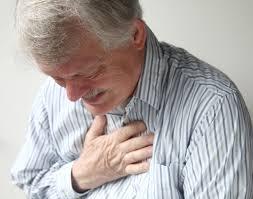
Acute chest pain and ECG need for immediate coronary angiography?
|
|
|
- Anabel Hudson
- 5 years ago
- Views:
Transcription
1 Acute chest pain and ECG need for immediate coronary angiography? Kjell Nikus, MD, PhD Heart Center, Tampere University Hospital, Finland and Samuel Sclarovsky, MD, PhD Tel Aviv University, Israel


2 There are clear cases

3 But also more challenging ones
4 FACTORS AFFECTING ISCHEMIC ECG/ EGM CHANGES Pathophysiologic mechanism Supply vs. demand ischemia Total occlusion, stenosis, spasm Duration of ischemia Transmural vs. subendocardial ischemia Severity of ischemia Myocardial protection (collateral flow, preconditioning, second artery) Localization vs. electrode(s) properties of volume conductor (distance etc.), vector direction Baseline ECG changes à Importance of comparison (BBB, PM, LVH, WPW, rotation, horizontal vs. vertical heart) Variation in coronary anatomy
5 Timing of ECG changes in coronary occlusion Stage I Acute occlusion The presenting ECG may be recorded at any of these stages! Stage II Reperfusion Stage III Restoration of cell metabolism
6 STelevation J point
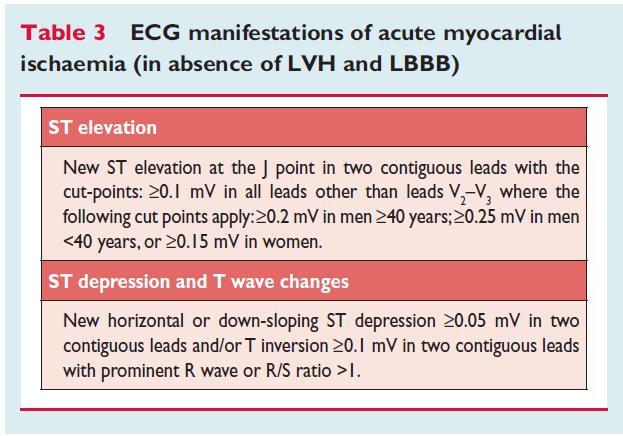
7 The definition of ST elevation is clear, but not all ST elevation is STEMI and on the other hand, acute coronary artery occlusion may be present without ST elevations fulfilling the criteria defined in the guidelines
8 NISTE (non-ischemic ST elevation) Huang HD et al JECG 2011 Chung S-L et al. Am J Emerg Med 2013


9 Chronic remodeling, for example in aortic stenosis: primary change ST depression V5-V6, reciprocal STelevations in V1-V2


10 Male 71 y. Chest pain, elevated CRP No fever or obvious infection
11
12 Some causes for STEMIs not recognized in the emergency department Sometimes the primary ischemic change (=ST-elevation) may be less evident than the secondary (reciprocal) ST depressions ST-elevations may be mild and appear in only 1 lead (Birnbaum Y et al. Eur Heart J 1993) Old Q-wave MI
13 Borderline ST-elevations in the leads II, III and avf Note reciprocal ST changes in avl
")

14 STEMI that could be misdiagnosed as NSTEMI (diagonal branch occlusion) Eight leads with ST depression Primary change is ST-elevation in I, avl 50 mm/sec


15 Very proximal LAD occlusions may have ST elevations only in V1-V2 (and avr) avl pattern ~3/4 avr/v1 pattern ~1/4 Eskola MJ et al. Int J Cardiol 2009
16 A working group proposed a pathophysiologic classification of ACS instead of categorical classification based strictly on the ECG presentations
17


18 A. Transmural ischemia ST elevation Typical STEMI-equivalent: mirror-image T-wave change Prominent T waves T-wave inversion (post-ischemic finger prints )
19 B. Subendocardial ischemia ST/T changes Circumferential ischemia Regional ischemia Normal ECG Confounding factors
20 The most recent international STEMI guidelines point out the importance of recognizing atypical ECG presentations

21 ESC STEMI guidelines 2012
22 Non-ST elevation acute coronary syndrome indications for immediate angiography?
23
24
25 1. Mirror-image STEMI

26 Left circumflex occlusion Sensitivity of the ECG to detect acute coronary occlusion: LAD 85-90% RCA 70-90% LCx 32-50% Role of additional leads Neill J et al. Coron Art Dis 2010 Krishnaswamy et al. Am Heart J 2009

27 n= randomized studies Krishnaswamy et al. Am Heart J 2009


28 ST-depression V2-V4 posterior leads may help in detecting STEMI V8

29 Wung S-F. Am J Crit Care 2007

30 ESC STEMI guidelines 2012
31 2. Circumferential (global) subendocardial ischemia

32 1. Clinical picture indicating acute coronary syndrome 2. Widespread ST-depressions ( 6 leads) 3. Maximal ST-depression V4-V5 4. Negative T V4-V5 5. ST-elevation 0.5 mm avr 6. Transient changes Less specific in case of tachycardia or LVH ECG 50 mm/sec

33 Nikus K ym. Ann Med. 2012

34 ESC STEMI guidelines 2012
35 ST DEPRESSION AND INVERTED ASYMMETRIC T WAVES IN PATIENTS WITHOUT TACHYCARDIA Extensive ischemia causes global reduction in coronary blood flow * This results in impaired relaxation of the left ventricle * * The resulting increase in LVEDP induces severe subendocardial ischemia Resulting in a distinct ECG pattern *Palacios I, Morvell SB, Powel WJ. Circulation 1976; 39:744 * * Baim DS, Grossman W. Grossman s cardiac catheterisation, angiography, and intervention. Lippincott Williams & Wilkins, 2001; 382. Visner MS, Arentzen CE, Parrish DG et al. Circulation 1985; 71: Sclarovsky S. Electrocardiography of acute myocardial ischaemic syndromes. London: Martin Dunitz, 1999:10
36 ST DEPRESSION WITH POSITIVE T WAVE Regional, non-extensive, subendocardial ischemia may manifest as ST depression * Tall and peaked T waves * * Probably caused by high extracellular potassium, related to hyperpolarization of myocytes Due to an opening of the K-ATP-channel *Guyton R, McClenathan JH, Newman G et al. Am J Cardiol 1977;40:373 * *Sclarovsky S, Birnbaum Y, Solodky Y et al. Int J Cardiol 1994;46:37-47 Katz AM. Physiology of the heart. 3 rd ed. Lippincott Williams & Wilkins, 2001: 644 Kondo T, Kubota I, Tachibana H et al. Cardiovasc Res 1996; 31:
37 Levine and Ford described for the first time circumferential subendocardial infarction: the clinical picture, ECG, myocardial and coronary anatomy. (Levine H ; Ford R. Circulation 1950;1:246-62) 5 out of 6 cases were due to mechanical or atherosclerotic obstruction of the left main coronary artery, one had severe 3-vessel disease. No one could reproduce this type of MI in animal experiments Levine was so convinced of his findings that he said: Nature, it seems, can fulfill the conditions of the experiment much more readily than can a physiologist


38 Unstable Angina With ST Segment Depression: With Negative T-Wave Versus Positive T Wave Sclarovsky S. Electrocardiography of acute myocardial ischaemic syndromes. London: Martin Dunitz Sclarovsky S al. Am Heart J 1988;116:933-41
39 The significance of T-wave direction in ACS with ST depression

40 The significance of T-wave direction in ACS with ST depression
41 Circumferential subendocardial ischemia is an independent marker of poor outcome in ACS Nikus K et al. Ann Med. 2012
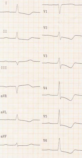
42 3. T-wave changes


43 Old ECG Chest pain ECG 50 mm/sec
 and")
 At least:")

44 LAD occluded (stent thrombosis) Post-PCI RCA New prominent T waves V1-V4 (=LAD) and symptoms compatible with acute MI à consider acute angiography (STEMI protocol) At least: follow-up ECG within min But: Hyperkalemia, individual differences in T-wave amplitude

45 Sclarovsky-Birnbaum grade of ischemia Sclarovsky S et al. Isr J Med Sci 1990;26:525-33

46 Grade 1 ischemia: slow development of Q waves due to well protected myocardium Sclarovsky S. Electrocardiography of acute myocardial ischaemic syndromes. London: Martin Dunitz

47 ACCF/AHA STEMI guidelines 2013

48 Prominent T wave and subtotal LAD occlusion Courtesy: Zhan Zhong-qun

49 Regional subendocardial ischemia -ST depression -Positive T wave Sclarovsky S. Electrocardiography of acute myocardial ischaemic syndromes. London: Martin Dunitz
50 Persistent hyperacute T wave


51 Regional subendocardial ischemia progressing to transmural ischemia grade Ventricular fibrillation Resuscitation
52
53 Despite acute coronary occlusion the 12-lead ECG may be normal or without new changes ECG not recorded during symptoms Distal occlusion of the left circumflex coronary artery (LCx) Small area of ischemia (side branch occlusion) LBBB or non-specific intraventricular conduction delay (QRS>120 ms) Pacemaker ECG
54 Final remarks From an ECG standpoint, early signs of acute STEMI (~ sudden acute coronary artery occlusion) are: hyper-acute prominent T waves, ST elevation without Q waves or T-wave inversions, and ST depression in V1/V2-V3/V4 (mirror-image STEMI equivalent)
55 Final remarks Due to the sometimes very dynamic ECG changes, also later signs of the evolving ischemia/infarction process may be present early after symptom onset Telecardiology within regional STEMI networks are recommended to improve the diagnostics and to shorten ECG to device times
Preface: Wang s Viewpoints
 AHA/ACCF/HRS Recommendations for the Standardization and Interpretation of the Electrocardiogram: Part IV, Ischemia and Infarction Presented by: WANG, TZONG LUEN, MD, PhD, JM, FACC, FESC, FCAPSC Professor,
ECG in coronary artery disease. By Sura Boonrat Central Chest Institute
 ECG in coronary artery disease By Sura Boonrat Central Chest Institute EKG P wave = Atrium activation PR interval QRS = Ventricle activation T wave= repolarization J-point EKG QT interval Abnormal repolarization
Myocardial Infarction. Reading Assignment (p66-78 in Outline )
") Myocardial Infarction Reading Assignment (p66-78 in Outline ) Objectives 1. Why do ST segments go up or down in ischemia? 2. STEMI locations and culprit vessels 3. Why 15-lead ECGs? 4. What s up with avr?
Myocardial Infarction Reading Assignment (p66-78 in Outline ) Objectives 1. Why do ST segments go up or down in ischemia? 2. STEMI locations and culprit vessels 3. Why 15-lead ECGs? 4. What s up with avr?
12 Lead ECG Interpretation: Color Coding for MI s
 12 Lead ECG Interpretation: Color Coding for MI s Anna E. Story, RN, MS Director, Continuing Professional Education Critical Care Nurse Online Instructional Designer 2004 Anna Story 1 Objectives review
ECG Diagnosis and Classification of Acute Coronary Syndromes
 REVIEW ARTICLE ECG Diagnosis and Classification of Acute Coronary Syndromes Yochai Birnbaum, M.D.,, James Michael Wilson, M.D.,, Miquel Fiol, M.D., Antonio Bayés de Luna, M.D., Markku Eskola, M.D., and
Different ECG patterns at presentation in ACS. D. Goldwasser F. Molina A. Bayes de Luna
 Different ECG patterns at presentation in ACS D. Goldwasser F. Molina A. Bayes de Luna Acute Coronary syndromes: The importance of the ECG There are two types of ACS: STE- ACS and Non STE-ACS The most
Preface: Wang s Viewpoints
 AHA/ACCF/HRS Recommendations for the Standardization and Interpretation of the Electrocardiogram: Ischemia and Infarction 103.10.07 Presented by: WANG, TZONG LUEN, MD, PhD, JM, FACC, FESC, FCAPSC Professor,
12 LEAD EKG BASICS. By: Steven Jones, NREMT P CLEMC
 12 LEAD EKG BASICS By: Steven Jones, NREMT P CLEMC ECG Review Waves and Intervals P wave: the sequential activation (depolarization) of the right and left atria QRS complex: right and left ventricular
Ischemic heart disease
 Ischemic heart disease Introduction In > 90% of cases: the cause is: reduced coronary blood flow secondary to: obstructive atherosclerotic vascular disease so most of the time it is called: coronary artery
Electrocardiography. Hilal Al Saffar College of Medicine,Baghdad University
 Electrocardiography Hilal Al Saffar College of Medicine,Baghdad University Which of the following is True 1. PR interval, represent the time taken for the impulse to travel from SA node to AV nose. 2.
12 Lead Electrocardiogram (ECG) PFN: SOMACL17. Terminal Learning Objective. References
 PFN: SOMACL17. Terminal Learning Objective. References") 12 Lead Electrocardiogram (ECG) PFN: SOMACL17 Slide 1 Terminal Learning Objective Action: Communicate knowledge of 12 Lead Electrocardiogram (ECG) Condition: Given a lecture in a classroom environment
12 Lead Electrocardiogram (ECG) PFN: SOMACL17 Slide 1 Terminal Learning Objective Action: Communicate knowledge of 12 Lead Electrocardiogram (ECG) Condition: Given a lecture in a classroom environment
UPDATE ON THE MANAGEMENTACUTE CORONARY SYNDROME. DR JULES KABAHIZI, Psc (Rwa) Lt Col CHIEF CONSULTANT RMH/KFH 28 JUNE18
 Lt Col CHIEF CONSULTANT RMH/KFH 28 JUNE18") UPDATE ON THE MANAGEMENTACUTE CORONARY SYNDROME DR JULES KABAHIZI, Psc (Rwa) Lt Col CHIEF CONSULTANT RMH/KFH 28 JUNE18 INTRODUCTION The clinical entities that comprise acute coronary syndromes (ACS)-ST-segment
UPDATE ON THE MANAGEMENTACUTE CORONARY SYNDROME DR JULES KABAHIZI, Psc (Rwa) Lt Col CHIEF CONSULTANT RMH/KFH 28 JUNE18 INTRODUCTION The clinical entities that comprise acute coronary syndromes (ACS)-ST-segment
Electrocardiography for Healthcare Professionals. Chapter 14 Basic 12-Lead ECG Interpretation
 Electrocardiography for Healthcare Professionals Chapter 14 Basic 12-Lead ECG Interpretation 2012 The Companies, Inc. All rights reserved. Learning Outcomes 14.1 Discuss the anatomic views seen on a 12-lead
Solutions for Every Day Problems Cardiologists and the ECG: Are We Really That Good at It? Part II Daniel José Piñeiro Profesor Titular de Medicina,
 Solutions for Every Day Problems Cardiologists and the ECG: Are We Really That Good at It? Part II Daniel José Piñeiro Profesor Titular de Medicina, Universidad de Buenos Aires, Argentina Member, Membership
By the end of this lecture, you will be able to: Understand the 12 lead ECG in relation to the coronary circulation and myocardium Perform an ECG
 By the end of this lecture, you will be able to: Understand the 12 lead ECG in relation to the coronary circulation and myocardium Perform an ECG recording Identify the ECG changes that occur in the presence
INTERPRETAZIONE ECG NEL PAZIENTE CON SOSPETTO STEMI
 INTERPRETAZIONE ECG NEL PAZIENTE CON SOSPETTO STEMI Giacomo Veronese Scuola di Specializzazione Medicina d Emergenza e Urgenza Università Milano-Bicocca Siete d accordo se vi propongo per una relazione..
Acute Coronary Syndromes. Disclosures
 Acute Coronary Syndromes Disclosures I work for Virginia Garcia Memorial Health Center, Beaverton, OR. Jon Tardiff, BS, PA-C OHSU Clinical Assistant Professor And I am a medical editor for Jones & Bartlett
12 Lead EKG Chapter 4 Worksheet
 Match the following using the word bank. 1. A form of arteriosclerosis in which the thickening and hardening of the vessels walls are caused by an accumulation of fatty deposits in the innermost lining
ECG pre-reading manual. Created for the North West Regional EMET training program
 ECG pre-reading manual Created for the North West Regional EMET training program Author:- Dr Juan Carlos Ascencio-Lane juan.ascencio-lane@ths.tas.gov.au 1 Disclaimer This handbook has been created for
ACUTE CORONARY SYNDROME
 12 LEAD ECG INTERPRETATION in ACUTE CORONARY SYNDROME WAYNE W RUPPERT, CVT, CCCC, NREMT-P Cardiovascular Clinical Coordinator Bayfront Health Seven Rivers Crystal River, FL Education Specialist St. Joseph
The Fundamentals of 12 Lead EKG. ECG Recording. J Point. Reviewing the Cardiac Conductive System. Dr. E. Joe Sasin, MD Rusty Powers, NRP
 The Fundamentals of 12 Lead EKG Dr. E. Joe Sasin, MD Rusty Powers, NRP SA Node Intranodal Pathways AV Junction AV Fibers Bundle of His Septum Bundle Branches Purkinje System Reviewing the Cardiac Conductive
3/4/2018. March Martina Frost, PA C Desert Cardiology. Electricity moving towards/away from electrode create downward/upward directions of waves
 March 2018 Martina Frost, PA C Desert Cardiology Electricity moving towards/away from electrode create downward/upward directions of waves Frontal view Limb leads: I, II, III, avl, avf, (avr) Horizontal
12 Lead ECG Interpretation
 12 Lead ECG Interpretation Julie Zimmerman, MSN, RN, CNS, CCRN Significant increase in mortality for every 15 minutes of delay! N Engl J Med 2007;357:1631-1638 Who should get a 12-lead ECG? Also include
Acute Coronary Syndromes Unstable Angina Non ST segment Elevation MI (NSTEMI) ST segment Elevation MI (STEMI)
 ST segment Elevation MI (STEMI)") Leanna R. Miller, RN, MN, CCRN-CSC, PCCN-CMC, CEN, CNRN, CMSRN, NP Education Specialist LRM Consulting Nashville, TN Objectives Evaluate common abnormalities that mimic myocardial infarction. Identify
Leanna R. Miller, RN, MN, CCRN-CSC, PCCN-CMC, CEN, CNRN, CMSRN, NP Education Specialist LRM Consulting Nashville, TN Objectives Evaluate common abnormalities that mimic myocardial infarction. Identify
12 Lead ECGs: Ischemia, Injury & Infarction. Kevin Handke NRP, FP-C, CCP, CMTE STEMI Coordinator Flight Paramedic
 12 Lead ECGs: Ischemia, Injury & Infarction Kevin Handke NRP, FP-C, CCP, CMTE STEMI Coordinator Flight Paramedic None Disclosures Objectives Upon completion of this program the learner will be able to
About T waves
 About T waves - 2014 Dr. Andres R. Pérez Riera The T waves is a positive deflection after each QRS complex. It represents ventricular repolarization The T wave represents the unconcealed potential differences
Marcin Dada, MD December 03, 2013
 STEMI Imposters Marcin Dada, MD December 03, 2013 Marcin Dada, MD Associate Director, Chest Pain Center Hartford Hospital, Hartford, CT Member, AHA Mission Lifeline Steering Committee Outline of Topics
Chapter 2 Practical Approach
 Chapter 2 Practical Approach There are beginners in electrocardiogram (ECG) analysis who are fascinated by a special pattern (e.g., a bundle-branch block or a striking Q wave) and thereby overlook other
Case 1. Case 2. Case 3
 Case 1 The correct answer is D. Occasionally, the Brugada syndrome can present similar morphologies to A and also change depending on the lead position but in the Brugada pattern the r is wider and ST
Section V. Objectives
 Section V Landscape of an MI Objectives At the conclusion of this presentation the participant will be able to Outline a systematic approach to 12 lead ECG interpretation Demonstrate the process for determining
ECG Basics Sonia Samtani 7/2017 UCI Resident Lecture Series
 ECG Basics Sonia Samtani 7/2017 UCI Resident Lecture Series Agenda I. Introduction II.The Conduction System III.ECG Basics IV.Cardiac Emergencies V.Summary The Conduction System Lead Placement avf Precordial
12 Lead Interpretation
 12 Lead Interpretation Objectives Ischemia, injury and infarction ECG complex review J point ST segment STEMI recognition Ischemia to Infarct Infarction is an evolving process As the infarct evolves ECG
REtrive. REpeat. RElearn Design by. Test-Enhanced Learning based ECG practice E-book
 Test-Enhanced Learning Test-Enhanced Learning Test-Enhanced Learning Test-Enhanced Learning based ECG practice E-book REtrive REpeat RElearn Design by S I T T I N U N T H A N G J U I P E E R I Y A W A
Common pitfalls in the interpretation of electrocardiograms from patients with acute coronary syndromes with narrow QRS: a consensus report
 Available online at www.sciencedirect.com Journal of Electrocardiology 45 (2012) 463 475 www.jecgonline.com Common pitfalls in the interpretation of electrocardiograms from patients with acute coronary
Cardiac Ischemia ECG Workshop
 Cardiac Ischemia ECG Workshop Classic, Confusing, and Confounding Patterns Amal Mattu, MD, NE Professor and Vice Chair Department of Emergency Medicine University of Maryland School of Medicine amalmattu@comcast.net
12 Lead ECG. Presented by Rebecca Sevigny BSN, RN Professional Practice & Development Dept.
 12 Lead ECG Presented by Rebecca Sevigny BSN, RN Professional Practice & Development Dept. Two Main Coronary Arteries RCA LCA which branches into Left Anterior Descending Circumflex Artery Two Main Coronary
Electrocardiographic Diagnosis of ST-elevation Myocardial Infarction
 Cardiol Clin 24 (2006) 343 365 Electrocardiographic Diagnosis of ST-elevation Myocardial Infarction Shaul Atar, MD, Alejandro Barbagelata, MD, Yochai Birnbaum, MD* Division of Cardiology, University of
Acute Myocardial Infarction
 Acute Myocardial Infarction Hafeza Shaikh, DO, FACC, RPVI Lourdes Cardiology Services Asst.Program Director, Cardiology Fellowship Associate Professor, ROWAN-SOM Acute Myocardial Infarction Definition:
STAT 12 Lead ECG Workshop: Basics & ACS
 STAT 12 Lead ECG Workshop: Basics & ACS Part 2: Acute Coronary Syndrome WAYNE W RUPPERT, CVT, CCCC, NREMT-P Cardiovascular Coordinator Bayfront Health Seven Rivers Crystal River, Florida Interventional
F or a long time the 12-lead electrocardiogram
 490 REVIEW The electrocardiogram in ST elevation acute myocardial infarction: correlation with coronary anatomy and prognosis Y Birnbaum, B J Drew... The electrocardiogram is considered an essential part
Basic electrocardiography reading. R3 lee wei-chieh
 Basic electrocardiography reading R3 lee wei-chieh The Normal Conduction System Lead Placement avf Limb Leads Precordial Leads Interpretation Rate Rhythm Interval Axis Chamber abnormality QRST change What
TOPICS IN EMERGENCY MEDICINE SEMI-FINAL
 RISK ASSESSMENT IN PATIENTS WITH CHEST PAIN Nora Goldschlager, M.D. FACP, FACC, FAHA, FHRS Cardiology - San Francisco General Hospital UCSF Disclosures: None 1 CHEST PAIN NOT DUE TO MYOCARDIAL ISCHEMIA
Heart disease remains the leading cause of morbidity and mortality in industrialized nations. It accounts for nearly 40% of all deaths in the United
 Heart disease remains the leading cause of morbidity and mortality in industrialized nations. It accounts for nearly 40% of all deaths in the United States, totaling about 750,000 individuals annually
10 ECGs No Practitioner Can Afford to Miss. Objectives
 10 ECGs No Practitioner Can Afford to Miss Mary L. Dohrmann, MD Professor of Clinical Medicine Division of Cardiovascular Medicine University of Missouri School of Medicine No disclosures Objectives 1.
Myocardial infarction
 CHAPTER-I CARDIOVASCULAR SYSTEM Myocardial infarction SUB: PHARMACOTHERAPEUTICS-I CODE:T0820006 Dr. Venugopal Pharm.D Assistant Professor Department of Pharm.D Kriahna Teja Pharmacy College,Tirupati. Definition
A few new tools for better detection and understanding of STEMIs in the field.
 A few new tools for better detection and understanding of STEMIs in the field. Let s talk, prep and placement. Try to shoot for quality, consistency and no artifact! (looking sometimes for 1 or 2 mm changes)
Acute Myocardial Infarction. Willis E. Godin D.O., FACC
 Acute Myocardial Infarction Willis E. Godin D.O., FACC Acute Myocardial Infarction Definition: Decreased delivery of oxygen and nutrients to the myocardium Myocardial tissue necrosis causing irreparable
Disclosure. 3. ST depression indicative of ischemia is most commonly observed in leads: 1. V1-V2. 2. I and avl 3. V
 Interpreting Stress Induced Ischemia by ECG, Bundle Branch Block & Arrhythmias Disclosure Gregory S Thomas MD, MPH Medical Director, MemorialCare Heart & Vascular Institute, Long Beach Memorial Astellas
15 th Sukaman Memorial Lecture ST Segment Elevation: New Electrocardiographic Insights in 2014
 DOI 10.7603/s40602-016-0006-3 ASEAN Heart Journal http://www.globalsciencejournals.com/journal/40602 Vol. 24, no.1, 98 105 (2016) ISSN: 2315-4551 15 th Sukaman Memorial Lecture ST Segment Elevation: New
12 Lead ECG Workshop. Virginia Hass, DNP, FNP-C, PA-C Kim Newlin, CNS, ANP-C, FPCNA. California Association of Nurse Practitioners March 18, 2016
 12 Lead ECG Workshop Virginia Hass, DNP, FNP-C, PA-C Kim Newlin, CNS, ANP-C, FPCNA California Association of Nurse Practitioners March 18, 2016 Learning Objectives Identify key changes on the ECG which
Topic. Updates on Definition of Myocardial Infarction
 Topic Updates on Definition of Myocardial Infarction In the past, general consensus for MI? Definition of MI by WHO - Combination of 2 of 3 characteristics - 1. Typical Symptoms 2. Enzyme Rise 3. Typical
A Review of Cardiac Pathophysiology and EKG. Jamie Dyson PT, DPT Kathy Swanick PT, DPT, OCS
 A Review of Cardiac Pathophysiology and EKG Jamie Dyson PT, DPT Kathy Swanick PT, DPT, OCS Cardiac Pathophysiology Coronary Artery Disease Congestive Heart Failure Valvular Heart Disease Athletic Heart
Appendix D Output Code and Interpretation of Analysis
 Appendix D Output Code and Interpretation of Analysis 8 Arrhythmia Code No. Description 8002 Marked rhythm irregularity 8110 Sinus rhythm 8102 Sinus arrhythmia 8108 Marked sinus arrhythmia 8120 Sinus tachycardia
FFR vs icecg in Coronary Bifurcations FIESTA ClinicalTrials.gov Identifier: NCT
 FFR vs icecg in Coronary Bifurcations FIESTA ClinicalTrials.gov Identifier: NCT01724957 Dobrin Vassilev MD, PhD Assoc. Prof. in Cardiology Head Cardiology Clinic, Alexandrovska University Hospital Medical
Essam Mahfouz, MD. Professor of Cardiology, Mansoura University
 By Essam Mahfouz, MD. Professor of Cardiology, Mansoura University Agenda Definitions Classifications Epidemiology Risk stratification What is new? What is MI? Myocardial infarction is the death of part
12-Lead ECG Interpretation. Kathy Kuznar, RN, ANP
 12-Lead ECG Interpretation Kathy Kuznar, RN, ANP The 12-Lead ECG Objectives Identify the normal morphology and features of the 12- lead ECG. Perform systematic analysis of the 12-lead ECG. Recognize abnormalities
Electrocardiographic abnormalities encountered in acute myocardial infarction
 40 J Accid Emerg Med 2000;17:40 45 ECG NTERPRETATON FOR THE EMERGENCY DEPARTMENT Department of Emergency Medicine, University of Virginia School of Medicine, Charlottesville, Virginia, USA W J Brady Department
EAE Teaching Course. Magnetic Resonance Imaging. Competitive or Complementary? Sofia, Bulgaria, 5-7 April F.E. Rademakers
 EAE Teaching Course Magnetic Resonance Imaging Competitive or Complementary? Sofia, Bulgaria, 5-7 April 2012 F.E. Rademakers Complementary? Of Course N Engl J Med 2012;366:54-63 Clinical relevance Treatment
12 Lead EKG. The Basics
 12 Lead EKG The Basics Objectives Demonstrate proper 12 EKG lead placement Determine electrical axis Identify ST and T wave changes as they relate to myocardial ischemia Describe possible complications
Pennsylvania Academy of Family Physicians Foundation & UPMC 43rd Refresher Course in Family Medicine CME Conference March 10-13, 2016
 Pennsylvania Academy of Family Physicians Foundation & UPMC 43rd Refresher Course in Family Medicine CME Conference March 10-13, 2016 Disclosures: EKG Workshop Louis Mancano, MD Speaker has no disclosures
Features of electrocardiogram in patients with stenosis of the proximal right coronary artery
 ORIGINAL ARTICLE Korean J Intern Med 2017;32:277-285 Features of electrocardiogram in patients with stenosis of the proximal right coronary artery Moo Seong Koh, Jae Hoon Lee, Jin Woo Jeong, and Jun Young
Prediction of the Site of Coronary Artery Lesion in Acute Inferior Myocardial Infarction with Right Sided Precordial Lead (V4r)
") Prediction of the Site of Coronary Artery Lesion in Acute Inferior Myocardial Infarction with Right Sided Precordial Lead (V4r) MS Alam, M Ullah, SU Ulabbi, MM Haque, R Uddin, MS Mamun, AAS Majumder National
Prediction of the Site of Coronary Artery Lesion in Acute Inferior Myocardial Infarction with Right Sided Precordial Lead (V4r) MS Alam, M Ullah, SU Ulabbi, MM Haque, R Uddin, MS Mamun, AAS Majumder National
Common Codes for ICD-10
 Common Codes for ICD-10 Specialty: Cardiology *Always utilize more specific codes first. ABNORMALITIES OF HEART RHYTHM ICD-9-CM Codes: 427.81, 427.89, 785.0, 785.1, 785.3 R00.0 Tachycardia, unspecified
DR QAZI IMTIAZ RASOOL OBJECTIVES
 PRACTICAL ELECTROCARDIOGRAPHY DR QAZI IMTIAZ RASOOL OBJECTIVES Recording of electrical events in heart Established electrode pattern results in specific tracing pattern Health of heart i. e. Anatomical
Please check your answers with correct statements in answer pages after the ECG cases.
 ECG Cases ECG Case 1 Springer International Publishing AG, part of Springer Nature 2018 S. Okutucu, A. Oto, Interpreting ECGs in Clinical Practice, In Clinical Practice, https://doi.org/10.1007/978-3-319-90557-0
All About STEMIs. Presented By: Brittney Urvand, RN, BSN, CCCC. Essentia Health Fargo Cardiovascular Program Manager.
 All About STEMIs Presented By: Brittney Urvand, RN, BSN, CCCC Essentia Health Fargo Cardiovascular Program Manager Updated 10/2/2018 None Disclosures Objectives Identify signs and symptoms of a heart attack
ECG Workshop. Nezar Amir
 ECG Workshop Nezar Amir Myocardial Ischemia ECG Infarct ECG in STEMI is dynamic & evolving Common causes of ST shift Infarct Localisation Left main artery occlusion: o diffuse ST-depression with ST elevation
Other 12-Lead ECG Findings
 Other 12-Lead ECG Findings Left Atrial Enlargement Left atrial enlargement is illustrated by increased P wave duration in lead II, top ECG, and by the prominent negative P terminal force in lead V1, bottom
Family Medicine for English language students of Medical University of Lodz ECG. Jakub Dorożyński
 Family Medicine for English language students of Medical University of Lodz ECG Jakub Dorożyński Parts of an ECG The standard ECG has 12 leads: six of them are considered limb leads because they are placed
ECG Cases and Questions. Ashish Sadhu, MD, FHRS, FACC Electrophysiology/Cardiology
 ECG Cases and Questions Ashish Sadhu, MD, FHRS, FACC Electrophysiology/Cardiology 32 yo female Life Insurance Physical 56 yo male with chest pain Terminology Injury ST elevation Ischemia T wave inversion
Masqueraders of STEMI
 Masqueraders of STEMI Steven M. Costa, M.D. Assistant Professor Department of Medicine Division of Cardiology Scott & White Memorial Hospital and Clinic Texas A&M University Health Science Center Disclosures
Electrical System Overview Electrocardiograms Action Potentials 12-Lead Positioning Values To Memorize Calculating Rates
 Electrocardiograms Electrical System Overview James Lamberg 2/ 74 Action Potentials 12-Lead Positioning 3/ 74 4/ 74 Values To Memorize Inherent Rates SA: 60 to 100 AV: 40 to 60 Ventricles: 20 to 40 Normal
Foundations EKG I - Unit 1 Summary
 Foundations EKG I - Unit 1 Summary The accurate diagnosis of ST elevation myocardial infarction (STEMI) is one of the most time critical duties in the practice of EM. Diagnosis is not always easy so guidelines
CME Article Brugada pattern masking anterior myocardial infarction
 Electrocardiography Series Singapore Med J 2011; 52(9) : 647 CME Article Brugada pattern masking anterior myocardial infarction Seow S C, Omar A R, Hong E C T Cardiology Department, National University
also aid the clinician in recognizing both the obvious and subtle abnormalities that may help guide therapy.
 Karen Lieberman, MS, CRNP f the many diagnostic tools used to screen for and evaluate cardiac abnormalities, the 12-lead electrocardiogram (ECG) is among the most basic. This inexpensive and noninvasive
Received: 8 January 2014 Accepted: 10 June 2014 Published: 23 September 2014
 Original Article Medical Journal of the Islamic Republic of Iran (MJIRI) Iran University of Medical Sciences The relation of ST segment deviations in 12-lead conventional Electrocardiogram, right and posterior
402 Index. B β-blockers, 4, 5 Bradyarrhythmias, 76 77
 Index A Acquired immunodeficiency syndrome (AIDS), 126, 163 Action potentials, 1, 5, 27 Acute coronary syndromes, 123t, 129 Adenosine, intravenous, 277 Alcohol abuse, as T wave inversion cause, 199 Aneurysm,
ECG ABNORMALITIES D R. T AM A R A AL Q U D AH
 ECG ABNORMALITIES D R. T AM A R A AL Q U D AH When we interpret an ECG we compare it instantaneously with the normal ECG and normal variants stored in our memory; these memories are stored visually in
Ischemic Heart Disease
 Ischemic Heart Disease Dr Rodney Itaki Lecturer Division of Pathology University of Papua New Guinea School of Medicine & Health Sciences Division of Pathology General Consideration Results from partial
Introduction to Electrocardiography
 Introduction to Electrocardiography Class Objectives: Introduction to ECG monitoring Discuss principles of interpretation Identify the components and measurements of the ECG ECG analysis ECG Monitoring
Correlation of ECG Changes with Coronary Angiographic Findings in Acute Inferior Myocardial Infarction
 Original Article Correlation of ECG Changes with Coronary Angiographic Findings in Acute Inferior Myocardial Infarction Mamun KSA 1, Awal A 2, Murshed AKMM 3 Abstract The determination of infarct related
ECG Interpretation Cat Williams, DVM DACVIM (Cardiology)
") ECG Interpretation Cat Williams, DVM DACVIM (Cardiology) Providing the best quality care and service for the patient, the client, and the referring veterinarian. GOAL: Reduce Anxiety about ECGs Back to
ECG Interpretation Cat Williams, DVM DACVIM (Cardiology) Providing the best quality care and service for the patient, the client, and the referring veterinarian. GOAL: Reduce Anxiety about ECGs Back to
Transient Right Axis Deviation During Acute Anterior Wall Infarction or Ischemia: Electrocardiographic and Angiographic Correlation
 JACC Vol. 8, No.1 27 Transient Right Axis Deviation During Acute Anterior Wall Infarction or Ischemia: Electrocardiographic and Angiographic Correlation SAMUEL SCLAROVSKY, MD, ALEX SAGlE, MD, BORIS STRASBERG,
Chapter 76 Acute Coronary Syndromes Part A
 Chapter 76 Acute Coronary Syndromes Part A Episode Overview: 1. Define Stable Angina, UA, AMI 2. Describe the pathophysiology of AMI 3. What are the components of prehospital management of AMI 4. List
Left posterior hemiblock (LPH)/
/") ECG OF THE MONTH Left Postero-inferior Depolarization Delay Keywords Electrocardiography Intraventricular conduction delay, Inferoposterior hemiblock, Left posterior fascicular block, Left posterior hemiblock
ECG OF THE MONTH Left Postero-inferior Depolarization Delay Keywords Electrocardiography Intraventricular conduction delay, Inferoposterior hemiblock, Left posterior fascicular block, Left posterior hemiblock
MRI ACS-ben. Tamás Simor MD, PhD, Med Hab. University of Pécs, Heart Institute
 MRI ACS-ben Tamás Simor MD, PhD, Med Hab Time Course of Changes in Infarct Size, Viable Myocardium, and LV Mass After Reperfused and Nonreperfused MI Blue lines denote reperfused myocardial infarction
ECG Interpretation. Best to have a system to methodically evaluate ECG (from Dubin) * Rate * Rhythm * Axis * Intervals * Hypertrophy * Infarction
 * Rate * Rhythm * Axis * Intervals * Hypertrophy * Infarction") ECG to save Babies ECG Interpretation Best to have a system to methodically evaluate ECG (from Dubin) * Rate * Rhythm * Axis * Intervals * Hypertrophy * Infarction Electrical Activity in the heart 5 events
ECG to save Babies ECG Interpretation Best to have a system to methodically evaluate ECG (from Dubin) * Rate * Rhythm * Axis * Intervals * Hypertrophy * Infarction Electrical Activity in the heart 5 events
ECG CONVENTIONS AND INTERVALS
 1 ECG Waveforms and Intervals ECG waveforms labeled alphabetically P wave== represents atrial depolarization QRS complex=ventricular depolarization ST-T-U complex (ST segment, T wave, and U wave)== V repolarization.
Three most relevant tools available to an emergency
 CASE REPORT ST-segment Depression: All are Not Created Equal! Sonia Mishra 1, Ajay Mishra 2, Jagdish Mishra 3 1 Upstate Cardiology, Batavia 14020, New York, 2 Georgetown University, Washington, DC 20057
Imaging ischemic heart disease: role of SPECT and PET. Focus on Patients with Known CAD
 Imaging ischemic heart disease: role of SPECT and PET. Focus on Patients with Known CAD Hein J. Verberne Academic Medical Center, University of Amsterdam, Amsterdam, Netherlands International Conference
ST SEGMENT IN LEAD A VR IN ACUTE INFERIOR MYOCARDIAL INFARCTION
 ST SEGMENT IN LEAD A VR IN ACUTE INFERIOR MYOCARDIAL INFARCTION Pages with reference to book, From 365 To 366 Shehbaz A. Kureshi, Yoshiharu Yonekura, Yutaka Konishi, Kanji Torizuka ( Kyoto University School
DIAGNOSTIC CRITERIA OF AMI/ACS
 DIAGNOSTIC CRITERIA OF AMI/ACS Diagnostic criteria are used to validate clinical diagnoses. Those used in epidemiological studies are here below reported. 1. MONICA - Monitoring trends and determinants
Fourth Universal Definition of Myocardial Infarction (2018)
") EUSEM, Glasgow 2018 Fourth Universal Definition of Myocardial Infarction (2018) Univ.-Prof. Dr. Martin Möckel, Division of Emergency and Acute Medicine, Charité Universitätsmedizin Berlin 2 1 History of
EUSEM, Glasgow 2018 Fourth Universal Definition of Myocardial Infarction (2018) Univ.-Prof. Dr. Martin Möckel, Division of Emergency and Acute Medicine, Charité Universitätsmedizin Berlin 2 1 History of
Reciprocal ST depression in acute myocardial infarction
 Reciprocal ST depression in acute myocardial infarction Br Heart J 1985; 54: 479-83 OLUSOLA ODEMUYIWA, IAN PEART, CATHERINE ALBERS, ROGER HALL From the Royal Victoria Infirmary, Newcastle upon Tyne SUMMARY
ST SEGMENT THE UPS AND THE DOWNS
 07 October 2016 No. 23 ST SEGMENT THE UPS AND THE DOWNS M Nontshe Moderator: S Ramcharan School of Clinical Medicine Discipline of Anaesthesiology and Critical Care CONTENTS ST SEGMENT, THE UPS AND DOWNS...
Nstemi But Stemi-De Winters Sign
 Cardiology and Angiology: An International Journal 3(3): 162-166, 2015, Article no.ca.2015.015 ISSN: 2347-520X SCIENCEDOMAIN international www.sciencedomain.org Nstemi But Stemi-De Winters Sign Prem Krishna
Atherosclerotic Heart Disease: Coronary Vessels, EKG Localization of STEMI and Complications/Derivatives for USMLE Step One
 Atherosclerotic Heart Disease: Coronary Vessels, EKG Localization of STEMI and Complications/Derivatives for USMLE Step One Howard J. Sachs, MD Associate Professor of Medicine University of Massachusetts
TYPE II MI. KC ACDIS LOCAL CHAPTER March 8, 2016
 TYPE II MI KC ACDIS LOCAL CHAPTER March 8, 2016 TYPE 2 MI DEFINITION: Acute coronary syndrome (ACS) encompasses a continuum of myocardial ischemia and infarction, which can make the diagnostic and coding
Hot Topics in Cardiac Arrest. Should the patient go To the Cath Lab?
 Hot Topics in Cardiac Arrest Should the patient go To the Cath Lab? Tim Russert 1950-2008 Host of NBC s Meet the Press Sudden Cardiac Arrest : Autopsy showed plaque rupture in his LAD ( per LA Times,
Ekg pra pr c a tice D.HAMMOUDI.MD
 Ekg practice D.HAMMOUDI.MD Anatomy Revisited RCA (Right Coronary Artery) Right ventricle Inferior wall of LV Posterior wall of LV (75%) SA Node (60%) AV Node (>80%) LCA (Left Coronary Artery) Septal wall