Atherosclerotic Heart Disease: Coronary Vessels, EKG Localization of STEMI and Complications/Derivatives for USMLE Step One
|
|
|
- Kathlyn Baldwin
- 5 years ago
- Views:
Transcription

1 Atherosclerotic Heart Disease: Coronary Vessels, EKG Localization of STEMI and Complications/Derivatives for USMLE Step One Howard J. Sachs, MD Associate Professor of Medicine University of Massachusetts Medical School
2 They will never ask you to name a vessel Name this vessel
3 They will never ask you to name a vessel They will ask you the manifestations of that blocked vessel.

4 They will ask you the manifestations of that blocked vessel. Well, they won t actually say manifestations of a blocked vessel. They ll say chest pain and ST segment (elevation/depression) in leads
5 CT Angiogram, Coronary Vessels Blood in LV
6 CT Angiogram, Coronary Vessels Left Main
7 CT Angiogram, Coronary Vessels Ramus Intermedius (left main trifurcation)
8 Lateral Wall by LCX Anterior Wall by LAD Inferior Wall by RCA
9 Lateral Wall by LCX Anterior Wall by LAD Posterior Wall by RCA
10 RCA LCX LAD Lateral Wall by LCX Anterior Wall by LAD Inferior Wall by RCA

11 RCA R C A P D A S e p LAD t LAD u Anterior m Wall by LAD 1/3 2/3 LCX Lateral Wall by LCX Inferior Wall by RCA
12
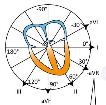
13 II III avf III II avf
14 II III avf

15 II III avf
16 II III avf
 wall will have ST depressions (opposite")
17 II III avf Reciprocal Changes: If the inferior wall has ST elevations, the anterior (contralateral leads) wall will have ST depressions (opposite vectors)
18 Reciprocal Changes Present in Ischemia/Infarction II III avf Acute Pericarditis: diffuse (inferior, anterior and lateral) ST-segment elevation devoid of reciprocal changes

19 Patient experiences STEMI 3 days ago. Now with sharp chest pain relieved while leaning forward. VSS; lungs clear; cor scratchy heart sound. Choose the correct diagnosis?: A. Acute pericarditis B. Extension of myocardial infarction

20 II III avf Derivatives: RCA occlusion/imi (inferior wall MI)

21 RCA occlusion (IMI) Bradyarrhythmias Sinus Bradycardia 2 nd Degree Heart Block
22 RCA occlusion (IMI) Bradyarrhythmias Sinus Bradycardia 2 nd Degree Heart Block Patient with chest pain and elevated troponin. Rhythm strip shown. Occlusion of which vessel is most likely? EKG will show elevations in which leads?
23 RCA occlusion (IMI) Bradyarrhythmias Sinus Bradycardia 2 nd Degree Heart Block Patient with chest pain and elevated troponin. Rhythm strip shown. Occlusion of which vessel is most likely RCA (or PDA) EKG will show elevations in which leads II, III, avf
24 RCA Posterior Descending Artery Dominance (or Co-dominance) ~80% ~10%

")
25 RCA PDA Dominance (Posterior 1/3 Septum) ~80% ~10%
26 RV Infarct Left Ventricle: EF? PCWP? Lung exam?
27 RV Infarct Left Ventricle: EF: normal PCWP: normal Lung exam: clear


28 JVP Q. Patient with cardiogenic shock and normal EF/PCWP with clear lungs RV Infarct


29 RCA RV infarct Hypotension Elevated CVP Clear lungs High index of suspicion Very pre-load dependent: push fluids, avoid nitrates Right sided chest leads
30 Coronary Vessels, EKG Localization of STEMI and Derivatives for USMLE Step One Right Coronary Artery [inferior (posterior) wall] Leads: II, III, avf Sinoatrial and AV Nodes bradyarrhythmia Dominance: supplies the posterior descending a. Posterior 1/3 of septum RV infarct: cardiogenic shock without LV EDV Left Circumflex (lateral wall) Leads: I, avl, V5,6 Dominance Territorial pattern Left Anterior Descending Leads: V2,3,4 Major MI, Major Complications (for the boards) Rupture: LV free wall tamponade; Papillary muscle mitral regurgitation, CHF Cardiogenic shock Special Notes: Reciprocal changes
31 LCX Lateral Wall I, avl, V5, 6
32 LCx I, avl, V5, 6 Lateral Wall
33 Take homes for Left Circumflex: 1. Dominance: infrequent; if dominant, supplies PDA (and posterior 1/3 septum) 2. Territorial pattern: overall pattern of elevations in acute pericarditis


34 Take homes for Left Circumflex: 1. Dominance: infrequent; if dominant, supplies PDA (and posterior 1/3 septum) 2. Territorial pattern: overall pattern of elevations in acute pericarditis Acute Pericarditis
 2.")
35 Take homes for Left Circumflex: 1. Dominance: infrequent; if dominant, supplies PDA (and posterior 1/3 septum) 2. Territorial pattern: overall pattern of elevations in acute pericarditis 3. Occlusion: lateral wall infarction


36 Anterior Wall, LAD V2-4
37 Anterior Wall, LAD V2-4

38 Look at limb leads for reciprocal s in AWMI Anterior Wall, LAD V2-4
39 Anterior Wall Infarction (STEMI) LAD, V2-4
40 Key Derivatives: Anterior Wall Infarction (STEMI) LAD, V2-4 LV Free Wall Rupture Macrophage Phase Papillary Muscle
 LAD, V2-4")

41 Key Derivatives: Anterior Wall Infarction (STEMI) LAD, V2-4 Macrophage Phase Day 5 post-awmi Cardiogenic Shock Hemopericardium Pulsus Paradoxus


42 Key Derivatives: Anterior Wall Infarction (STEMI) LAD, V2-4 Day 5 post-awmi New Onset CHF M Harsh, holosystolic axilla Macrophage Phase


43 Key Derivatives: Anterior Wall Infarction (STEMI) LAD, V2-4 Cardiogenic Shock
44 Key Derivatives: Anterior Wall Infarction (STEMI) LAD, V2-4 LV Aneurysm


45 Key Derivatives: Anterior Wall Infarction (STEMI) LAD, V2-4 Mural Thrombus LV Aneurysm
46 RCA LCX LAD Lateral Wall by LCX Anterior Wall by LAD Inferior Wall by RCA

47 RCA LCX LAD Lateral Wall by LCX Anterior Wall by LAD Inferior Wall by RCA
48 Coronary Vessels, EKG Localization of STEMI and Derivatives for USMLE Step One Right Coronary Artery [inferior (posterior) wall] Leads: II, III, avf Sinoatrial and AV Nodes bradyarrhythmia Dominance: supplies the posterior descending a. Posterior 1/3 of septum RV infarct: cardiogenic shock without LV EDV Left Circumflex (lateral wall) Leads: I, avl, V5,6 Dominance Territorial pattern Left Anterior Descending Leads: V2,3,4 Major MI, Major Complications (for the boards) Rupture: LV free wall tamponade; Papillary muscle mitral regurgitation, CHF Cardiogenic shock LV aneurysm and sequelae Special Notes: Reciprocal changes and acute pericarditis
49 Atherosclerotic Heart Disease: Coronary Vessels, EKG Localization of STEMI and Complications/Derivatives for USMLE Step One Howard J. Sachs, MD Associate Professor of Medicine University of Massachusetts Medical School
12 Lead EKG. The Basics
 12 Lead EKG The Basics Objectives Demonstrate proper 12 EKG lead placement Determine electrical axis Identify ST and T wave changes as they relate to myocardial ischemia Describe possible complications
Section V. Objectives
 Section V Landscape of an MI Objectives At the conclusion of this presentation the participant will be able to Outline a systematic approach to 12 lead ECG interpretation Demonstrate the process for determining
12 Lead ECGs: Ischemia, Injury & Infarction. Kevin Handke NRP, FP-C, CCP, CMTE STEMI Coordinator Flight Paramedic
 12 Lead ECGs: Ischemia, Injury & Infarction Kevin Handke NRP, FP-C, CCP, CMTE STEMI Coordinator Flight Paramedic None Disclosures Objectives Upon completion of this program the learner will be able to
All About STEMIs. Presented By: Brittney Urvand, RN, BSN, CCCC. Essentia Health Fargo Cardiovascular Program Manager.
 All About STEMIs Presented By: Brittney Urvand, RN, BSN, CCCC Essentia Health Fargo Cardiovascular Program Manager Updated 10/2/2018 None Disclosures Objectives Identify signs and symptoms of a heart attack
Myocardial Infarction
 Myocardial Infarction MI = heart attack Defined as necrosis of heart muscle resulting from ischemia. A very significant cause of death worldwide. of these deaths, 33% -50% die before they can reach the
Ischemic heart disease
 Ischemic heart disease Introduction In > 90% of cases: the cause is: reduced coronary blood flow secondary to: obstructive atherosclerotic vascular disease so most of the time it is called: coronary artery
12 Lead ECG Interpretation
 12 Lead ECG Interpretation Julie Zimmerman, MSN, RN, CNS, CCRN Significant increase in mortality for every 15 minutes of delay! N Engl J Med 2007;357:1631-1638 Who should get a 12-lead ECG? Also include
1) Severe, crushing substernal chest pain 2) radiate to the neck, jaw, epigastrium, or left arm. 3- rapid and weak pulse 4- nausea (posterior MI).
 Severe, crushing substernal chest pain 2) radiate to the neck, jaw, epigastrium, or left arm. 3- rapid and weak pulse 4- nausea (posterior MI).") 1) Severe, crushing substernal chest pain 2) radiate to the neck, jaw, epigastrium, or left arm. 3- rapid and weak pulse 4- nausea (posterior MI). 5- cardiogenic shock (massive MIs >40% of the left ventricle)
1) Severe, crushing substernal chest pain 2) radiate to the neck, jaw, epigastrium, or left arm. 3- rapid and weak pulse 4- nausea (posterior MI). 5- cardiogenic shock (massive MIs >40% of the left ventricle)
12 Lead ECG Interpretation: Color Coding for MI s
 12 Lead ECG Interpretation: Color Coding for MI s Anna E. Story, RN, MS Director, Continuing Professional Education Critical Care Nurse Online Instructional Designer 2004 Anna Story 1 Objectives review
ECG in coronary artery disease. By Sura Boonrat Central Chest Institute
 ECG in coronary artery disease By Sura Boonrat Central Chest Institute EKG P wave = Atrium activation PR interval QRS = Ventricle activation T wave= repolarization J-point EKG QT interval Abnormal repolarization
12 th Annual West Virginia ACC Meeting April 8, 2017
 12 th Annual West Virginia ACC Meeting April 8, 2017 Rameez Sayyed, M.D., FACC, FSCAI Associate professor of Medicine Program Director for interventional cardiology Marshall University Joan C. Edwards
The Fundamentals of 12 Lead EKG. ECG Recording. J Point. Reviewing the Cardiac Conductive System. Dr. E. Joe Sasin, MD Rusty Powers, NRP
 The Fundamentals of 12 Lead EKG Dr. E. Joe Sasin, MD Rusty Powers, NRP SA Node Intranodal Pathways AV Junction AV Fibers Bundle of His Septum Bundle Branches Purkinje System Reviewing the Cardiac Conductive
Understanding the 12-lead ECG, part II
 Bundle-branch blocks Understanding the 12-lead ECG, part II Most common electrocardiogram (ECG) abnormality Appears as a wider than normal S complex Occurs when one of the two bundle branches can t conduct
Common Codes for ICD-10
 Common Codes for ICD-10 Specialty: Cardiology *Always utilize more specific codes first. ABNORMALITIES OF HEART RHYTHM ICD-9-CM Codes: 427.81, 427.89, 785.0, 785.1, 785.3 R00.0 Tachycardia, unspecified
Preface: Wang s Viewpoints
 AHA/ACCF/HRS Recommendations for the Standardization and Interpretation of the Electrocardiogram: Part IV, Ischemia and Infarction Presented by: WANG, TZONG LUEN, MD, PhD, JM, FACC, FESC, FCAPSC Professor,
12 Lead Electrocardiogram (ECG) PFN: SOMACL17. Terminal Learning Objective. References
 PFN: SOMACL17. Terminal Learning Objective. References") 12 Lead Electrocardiogram (ECG) PFN: SOMACL17 Slide 1 Terminal Learning Objective Action: Communicate knowledge of 12 Lead Electrocardiogram (ECG) Condition: Given a lecture in a classroom environment
12 Lead Electrocardiogram (ECG) PFN: SOMACL17 Slide 1 Terminal Learning Objective Action: Communicate knowledge of 12 Lead Electrocardiogram (ECG) Condition: Given a lecture in a classroom environment
A walk through a STEMI
 A walk through a STEMI M.M. s Story Kim Robison Ashley Corcoran Situation M.M. is an 82 year old male brought in by private vehicle on 10/22/17 to the Emergency Department Pt. c/o left arm numbness, pain
UPDATE ON THE MANAGEMENTACUTE CORONARY SYNDROME. DR JULES KABAHIZI, Psc (Rwa) Lt Col CHIEF CONSULTANT RMH/KFH 28 JUNE18
 Lt Col CHIEF CONSULTANT RMH/KFH 28 JUNE18") UPDATE ON THE MANAGEMENTACUTE CORONARY SYNDROME DR JULES KABAHIZI, Psc (Rwa) Lt Col CHIEF CONSULTANT RMH/KFH 28 JUNE18 INTRODUCTION The clinical entities that comprise acute coronary syndromes (ACS)-ST-segment
UPDATE ON THE MANAGEMENTACUTE CORONARY SYNDROME DR JULES KABAHIZI, Psc (Rwa) Lt Col CHIEF CONSULTANT RMH/KFH 28 JUNE18 INTRODUCTION The clinical entities that comprise acute coronary syndromes (ACS)-ST-segment
ECG Basics Sonia Samtani 7/2017 UCI Resident Lecture Series
 ECG Basics Sonia Samtani 7/2017 UCI Resident Lecture Series Agenda I. Introduction II.The Conduction System III.ECG Basics IV.Cardiac Emergencies V.Summary The Conduction System Lead Placement avf Precordial
Acute Myocardial Infarction
 Acute Myocardial Infarction Hafeza Shaikh, DO, FACC, RPVI Lourdes Cardiology Services Asst.Program Director, Cardiology Fellowship Associate Professor, ROWAN-SOM Acute Myocardial Infarction Definition:
Ischemic heart disease (IHD) Dr: Salah Ahmed
 Dr: Salah Ahmed") Ischemic heart disease (IHD) Dr: Salah Ahmed The coronaries: 1- Left anterior descending coronary artery: - supplies anterior portion of LV, anterior 2/3 of IVS - accounts for 40-50% of coronary artery
Ischemic heart disease (IHD) Dr: Salah Ahmed The coronaries: 1- Left anterior descending coronary artery: - supplies anterior portion of LV, anterior 2/3 of IVS - accounts for 40-50% of coronary artery
Myocardial Infarction. Reading Assignment (p66-78 in Outline )
") Myocardial Infarction Reading Assignment (p66-78 in Outline ) Objectives 1. Why do ST segments go up or down in ischemia? 2. STEMI locations and culprit vessels 3. Why 15-lead ECGs? 4. What s up with avr?
Myocardial Infarction Reading Assignment (p66-78 in Outline ) Objectives 1. Why do ST segments go up or down in ischemia? 2. STEMI locations and culprit vessels 3. Why 15-lead ECGs? 4. What s up with avr?
2/14/2018 CASE STUDIES: COMPLICATIONS OF MYOCARDIAL INFARCTION. Judy Hung, MD Cardiology Division Massachusetts General Hospital Boston, MA CASE 1
 CASE STUDIES: COMPLICATIONS OF MYOCARDIAL INFARCTION Judy Hung, MD Cardiology Division Massachusetts General Hospital Boston, MA CASE 1 1 PRESENTATION 57 yo male with a past medical history of hypertension
ECG Workshop. Nezar Amir
 ECG Workshop Nezar Amir Myocardial Ischemia ECG Infarct ECG in STEMI is dynamic & evolving Common causes of ST shift Infarct Localisation Left main artery occlusion: o diffuse ST-depression with ST elevation
DAY1_CARDIOVASCULAR PRACTICE QUESTIONS
 DAY1_CARDIOVASCULAR PRACTICE QUESTIONS 1 P age 1. A 59-year-old male is admitted complaining of chest pain and dyspnea. ST elevation and T-wave inversion were seen on the ECG in V2, V3, and V4. IV thrombolytic
12 Lead ECG. Presented by Rebecca Sevigny BSN, RN Professional Practice & Development Dept.
 12 Lead ECG Presented by Rebecca Sevigny BSN, RN Professional Practice & Development Dept. Two Main Coronary Arteries RCA LCA which branches into Left Anterior Descending Circumflex Artery Two Main Coronary
Pulmonary-Vascular Disease. Howard J. Sachs, MD.
 Pulmonary-Vascular Disease Howard J. Sachs, MD www.12daysinmarch.com Dyspnea Cardiac Pulmonary CAD Pump Failure Chest Wall Airways Valve Disease Pericardial Disease Alveoli Interstitium Rhythm Disturbance
Echocardiography as a diagnostic and management tool in medical emergencies
 Echocardiography as a diagnostic and management tool in medical emergencies Frank van der Heusen MD Department of Anesthesia and perioperative Care UCSF Medical Center Objective of this presentation Indications
By the end of this lecture, you will be able to: Understand the 12 lead ECG in relation to the coronary circulation and myocardium Perform an ECG
 By the end of this lecture, you will be able to: Understand the 12 lead ECG in relation to the coronary circulation and myocardium Perform an ECG recording Identify the ECG changes that occur in the presence
Acute Coronary Syndromes
 Acute Coronary Syndromes Kelly Kreimer, MSN and Shane Johnson, RN, BSN, CCRN Cardiovascular Circulatory System Heart-Pump Arteries-regulate blood supply Capillaries-exchange of cell nutrients and wastes
ST ELEVATION MYOCARDIAL INFARCTION (STEMI) Gordon Kritzer, MD, FACC Virginia Mason Medical Center, Seattle
 Gordon Kritzer, MD, FACC Virginia Mason Medical Center, Seattle") ST ELEVATION MYOCARDIAL INFARCTION (STEMI) Gordon Kritzer, MD, FACC Virginia Mason Medical Center, Seattle STEMI ACS and STEMI History and physical, differential diagnosis ECGs Initial Therapy and management
ST ELEVATION MYOCARDIAL INFARCTION (STEMI) Gordon Kritzer, MD, FACC Virginia Mason Medical Center, Seattle STEMI ACS and STEMI History and physical, differential diagnosis ECGs Initial Therapy and management
Form 4: Coronary Evaluation
 Form : Coronary Evaluation Print this Form t Started Date of Coronary Evaluation Coronary Evaluation Indication for Coronary Evaluation Check only one. Angio NOT DONE: n invasive test performed Followup
Abnormal, Autoquant Adenosine Myocardial Perfusion Heart Imaging. ID: GOLD Date: Age: 46 Sex: M John Doe Phone (310)
") Background: Reason: preoperative assessment of CAD, Shortness of Breath Symptom: atypical chest pain Risk factors: hypertension Under influence: a beta blocker Medications: digoxin Height: 66 in. Weight:
Background: Reason: preoperative assessment of CAD, Shortness of Breath Symptom: atypical chest pain Risk factors: hypertension Under influence: a beta blocker Medications: digoxin Height: 66 in. Weight:
Part One Objectives. Don t Worry About It. All done for you Paper Speed 25 mm/sec Calibration 1 mv charge over 20 ms = 10 mm tall Lincoln Hat
 12-lead and ACS Review North Lyon Refresher Part One Objectives 12 lead ECG Basics Anatomy and Physiology STEMI Diagnosis Types of MI ACS Review STEMI System and Interventional Cardiology Review The Value
REtrive. REpeat. RElearn Design by. Test-Enhanced Learning based ECG practice E-book
 Test-Enhanced Learning Test-Enhanced Learning Test-Enhanced Learning Test-Enhanced Learning based ECG practice E-book REtrive REpeat RElearn Design by S I T T I N U N T H A N G J U I P E E R I Y A W A
Basic Coronary Angiography DAVID SHAVELLE MD
 Basic Coronary Angiography DAVID SHAVELLE MD Basic Coronary Angiography: Take Home Points Cardiovascular Medicine Boards and Clinical Practice Understand normal coronary anatomy Understand different imaging
Arrhythmic Complications of MI. Teferi Mitiku, MD Assistant Clinical Professor of Medicine University of California Irvine
 Arrhythmic Complications of MI Teferi Mitiku, MD Assistant Clinical Professor of Medicine University of California Irvine Objectives Brief overview -Pathophysiology of Arrhythmia ECG review of typical
Electrocardiography. Hilal Al Saffar College of Medicine,Baghdad University
 Electrocardiography Hilal Al Saffar College of Medicine,Baghdad University Which of the following is True 1. PR interval, represent the time taken for the impulse to travel from SA node to AV nose. 2.
Form 4: Coronary Evaluation
 Patient Details Hidden Show Show/Hide Annotations Form : Coronary Evaluation Print this Form t Started Date of Coronary Evaluation Coronary Evaluation Indication for Coronary Evaluation Check only one.
Mechanical Complications of Acute Myocardial Infarction. Chaitan K. Narsule
 Mechanical Complications of Acute Myocardial Infarction Chaitan K. Narsule Introduction Outline Rupture of ventricular septum Mitral regurgitation from rupture of papillary muscle Rupture of ventricular
Cindy Stephens, MSN, ANP Kelly Walker, MS, ACNP Peter Cohn, MD, FACC
 Cindy Stephens, MSN, ANP Kelly Walker, MS, ACNP Peter Cohn, MD, FACC Define Acute Coronary syndromes Explain the Cause Assessment, diagnosis and therapy Reperfusion for STEMI Complications to look for
Acute Coronary Syndromes Unstable Angina Non ST segment Elevation MI (NSTEMI) ST segment Elevation MI (STEMI)
 ST segment Elevation MI (STEMI)") Leanna R. Miller, RN, MN, CCRN-CSC, PCCN-CMC, CEN, CNRN, CMSRN, NP Education Specialist LRM Consulting Nashville, TN Objectives Evaluate common abnormalities that mimic myocardial infarction. Identify
Leanna R. Miller, RN, MN, CCRN-CSC, PCCN-CMC, CEN, CNRN, CMSRN, NP Education Specialist LRM Consulting Nashville, TN Objectives Evaluate common abnormalities that mimic myocardial infarction. Identify
A Diagnostic Dilemma saved by sound
 A Diagnostic Dilemma saved by sound Dr Syam Ravindranath MBBS DNB, Dr Ash Mukherjee FCEM FACEM We p r e s e n t a d i a g n o s t i c a l l y c h a l l e n g i n g s c e n a r i o in a 59 y e a r old f
Blood supply of the Heart & Conduction System. Dr. Nabil Khouri
 Blood supply of the Heart & Conduction System Dr. Nabil Khouri Arterial supply of Heart Right coronary artery Left coronary artery 3 Introduction: Coronary arteries - VASAVASORUM arising from aortic sinuses
Case based learning: CMR in Heart Failure
 Case based learning: CMR in Heart Failure Milind Y Desai, MD FACC FAHA FESC Associate Professor of Medicine Heart and Vascular Institute, Cleveland Clinic Cleveland, OH Disclosures: none Use of Gadolinium
Complications of Myocardial Infarction
 Complications of Myocardial Infarction Sunil Mankad, MD, FACC, FCCP, FASE Associate Professor of Medicine Mayo Clinic College of Medicine Director, Transesophageal Echocardiography Associate Director,
DR QAZI IMTIAZ RASOOL OBJECTIVES
 PRACTICAL ELECTROCARDIOGRAPHY DR QAZI IMTIAZ RASOOL OBJECTIVES Recording of electrical events in heart Established electrode pattern results in specific tracing pattern Health of heart i. e. Anatomical
_FT 4. _Ci4 - ;,..., r. _ L.1_4±J-1 +.1_77 di _-_,..i , lt.! -7H :-,-...i-2 Litt drri "94. _Ia. :7...-t. ' ; r., r - .J...
 rr 7 :,Liir 6 E I it i, r i s : al, r I Electrocardiography Series Singapore Med 20, 52() 3 C M E Article ECG ST segment elevation in patients with chest pain Poh K K, Tan H C, Teo S G Cardiac Department,
CARDIAC AND CORONARY ARTERY ANATOMY NO DISCLOSURES. Axial Anatomy of Heart. Axial Anatomy of Heart. Axial Anatomy of Heart
 CARDIAC AND CORONARY ARTERY ANATOMY NO DISCLOSURES NASCI MEETING, ORLANDO FLORIDA 2009 KOSTAKI G. BIS, MD, FACR DEPARTMENT OF RADIOLOGY WILLIAM BEAUMONT HOSPITAL Royal Oak, Michigan OBJECTIVES CARDIAC
Year 1 Peer Based Learning 2018 Cardiovascular System
 Please note this learning resource has been produced by the GUMS Academic Team. It is possible that there are some minor errors in the questions/answers, and other possible answers that are not included
Pennsylvania Academy of Family Physicians Foundation & UPMC 43rd Refresher Course in Family Medicine CME Conference March 10-13, 2016
 Pennsylvania Academy of Family Physicians Foundation & UPMC 43rd Refresher Course in Family Medicine CME Conference March 10-13, 2016 Disclosures: EKG Workshop Louis Mancano, MD Speaker has no disclosures
Case 1. Case 2. Case 3
 Case 1 The correct answer is D. Occasionally, the Brugada syndrome can present similar morphologies to A and also change depending on the lead position but in the Brugada pattern the r is wider and ST
TELEMETRY EXAM. 1. Match the numeral in the right column with the correct drug in the left column. a. Pronestyl 1. Hypotension
 TELEMETRY EXAM 1. Match the numeral in the right column with the correct drug in the left column. a. Pronestyl 1. Hypotension b. Digitalis 2. Tachycardias c. Potassium 3. Assists in repolarization d. Inderal
Basic electrocardiography reading. R3 lee wei-chieh
 Basic electrocardiography reading R3 lee wei-chieh The Normal Conduction System Lead Placement avf Limb Leads Precordial Leads Interpretation Rate Rhythm Interval Axis Chamber abnormality QRST change What
STAT 12 Lead ECG Workshop: Basics & ACS
 STAT 12 Lead ECG Workshop: Basics & ACS Part 2: Acute Coronary Syndrome WAYNE W RUPPERT, CVT, CCCC, NREMT-P Cardiovascular Coordinator Bayfront Health Seven Rivers Crystal River, Florida Interventional
Anatomy of the Heart and the. ICD-10 Codes
 Anatomy of the Heart and the Diseases ICD-10 Codes Sharon J. Oliver CPC, CPMA, CPC-I All Rights Reserved 1 Anatomy of the Heart Pulmonary Tricuspid (AV) Valve Mitral Aortic Semilunar Valve Chordae Tendineae
Acute Myocardial Infarction. Willis E. Godin D.O., FACC
 Acute Myocardial Infarction Willis E. Godin D.O., FACC Acute Myocardial Infarction Definition: Decreased delivery of oxygen and nutrients to the myocardium Myocardial tissue necrosis causing irreparable
Role of echocardiography in the assessment of ischemic heart disease 분당서울대학교병원윤연이
 Role of echocardiography in the assessment of ischemic heart disease 분당서울대학교병원윤연이 Outline Evaluation of Chest pain Evaluation of MI complications Prediction of Outcomes Evaluation of Chest pain Evaluation
NOT ANOTHER TALK ABOUT A - FIB
 NOT ANOTHER TALK ABOUT A - FIB CASES KUDOS AND A CHALLENGE Case 1 67 y/o female s/p R mastectomy 3 months earlier Second course of adjuvant chemotherapy Muga scan E.F. 35% What do we do next? Case 1 Cardiology
Family Medicine for English language students of Medical University of Lodz ECG. Jakub Dorożyński
 Family Medicine for English language students of Medical University of Lodz ECG Jakub Dorożyński Parts of an ECG The standard ECG has 12 leads: six of them are considered limb leads because they are placed
When Aspiration Thrombectomy Does Not Work? A A R O N W O N G N A T I O N A L H E A R T C E N T R E S I N G A P O R E
 When Aspiration Thrombectomy Does Not Work? A A R O N W O N G N A T I O N A L H E A R T C E N T R E S I N G A P O R E Thrombus in STEMI Over 70% of STEMI patients has angiographic evidence of thrombus
EAE Teaching Course. Magnetic Resonance Imaging. Competitive or Complementary? Sofia, Bulgaria, 5-7 April F.E. Rademakers
 EAE Teaching Course Magnetic Resonance Imaging Competitive or Complementary? Sofia, Bulgaria, 5-7 April 2012 F.E. Rademakers Complementary? Of Course N Engl J Med 2012;366:54-63 Clinical relevance Treatment
Form 4: Coronary Evaluation
 Page of 7 Patient Details Hidden Show Show/Hide Annotations Stickies: Toggle All Toggle Open Toggle Resolved Form : Coronary Evaluation Print this Form t Started Date of Coronary Evaluation Coronary Evaluation
12 Lead ECGs: Ischemia, Injury, Infarction
 12 Lead ECGs: Ischemia, Injury, Infarction This course has been awarded four (4) contact hours. This course expires on March 31, 2019. Copyright 2015 by RN.com. All Rights Reserved. Reproduction and distribution
ECGs: Everything a finalist needs to know. Dr Amy Coulden As part of the Simply Finals series
 ECGs: Everything a finalist needs to know Dr Amy Coulden As part of the Simply Finals series Aims and objectives To be able to interpret basic ECG abnormalities To be able to recognise commonly tested
Blunt trauma, Chest contusion, Acute myocardial infarction
 Case Reports A Case of Blunt Chest Trauma Induced Acute Myocardial Infarction Involving Two Vessels Chao-Hung LAI, 1 MD, Tsochiang MA, 2 PhD, Ting-Chuan CHANG, 1 MD, Mu-Hsin CHANG, 1 MD, Pesus CHOU, 3
INTERESTING CASES OF CT CORONARY ANGIOGRAPHY. Dr. Khushboo Singhania, III year DNB, Saifee Hospital
 INTERESTING CASES OF CT CORONARY ANGIOGRAPHY Dr. Khushboo Singhania, III year DNB, Saifee Hospital CASE 1 HISTORY 32 year old male patient No co-morbidities Occasional angina- like chest pain in the past
Ischemic Heart Diseases. Dr. Nabila Hamdi MD, PhD
 Ischemic Heart Diseases Dr. Nabila Hamdi MD, PhD ILOs Compare and contrast the different types of angina regarding their pathogenesis, clinical manifestations and evolution. Discuss myocardial infarct,
A Review of Cardiac Pathophysiology and EKG. Jamie Dyson PT, DPT Kathy Swanick PT, DPT, OCS
 A Review of Cardiac Pathophysiology and EKG Jamie Dyson PT, DPT Kathy Swanick PT, DPT, OCS Cardiac Pathophysiology Coronary Artery Disease Congestive Heart Failure Valvular Heart Disease Athletic Heart
Form 4: Coronary Evaluation
 Page of 8 Patient Details Hidden Show Show/Hide Annotations Stickies: Toggle All Toggle Open Toggle Resolved Form : Coronary Evaluation Toggle Question Year/Info Print this Form t Started Date of Coronary
Myocardial infarction
 CHAPTER-I CARDIOVASCULAR SYSTEM Myocardial infarction SUB: PHARMACOTHERAPEUTICS-I CODE:T0820006 Dr. Venugopal Pharm.D Assistant Professor Department of Pharm.D Kriahna Teja Pharmacy College,Tirupati. Definition
It is what you will see most in practice and what you need to know thoroughly.
 Acute Myocardial Infarction: Pre-hospital Issues 2 CEUs By: Michelle E. Duffelmeyer, MD INTRODUCTION An in depth review of the pathophysiology, symptomatology, and treatment modalities for acute MI. It
High Yield Associations Cardio for Step 1. Amanda Krauss, Adee Elhamdani
 High Yield Associations Cardio for Step 1 Amanda Krauss, Adee Elhamdani EKGs Irregularly irregular with no p waves and inconsistent RR intervals High Yield new onset MC risk factors Regular PP intervals
12 Lead Interpretation
 12 Lead Interpretation Objectives Ischemia, injury and infarction ECG complex review J point ST segment STEMI recognition Ischemia to Infarct Infarction is an evolving process As the infarct evolves ECG
Acute Coronary Syndromes. Disclosures
 Acute Coronary Syndromes Disclosures I work for Virginia Garcia Memorial Health Center, Beaverton, OR. Jon Tardiff, BS, PA-C OHSU Clinical Assistant Professor And I am a medical editor for Jones & Bartlett
New York Science Journal 2016;9(12)
") Evaluation of Different ECG Parameters to Predict the Culprit Artery and Site of occlusion in patient with Acute Inferior Wall Myocardial Infarction Mamdouh Helmy Eltahan 1, Ali Ali Ramzy 2, Ashraf Mohamed
Evaluation of Different ECG Parameters to Predict the Culprit Artery and Site of occlusion in patient with Acute Inferior Wall Myocardial Infarction Mamdouh Helmy Eltahan 1, Ali Ali Ramzy 2, Ashraf Mohamed
Heart failure congestive heart failure, or CHF
 Heart failure Heart failure (also called congestive heart failure, or CHF) is a frequent end point of many of the conditions In the United States alone, CHF affects nearly 5 million individuals annually,
Section 3 and 4. Objectives. Bundle Branches 10/9/2018. LBBB, RBBB Bifascicular, Trifascicular Block
 Section 3 and 4 LBBB, RBBB Bifascicular, Trifascicular Block Objectives At the conclusion of this presentation the participant will be able to Outline a systematic approach to 12 lead ECG interpretation
ADVANCED ASSESSMENT Cardiovascular System
 ONTARIO BASE HOSPITAL GROUP QUIT ADVANCED ASSESSMENT Cardiovascular System 2007 Ontario Base Hospital Group ADVANCED ASSESSMENT Cardiovascular System AUTHORS Mike Muir AEMCA, ACP, BHSc Paramedic Program
MI Acute occlusion of the proximal left anterior descending (LAD) artery is the cause of 40% to 50% of all MIs. *
 artery is the cause of 40% to 50% of all MIs. *") MI *33% -50% die before hospital lethal arrhythmia Sudden Cardiac Death. * Arrhythmias are caused by electrical abnormalities of the ischemic myocardium and conduction system. *Acute occlusion of the proximal
MI *33% -50% die before hospital lethal arrhythmia Sudden Cardiac Death. * Arrhythmias are caused by electrical abnormalities of the ischemic myocardium and conduction system. *Acute occlusion of the proximal
Assessment of plaque morphology by OCT in patients with ACS
 Assessment of plaque morphology by OCT in patients with ACS Takashi Akasaka, M.D. Department of Cardiovascular Medicine Wakayama, Japan Unstable plaque Intima Lipid core Plaque rupture and coronary events
CORONARY ARTERY DISEASES
 CORONARY ARTERY DISEASES It has been estimated that over one third of the population eventually will die of CAD, and 20% will develop symptoms when younger than age 60 years. ANATOMY OF THE CORONARY ARTERIES
Coronary arteriography in complicated acute myocardial infarction; clinical and angiographic correlates
 Coronary arteriography in complicated acute myocardial ; clinical and angiographic correlates Luis M. de la Fuente, M.D. Buenos Aires, Argentina From January 1979 to June 30, 1979, we performed coronary
AP2 Lab 3 Coronary Vessels, Valves, Sounds, and Dissection
 AP2 Lab 3 Coronary Vessels, Valves, Sounds, and Dissection Project 1 - BLOOD Supply to the Myocardium (Figs. 18.5 &18.10) The myocardium is not nourished by the blood while it is being pumped through the
Ekg pra pr c a tice D.HAMMOUDI.MD
 Ekg practice D.HAMMOUDI.MD Anatomy Revisited RCA (Right Coronary Artery) Right ventricle Inferior wall of LV Posterior wall of LV (75%) SA Node (60%) AV Node (>80%) LCA (Left Coronary Artery) Septal wall
Prediction of the Site of Coronary Artery Lesion in Acute Inferior Myocardial Infarction with Right Sided Precordial Lead (V4r)
") Prediction of the Site of Coronary Artery Lesion in Acute Inferior Myocardial Infarction with Right Sided Precordial Lead (V4r) MS Alam, M Ullah, SU Ulabbi, MM Haque, R Uddin, MS Mamun, AAS Majumder National
Prediction of the Site of Coronary Artery Lesion in Acute Inferior Myocardial Infarction with Right Sided Precordial Lead (V4r) MS Alam, M Ullah, SU Ulabbi, MM Haque, R Uddin, MS Mamun, AAS Majumder National
12/18/2009 Resting and Maxi Resting and Max mal Coronary Blood Flow 2
 Coronary Artery Pathophysiology ACS / AMI LeRoy E. Rabbani, MD Director, Cardiac Inpatient Services Director, Cardiac Intensive Care Unit Professor of Clinical Medicine Major Determinants of Myocardial
12-Lead ECG Interpretation. Kathy Kuznar, RN, ANP
 12-Lead ECG Interpretation Kathy Kuznar, RN, ANP The 12-Lead ECG Objectives Identify the normal morphology and features of the 12- lead ECG. Perform systematic analysis of the 12-lead ECG. Recognize abnormalities
10 ECGs No Practitioner Can Afford to Miss. Objectives
 10 ECGs No Practitioner Can Afford to Miss Mary L. Dohrmann, MD Professor of Clinical Medicine Division of Cardiovascular Medicine University of Missouri School of Medicine No disclosures Objectives 1.
Coronary Anomalies & Hemodynamic Identification
 Coronary Anomalies & Hemodynamic Identification David Stultz, MD Cardiology Fellow, PGY 6 May 2, 2006 Anomaly #1 Anomaly #2 Anomaly #3 Figure 18-27 Anomalous origin of the left circumflex artery.
Hanna K. Al-Makhamreh, M.D., FACC Interventional Cardiologist
 Hanna K. Al-Makhamreh, M.D., FACC Interventional Cardiologist Introduction. Basic Life Support (BLS). Advanced Cardiac Life Support (ACLS). Cardiovascular diseases (CVDs) are the number one cause of death
Preface: Wang s Viewpoints
 AHA/ACCF/HRS Recommendations for the Standardization and Interpretation of the Electrocardiogram: Ischemia and Infarction 103.10.07 Presented by: WANG, TZONG LUEN, MD, PhD, JM, FACC, FESC, FCAPSC Professor,
ACUTE CORONARY SYNDROME
 12 LEAD ECG INTERPRETATION in ACUTE CORONARY SYNDROME WAYNE W RUPPERT, CVT, CCCC, NREMT-P Cardiovascular Clinical Coordinator Bayfront Health Seven Rivers Crystal River, FL Education Specialist St. Joseph
Cardiogenic Shock. Carlos Cafri,, MD
 Cardiogenic Shock Carlos Cafri,, MD SHOCK= Inadequate Tissue Mechanisms: Perfusion Inadequate oxygen delivery Release of inflammatory mediators Further microvascular changes, compromised blood flow and
, David Stultz, MD.
 http://www.dilbert.com EKG Rounds Handouts available at http://www.drstultz.com January 5, 2004 David Stultz, MD Cardiology Fellow, PGY 4 Overview of Topics How to read an EKG Normal EKG Determination
Matters of the Heart: Comprehensive Cardiology SARAH BEANLANDS RN BSCN MSC
 Matters of the Heart: Comprehensive Cardiology SARAH BEANLANDS RN BSCN MSC Who am I? Class Outline Gross anatomy of the heart Trip around the heart Micro anatomy: cellular and tissue level Introduction
Hot Topics in Cardiac Arrest. Should the patient go To the Cath Lab?
 Hot Topics in Cardiac Arrest Should the patient go To the Cath Lab? Tim Russert 1950-2008 Host of NBC s Meet the Press Sudden Cardiac Arrest : Autopsy showed plaque rupture in his LAD ( per LA Times,
A patient with non-q wave acute inferior myocardial infarction. Citation Hong Kong Practitioner, 1997, v. 19 n. 4, p
 Title A patient with non-q wave acute inferior myocardial infarction Author(s) Ng, W; Wong, CK; Lau, CP Citation Hong Kong Practitioner, 1997, v. 19 n. 4, p. 199-202 ssued Date 1997 URL http://hdl.handle.net/10722/45037
Electrocardiography for Healthcare Professionals. Chapter 14 Basic 12-Lead ECG Interpretation
 Electrocardiography for Healthcare Professionals Chapter 14 Basic 12-Lead ECG Interpretation 2012 The Companies, Inc. All rights reserved. Learning Outcomes 14.1 Discuss the anatomic views seen on a 12-lead