CARDIOVASCULAR PHYSIOLOGY
|
|
|
- Dortha Holland
- 5 years ago
- Views:
Transcription
1 CARDIOVASCULAR PHYSIOLOGY LECTURE 4 Cardiac cycle Polygram - analysis of cardiac activity Ana-Maria Zagrean MD, PhD
2 The Cardiac Cycle - definitions: the sequence of electrical and mechanical events that repeats with every heartbeat OR the period of time from the beginning of one heartbeat to the beginning of the next one OR a sequence of filling and pumping - the duration of the cardiac cycle is the reciprocal of the heart rate: CC duration is dependent on HR =0.8 s for a HR=75 beats/min 1 s for a HR=60 beats/min
3
, pumping blood via the AV valves into the ventricles.")
. This is followed by relaxation (diastole) of the ventricles, and the semilunar valves shut.")
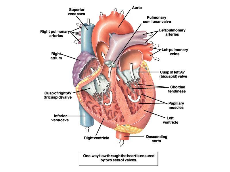
4 The Cardiac Cycle Note the valves (blue) which control the one way direction of blood flow; also, the tendons prevent the AV valves from turning inside-out. As the animation starts, the atria fill, then contract (atrial systole), pumping blood via the AV valves into the ventricles. Then the ventricles contract (ventricular systole), causing the AV valves to shut and the semicircular valves to open, allowing blood out of the heart (normally, A and V do not contract at the same time). This is followed by relaxation (diastole) of the ventricles, and the semilunar valves shut. The cycles then repeats itself

5 The Cardiac Cycle The closing and opening of the cardiac valves define 4 main phases of the CC 1. Inflow phase: the inlet valve is open and the outlet valve is closed; AV valve closure terminates phase 1 2. Isovolumetric contraction: both valves are closed, with no blood flow; Semilunar valve opening terminates phase Outflow phase: the outlet valve is open and the inlet valve is closed; Semilunar valve closing terminates phase 3 4. Isovolumetric relaxation: both valves are closed, with no blood flow; AV valve opening terminates phase = systole (ventricles contract), about 0.3 sec for a HR =75 beats /min 4+1 = diastole (ventricles relax), about 0.5 sec from a CC of 0.8 sec, HT=75 beats/min
 8.")
6 The Cardiac Cycle 1. AS: pumping the blood into V during the last part of the VD 2. VS: isovolumic contraction 3. VS: rapid ventricular ejection 4. VS: reduced ventricular ejection 5. VD: isovolumic ventricular relaxation 6. VD: rapid ventricular filling 7. VD: slow/reduced ventricular filling (diastasis) 8. AD: during all the VS and part of the VD A for atrial, V for ventricular, S for systole, D for diastole

7 The Cardiac Cycle

8 Changes in ventricular volumes, pressures and flow during the phases of the cardiac cycle The placement of catheters used for pressure measurements in the right heart.
9 Atrial Systole - immediately the P wave on the ECG - 0,1 sec at a HR of 75 bats/min - functional significance: Atrium = primer pump for the ventricle: contributes to, but is not essential for ventricular filling (<20% from stroke volume in a person at rest and up to 40% during heavy exercise) - causes the 4 th heart sound (phonocardiogram) - venous pulse: a wave (jugulogram)
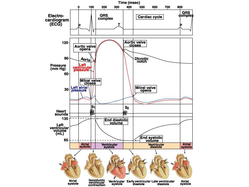
10 Volume (ml) Presure (mmhg) Isovolumic Isovolumic Rapid inflow contraction Ejection relaxation Diastasis Atrial systole 120 Ao C Aortic pressure M C Ao O M O a c v Atrial pressure Ventricular pres Ventricular volume S 1 S 2 S 3 S 4 S 1 Electrocardiogram Phonocardiogram Polygram: events of the cardiac cycle for left ventricular function
11 Atrial Diastole - 0,7 sec for 75 beats/min - changes in diastolic intra-atrial pressure: - physiological changes: c and v waves - pathological changes (valves pathology)
12 Volume (ml) Presure (mmhg) Isovolumic Isovolumic Rapid inflow contraction Ejection relaxation Diastasis Atrial systole 120 Ao C Aortic pressure M C Ao O a c v M O a c v Atrial pressure Ventricular pres Ventricular volume S 1 S 2 S 3 S 4 S 1 Electrocardiogram Phonocardiogram Polygram: events of the cardiac cycle for left ventricular function
13 - 0,27 s; - phases: Ventriculare systole 1. isovolumic contraction AV valves close, V contracts with closed valves increase in intraventricular pressure that will cause opening of semilunar valves 2. rapid ejection; 70% ejection accelerated decrease in ventricular volume 3. decreased/slow ejection; 30% ejection slow decrease in ventricular volume decrease ventricular and aortic pressures - results in ejection of 70 ml of blood = stroke volume (leaving another 50 ml in the ventricle end systolic volume)
14 Isovolumic Contraction Ventricles are closed chambers
15 Ventricular ejection When P LV > 80 mmhg P RV > 8 mmhg Rapid (1/3; 70%) ejection Slow (2/3; 30%) ejection
16 Volume (ml) Presure (mmhg) Isovolumic Isovolumic Rapid inflow contraction Ejection relaxation Diastasis Atrial systole 120 Ao C Aortic pressure M C Ao O M O a c v Atrial pressure Ventricular pres Ventricular volume S 1 S 2 S 3 S 4 S 1 Electrocardiogram Phonocardiogram Polygram: events of the cardiac cycle for left ventricular function
17 Ventricular diastole - 0,53 s (decreases with increase in HR) - phases: - isovolumic relaxation (all valves closed, pressure falls rapidly in the ventricle) - rapid ventricular filling - slow ventricular filling (diastasis) - late ventricular filling determined by atrial systole
18 Isovolumic Relaxation
19 Ventricular filling
20 Volume (ml) Presure (mmhg) Isovolumic Isovolumic Rapid inflow contraction Ejection relaxation Diastasis Atrial systole 120 Ao C Aortic pressure M C Ao O M O a c v Atrial pressure Ventricular pres Ventricular volume S 1 S 2 S 3 S 4 S 1 Electrocardiogram Phonocardiogram Polygram: events of the cardiac cycle for left ventricular function
21

22 Comparison of the dynamics of the left and right ventricles

23 Comparison of the dynamics of the left and right ventricles
 Phase 1 of the CC has three subparts: -rapid ventricular filling, -decreased ventricular filling, -atrial systole (last")
24 Mechanical, electrical, acoustic, and echocardiographic events in the cardiac cycle. Here the cardiac cycle begins with atrial contraction (AS) Phase 1 of the CC has three subparts: -rapid ventricular filling, -decreased ventricular filling, -atrial systole (last stage of ventricular filling); Phase 3 has two subparts: -rapid and decreased ventricular ejection
25 Polygram - Analysis of Cardiac activity Electrical activity measured by electrocardiography Mechanical activity evaluated by: 1. Atrial pressure curve and venous pressure: recorded at jugular vein level (jugulogram) 2. Aortic pressure curve: recorded at carotid artery level (carotidogram) 3. Phonocardiography: record of the heart sounds 4. Ventricular volume: evaluated by apexocardiogram

26 Pressure waves in veins systemic veins have pressure waves - venous pulse: (1) retrograde action of the heartbeat during the cardiac cycle, (2) the respiratory cycle (3) the contraction of skeletal muscles. Jugular vein, has a complex pulse wave synchronized to the cardiac cycle: 3 peaks, labeled a, c, and v 3 minima, labeled av, x, and y.

27 Pressure transients in the jugular vein pulse reflect events in the cardiac cycle: a peak - caused by the contraction of the right atrium. av minimum is due to relaxation of the right atrium and closure of the tricuspid valve. c peak reflects the pressure rise in the right ventricle early during systole and the resultant protruding of the tricuspid valve-which has just closed-into the right atrium. x minimum occurs as the ventricle contracts and shortens during the ejection phase, later in systole. The shortening heart-with tricuspid valve still closed-pulls on and therefore elongates the veins, lowering their pressure. v peak is related to filling of the right atrium against a closed tricuspid valve, which causes right atrial pressure to slowly rise. As the tricuspid valve opens, the v peak begins to decline. y minimum reflects a fall in right atrial pressure during rapid ventricular filling, as blood leaves the right atrium through an open tricuspid valve and enters the right ventricle. The increase in venous pressure after the y minimum occurs as venous return continues in the face of reduced ventricular filling.

28 Effect of the Respiratory Cycle During inspiration, the diaphragm descends, causing intrathoracic pressure (and therefore the pressure inside the thoracic vessels) to decrease and intra-abdominal pressure to increase the venous return from the head and upper extremities transiently increases, as low-pressure vessels literally draw blood into the thoracic cavity. Simultaneously, the venous flow decreases from the lower extremities because of the relatively high pressure of the abdominal veins during inspiration. Therefore, during inspiration, pressure in the jugular vein falls while pressure in the femoral vein rises.
29
30 Polygram - Analysis of Cardiac activity Electrical activity measured by electrocardiography Mechanical activity evaluated by: 1. Atrial pressure curve and venous pressure: recorded at jugular vein level (jugulogram) 2. Aortic pressure curve: recorded at carotid artery level (carotidogram) 3. Phonocardiography: record of the heart sounds 4. Ventricular volume: evaluated by apexocardiogram

31 Cardiac Cycle causes flow waves in aorta and peripheral vessels With the closing & opening of pulmonary and aortic valves, blood flow and blood velocity across these valves oscillate from near zero, when the valves are closed, to high values, when the valves are open. Blood flow in the aortic arch actually oscillates between slightly negative and highly positive values. Flow (A) and pressure (B) profiles in the aorta and smaller vessels. Pressure in the aortic arch typically oscillates between ~ mm Hg. Phasic changes in pressure and flow also occur in the peripheral arteries. Arterial pressure is usually measured in a large artery, such as the brachial artery the measured systolic and diastolic arterial pressures, as well as the pulse pressure and mean arterial pressure, closely approximate the corresponding aortic pressures.
32 Polygram - Analysis of Cardiac activity Electrical activity measured by electrocardiography Mechanical activity evaluated by: 1. Atrial pressure curve and venous pressure: recorded at jugular vein level (jugulogram) 2. Aortic pressure curve: recorded at carotid artery level (carotidogram) 3. Phonocardiography: record of the heart sounds 4. Ventricular volume: evaluated by apexocardiogram
33 Heart sounds and phonocardiography Heart sounds are relatively brief, discrete auditory vibrations of varying intensity (loudness), frequency (pitch), and quality (timbre). The first heart sound identifies the onset of ventricular systole, and the second heart sound identifies the onset of diastole. These two auscultatory events establish a framework within which other heart sounds and murmurs can be placed and timed. Listening to the sounds of the body with the aid of a stethoscope is called auscultation. The stethoscope can detect leaks in the valves that permit jets of blood to flow backward across the valvular orifice (i.e., regurgitation) as well as stenotic lesions that narrow the valve opening, forcing the blood to pass through a narrower space (i.e., stenosis). During certain parts of the cardiac cycle, blood passing through either regurgitant or stenotic lesions makes characteristic sounds that are called murmurs. Phonocardiogram: the recording of the auscultatory cardiac activity, using a transducer placed on the thorax. The movement of the valve leaflets can be detected by echocardiography.


34 Chest Surface Areas for Auscultation of Normal Heart Sounds The primary aortic area: 2 nd right intercostal space, adjacent to the sternum. The secondary aortic area: 3 rd left intercostal space, adjacent to the sternum (known as Erb area). The pulmonary area: 2 nd left intercostal space The tricuspid area: 4 th & 5 th intercostal spaces, adjacent to the left sternal border. The mitral area at the cardiac apex: 5 th left intercostal space, on the medioclavicular line.
35
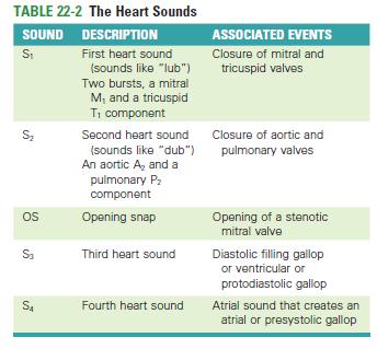
36 The first heart sound (S1) systolic sound the lub appears at sec after the beginning of the QRS complex vibrations are low in pitch and relatively long-lasting - lasts ~ sec; frequency ~ Hz; produced, in this order, by: closing of the mitral valve closing of the tricuspid valve opening of the pulmonar valve opening of the aortic valve.
37 The second heart sound (S2) diastolic sound the dub appears in the terminal period of the T wave lasts s produced, in this order, by: closing of the aortic valve, closing of the pulmonic valve, opening of the tricuspid valve, opening of the mitral valve. heard like a rapid snap because these valves close rapidly, and the surroundings vibrate for a short period physiologic splitting that varies with respiration (wider splitting with inspiration) Split S2 Inspiration Expiration Normal or physiologic
38 Split S2 Audible respiratory splitting means > 30 msec difference in the timing of the aortic (A 2 ) and pulmonic (P 2 ) components of the second heart sound. Splitting of S 2 is best heard over the 2 nd left intercostal space The normal P 2 is often softer than A 2 and rarely audible at apex Inspiration accentuates the splitting of S 2. Split S2 Inspiration Expiration Normal or physiologic
39 The third heart sound (S3) occurs in early diastole (at the beginning of the middle third of diastole) when rapid filling of the ventricles results in recoil of ventricular walls that have a limited distensibility lasts sec protodiastolic sound or gallop A gallop rhythm is a grouping of three heart sounds that together sound like hoofs of a galloping horse. The addition of an S3 to the physiological S1 and S2 creates a three-sound sequence, S1-S2-S3, that is termed a protodiastolic gallop or ventricular gallop. it is normal in children and individuals with a thin thoracic wall occasionally heard as a weak, rumbling sound
40 The fourth heart sound (S4) presystolic sound: appears at 0.04 s after the P wave (late diastolic) lasts s caused by the blood flow that hits the ventricular wall during the atrial systole. physiological only in small children, if heard in other conditions it is a sign of reduced ventricular compliance. addition of an S 4 produces another three-sound sequence, S 4 - S 1 -S 2, which is a presystolic gallop rhythm or atrial gallop during tachycardia S4-S1 can fuse, producing a summation gallop
41 EC=ejection click: most common early systolic sound; Results from abrupt halting of semilunar valves OS=opening snap: high-frequency early diastolic sound (occurs msec after A2) associated with mitral stenosis (stiffening of the mitral valve); sound due to abrupt deceleration of mitral leaflets sound with associated murmur.

.")
42 Phonocardiogram The duration of S1, S2 is slightly more than 0.10 sec. S1 ~ 0.14 sec S2 ~ 0.11 sec. (the semilunar valves are more taut than the A-V valves, so that they vibrate for a shorter time than do the A-V valves). The audible range of frequency (pitch) in the first and second heart sounds: ~ 40 cycles/sec up above 500 cycles/sec.
43 Polygram - Analysis of Cardiac activity Electrical activity measured by electrocardiography Mechanical activity evaluated by: 1. Phonocardiography: record of the heart sounds 2. Atrial pressure curve: recorded at jugular vein level (jugulogram) 3. Ventricular volume: evaluated by apexocardiogram 4. Aortic pressure curve: recorded at carotid artery level (carotidogram)
44 Volume (ml) Presure (mmhg) Isovolumic Isovolumic Rapid inflow contraction Ejection relaxation Diastasis Atrial systole AoC Aortic pressure MC Ao O MO a c v Atrial pressure Ventricular pres Ventricular volume S 1 S 2 S 3 S 4 S 1 Electrocardiogram Phonocardiogram Polygram: events of the cardiac cycle for left ventricular function
.")


45 ATRIAL SYSTOLE (The end of ventricular diastole) Heart: During atrial systole the atrium contracts and tops off the volume in the ventricle with only a small amount of blood. Atrial contraction is complete before the ventricle begins to contract. Atrial pressure: The "a" wave occurs when the atrium contracts, increasing atrial pressure (yellow). Blood arriving at the heart cannot enter the atrium so it flows back up the jugular vein, causing the first discernible wave in the jugular venous pulse. Atrial pressure drops when the atria stop contracting. ECG: An impulse arising from the SA node results in depolarization and contraction of the atria. The P wave is due to this atrial depolarization. The PR segment is electrically quiet as the depolarization proceeds to the AV node. This brief pause before contraction allows the ventricles to fill completely with blood. Heart sounds: A fourth heart sound (S4) is abnormal and is associated with the end of atrial emptying after atrial contraction. It occurs with hypertrophic congestive heart failure, massive pulmonary embolism or tricuspid incompetence.
.")
 exceeds the pressure in the atria (yellow).")
46 ISOVOLUMETRIC CONTRACTION The beginning of systole Heart: The atrioventricular (AV) valves close at the beginning of this phase. Electrically, ventricular systole is defined as the interval between the QRS complex and the end of the T wave (the Q-T interval). Mechanically, ventricular systole is defined as the interval between the closing of the AV valves and the opening of the semilunar valves (aortic and pulmonary valves). Pressures & Volume: The AV valves close when the pressure in the ventricles (red) exceeds the pressure in the atria (yellow). As the ventricles contract isovolumetrically - - their volume does not change (white ) -- the pressure inside increases, approaching the pressure in the aorta and pulmonary arteries (green). ECG: The electrical impulse propagates from the AV node through the His bundle and Purkinje system to allow the ventricles to contract from the apex of the heart towards the base. The QRS complex is due to ventricular depolarization, and it marks the beginning of ventricular systole. It is so large that it masks the underlying atrial repolarization signal. Heart sounds: The first heart sound (S1, "lub") is due to the closing AV valves and associated blood turbulence.
 exceeds the pressure in the")
. As more blood enters the arteries, pressure there builds until the flow of blood reaches a peak.")

47 RAPID EJECTION Heart: The semilunar (aortic and pulmonary) valves open at the beginning of this phase. Pressures & Volume: While the ventricles continue contracting, the pressure in the ventricles (red) exceeds the pressure in the aorta and pulmonary arteries (green); the semilunar valves open, blood exits the ventricles, and the volume in the ventricles decreases rapidly ( white). As more blood enters the arteries, pressure there builds until the flow of blood reaches a peak. The "c" wave of atrial pressure is not normally discernible in the jugular venous pulse. Right ventricular contraction pushes the tricuspid valve into the atrium and increases atrial pressure, creating a small wave into the jugular vein. It is normally simultaneous with the carotid pulse. ECG: Heart sounds:
, blood flow out of the ventricles decreases and ventricular volume decreases more")

48 REDUCED EJECTION The end of systole Heart: At the end of this phase the semilunar (aortic and pulmonary) valves close. Pressures & Volume: After the peak in ventricular and arterial pressures (red and green), blood flow out of the ventricles decreases and ventricular volume decreases more slowly (white ). When the pressure in the ventricles falls below the pressure in the arteries, blood in the arteries begins to flow back toward the ventricles and causes the semilunar valves to close. This marks the end of ventricular systole mechanically. ECG: The T wave is due to ventricular repolarization. The end of the T wave marks the end of ventricular systole electrically. Heart sounds:
.")
 is at a minimum and is ready to be filled again with blood.")
49 ISOVOLUMETRIC RELAXATION The beginning of diastole Heart: At the beginning of this phase the AV valves are closed. Pressures & Volume: Throughout this and the previous two phases, the atrium in diastole has been filling with blood on top of the closed AV valve, causing atrial pressure to rise gradually (yellow). The "v" wave is due to the back flow of blood after it hits the closed AV valve. It is the second discernible wave of the jugular venous pulse. The pressure in the ventricles (red) continues to drop. Ventricular volume (white ) is at a minimum and is ready to be filled again with blood. ECG: Heart sounds: The second heart sound (S2, "dup") occurs when the semilunar (aortic and pulmonary) valves close. S2 is normally split because the aortic valve closes slightly earlier than the pulmonary valve.

")
 is usually")

50 RAPID VENTRICULAR FILLING Heart: Once the AV valves open, blood that has accumulated in the atria flows rapidly into the ventricles. Pressures & Volume: Ventricular volume ( white white) increases rapidly as blood flows from the atria into the ventricles. ECG: Heart sounds: A third heart sound (S3) is usually abnormal and is due to rapid passive ventricular filling. It occurs in dilated congestive heart failure, myocardial infarction, or mitral incompetence.
 Heart: Pressures &")
 increases more slowly now.")

51 REDUCED VENTRICULAR FILLING (DIASTASIS) Heart: Pressures & Volume: Ventricular volume ( white ) increases more slowly now. The ventricles continue to fill with blood until they are nearly full. ECG: Heart sounds:
CARDIAC CYCLE CONTENTS. Divisions of cardiac cycle 11/13/13. Definition. Badri Paudel GMC
 CARDIAC CYCLE Badri Paudel GMC CONTENTS Ø DEFINATION Ø DIVISION OF CARDIAC CYCLE Ø SUB DIVISION AND DURATION OF CARDIAC CYCLE Ø SYSTOLE Ø DIASTOLE Ø DESCRIPTION OF EVENTS OF CARDIAC CYCLE Ø SUMMARY Ø ELECTROCARDIOGRAPHY
CV Lecture. Heart as a pump cardiac performance Polygram - analysis of cardiac activity. Dr. Ana-Maria Zagrean
 CV Lecture Heart as a pump cardiac performance Polygram - analysis of cardiac activity Dr. Ana-Maria Zagrean Coronary Circulation Left coronary artery: for the anterior & left lateral portions of LV. Right
2. The heart sounds are produced by a summed series of mechanical events, as follows:
 Heart Sounds. Phonocardiography 1 Objectives 1. Phonocardiography - Definition 2. What produces the heart sounds 3. Where to listen for the heart sounds 4. How to record a phonocardiogram 5. Normal heart
The Cardiac Cycle Clive M. Baumgarten, Ph.D.
 The Cardiac Cycle Clive M. Baumgarten, Ph.D. OBJECTIVES: 1. Describe periods comprising cardiac cycle and events within each period 2. Describe the temporal relationships between pressure, blood flow,
Heart sounds and murmurs. Dr. Szathmári Miklós Semmelweis University First Department of Medicine 15. Oct
 Heart sounds and murmurs Dr. Szathmári Miklós Semmelweis University First Department of Medicine 15. Oct. 2013. Conditions for auscultation of the heart Quiet room Patient comfortable Chest fully exposed
Electrical Conduction
 Sinoatrial (SA) node Electrical Conduction Sets the pace of the heartbeat at 70 bpm AV node (50 bpm) and Purkinje fibers (25 40 bpm) can act as pacemakers under some conditions Internodal pathway from
Objectives of the Heart
 Objectives of the Heart Electrical activity of the heart Action potential EKG Cardiac cycle Heart sounds Heart Rate The heart s beat separated into 2 phases Relaxed phase diastole (filling of the chambers)
Cardiac Cycle MCQ. Professor of Cardiovascular Physiology. Cairo University 2007
 Cardiac Cycle MCQ Abdel Moniem Ibrahim Ahmed, MD Professor of Cardiovascular Physiology Cairo University 2007 1- Regarding the length of systole and diastole: a- At heart rate 75 b/min, the duration of
Pathological Arrhythmias/ Tachyarrhythmias
 Pathological Arrhythmias/ Tachyarrhythmias caused by: 1.Ectopic focus: Extrasystole or premature beat. If discharge is occasional. Can be: Atrial Extrasystole Vevtricular Extrasystole 2.Cardiac Arrhythmia
Cardiac Cycle. Each heartbeat is called a cardiac cycle. First the two atria contract at the same time.
 The Heartbeat Cardiac Cycle Each heartbeat is called a cardiac cycle. First the two atria contract at the same time. Next the two ventricles contract at the same time. Then all the chambers relax. http://www.youtube.com/watch?v=frd3k6lkhws
PHONOCARDIOGRAPHY (PCG)
") PHONOCARDIOGRAPHY (PCG) The technique of listening to sounds produced by the organs and vessels of the body is called auscultation. The areas at which the heart sounds are heard better are called auscultation
PHONOCARDIOGRAPHY (PCG) The technique of listening to sounds produced by the organs and vessels of the body is called auscultation. The areas at which the heart sounds are heard better are called auscultation
Cardiac Ausculation in the Elderly
 Cardiac Ausculation in the Elderly 박성하 신촌세브란스병원심장혈관병원심장내과 Anatomy Surface projection of the Heart and Great Vessels Evaluating pulsation Superior vena cava Rt. pulmonary artery Right atrium Right ventricle
CARDIAC EXAMINATION MINI-QUIZ
 CARDIAC EXAMINATION MINI-QUIZ 1. Sitting bolt upright, your dyspneic (short of breath) patient has visible jugular venous pulsations to the angle of his jaw, which is 12 cm above his sternal angle. What
CARDIOLOGY FOR DUMMIES
 CARDIOLOGY FOR DUMMIES Prof. dr. Gunther van Loon, DVM, PhD, Dipl ECEIM Dept. of Large Animal Internal Medicine, Fac. of Veterinary Medicine, Ghent University, Salisburylaan 133, B-9820 Merelbeke, Belgium
4. The two inferior chambers of the heart are known as the atria. the superior and inferior vena cava, which empty into the left atrium.
 Answer each statement true or false. If the statement is false, change the underlined word to make it true. 1. The heart is located approximately between the second and fifth ribs and posterior to the
Collin County Community College
 Collin County Community College BIOL. 2402 Anatomy & Physiology WEEK 5 The Heart 1 The Heart Beat and the EKG 2 1 The Heart Beat and the EKG P-wave = Atrial depolarization QRS-wave = Ventricular depolarization
The Heart and Cardiovascular System
 The Heart and Cardiovascular System What you will learn The location of the heart 3 layers and covering of the heart Explain the function of the heart as 2 separate pumps Identify the 4 chambers of the
Chapter 18 - Heart. I. Heart Anatomy: size of your fist; located in mediastinum (medial cavity)
") Chapter 18 - Heart I. Heart Anatomy: size of your fist; located in mediastinum (medial cavity) A. Coverings: heart enclosed in double walled sac called the pericardium 1. Fibrous pericardium: dense connective
Chapter 18 - Heart I. Heart Anatomy: size of your fist; located in mediastinum (medial cavity) A. Coverings: heart enclosed in double walled sac called the pericardium 1. Fibrous pericardium: dense connective
Lab #3: Electrocardiogram (ECG / EKG)
") Lab #3: Electrocardiogram (ECG / EKG) An introduction to the recording and analysis of cardiac activity Introduction The beating of the heart is triggered by an electrical signal from the pacemaker. The
Lab #3: Electrocardiogram (ECG / EKG) An introduction to the recording and analysis of cardiac activity Introduction The beating of the heart is triggered by an electrical signal from the pacemaker. The
THE CARDIOVASCULAR SYSTEM. Heart 2
 THE CARDIOVASCULAR SYSTEM Heart 2 PROPERTIES OF CARDIAC MUSCLE Cardiac muscle Striated Short Wide Branched Interconnected Skeletal muscle Striated Long Narrow Cylindrical PROPERTIES OF CARDIAC MUSCLE Intercalated
1. how a careful cardiovascular evaluation can accurately assess pathology and physiology at the bedside, and
 This program will demonstrate: 1. how a careful cardiovascular evaluation can accurately assess pathology and physiology at the bedside, and 2. the importance of integrating this information with selected
CARDIOVASCULAR SYSTEM
 CARDIOVASCULAR SYSTEM Overview Heart and Vessels 2 Major Divisions Pulmonary Circuit Systemic Circuit Closed and Continuous Loop Location Aorta Superior vena cava Right lung Pulmonary trunk Base of heart
Cardiovascular system
 BIO 301 Human Physiology Cardiovascular system The Cardiovascular System: consists of the heart plus all the blood vessels transports blood to all parts of the body in two 'circulations': pulmonary (lungs)
The Mammalian Circulatory System
 The Mammalian Heart The Mammalian Circulatory System Recall: What are the 3 cycles of the mammalian circulatory system? What are their functions? What are the three main vessel types in the mammalian circulatory
12.2 Monitoring the Human Circulatory System
 12.2 Monitoring the Human Circulatory System Video 1: 3D Animation of Heart Pumping Blood blood flow through the heart... Video 2: Hank Reviews Everything on the Heart https://www.youtube.com/watch?v=x9zz6tcxari
(D) (E) (F) 6. The extrasystolic beat would produce (A) increased pulse pressure because contractility. is increased. increased
 (E) (F) 6. The extrasystolic beat would produce (A) increased pulse pressure because contractility. is increased. increased") Review Test 1. A 53-year-old woman is found, by arteriography, to have 5% narrowing of her left renal artery. What is the expected change in blood flow through the stenotic artery? Decrease to 1 2 Decrease
Review Test 1. A 53-year-old woman is found, by arteriography, to have 5% narrowing of her left renal artery. What is the expected change in blood flow through the stenotic artery? Decrease to 1 2 Decrease
37 1 The Circulatory System
 H T H E E A R T 37 1 The Circulatory System The circulatory system and respiratory system work together to supply cells with the nutrients and oxygen they need to stay alive. a) The respiratory system:
IB TOPIC 6.2 THE BLOOD SYSTEM
 IB TOPIC 6.2 THE BLOOD SYSTEM THE BLOOD SYSTEM TERMS TO KNOW circulation ventricle artery vein 6.2.U1 - Arteries convey blood at high pressure from the ventricles to the tissues of the body Circulation
Do Now. Get out work from last class to be checked
 Do Now Get out work from last class to be checked Heart Actions Cardiac Cycle: One complete heartbeat. The contraction of a heart chamber is called systole and the relaxation of a chamber is called diastole.
The Cardiovascular System
 Essentials of Human Anatomy & Physiology Elaine N. Marieb Slides 11.1 11.19 Seventh Edition Chapter 11 The Cardiovascular System Functions of the Cardiovascular system Function of the heart: to pump blood
Heart Pump and Cardiac Cycle. Faisal I. Mohammed, MD, PhD
 Heart Pump and Cardiac Cycle Faisal I. Mohammed, MD, PhD 1 Objectives To understand the volume, mechanical, pressure and electrical changes during the cardiac cycle To understand the inter-relationship
10. Thick deposits of lipids on the walls of blood vessels, called, can lead to serious circulatory issues. A. aneurysm B. atherosclerosis C.
 Heart Student: 1. carry blood away from the heart. A. Arteries B. Veins C. Capillaries 2. What is the leading cause of heart attack and stroke in North America? A. alcohol B. smoking C. arteriosclerosis
SIKLUS JANTUNG. Rahmatina B. Herman
 SIKLUS JANTUNG Rahmatina B. Herman The Cardiac Cycle Definition: The cardiac events that occur from the beginning of one heartbeat to the beginning of the next The cardiac cycle consists of: - Diastole
HISTORY. Question: How do you interpret the patient s history? CHIEF COMPLAINT: Dyspnea of two days duration. PRESENT ILLNESS: 45-year-old man.
 HISTORY 45-year-old man. CHIEF COMPLAINT: Dyspnea of two days duration. PRESENT ILLNESS: His dyspnea began suddenly and has been associated with orthopnea, but no chest pain. For two months he has felt
The cardiovascular system is composed of the heart and blood vessels that carry blood to and from the body s organs. There are 2 major circuits:
 1 The cardiovascular system is composed of the heart and blood vessels that carry blood to and from the body s organs. There are 2 major circuits: pulmonary and systemic. The pulmonary goes out to the
Human Cardiovascular Physiology: Blood Pressure and Pulse Determinations
 ighapmlre33apg269_274 5/12/04 3:10 PM Page 269 impos03 302:bjighapmL:ighapmLrevshts:layouts: NAME Human Cardiovascular Physiology: Blood Pressure and Pulse Determinations LAB TIME/DATE REVIEW SHEET exercise
Cardiovascular System
 Cardiovascular System Chapter 8 1 Cardiovascular System Functions: pump saturated oxygenated blood into arterial system cells pump desaturated deoxygenated blood to lungs via veins for reoxygenation Heart
IP: Regulation of Cardiac Output
 ANP 1105D Winter 2013 Assignment 9: The Heart, part 2: Chap... Assignment 9: The Heart, part 2: Chapter 18 Signed in as Alex Sokolowski Help Close Resources Due: 11:59pm on Monday, March 25, 2013 Note:
Cardiovascular System Notes: Physiology of the Heart
 Cardiovascular System Notes: Physiology of the Heart Interesting Heart Fact Capillaries are so small it takes ten of them to equal the thickness of a human hair. Review What are the 3 parts of the cardiovascular
CRITICAL THINKING QUESTIONS AND ANSWERS AND CYCLE 2 LAB EXAM TEMPLATE. There are two main mechanisms that work in conjunction to return the blood
 CRITICAL THINKING QUESTIONS AND ANSWERS AND CYCLE 2 LAB EXAM TEMPLATE There are two main mechanisms that work in conjunction to return the blood THE CARDIAC PUMP 1) The forward pull(vis a fronte) This
Occurrence of the First Heart Sound and the Opening Snap in Mitral Stenosis
 The Effect of Cycle Length on the Time of Occurrence of the First Heart Sound and the Opening Snap in Mitral Stenosis By ADDISON L. MESSER, M.D., TIMOTHY B. COUNIHAN, M.D., MAURICE B. RAPPAPORT, E.E.,
The Heart. Happy Friday! #takeoutyournotes #testnotgradedyet
 The Heart Happy Friday! #takeoutyournotes #testnotgradedyet Introduction Cardiovascular system distributes blood Pump (heart) Distribution areas (capillaries) Heart has 4 compartments 2 receive blood (atria)
Practice Exercises for the Cardiovascular System
 Practice Exercises for the Cardiovascular System On the diagram below, color the oxygen-rich blood red and the oxygen-poor blood blue. Label the parts: Continued on the next page... Label the parts on
current, and acting like
 Heart 10 IV. HEART PHYSIOLOGY - How the heart beats. How the heart depolarizes the myocardium, which leads to a contraction. A) INTRINSIC CONTROL - Heart controls its own rhythm. HOW? The presence of gap
Cardiovascular System Notes: Heart Disease & Disorders
 Cardiovascular System Notes: Heart Disease & Disorders Interesting Heart Facts The Electrocardiograph (ECG) was invented in 1902 by Willem Einthoven Dutch Physiologist. This test is still used to evaluate
IB TOPIC 6.2 THE BLOOD SYSTEM
 IB TOPIC 6.2 THE BLOOD SYSTEM TERMS TO KNOW circulation ventricle artery vein THE BLOOD SYSTEM 6.2.U1 - Arteries convey blood at high pressure from the ventricles to the tissues of the body Circulation
Circulation: Chapter 25. Cardiac Output. The Mammalian Heart Fig Right side of the heart
 Circulation: Chapter 25 1. Limits of Diffusion A. Small organisms use diffusion B. rapid over small distances 2. Most animals have circulatory systems A. Blood B. Pump (Heart) or propulsive structures
SMALL GROUP SESSION 19 January 30 th or February 1st. Groups 1-12: Cardiac Case and Cardiac Exam Workshop
 SMALL GROUP SESSION 19 January 30 th or February 1st Groups 1-12: Cardiac Case and Cardiac Exam Workshop Readings: Complete the cardiac examination tutorial on the POM1 web site. Optional: http://medicine.ucsd.edu/clinicalmed/heart.htm
HISTORY. Question: What category of heart disease is suggested by the fact that a murmur was heard at birth?
 HISTORY 23-year-old man. CHIEF COMPLAINT: Decreasing exercise tolerance of several years duration. PRESENT ILLNESS: The patient is the product of an uncomplicated term pregnancy. A heart murmur was discovered
Approximately the size of your fist Location. Pericardial physiology
 Heart Anatomy Approximately the size of your fist Location Superior surface of diaphragm Left of the midline Anterior to the vertebral column, posterior to the sternum Wednesday, March 28, 2012 Muscle
Large Arteries of Heart
 Cardiovascular System (Part A-2) Module 5 -Chapter 8 Overview Arteries Capillaries Veins Heart Anatomy Conduction System Blood pressure Fetal circulation Susie Turner, M.D. 1/5/13 Large Arteries of Heart
Pearson's Comprehensive Medical Assisting Administrative and Clinical Competencies
 Pearson's Comprehensive Medical Assisting Administrative and Clinical Competencies THIRD EDITION CHAPTER 27 The Cardiovascular System Lesson 1: Overview of the Cardiovascular System Lesson Objectives Upon
The HEART. What is it???? Pericardium. Heart Facts. This muscle never stops working It works when you are asleep
 This muscle never stops working It works when you are asleep The HEART It works when you eat It really works when you exercise. What is it???? Located between the lungs in the mid thoracic region Apex
The Cardiovascular System. Chapter 15. Cardiovascular System FYI. Cardiology Closed systemof the heart & blood vessels. Functions
 Chapter 15 Cardiovascular System FYI The heart pumps 7,000 liters (4000 gallons) of blood through the body each day The heart contracts 2.5 billion times in an avg. lifetime The heart & all blood vessels
Physiology of the Heart Delmar Learning, a Division of Thomson Learning, Inc.
 Physiology of the Heart 2004 Delmar Learning, a Division of Thomson Learning, Inc. Physiology of the Heart State Standards 35) Outline the structure and functions of the anatomy of the cardiovascular system,
Chapter 13 The Cardiovascular System: Cardiac Function
 Chapter 13 The Cardiovascular System: Cardiac Function Overview of the Cardiovascular System The Path of Blood Flow through the Heart and Vasculature Anatomy of the Heart Electrical Activity of the Heart
Cardiovascular Physiology
 Cardiovascular Physiology The mammalian heart is a pump that pushes blood around the body and is made of four chambers: right and left atria and right and left ventricles. The two atria act as collecting
Electrocardiogram and Heart Sounds
 Electrocardiogram and Heart Sounds Five physiologic properties of cardiac muscle Automaticity: SA node is the primary pacemaker of the heart, but any cells in the conduction system can initiate their
The Cardiovascular System
 The Cardiovascular System The Cardiovascular System A closed system of the heart and blood vessels The heart pumps blood Blood vessels allow blood to circulate to all parts of the body The function of
HISTORY. Question: What category of heart disease is suggested by this history? CHIEF COMPLAINT: Heart murmur present since early infancy.
 HISTORY 18-year-old man. CHIEF COMPLAINT: Heart murmur present since early infancy. PRESENT ILLNESS: Although normal at birth, a heart murmur was heard at the six week check-up and has persisted since
The Circulatory System. The Heart, Blood Vessels, Blood Types
 The Circulatory System The Heart, Blood Vessels, Blood Types The Closed Circulatory System Humans have a closed circulatory system, typical of all vertebrates, in which blood is confined to vessels and
PART I: HEART ANATOMY
 Lab 7: Heart Sounds and Blood Pressure PART I: HEART ANATOMY a) You should be able to identify the following structures on an adult human heart diagram. the 4 chambers the bicuspid (mitral) and tricuspid
Heart. Structure Physiology of blood pressure and heartbeat
 Heart Structure Physiology of blood pressure and heartbeat Location and Anatomy Location and Anatomy Pericardial cavity: surrounds, isolates, and anchors heart Parietal pericardium lined with serous membrane
CARDIOVASCULAR PHYSICAL EXAMINATION
 CARDIOVASCULAR PHYSICAL EXAMINATION Clarke Atkins, DVM, Diplomate, ACVIM (Internal Medicine and Cardiology) Jane Lewis Seaks Distinguished Professor Emeritus North Carolina State University, College of
BME 5742 Bio-Systems Modeling and Control. Lecture 41 Heart & Blood Circulation Heart Function Basics
 BME 5742 Bio-Systems Modeling and Control Lecture 41 Heart & Blood Circulation Heart Function Basics Dr. Zvi Roth (FAU) 1 Pumps A pump is a device that accepts fluid at a low pressure P 1 and outputs the
Tracheal normal sound heard over trachea loud tubular quality high-pitched expiration equal to or slightly longer than inspiration
 = listening for sounds produced in the body over chest to ID normal & abnormal lung sounds all BS made by turbulent flow in the airways useful in making initial D & evaluating effects of R 4 characteristics
Topic 6: Human Physiology
 Topic 6: Human Physiology 6.2 The Blood System D.4 The Heart Essential Questions: 6.2 The blood system continuously transports substances to cells and simultaneously collects waste products. D.3 The chemical
Circulatory System Notes
 Circulatory System Notes Functions of Circulatory System A. Transports B. Transports C. Transports D. Transports E. of fluids F. G. Regulate temperature H. Blood clotting Characteristics of various blood
Circulatory Systems. All cells need to take in nutrients and expel metabolic wastes.
 Circulatory Systems All cells need to take in nutrients and expel metabolic wastes. Single celled organisms: nutrients from the environment can diffuse (or be actively transported) directly in to the cell
AP2 Lab 3 Coronary Vessels, Valves, Sounds, and Dissection
 AP2 Lab 3 Coronary Vessels, Valves, Sounds, and Dissection Project 1 - BLOOD Supply to the Myocardium (Figs. 18.5 &18.10) The myocardium is not nourished by the blood while it is being pumped through the
Ch 19: Cardiovascular System - The Heart -
 Ch 19: Cardiovascular System - The Heart - Give a detailed description of the superficial and internal anatomy of the heart, including the pericardium, the myocardium, and the cardiac muscle. Trace the
CAMOSUN COLLEGE BIOLOGY 144 (2010) LABS
 LABS") LAB 8: CARDIOVASCULAR PHYSIOLOGY PART 1. HEART SOUNDS AND PULSE DETERMINATIONS Introduction Two distinct sounds can be heard during each cardiac cycle. These sounds are commonly described as lub and dup
LAB 8: CARDIOVASCULAR PHYSIOLOGY PART 1. HEART SOUNDS AND PULSE DETERMINATIONS Introduction Two distinct sounds can be heard during each cardiac cycle. These sounds are commonly described as lub and dup
Cardiovascular System- Heart. Miss Wheeler Unit 8
 Cardiovascular System- Heart Miss Wheeler Unit 8 Overview CARDIOVASCULAR SYSTEM heart vessels Made up of heart, blood vessels, and blood Functions Heart- pump blood Vessels- (veins, arteries, capillaries)
Circulation. Circulation = is a process used for the transport of oxygen, carbon! dioxide, nutrients and wastes through-out the body
 Circulation Circulation = is a process used for the transport of oxygen, carbon! dioxide, nutrients and wastes through-out the body Heart = muscular organ about the size of your fist which pumps blood.
Biology Unit 3 The Human Heart P
 Biology 2201 Unit 3 The Human Heart P 314-321 Structure and Function of the Human Heart Structure of the Human Heart Has four Chambers (2 Atria and 2 Ventricles) Made of Cardiac Muscle Found in Chest Cavity
THE HEART. A. The Pericardium - a double sac of serous membrane surrounding the heart
 THE HEART I. Size and Location: A. Fist-size weighing less than a pound (250 to 350 grams). B. Located in the mediastinum between the 2 nd rib and the 5 th intercostal space. 1. Tipped to the left, resting
The Cardiovascular System Part I: Heart Outline of class lecture After studying part I of this chapter you should be able to:
 The Cardiovascular System Part I: Heart Outline of class lecture After studying part I of this chapter you should be able to: 1. Describe the functions of the heart 2. Describe the location of the heart,
Outline. Electrical Activity of the Human Heart. What is the Heart? The Heart as a Pump. Anatomy of the Heart. The Hard Work
 Electrical Activity of the Human Heart Oguz Poroy, PhD Assistant Professor Department of Biomedical Engineering The University of Iowa Outline Basic Facts about the Heart Heart Chambers and Heart s The
Lab 16. The Cardiovascular System Heart and Blood Vessels. Laboratory Objectives
 Lab 16 The Cardiovascular System Heart and Blood Vessels Laboratory Objectives Describe the anatomical structures of the heart to include the pericardium, chambers, valves, and major vessels. Describe
The Heart and Heart Disease
 The Heart and Heart Disease Illustration of the heart by Leonardo DaVinci heart-surgeon.com/ history.html 2/14/2010 1 I. Location, Size and Position of the Heart A. Triangular organ located 1. of mass
1. Name the components of the formed elements in the blood and mention one major function of each of them.
 CLASS XI BIOLOGY Body Fluids And Circulation 1. Name the components of the formed elements in the blood and mention one major function of each of them. Formed Elements in Blood And Their Functions: Erythrocytes
Cardiovascular System
 Cardiovascular System The Heart Cardiovascular System The Heart Overview What does the heart do? By timed muscular contractions creates pressure gradients blood moves then from high pressure to low pressure
UNIT 08: ASSESSMENT OF CARDIOVASCULAR SYSTEM
 In The Name of God (A PROJECT OF NEW LIFE COLLEGE OF NURSING KARACHI) UNIT 08: ASSESSMENT OF CARDIOVASCULAR SYSTEM Shahzad Bashir RN, BScN, DCHN Instructor New Life College of Nursing December 01, 2015
Cardiac Examination. Pediatrics Clinical Examination
 Pediatrics Clinical Examination Symptoms of Cardiovascular Affection: Cardiac Examination 1. Perinatal history: Maternal DM, cyanosis, respiratory distress 2. Symptoms of lung congestion: Poor interrupted
Anatomy & Physiology of Cardiovascular System. Chapter 18 & 19
 Anatomy & Physiology of Cardiovascular System Chapter 18 & 19 Objectives..cont 1. Discuss the physiological stages of cardiac muscle contraction. 2. Trace a typical ECG and label each wave or complex 3.
Principles of Biomedical Systems & Devices. Lecture 8: Cardiovascular Dynamics Dr. Maria Tahamont
 Principles of Biomedical Systems & Devices Lecture 8: Cardiovascular Dynamics Dr. Maria Tahamont Review of Cardiac Anatomy Four chambers Two atria-receive blood from the vena cave and pulmonary veins Two
Lab Activity 23. Cardiac Anatomy. Portland Community College BI 232
 Lab Activity 23 Cardiac Anatomy Portland Community College BI 232 Cardiac Muscle Histology Branching cells Intercalated disc: contains many gap junctions connecting the adjacent cell cytoplasm, creates
Matters of the Heart: Comprehensive Cardiology SARAH BEANLANDS RN BSCN MSC
 Matters of the Heart: Comprehensive Cardiology SARAH BEANLANDS RN BSCN MSC Who am I? Class Outline Gross anatomy of the heart Trip around the heart Micro anatomy: cellular and tissue level Introduction
The Cardiovascular System
 PowerPoint Lecture Slide Presentation by Patty Bostwick-Taylor, Florence-Darlington Technical College The Cardiovascular System 11 PART A The Cardiovascular System A closed system of the heart and blood
Chapter 20: Cardiovascular System: The Heart
 Chapter 20: Cardiovascular System: The Heart I. Functions of the Heart A. List and describe the four functions of the heart: 1. 2. 3. 4. II. Size, Shape, and Location of the Heart A. Size and Shape 1.
Cardiovascular System: The Heart
 Cardiovascular System: The Heart I. Anatomy of the Heart (See lab handout for terms list) A. Describe the size, shape and location of the heart B. Describe the structure and function of the pericardium
Introduction. Circulation
 Introduction Circulation 1- Systemic (general) circulation 2- Pulmonary circulation carries oxygenated blood to all parts of the body carries deoxygenated blood to the lungs From Lt. ventricle aorta From
Circulatory system of mammals
 Circulatory system of mammals Explain the cardiac cycle and its initiation Discuss the internal factors that control heart action Blood flows through the heart as a result of pressure differences Blood
11/10/2014. Muscular pump Two atria Two ventricles. In mediastinum of thoracic cavity 2/3 of heart's mass lies left of midline of sternum
 It beats over 100,000 times a day to pump over 1,800 gallons of blood per day through over 60,000 miles of blood vessels. During the average lifetime, the heart pumps nearly 3 billion times, delivering
Heart. Heart 2-Tunica media: middle layer (media ='middle') muscle fibers (smooth or cardiac).
 muscle fibers (smooth or cardiac).") t. innermost lumenal General Circulatory system heart and blood vessels walls have 3 layers (inside to outside) 1-Tunica interna: aka tunica intima layer--lumenal layer epithelium--endothelium simple squamous
t. innermost lumenal General Circulatory system heart and blood vessels walls have 3 layers (inside to outside) 1-Tunica interna: aka tunica intima layer--lumenal layer epithelium--endothelium simple squamous
Health Science 20 Circulatory System Notes
 Health Science 20 Circulatory System Notes Functions of the Circulatory System The circulatory system functions mainly as the body s transport system. It transports: o Oxygen o Nutrients o Cell waste o
Heart and Neck Vessels
 CHAPTER 12 Heart and Neck Vessels ANATOMY The precordium is the area on the anterior chest overlying the heart and great vessels. The heart extends from the second to the fifth intercostal space, and from
Major Function of the Cardiovascular System. Transportation. Structures of the Cardiovascular System. Heart - muscular pump
 Structures of the Cardiovascular System Heart - muscular pump Blood vessels - network of tubes Blood - liquid transport vehicle brachiocephalic trunk superior vena cava right pulmonary arteries right pulmonary