CV Lecture. Heart as a pump cardiac performance Polygram - analysis of cardiac activity. Dr. Ana-Maria Zagrean
|
|
|
- Sara Cox
- 6 years ago
- Views:
Transcription
1 CV Lecture Heart as a pump cardiac performance Polygram - analysis of cardiac activity Dr. Ana-Maria Zagrean


2 Coronary Circulation Left coronary artery: for the anterior & left lateral portions of LV. Right coronary artery for most of the RV and the posterior part of the LV. - epicardial arteries on the surface of the heart; - intramuscular arteries penetrate from the surface into the cardiac muscle mass; compressed during systole - subendocardial arterial plexus -! inner 0.1 mm of the endocardial surface is also nourished directly from the intracardiac blood
; - from the RV returns through small anterior")

3 Coronary venous blood flow: Coronary Circulation - from the LV returns to the RA by way of the coronary sinus (~75% of the total coronary blood flow); - from the RV returns through small anterior cardiac veins that flow directly into the RA. -! a very small amount of coronary venous blood also flows back into the heart through very minute thebesian veins, which empty directly into all chambers of the heart.

4 Collateral Circulation in the Heart In a normal heart, almost no large communications exist among the larger coronary arteries. But many anastomoses do exist among the smaller arteries sized µm in diameter. The degree of damage to the heart muscle (secondary to atherosclerotic coronary constriction or by sudden coronary occlusion) is determined to a great extent by the degree of collateral circulation that has already developed or that can open within minutes after the occlusion. Minute anastomoses in the normal coronary arterial system
5 Coronary Blood Flow (CBF) In resting conditions ~ 225 ml/min in adults or 4 5 % of the total CO. During strenuous exercise: 4-7 fold increase in CO, together with increased arterial pressure 6-9 fold increased work output of the heart, with only 3-4 times increase in CBF! increase of the ratio (heart energy expenditure / CBF) shows a relative deficiency of coronary blood supply the need for increasing the "efficiency" of cardiac utilization of energy. Phasic flow of blood through the coronary capillaries of the LV during cardiac systole and diastole: strong compression of the LV muscle around the intramuscular vessels during systolic contraction (for the RV the phasic changes are partial, because the force of contraction of the RV muscle is far less than that of the LV).
6 Control of Coronary Blood Flow 1. Regulation through Local Muscle Metabolism Metabolic factors, especially myocardial oxygen consumption/oxygen demand, are the major controllers of myocardial blood flow. Normally ~70% of the oxygen in the coronary arterial blood is removed as the blood flows through the heart muscle little additional oxygen can be supplied to the heart musculature the need to increase the coronary blood flow, through local arteriolar vasodilation, proportional to cardiac muscle metabolism/degree of activity. Vasodilator substances released from the muscle cells in response to increased metabolism: - Adenosine: low oxygen conc. in the muscle cells ATP degrades to adenosine monophosphate further degraded to adenosine adenosine release into the tissue fluids of the heart muscle vasodilation (action maintained for only 1-3 hrs). Most of adenosine is reabsorbed into the cardiac cells to be reused. - Other vasodilators: adenosine phosphate compounds, K+, H+, CO 2, bradykinin, prostaglandins, nitric oxide.
7 Control of Coronary Blood Flow 2. Autonomic Nervous Control of CBF: Direct & Indirect effects Direct effects: action of Ach (vagus nerves) and NE/E (sympathetic nerves) on the coronary vessels Ach has a direct effect to dilate the coronary arteries, even the distribution of vagal nerve fibers to the ventricular coronary system is reduced. NE has either vascular constrictor or dilator effects, depending on type of receptors: constrictor - alpha receptors, more on epicardial coronary vessels Dilator - beta receptors, more on intramuscular arteries. Both alpha and beta receptors exist in the coronary vessels sympathetic stimulation cause slight overall coronary constriction or dilation, but usually constriction. Excess sympathetic drive severe alpha vasoconstrictor effects vasospastic myocardial ischemia. Indirect effects: secondary changes in coronary blood flow caused by activity of the heart, mostly opposite to the direct effects Sympathetic stimulation increases both HR and contractility increases the rate of metabolism vasodilation of coronary vessels through local blood flow regulatory mechanisms blood flow increases. Vagal stimulation slows the heart and has a slight depressive effect on heart contractility decrease cardiac oxygen consumption
8 Particularities of Cardiac Muscle Metabolism - derives mainly from oxidative metabolism of fatty acids (70%) and, to a lesser extent, of lactate and glucose (anaerobic conditions, ischemic cardiac pain due to production of lactate and ph decrease) - is measured by the rate of oxygen consumption in the heart - is used to provide the work of contraction. - >95% of the metabolic energy is used to form ATP in the mitochondria. ATP is then used for cardiac muscular contraction and other cellular functions. -in severe coronary ischemia, ATP degrades to adenosine diphosphate adenosine monophosphate adenosine to provide dilation of the coronary arterioles during coronary hypoxia. Adenosine diffusion from the muscle cells into the circulating blood with serious cellular consequence. Within 30 min. of severe coronary ischemia, about one half of the adenine base can be lost from the affected cardiac muscle cells. New synthesis of adenine is only possible at a rate of 2%/hour. For a coronary ischemia that persisted for 30 minutes, relief of the ischemia may be too late for the cardiac cells to survive.
9 Chemical energy required for cardiac contraction Efficiency of cardiac contraction - most of the expended chemical energy is converted into heat (75-80%) - a much smaller portion is converted into work output (WO) (20-25%). Efficiency of cardiac contraction = WO / total chemical energy expenditure Maximum efficiency of the normal heart ~ %. In heart failure, it decreases to as low as 5-10 %.
10 Cardiac Work Output Stroke work output of the heart - amount of energy converted to work / heartbeat. Minute work output - total amount of energy converted to work /1 minute (stroke work output x HR) Work output (WO) of the heart is used: 1) to move the blood from the low-pressure veins to the high-pressure arteries - volume-pressure work or external work (EW) (WO LV ~ 6 x WO RV, given the different systolic press. in the 2 pumps). 2) a minor proportion of energy is used to accelerate the blood to its velocity of ejection through the aortic and pulmonary valves kinetic energy of blood flow: ~ mass of blood ejected x v ejection 2. normally 1% of WO, up to 50% in Aortic Stenosis

11 Volume-pressure curves for the left ventricle Graphical Analysis of Ventricular Pumping. Relationship between LV volume and intraventricular pressure during diastole and systole. Red lines show the "volume-pressure diagram", demonstrating changes in intraventricular volume and pressure during the normal cardiac cycle. EW, external work (the area subtended by the volume-pressure diagram).
12 "Volume-Pressure Diagram" during the cardiac cycle: Volume-pressure curves for the LV - diastolic pressure curve: *shows gradual filling of LV up to the end-diastolic pressure (EDP) *pressure greatly rises after 150 ml ventricular filling (no more stretch, pericardial limit) - systolic pressure curve: *shows systolic pressure during LV contraction at each volume of filling; *increases even at low ventricular volumes *reaches a maximum ( mmhg for the LV, and mmhg for the RV) at ml. * for volumes > 170 ml, the systolic pressure actually decreases (actin and myosin filaments interrelation decreases)
13 The 4 phases of the "volume-pressure diagram", during the normal cardiac cycle. Phase I: Period of filling. -initial ventricular volume ~45 ml (end-systolic volume), diastolic pressure ~0 mm Hg. -ventricular volume normally increases with 70 ml, up to ~115 ml (end-diastolic volume), and the diastolic pressure rise to about 5 mm Hg. Phase II: Period of isovolumic contraction. -volume of the ventricle constant (all valves closed) ~115 ml, the pressure inside the ventricle increases to equal the pressure in the aorta, at ~80 mm Hg. Phase III: Period of ejection. -systolic pressure rises higher during contraction of the ventricle (up to ~120 mmhg), while the volume of the ventricle decreases during ejection. Phase IV: Period of isovolumic relaxation. -aortic valve closes, no change in volume (~45 ml ESV), decrease of ventricular pressure back to diastolic pressure (~0 mm Hg).
14 Left Ventricular Pressure D D D C C C EDV normal EDV A A A B B B Left Ventricular Volume Note the change in External Work
15 Preload and Afterload Preload - the degree of tension on the muscle when it begins to contract. - is usually considered to be the end-diastolic pressure when the ventricle has become filled. - depends on the incoming blood in the RA = venous return Afterload - the load against which the muscle exerts its contractile force. - is the systolic pressure in the artery leading from the ventricle, (relation with the vascular resistance).
16 RA LV Cardiovascular unit Cardiac activity regulation A. Intrinsic/local regulation of the heart B. Systemic regulation (nervous, humoral, integrated) CV Integration Heart activity Vascular tone
17 A. Intrinsic/local regulation Heart activity regulation 1. Ca 2+ role - Ca 2+ homeostasis - Ca 2+ regulation factors 2. Frank-Starling low of the heart Microcirculation regulation 1. Myogenic autoregulation 2. Metabolic factors 3. Chemical messengers B. Systemic regulation Short-term regulation - Nervous reflex regulation Long-term regulation - Humoral regulation Integrated regulation

18 A. Intrinsic/Local Regulation Heart activity regulation 1. Ca 2+ role - Ca 2+ homeostasis - excitation-contraction coupling, intracellular calcium - membrane transport systems for Ca 2+ Ca 2+ pump Na + /K + pump Ca 2+ channels Na + /Ca 2+ exchanger Calsequestrin Sarcoplasmic reticulum (-) phospholamban Ca 2+ pump (SERCA)
19
20 2. Frank-Starling low of the heart: Within physiological limits, the heart pumps all the blood that returns to it. - Preload: the wall tension that corresponds to ED pressure venous return - skeletal mm pump & respiratory pump - sympathetic constriction of veins EDV - length of sarcomere at beginning of contraction; length-tension relationship in cardiac muscle optimal sarcomere lengths max. no. of A-M cross-bridges, troponin affinity for Ca increase Ca uptake from ECF and release from SR - Afterload arterial blood pressure - Inotropic state of the heart - Stretch of the right atrial wall directly increases the heart rate by % increase the amount of blood pumped each minute

21 The tension generated (force) is directly proportional to the initial length of the muscle fiber. Length-Tension Relationship Factors that influence this relationship: Intracellular Ca 2+ Changes in force due to fiber length Changes in force created by catecholamines discharges The ability of stretched muscle, up to an optimal length, to contract with increased work output is characteristic of all striated muscle.
 Isovolumic relaxation ejection filling Aortic valves open (Afterload- arterial pressure) Isovolumic contraction (Preload EDP, degree of stretch in the resting state) Stroke")
22 Pressure-Volume curve for the left ventricle during one cardiac cycle. (EDV-ESV) Isovolumic relaxation ejection filling Aortic valves open (Afterload- arterial pressure) Isovolumic contraction (Preload EDP, degree of stretch in the resting state) Stroke volume is determined by: 1) preload, 2) afterload (arterial pressure) and 3) intrinsic inotropic state of the myocardium.
23 Left Ventricular Pressure Frank-Starling law of the heart End-systolic pressure-volume relation D D D C C C EDV Normal EDV EDV A A A B B B Left Ventricular Volume
24 B. Systemic Regulation - Short-term regulation Nervous reflex regulation: fast response - Long-term regulation Humoral regulation - Integrated regulation

 vasodilatation")
 - vasodilatation by b2 Rec (E in heart, skeletal mm > fight or flight")
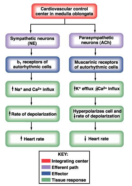
25 Nervous reflex regulation: Autonomic Nervous Regulation S and PS branches of the autonomic nervous system influence HR and AV node conduction through antagonistic control PS: 70 beats/min-[ SAN: intrinsic rate of /min ]- S: >100/min /min (Ach, muscarinic rec) (NE, b1 rec) PS tone - decrease HR and AV conduction; vagal escape - strong vagal stimulation can decrease the strength of heart muscle contraction by % - nitric oxide (NO) vasodilatation S tone - increase HR, AV conduction and contractility (b1 Rec) - normally S discharge continuously at a slow rate 30% CO - determine vasoconstriction by a1 Rec (NE) - vasodilatation by b2 Rec (E in heart, skeletal mm > fight or flight response)

")
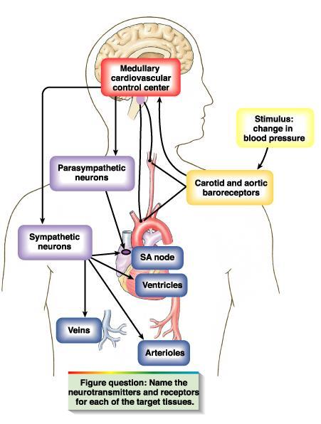
26 Distribution of the autonomic nervous system in myocardium Cardiac output can be increased more than 100% by sympathetic stimulation, and can be decreased to almost zero by vagal (parasympathetic) stimulation.

27 Effect on the cardiac output of different degrees of sympathetic or parasympathetic stimulation. The picture shows relation between RA pressure at the input of the right heart and CO from the LV into the aorta. CO changes caused by nerve stimulation result both from changes in heart rate and from changes in contractile strength of the heart.
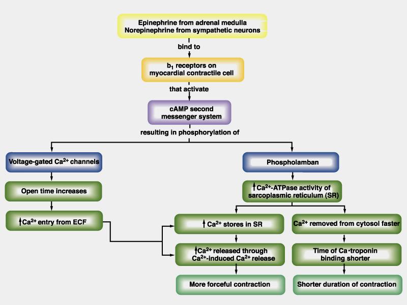
28 Effect of NE on contractility of the heart
29

30 Factors that affect cardiac output
31 Polygram - Analysis of Cardiac activity Electrical activity measured by electrocardiography Mechanical activity evaluated by: 1. Atrial pressure curve and venous pressure: recorded at jugular vein level (jugulogram) 2. Aortic pressure curve: recorded at carotid artery level (carotidogram) 3. Phonocardiography: record of the heart sounds 4. Ventricular volume: evaluated by apexocardiogram
 the cardiac cycle begins with atrial contraction; (2) phase 1 of the cardiac cycle has three subparts: rapid ventricular filling, decreased ventricular filling, and")
32 Mechanical, electrical, acoustic, and echocardiographic events in the cardiac cycle. (1) the cardiac cycle begins with atrial contraction; (2) phase 1 of the cardiac cycle has three subparts: rapid ventricular filling, decreased ventricular filling, and atrial systole; (3) phase 3 has two subparts: rapid and decreased ventricular ejection.

33 Pressure Waves in Veins systemic veins have pressure waves - venous pulse: (1) retrograde action of the heartbeat during the cardiac cycle, (2) the respiratory cycle (3) the contraction of skeletal muscles. Jugular vein, has a complex pulse wave synchronized to the cardiac cycle: 3 peaks, labeled a, c, and v 3 minima, labeled av, x, and y.

34 Pressure transients in the jugular vein pulse reflect events in the cardiac cycle: a peak - caused by the contraction of the right atrium. av minimum is due to relaxation of the right atrium and closure of the tricuspid valve. c peak reflects the pressure rise in the right ventricle early during systole and the resultant protruding of the tricuspid valve-which has just closed-into the right atrium. x minimum occurs as the ventricle contracts and shortens during the ejection phase, later in systole. The shortening heart-with tricuspid valve still closed-pulls on and therefore elongates the veins, lowering their pressure. v peak is related to filling of the right atrium against a closed tricuspid valve, which causes right atrial pressure to slowly rise. As the tricuspid valve opens, the v peak begins to decline. y minimum reflects a fall in right atrial pressure during rapid ventricular filling, as blood leaves the right atrium through an open tricuspid valve and enters the right ventricle. The increase in venous pressure after the y minimum occurs as venous return continues in the face of reduced ventricular filling.
35 Effect of the Respiratory Cycle During inspiration, the diaphragm descends, causing intrathoracic pressure (and therefore the pressure inside the thoracic vessels) to decrease and intra-abdominal pressure to increase the venous return from the head and upper extremities transiently increases, as lowpressure vessels literally suck blood into the thoracic cavity. Simultaneously, the venous flow decreases from the lower extremities because of the relatively high pressure of the abdominal veins during inspiration. Therefore, during inspiration, pressure in the jugular vein falls while pressure in the femoral vein rises.
36 Polygram - Analysis of Cardiac activity Electrical activity measured by electrocardiography Mechanical activity evaluated by: 1. Atrial pressure curve and venous pressure: recorded at jugular vein level (jugulogram) 2. Aortic pressure curve: recorded at carotid artery level (carotidogram) 3. Phonocardiography: record of the heart sounds 4. Ventricular volume: evaluated by apexocardiogram

37 Cardiac Cycle causes flow waves in aorta and peripheral vessels Flow (A) and pressure (B) profiles in the aorta and smaller vessels. With the closing & opening of pulmonary and aortic valves, blood flow and blood velocity across these valves oscillate from near zero, when the valves are closed, to high values, when the valves are open. Blood flow in the aortic arch actually oscillates between slightly negative and highly positive values. Pressure in the aortic arch typically oscillates between ~ mm Hg. Phasic changes in pressure and flow also occur in the peripheral arteries. Arterial pressure is usually measured in a large artery, such as the brachial artery the measured systolic and diastolic arterial pressures, as well as the pulse pressure and mean arterial pressure, closely approximate the corresponding aortic pressures.

38 Comparison of the dynamics of the left and right ventricles.
39 Polygram - Analysis of Cardiac activity Electrical activity measured by electrocardiography Mechanical activity evaluated by: 1. Atrial pressure curve and venous pressure: recorded at jugular vein level (jugulogram) 2. Aortic pressure curve: recorded at carotid artery level (carotidogram) 3. Phonocardiography: record of the heart sounds 4. Ventricular volume: evaluated by apexocardiogram
40 Heart sounds and phonocardiography Heart sounds are relatively brief, discrete auditory vibrations of varying intensity (loudness), frequency (pitch), and quality (timbre). The first heart sound identifies the onset of ventricular systole, and the second heart sound identifies the onset of diastole. These two auscultatory events establish a framework within which other heart sounds and murmurs can be placed and timed. Listening to the sounds of the body with the aid of a stethoscope is called auscultation. The stethoscope can detect leaks in the valves that permit jets of blood to flow backward across the valvular orifice (i.e., regurgitation) as well as stenotic lesions that narrow the valve opening, forcing the blood to pass through a narrower space (i.e., stenosis). During certain parts of the cardiac cycle, blood passing through either regurgitant or stenotic lesions makes characteristic sounds that are called murmurs. Phonocardiogram: the recording of the auscultatory cardiac activity, using a transducer placed on the thorax. The movement of the valve leaflets can be detected by echocardiography.


41 Chest Surface Areas for Auscultation of Normal Heart Sounds The primary aortic area: 2 nd right intercostal space, adjacent to the sternum. The secondary aortic area: 3 rd left intercostal space, adjacent to the sternum (known as Erb area). The pulmonary area: 2 nd left intercostal space The tricuspid area: 4 th & 5 th intercostal spaces, adjacent to the left sternal border. The mitral area at the cardiac apex: 5 th left intercostal space, on the medioclavicular line.
42 The first heart sound (S1) systolic sound the lub appears at sec after the beginning of the QRS complex vibrations are low in pitch and relatively long-lasting - lasts ~ sec; frequency ~ Hz; produced, in this order, by : closing of the mitral valve, closing of the tricuspid valve, opening of the pulmonar valve, opening of the aortic valve.
43 The second heart sound (S2) diastolic sound the dub appears in the terminal period of the T wave lasts s produced, in this order, by: closing of the aortic valve, closing of the pulmonic valve, opening of the tricuspid valve, opening of the mitral valve. heard like a rapid snap because these valves close rapidly, and the surroundings vibrate for a short period physiologic splitting that varies with respiration (wider splitting with inspiration) Split S2 Inspiration Expiration Normal or physiologic
44 Split S2 Audible respiratory splitting means > 30 msec difference in the timing of the aortic (A 2 ) and pulmonic (P 2 ) components of the second heart sound. Splitting of S 2 is best heard over the 2 nd left intercostal space The normal P 2 is often softer than A 2 and rarely audible at apex Inspiration accentuates the splitting of S 2. Split S2 Inspiration Expiration Normal or physiologic
45 The third heart sound (S3) occurs in early diastole (at the beginning of the middle third of diastole) when rapid filling of the ventricles results in recoil of ventricular walls that have a limited distensibility lasts sec protodiastolic sound or gallop A gallop rhythm is a grouping of three heart sounds that together sound like hoofs of a galloping horse. The addition of an S3 to the physiological S1 and S2 creates a three-sound sequence, S1-S2-S3, that is termed a protodiastolic gallop or ventricular gallop. it is normal in children and individuals with a thin thoracic wall occasionally heard as a weak, rumbling sound
46 The fourth heart sound (S4) presystolic sound: appears at 0.04 s after the P wave (late diastolic) lasts s caused by the blood flow that hits the ventricular wall during the atrial systole. physiological only in small children, if heard in other conditions it is a sign of reduced ventricular compliance. addition of an S 4 produces another three-sound sequence, S 4 -S 1 -S 2, which is a presystolic gallop rhythm or atrial gallop during tachycardia S4-S1 can fuse, producing a summation gallop
47 EC=ejection click: most common early systolic sound; Results from abrupt halting of semilunar valves OS=opening snap: high-frequency early diastolic sound (occurs msec after A2) associated with mitral stenosis (stiffening of the mitral valve); sound due to abrupt deceleration of mitral leaflets sound with associated murmur.

.")
48 Phonocardiogram The duration of S1, S2 is slightly more than 0.10 sec. S1 ~ 0.14 sec S2 ~ 0.11 sec. (the semilunar valves are more taut than the A-V valves, so that they vibrate for a shorter time than do the A-V valves). The audible range of frequency (pitch) in the first and second heart sounds: ~ 40 cycles/sec up above 500 cycles/sec.
49 Polygram - Analysis of Cardiac activity Electrical activity measured by electrocardiography Mechanical activity evaluated by: 1. Phonocardiography: record of the heart sounds 2. Atrial pressure curve: recorded at jugular vein level (jugulogram) 3. Ventricular volume: evaluated by apexocardiogram 4. Aortic pressure curve: recorded at carotid artery level (carotidogram)
50 Volume (ml) Presure (mmhg) Isovolumic Isovolumic Rapid inflow contraction Ejection relaxation Diastasis Atrial systole AoC Aortic pressure MC Ao O MO a c v Atrial pressure Ventricular pres Ventricular volume S 1 S 2 S 3 S 4 S 1 Electrocardiogram Phonocardiogram Polygram: events of the cardiac cycle for left ventricular function
.")
51 ATRIAL SYSTOLE (The end of ventricular diastole) Heart: During atrial systole the atrium contracts and tops off the volume in the ventricle with only a small amount of blood. Atrial contraction is complete before the ventricle begins to contract. Atrial pressure: The "a" wave occurs when the atrium contracts, increasing atrial pressure (yellow). Blood arriving at the heart cannot enter the atrium so it flows back up the jugular vein, causing the first discernible wave in the jugular venous pulse. Atrial pressure drops when the atria stop contracting. ECG: An impulse arising from the SA node results in depolarization and contraction of the atria. The P wave is due to this atrial depolarization. The PR segment is electrically quiet as the depolarization proceeds to the AV node. This brief pause before contraction allows the ventricles to fill completely with blood. Heart sounds: A fourth heart sound (S4) is abnormal and is associated with the end of atrial emptying after atrial contraction. It occurs with hypertrophic congestive heart failure, massive pulmonary embolism or tricuspid incompetence.
.")
 exceeds the pressure in the atria (yellow).")
52 ISOVOLUMETRIC CONTRACTION The beginning of systole Heart: The atrioventricular (AV) valves close at the beginning of this phase. Electrically, ventricular systole is defined as the interval between the QRS complex and the end of the T wave (the Q-T interval). Mechanically, ventricular systole is defined as the interval between the closing of the AV valves and the opening of the semilunar valves (aortic and pulmonary valves). Pressures & Volume: The AV valves close when the pressure in the ventricles (red) exceeds the pressure in the atria (yellow). As the ventricles contract isovolumetrically - - their volume does not change (white ) -- the pressure inside increases, approaching the pressure in the aorta and pulmonary arteries (green). ECG: The electrical impulse propagates from the AV node through the His bundle and Purkinje system to allow the ventricles to contract from the apex of the heart towards the base. The QRS complex is due to ventricular depolarization, and it marks the beginning of ventricular systole. It is so large that it masks the underlying atrial repolarization signal. Heart sounds: The first heart sound (S1, "lub") is due to the closing AV valves and associated blood turbulence.
 exceeds the pressure in the aorta and pulmonary arteries (green); the semilunar valves open,")

53 RAPID EJECTION Heart: The semilunar (aortic and pulmonary) valves open at the beginning of this phase. Pressures & Volume: While the ventricles continue contracting, the pressure in the ventricles (red) exceeds the pressure in the aorta and pulmonary arteries (green); the semilunar valves open, blood exits the ventricles, and the volume in the ventricles decreases rapidly ( white). As more blood enters the arteries, pressure there builds until the flow of blood reaches a peak. The "c" wave of atrial pressure is not normally discernible in the jugular venous pulse. Right ventricular contraction pushes the tricuspid valve into the atrium and increases atrial pressure, creating a small wave into the jugular vein. It is normally simultaneous with the carotid pulse. ECG: Heart sounds:
, blood flow out of the ventricles decreases and ventricular volume decreases more")

54 REDUCED EJECTION The end of systole Heart: At the end of this phase the semilunar (aortic and pulmonary) valves close. Pressures & Volume: After the peak in ventricular and arterial pressures (red and green), blood flow out of the ventricles decreases and ventricular volume decreases more slowly (white ). When the pressure in the ventricles falls below the pressure in the arteries, blood in the arteries begins to flow back toward the ventricles and causes the semilunar valves to close. This marks the end of ventricular systole mechanically. ECG: The T wave is due to ventricular repolarization. The end of the T wave marks the end of ventricular systole electrically. Heart sounds:

.")

 is at a minimum and is ready to be filled again with blood.")
55 ISOVOLUMETRIC RELAXATION The beginning of diastole Heart: At the beginning of this phase the AV valves are closed. Pressures & Volume: Throughout this and the previous two phases, the atrium in diastole has been filling with blood on top of the closed AV valve, causing atrial pressure to rise gradually (yellow). The "v" wave is due to the back flow of blood after it hits the closed AV valve. It is the second discernible wave of the jugular venous pulse. The pressure in the ventricles (red) continues to drop. Ventricular volume (white ) is at a minimum and is ready to be filled again with blood. ECG: Heart sounds: The second heart sound (S2, "dup") occurs when the semilunar (aortic and pulmonary) valves close. S2 is normally split because the aortic valve closes slightly earlier than the pulmonary valve.

")
 is usually")

56 RAPID VENTRICULAR FILLING Heart: Once the AV valves open, blood that has accumulated in the atria flows rapidly into the ventricles. Pressures & Volume: Ventricular volume ( white white) increases rapidly as blood flows from the atria into the ventricles. ECG: Heart sounds: A third heart sound (S3) is usually abnormal and is due to rapid passive ventricular filling. It occurs in dilated congestive heart failure, myocardial infarction, or mitral incompetence.
 Heart:")


57 REDUCED VENTRICULAR FILLING (DIASTASIS) Heart: Pressures & Volume: Ventricular volume ( white ) increases more slowly now. The white ventricles continue to fill with blood until they are nearly full. ECG: Heart sounds:
CARDIOVASCULAR PHYSIOLOGY
 CARDIOVASCULAR PHYSIOLOGY LECTURE 4 Cardiac cycle Polygram - analysis of cardiac activity Ana-Maria Zagrean MD, PhD The Cardiac Cycle - definitions: the sequence of electrical and mechanical events that
CARDIAC CYCLE CONTENTS. Divisions of cardiac cycle 11/13/13. Definition. Badri Paudel GMC
 CARDIAC CYCLE Badri Paudel GMC CONTENTS Ø DEFINATION Ø DIVISION OF CARDIAC CYCLE Ø SUB DIVISION AND DURATION OF CARDIAC CYCLE Ø SYSTOLE Ø DIASTOLE Ø DESCRIPTION OF EVENTS OF CARDIAC CYCLE Ø SUMMARY Ø ELECTROCARDIOGRAPHY
2. The heart sounds are produced by a summed series of mechanical events, as follows:
 Heart Sounds. Phonocardiography 1 Objectives 1. Phonocardiography - Definition 2. What produces the heart sounds 3. Where to listen for the heart sounds 4. How to record a phonocardiogram 5. Normal heart
The Cardiac Cycle Clive M. Baumgarten, Ph.D.
 The Cardiac Cycle Clive M. Baumgarten, Ph.D. OBJECTIVES: 1. Describe periods comprising cardiac cycle and events within each period 2. Describe the temporal relationships between pressure, blood flow,
Electrical Conduction
 Sinoatrial (SA) node Electrical Conduction Sets the pace of the heartbeat at 70 bpm AV node (50 bpm) and Purkinje fibers (25 40 bpm) can act as pacemakers under some conditions Internodal pathway from
Cardiovascular Physiology. Heart Physiology. Introduction. The heart. Electrophysiology of the heart
 Cardiovascular Physiology Heart Physiology Introduction The cardiovascular system consists of the heart and two vascular systems, the systemic and pulmonary circulations. The heart pumps blood through
11/10/2014. Muscular pump Two atria Two ventricles. In mediastinum of thoracic cavity 2/3 of heart's mass lies left of midline of sternum
 It beats over 100,000 times a day to pump over 1,800 gallons of blood per day through over 60,000 miles of blood vessels. During the average lifetime, the heart pumps nearly 3 billion times, delivering
Chapter 13 The Cardiovascular System: Cardiac Function
 Chapter 13 The Cardiovascular System: Cardiac Function Overview of the Cardiovascular System The Path of Blood Flow through the Heart and Vasculature Anatomy of the Heart Electrical Activity of the Heart
THE CARDIOVASCULAR SYSTEM. Heart 2
 THE CARDIOVASCULAR SYSTEM Heart 2 PROPERTIES OF CARDIAC MUSCLE Cardiac muscle Striated Short Wide Branched Interconnected Skeletal muscle Striated Long Narrow Cylindrical PROPERTIES OF CARDIAC MUSCLE Intercalated
Cardiovascular system
 BIO 301 Human Physiology Cardiovascular system The Cardiovascular System: consists of the heart plus all the blood vessels transports blood to all parts of the body in two 'circulations': pulmonary (lungs)
Heart Pump and Cardiac Cycle. Faisal I. Mohammed, MD, PhD
 Heart Pump and Cardiac Cycle Faisal I. Mohammed, MD, PhD 1 Objectives To understand the volume, mechanical, pressure and electrical changes during the cardiac cycle To understand the inter-relationship
(D) (E) (F) 6. The extrasystolic beat would produce (A) increased pulse pressure because contractility. is increased. increased
 (E) (F) 6. The extrasystolic beat would produce (A) increased pulse pressure because contractility. is increased. increased") Review Test 1. A 53-year-old woman is found, by arteriography, to have 5% narrowing of her left renal artery. What is the expected change in blood flow through the stenotic artery? Decrease to 1 2 Decrease
Review Test 1. A 53-year-old woman is found, by arteriography, to have 5% narrowing of her left renal artery. What is the expected change in blood flow through the stenotic artery? Decrease to 1 2 Decrease
Cardiac Cycle MCQ. Professor of Cardiovascular Physiology. Cairo University 2007
 Cardiac Cycle MCQ Abdel Moniem Ibrahim Ahmed, MD Professor of Cardiovascular Physiology Cairo University 2007 1- Regarding the length of systole and diastole: a- At heart rate 75 b/min, the duration of
10/23/2017. Muscular pump Two atria Two ventricles. In mediastinum of thoracic cavity 2/3 of heart's mass lies left of midline of sternum
 It beats over 100,000 times a day to pump over 1,800 gallons of blood per day through over 60,000 miles of blood vessels. During the average lifetime, the heart pumps nearly 3 billion times, delivering
Approximately the size of your fist Location. Pericardial physiology
 Heart Anatomy Approximately the size of your fist Location Superior surface of diaphragm Left of the midline Anterior to the vertebral column, posterior to the sternum Wednesday, March 28, 2012 Muscle
CARDIOVASCULAR PHYSIOLOGY
 CARDIOVASCULAR PHYSIOLOGY LECTURE 5 Heart as a pump cardiac performance Coronary circulation. Particularities of the cardiac muscle metabolism. Ana-Maria Zagrean MD, PhD Chemical energy required for cardiac
Chapter 9, Part 2. Cardiocirculatory Adjustments to Exercise
 Chapter 9, Part 2 Cardiocirculatory Adjustments to Exercise Electrical Activity of the Heart Contraction of the heart depends on electrical stimulation of the myocardium Impulse is initiated in the right
The Cardiovascular System
 The Cardiovascular System The Cardiovascular System A closed system of the heart and blood vessels The heart pumps blood Blood vessels allow blood to circulate to all parts of the body The function of
*Generating blood pressure *Routing blood: separates. *Ensuring one-way blood. *Regulating blood supply *Changes in contraction
 *Generating blood pressure *Routing blood: separates pulmonary and systemic circulations *Ensuring one-way blood flow: valves *Regulating blood supply *Changes in contraction rate and force match blood
SIKLUS JANTUNG. Rahmatina B. Herman
 SIKLUS JANTUNG Rahmatina B. Herman The Cardiac Cycle Definition: The cardiac events that occur from the beginning of one heartbeat to the beginning of the next The cardiac cycle consists of: - Diastole
IB TOPIC 6.2 THE BLOOD SYSTEM
 IB TOPIC 6.2 THE BLOOD SYSTEM THE BLOOD SYSTEM TERMS TO KNOW circulation ventricle artery vein 6.2.U1 - Arteries convey blood at high pressure from the ventricles to the tissues of the body Circulation
10. Thick deposits of lipids on the walls of blood vessels, called, can lead to serious circulatory issues. A. aneurysm B. atherosclerosis C.
 Heart Student: 1. carry blood away from the heart. A. Arteries B. Veins C. Capillaries 2. What is the leading cause of heart attack and stroke in North America? A. alcohol B. smoking C. arteriosclerosis
Cardiovascular Physiology
 Cardiovascular Physiology Introduction The cardiovascular system consists of the heart and two vascular systems, the systemic and pulmonary circulations. The heart pumps blood through two vascular systems
Cardiovascular System: The Heart
 Cardiovascular System: The Heart I. Anatomy of the Heart (See lab handout for terms list) A. Describe the size, shape and location of the heart B. Describe the structure and function of the pericardium
4. The two inferior chambers of the heart are known as the atria. the superior and inferior vena cava, which empty into the left atrium.
 Answer each statement true or false. If the statement is false, change the underlined word to make it true. 1. The heart is located approximately between the second and fifth ribs and posterior to the
Principles of Biomedical Systems & Devices. Lecture 8: Cardiovascular Dynamics Dr. Maria Tahamont
 Principles of Biomedical Systems & Devices Lecture 8: Cardiovascular Dynamics Dr. Maria Tahamont Review of Cardiac Anatomy Four chambers Two atria-receive blood from the vena cave and pulmonary veins Two
IP: Regulation of Cardiac Output
 ANP 1105D Winter 2013 Assignment 9: The Heart, part 2: Chap... Assignment 9: The Heart, part 2: Chapter 18 Signed in as Alex Sokolowski Help Close Resources Due: 11:59pm on Monday, March 25, 2013 Note:
IB TOPIC 6.2 THE BLOOD SYSTEM
 IB TOPIC 6.2 THE BLOOD SYSTEM TERMS TO KNOW circulation ventricle artery vein THE BLOOD SYSTEM 6.2.U1 - Arteries convey blood at high pressure from the ventricles to the tissues of the body Circulation
Cardiovascular System
 Cardiovascular System The Heart Cardiovascular System The Heart Overview What does the heart do? By timed muscular contractions creates pressure gradients blood moves then from high pressure to low pressure
The Heart. Size, Form, and Location of the Heart. 1. Blunt, rounded point; most inferior part of the heart.
 12 The Heart FOCUS: The heart is composed of cardiac muscle cells, which are elongated, branching cells that appear striated. Cardiac muscle cells behave as a single electrical unit, and the highly coordinated
Pathological Arrhythmias/ Tachyarrhythmias
 Pathological Arrhythmias/ Tachyarrhythmias caused by: 1.Ectopic focus: Extrasystole or premature beat. If discharge is occasional. Can be: Atrial Extrasystole Vevtricular Extrasystole 2.Cardiac Arrhythmia
Collin County Community College
 Collin County Community College BIOL. 2402 Anatomy & Physiology WEEK 5 The Heart 1 The Heart Beat and the EKG 2 1 The Heart Beat and the EKG P-wave = Atrial depolarization QRS-wave = Ventricular depolarization
Heart sounds and murmurs. Dr. Szathmári Miklós Semmelweis University First Department of Medicine 15. Oct
 Heart sounds and murmurs Dr. Szathmári Miklós Semmelweis University First Department of Medicine 15. Oct. 2013. Conditions for auscultation of the heart Quiet room Patient comfortable Chest fully exposed
Chapter 18 - Heart. I. Heart Anatomy: size of your fist; located in mediastinum (medial cavity)
") Chapter 18 - Heart I. Heart Anatomy: size of your fist; located in mediastinum (medial cavity) A. Coverings: heart enclosed in double walled sac called the pericardium 1. Fibrous pericardium: dense connective
Chapter 18 - Heart I. Heart Anatomy: size of your fist; located in mediastinum (medial cavity) A. Coverings: heart enclosed in double walled sac called the pericardium 1. Fibrous pericardium: dense connective
Chapter 20: Cardiovascular System: The Heart
 Chapter 20: Cardiovascular System: The Heart I. Functions of the Heart A. List and describe the four functions of the heart: 1. 2. 3. 4. II. Size, Shape, and Location of the Heart A. Size and Shape 1.
Pearson's Comprehensive Medical Assisting Administrative and Clinical Competencies
 Pearson's Comprehensive Medical Assisting Administrative and Clinical Competencies THIRD EDITION CHAPTER 27 The Cardiovascular System Lesson 1: Overview of the Cardiovascular System Lesson Objectives Upon
Practice Exercises for the Cardiovascular System
 Practice Exercises for the Cardiovascular System On the diagram below, color the oxygen-rich blood red and the oxygen-poor blood blue. Label the parts: Continued on the next page... Label the parts on
BIOL 219 Spring Chapters 14&15 Cardiovascular System
 1 BIOL 219 Spring 2013 Chapters 14&15 Cardiovascular System Outline: Components of the CV system Heart anatomy Layers of the heart wall Pericardium Heart chambers, valves, blood vessels, septum Atrioventricular
Objectives of the Heart
 Objectives of the Heart Electrical activity of the heart Action potential EKG Cardiac cycle Heart sounds Heart Rate The heart s beat separated into 2 phases Relaxed phase diastole (filling of the chambers)
Lab #3: Electrocardiogram (ECG / EKG)
") Lab #3: Electrocardiogram (ECG / EKG) An introduction to the recording and analysis of cardiac activity Introduction The beating of the heart is triggered by an electrical signal from the pacemaker. The
Lab #3: Electrocardiogram (ECG / EKG) An introduction to the recording and analysis of cardiac activity Introduction The beating of the heart is triggered by an electrical signal from the pacemaker. The
BUSINESS. Articles? Grades Midterm Review session
 BUSINESS Articles? Grades Midterm Review session REVIEW Cardiac cells Myogenic cells Properties of contractile cells CONDUCTION SYSTEM OF THE HEART Conduction pathway SA node (pacemaker) atrial depolarization
Cardiac Cycle. Each heartbeat is called a cardiac cycle. First the two atria contract at the same time.
 The Heartbeat Cardiac Cycle Each heartbeat is called a cardiac cycle. First the two atria contract at the same time. Next the two ventricles contract at the same time. Then all the chambers relax. http://www.youtube.com/watch?v=frd3k6lkhws
The Heart. C h a p t e r. PowerPoint Lecture Slides prepared by Jason LaPres Lone Star College - North Harris
 C h a p t e r 20 The Heart PowerPoint Lecture Slides prepared by Jason LaPres Lone Star College - North Harris Copyright 2009 Pearson Education, Inc., publishing as Pearson Benjamin Cummings Introduction
PHYSIOLOGY MeQ'S (Morgan) All the following statements related to blood volume are correct except for: 5 A. Blood volume is about 5 litres. B.
 All the following statements related to blood volume are correct except for: 5 A. Blood volume is about 5 litres. B.") PHYSIOLOGY MeQ'S (Morgan) Chapter 5 All the following statements related to capillary Starling's forces are correct except for: 1 A. Hydrostatic pressure at arterial end is greater than at venous end.
PHYSIOLOGY MeQ'S (Morgan) Chapter 5 All the following statements related to capillary Starling's forces are correct except for: 1 A. Hydrostatic pressure at arterial end is greater than at venous end.
Cardiac Output (C.O.) Regulation of Cardiac Output
 Regulation of Cardiac Output") Cardiac Output (C.O.) Is the volume of the blood pumped by each ventricle per minute (5 Litre) Stroke volume: Is the volume of the blood pumped by each ventricle per beat. Stroke volume = End diastolic
Cardiac Output (C.O.) Is the volume of the blood pumped by each ventricle per minute (5 Litre) Stroke volume: Is the volume of the blood pumped by each ventricle per beat. Stroke volume = End diastolic
Collin County Community College. ! BIOL Anatomy & Physiology! WEEK 5. The Heart
 Collin County Community College! BIOL. 2402 Anatomy & Physiology! WEEK 5 The Heart 1 (1578-1657) A groundbreaking work in the history of medicine, English physician William Harvey s Anatomical Essay on
The Heart and Cardiovascular System
 The Heart and Cardiovascular System What you will learn The location of the heart 3 layers and covering of the heart Explain the function of the heart as 2 separate pumps Identify the 4 chambers of the
Cardiovascular System
 Cardiovascular System Purpose Transport oxygen and nutrients Take waste products away from tissues & organs Things we learned Blood pressure: the force of blood pushing against the walls of blood vessels
Human Cardiovascular Physiology: Blood Pressure and Pulse Determinations
 ighapmlre33apg269_274 5/12/04 3:10 PM Page 269 impos03 302:bjighapmL:ighapmLrevshts:layouts: NAME Human Cardiovascular Physiology: Blood Pressure and Pulse Determinations LAB TIME/DATE REVIEW SHEET exercise
-12. -Ensherah Mokheemer - ABDULLAH ZREQAT. -Faisal Mohammad. 1 P a g e
 -12 -Ensherah Mokheemer - ABDULLAH ZREQAT -Faisal Mohammad 1 P a g e In the previous lecture we talked about: - cardiac index: we use the cardiac index to compare the cardiac output between different individuals,
AnS SI 214 Practice Exam 2 Nervous, Muscle, Cardiovascular
 AnS SI 214 Practice Exam 2 Nervous, Muscle, Cardiovascular Select the best answer choice in the questions below. 1) On the electrocardiogram, repolarization of the atria is represented by the: A) P wave
The HEART. What is it???? Pericardium. Heart Facts. This muscle never stops working It works when you are asleep
 This muscle never stops working It works when you are asleep The HEART It works when you eat It really works when you exercise. What is it???? Located between the lungs in the mid thoracic region Apex
The Cardiovascular System
 Essentials of Human Anatomy & Physiology Elaine N. Marieb Slides 11.1 11.19 Seventh Edition Chapter 11 The Cardiovascular System Functions of the Cardiovascular system Function of the heart: to pump blood
Introduction. Circulation
 Introduction Circulation 1- Systemic (general) circulation 2- Pulmonary circulation carries oxygenated blood to all parts of the body carries deoxygenated blood to the lungs From Lt. ventricle aorta From
The Circulatory System. The Heart, Blood Vessels, Blood Types
 The Circulatory System The Heart, Blood Vessels, Blood Types The Closed Circulatory System Humans have a closed circulatory system, typical of all vertebrates, in which blood is confined to vessels and
Anatomy & Physiology of Cardiovascular System. Chapter 18 & 19
 Anatomy & Physiology of Cardiovascular System Chapter 18 & 19 Objectives..cont 1. Discuss the physiological stages of cardiac muscle contraction. 2. Trace a typical ECG and label each wave or complex 3.
Anatomy Review: The Heart Graphics are used with permission of A.D.A.M. Software, Inc. and Benjamin/Cummings Publishing Co.
 Anatomy Review: The Heart Graphics are used with permission of A.D.A.M. Software, Inc. and Benjamin/Cummings Publishing Co. Anatomy Views Label the diagrams of the heart below: Interactive Physiology Study
Heart. Structure Physiology of blood pressure and heartbeat
 Heart Structure Physiology of blood pressure and heartbeat Location and Anatomy Location and Anatomy Pericardial cavity: surrounds, isolates, and anchors heart Parietal pericardium lined with serous membrane
Physiology sheet #2. The heart composed of 3 layers that line its lumen and cover it from out side, these layers are :
 Physiology sheet #2 * We will talk in this lecture about cardiac muscle physiology, the mechanism and the energy sources of their contraction and intracellular calcium homeostasis. # Slide 4 : The heart
BIPN100 F15 Human Physiol I (Kristan) Lecture 14 Cardiovascular control mechanisms p. 1
 Lecture 14 Cardiovascular control mechanisms p. 1") BIPN100 F15 Human Physiol I (Kristan) Lecture 14 Cardiovascular control mechanisms p. 1 Terms you should understand: hemorrhage, intrinsic and extrinsic mechanisms, anoxia, myocardial contractility, residual
BIPN100 F15 Human Physiol I (Kristan) Lecture 14 Cardiovascular control mechanisms p. 1 Terms you should understand: hemorrhage, intrinsic and extrinsic mechanisms, anoxia, myocardial contractility, residual
The Cardiovascular System
 Chapter 18 Part A The Cardiovascular System 1/19/16 1 Annie Leibovitz/Contact Press Images Similarities of Cardiac and Skeletal Muscle RMP Ion concentration Deploarization Action Potential Repolarization
QUIZ/TEST REVIEW NOTES SECTION 1 CARDIAC MYOCYTE PHYSIOLOGY [CARDIOLOGY]
![QUIZ/TEST REVIEW NOTES SECTION 1 CARDIAC MYOCYTE PHYSIOLOGY [CARDIOLOGY]](/thumbs/96/126998162.jpg "QUIZ/TEST REVIEW NOTES SECTION 1 CARDIAC MYOCYTE PHYSIOLOGY [CARDIOLOGY]") QUIZ/TEST REVIEW NOTES SECTION 1 CARDIAC MYOCYTE PHYSIOLOGY [CARDIOLOGY] Learning Objectives: Describe the ionic basis of action potentials in cardiac contractile and autorhythmic cells Explain the relationship
QUIZ/TEST REVIEW NOTES SECTION 1 CARDIAC MYOCYTE PHYSIOLOGY [CARDIOLOGY] Learning Objectives: Describe the ionic basis of action potentials in cardiac contractile and autorhythmic cells Explain the relationship
Topic 6: Human Physiology
 Topic 6: Human Physiology 6.2 The Blood System D.4 The Heart Essential Questions: 6.2 The blood system continuously transports substances to cells and simultaneously collects waste products. D.3 The chemical
d) Cardiovascular System Higher Human Biology
 Cardiovascular System Higher Human Biology") d) Cardiovascular System Higher Human Biology What can your remember about the heart and blood vessels? What is the Cardiovascular System? The cardiovascular system, also known as the circulatory system,
d) Cardiovascular System Higher Human Biology What can your remember about the heart and blood vessels? What is the Cardiovascular System? The cardiovascular system, also known as the circulatory system,
Circulation: Chapter 25. Cardiac Output. The Mammalian Heart Fig Right side of the heart
 Circulation: Chapter 25 1. Limits of Diffusion A. Small organisms use diffusion B. rapid over small distances 2. Most animals have circulatory systems A. Blood B. Pump (Heart) or propulsive structures
During exercise the heart rate is 190 bpm and the stroke volume is 115 ml/beat. What is the cardiac output?
 The Cardiovascular System Part III: Heart Outline of class lecture After studying part I of this chapter you should be able to: 1. Be able to calculate cardiac output (CO) be able to define heart rate
The Heart. The Heart A muscular double pump. The Pulmonary and Systemic Circuits
 C H A P T E R 19 The Heart The Heart A muscular double pump circuit takes blood to and from the lungs Systemic circuit vessels transport blood to and from body tissues Atria receive blood from the pulmonary
Major Function of the Cardiovascular System. Transportation. Structures of the Cardiovascular System. Heart - muscular pump
 Structures of the Cardiovascular System Heart - muscular pump Blood vessels - network of tubes Blood - liquid transport vehicle brachiocephalic trunk superior vena cava right pulmonary arteries right pulmonary
Do Now. Get out work from last class to be checked
 Do Now Get out work from last class to be checked Heart Actions Cardiac Cycle: One complete heartbeat. The contraction of a heart chamber is called systole and the relaxation of a chamber is called diastole.
current, and acting like
 Heart 10 IV. HEART PHYSIOLOGY - How the heart beats. How the heart depolarizes the myocardium, which leads to a contraction. A) INTRINSIC CONTROL - Heart controls its own rhythm. HOW? The presence of gap
The cardiovascular system is composed of the heart and blood vessels that carry blood to and from the body s organs. There are 2 major circuits:
 1 The cardiovascular system is composed of the heart and blood vessels that carry blood to and from the body s organs. There are 2 major circuits: pulmonary and systemic. The pulmonary goes out to the
PHONOCARDIOGRAPHY (PCG)
") PHONOCARDIOGRAPHY (PCG) The technique of listening to sounds produced by the organs and vessels of the body is called auscultation. The areas at which the heart sounds are heard better are called auscultation
PHONOCARDIOGRAPHY (PCG) The technique of listening to sounds produced by the organs and vessels of the body is called auscultation. The areas at which the heart sounds are heard better are called auscultation
ADVANCED ASSESSMENT Cardiovascular System
 ONTARIO BASE HOSPITAL GROUP QUIT ADVANCED ASSESSMENT Cardiovascular System 2007 Ontario Base Hospital Group ADVANCED ASSESSMENT Cardiovascular System AUTHORS Mike Muir AEMCA, ACP, BHSc Paramedic Program
CARDIOVASCULAR SYSTEM
 CARDIOVASCULAR SYSTEM Overview Heart and Vessels 2 Major Divisions Pulmonary Circuit Systemic Circuit Closed and Continuous Loop Location Aorta Superior vena cava Right lung Pulmonary trunk Base of heart
Circulation. Circulation = is a process used for the transport of oxygen, carbon! dioxide, nutrients and wastes through-out the body
 Circulation Circulation = is a process used for the transport of oxygen, carbon! dioxide, nutrients and wastes through-out the body Heart = muscular organ about the size of your fist which pumps blood.
THE HEART Dr. Ali Ebneshahidi
 THE HEART Dr. Ali Ebneshahidi Functions is of the heart & blood vessels 1. The heart is an essential pumping organ in the cardiovascular system where the right heart pumps deoxygenated blood (returned
12.2 Monitoring the Human Circulatory System
 12.2 Monitoring the Human Circulatory System Video 1: 3D Animation of Heart Pumping Blood blood flow through the heart... Video 2: Hank Reviews Everything on the Heart https://www.youtube.com/watch?v=x9zz6tcxari
The Cardiovascular System
 Essentials of Human Anatomy & Physiology Elaine N. Marieb Seventh Edition Chapter 11 The Cardiovascular System Slides 11.1 11.19 Lecture Slides in PowerPoint by Jerry L. Cook The Cardiovascular System
Cardiovascular System Notes: Physiology of the Heart
 Cardiovascular System Notes: Physiology of the Heart Interesting Heart Fact Capillaries are so small it takes ten of them to equal the thickness of a human hair. Review What are the 3 parts of the cardiovascular
TEACH Lesson Plan Manual for Herlihy s The Human Body in Health and Illness 5 th edition
 TEACH Lesson Plan Manual for Herlihy s The Human Body in Health and Illness 5 th edition Chapter 17 Function of the Heart Lesson 17.1 Function of the Heart 1. Define cardiac cycle with respect to systole
The Cardiovascular System. Chapter 15. Cardiovascular System FYI. Cardiology Closed systemof the heart & blood vessels. Functions
 Chapter 15 Cardiovascular System FYI The heart pumps 7,000 liters (4000 gallons) of blood through the body each day The heart contracts 2.5 billion times in an avg. lifetime The heart & all blood vessels
Electrocardiogram and Heart Sounds
 Electrocardiogram and Heart Sounds Five physiologic properties of cardiac muscle Automaticity: SA node is the primary pacemaker of the heart, but any cells in the conduction system can initiate their
Cardiac physiology. b. myocardium -- cardiac muscle and fibrous skeleton of heart
 I. Heart anatomy -- general gross. A. Size/orientation - base/apex B. Coverings D. Chambers 1. parietal pericardium 2. visceral pericardium 3. Layers of heart wall a. epicardium Cardiac physiology b. myocardium
CARDIAC EXAMINATION MINI-QUIZ
 CARDIAC EXAMINATION MINI-QUIZ 1. Sitting bolt upright, your dyspneic (short of breath) patient has visible jugular venous pulsations to the angle of his jaw, which is 12 cm above his sternal angle. What
Ch 19: Cardiovascular System - The Heart -
 Ch 19: Cardiovascular System - The Heart - Give a detailed description of the superficial and internal anatomy of the heart, including the pericardium, the myocardium, and the cardiac muscle. Trace the
Cardiovascular System Notes: Heart Disease & Disorders
 Cardiovascular System Notes: Heart Disease & Disorders Interesting Heart Facts The Electrocardiograph (ECG) was invented in 1902 by Willem Einthoven Dutch Physiologist. This test is still used to evaluate
Using Figure 14.1, match the following: 1) Myelin sheath. 1) 2) Cell body of ANS preganglionic neuron. 2)
 Myelin sheath. 1) 2) Cell body of ANS preganglionic neuron. 2)") Practice Exam 1 AP 2 chapters 14 and 18 Name MATCHING: Match labeled areas with the appropriate terminology from the list below. Figure 14.1 Using Figure 14.1, match the following: 1) Myelin sheath. 1)
Practice Exam 1 AP 2 chapters 14 and 18 Name MATCHING: Match labeled areas with the appropriate terminology from the list below. Figure 14.1 Using Figure 14.1, match the following: 1) Myelin sheath. 1)
The Cardiovascular and Lymphatic Systems Cardiovascular System Blood Vessels Blood Vessels Arteries Arteries Arteries
 CH 12 The Cardiovascular and s The Cardiovascular and s OUTLINE: Cardiovascular System Blood Vessels Blood Pressure Cardiovascular System The cardiovascular system is composed of Blood vessels This system
Circulatory system ( 循环系统 )
") Circulatory system ( 循环系统 ) Circulatory system: heart + blood vessels Function: nutrient transportation and metabolites returning Blood: carrier Heart: pump Blood vessels: route, substance communications
Circulatory system ( 循环系统 ) Circulatory system: heart + blood vessels Function: nutrient transportation and metabolites returning Blood: carrier Heart: pump Blood vessels: route, substance communications
Chapter 20b Cardiac Physiology
 Chapter 20b Cardiac Physiology Heart Valve Mechanics The heart valve openand close because of pressure gradients. When pressure on one side is greater than the other, it pushes the valve open. For example,
The Cardiovascular System. Preview of Heart Action. The CV system provides oxygen & nutrients to tissues-removes wastes.
 The Cardiovascular System BIO 250 Human Anatomy & Physiology Preview of Heart Action http://www.youtube.com/watch?v=d3zdj gfddk0&nr=1 The CV system provides oxygen & nutrients to tissues-removes wastes.
- what other structures, besides the heart, does the mediastinum contain?
 Basic A & P II Dr. L. Bacha Chapter Outline (Martini & Nath 2010) An Introduction to the Cardiovascular System - read the paragraphs under this heading on page 580 The Heart is a Four Chambered Organ describe
THE HEART. A. The Pericardium - a double sac of serous membrane surrounding the heart
 THE HEART I. Size and Location: A. Fist-size weighing less than a pound (250 to 350 grams). B. Located in the mediastinum between the 2 nd rib and the 5 th intercostal space. 1. Tipped to the left, resting
P215 SPRING 2019: CIRCULATORY SYSTEM Chaps 13, 14 & 15: pp , , , I. Major Functions of the Circulatory System
 P215 SPRING 2019: CIRCULATORY SYSTEM Chaps 13, 14 & 15: pp 360-390, 395-404, 410-428 433-438, 441-445 I. Major Functions of the Circulatory System 1. 2. 3. 4. II. Structure of the Heart 1. atria 2. ventricles
37 1 The Circulatory System
 H T H E E A R T 37 1 The Circulatory System The circulatory system and respiratory system work together to supply cells with the nutrients and oxygen they need to stay alive. a) The respiratory system:
1. Which of the following conditions will result in a dilated, flaccid heart?
 1. Which of the following conditions will result in a dilated, flaccid heart? A) Excess calcium ions in the blood B) Excess potassium ions in the blood C) Excess sodium ions in the blood D) Increased sympathetic
Cardiovascular Physiology
 chapter 3 Cardiovascular Physiology I. CIRCUITRY OF THE CARDIOVASCULAR SYSTEM (FIGURE 3-1) A. Cardiac output of the left heart equals cardiac output of the right heart. Cardiac output from the left side
The Cardiovascular and Lymphatic Systems
 BIOLOGY OF HUMANS Concepts, Applications, and Issues Fifth Edition Judith Goodenough Betty McGuire 12 The Cardiovascular and Lymphatic Systems Lecture Presentation Anne Gasc Hawaii Pacific University and