CHAPTER 4. Thorax THORACIC CAVITY
|
|
|
- Timothy Carr
- 5 years ago
- Views:
Transcription
1 62 CHAPTER 4 Thorax THORACIC CAVITY CHEST WALL VERSUS THORACIC WALL BREAST (MAMMARY GLAND) SKELETAL COMPONENTS OF THE THORACIC WALL Vertebral Bodies Sternum Ribs MUSCULAR COMPONENTS OF THE ANTERIOR CHEST WALL Pectoralis Major Pectoralis Minor Subclavius The Clavipectoral Space MUSCULAR COMPONENTS OF THE THORACIC WALL Intercostal Muscles Subcostal Muscles and the Transversus Thoracis - Specializations of the Innermost Layer ARTERIES OF THE THORACIC WALL Posterior Intercostal Arteries Anterior Intercostal Arteries THE IMPORTANCE OF ANASTOMOSES BETWEEN ANTERIOR AND POSTERIOR INTERCOSTAL ARTERIES VEINS OF THE THORACIC WALL NERVES OF THE THORACIC WALL Intercostal Nerves SYMPATHETIC TRUNK ABDOMINAL DIAPHRAGM Innervation of the Diaphragm PLEURAL CAVITIES, PERICARDIAL CAVITY, AND MEDIASTINUM Further Development of the Pericardial Sac Further Development of the Pleural Sacs Areas of the Mediastinum THE HEART Right Atrium Right Ventricle Left Atrium Left Ventricle Conducting System of the Heart Arterial Supply to the Heart Right Coronary Artery Left Coronary (Left Main Coronary) Artery CORONARY DOMINANCE CORONARY ANASTOMOSES Venous Drainage of the Heart LUNGS Right Lung Left Lung Trachea and Large Bronchi Branches of the Right Principal Bronchus Branches of the Left Principal Bronchus Vasculature and Lymphatics of the Lung Arteries Veins SUPERIOR AND POSTERIOR MEDIASTINAL STRUCTURES Ascending Aorta and Aortic Arch Ligamentum Arteriosum Brachiocephalic (Innominate) Veins and Superior Vena Cava Thymus Trachea and Mainstem Bronchi Descending Thoracic Aorta Azygos and Hemiazygos Veins Thoracic Duct Esophagus Phrenic and Vagus Nerves Inferior Vena Cava INNERVATION OF THE INTERNAL ORGANS OF THE THORAX An Example--Innervation of the Heart REFERRED PAIN FROM THE HEART Innervation of the Other Internal Thoracic Organs Pain From the Pericardium and Pleura LYMPHATICS OF THE CHEST Nodes That Lie Along Vessels Axillary Nodes Internal Thoracic (Parasternal) Nodes Posterior Mediastinal Nodes Intercostal Nodes
2 63 Anterior Mediastinal Nodes Nodes That Do Not Lie Along Vessels Pulmonary, Bronchopulmonary, Tracheobronchial, and Tracheal Nodes SURGICAL NOMENCLATURE OF NODES DRAINING THE LUNGS Lateral (Middle) Diaphragmatic Nodes Drainage Routes of Thoracic Nodes Lymphatic Drainage of Specific Organs Skin and Superficial Fascia LYMPHATIC DRAINAGE OF THE BREAST Muscle of Chest Wall Internal Organs SURFACE ANATOMY OF THE CHEST Surface Landmarks of the Chest Bony Landmarks on Back of Chest Bony Landmarks on Front of Chest Heart Pulmonary Trunk Ascending Aorta and Aortic Arch Brachiocephalic Veins and Superior Vena Cava Inferior Vena Cava CARDIAC SHADOW Trachea and Mainstem Bronchi Lungs Right Lung Lobes of Right Lung AUSCULTATION OF RIGHT LUNG Left Lung Pleural Cavity Right Pleural Cavity Left Pleural Cavity Pleural Recesses PERICARDIOCENTESIS AND THORACENTESIS MEDIAN STERNOTOMY Esophagus THORACIC CAVITY The thorax is that part of the trunk bounded superiorly by the top edges of the 1st thoracic vertebra, 1st rib and sternum and bounded inferiorly by the abdominal diaphragm. It consists of a large cavity surrounded by body wall. Within the thoracic cavity are three fluid-filled sacs and some internal organs. The thoracic cavity is completely open superiorly, where it communicates with the neck. The abdominal diaphragm forms an inferior wall, but this muscle has several holes to allow passage of structures between the thoracic and abdominal cavities. The remaining walls (posterior, lateral, and anterior) of the thoracic cavity are composed of muscle and bone. The bones of the posterior wall are the vertebrae and posterior regions of the ribs. The muscles of the posterior wall are the intercostal and subcostal muscles. The lateral walls of the thoracic cavity are composed of ribs and intercostal muscles. The anterior wall is composed of ribs, costal cartilages, sternum, intercostal muscles, and the transversus thoracis muscle. CHEST WALL VERSUS THORACIC WALL A variety of muscles are applied to the outer surfaces of the structures that form the wall of the thoracic cavity. Some of these, like the intrinsic back muscles, are derived from thoracic dermomyotomes and, thus, are innervated by thoracic spinal nerves. Others are foreigners that have migrated to a position beneath the skin and superficial fascia of the chest. The posterior surface of the thoracic wall is covered by intrinsic back musculature and, more superficially, by the scapula and muscles attaching to this bone (see Chapter 3, pp ). On the lateral surface of the thoracic wall, from the 1st rib down to the 9th rib is the serratus anterior muscle, and below the 9th rib is the external abdominal oblique. Like the rhomboids, the serratus anterior is a girdle muscle derived from cells of cervical dermomyotomes. The external abdominal oblique is a muscle of the abdominal wall, although it is derived largely from thoracic dermomyotomes.
3 On the anterior surface of the thoracic wall above the level of the xiphisternal joint are the pectoralis major and pectoralis minor, muscles of the upper limb that have migrated onto the front of the chest. Below the level of the xiphisternal joint is the rectus abdominis, another abdominal wall muscle derived from lower thoracic dermomyotomes. None of the immigrant muscles just listed are said to form part of the thoracic wall, although one may say more generally that they contribute to the chest wall. The existence of these chest wall muscles means that virtually none of the actual thoracic wall can be palpated, or auscultated (i.e., listened to), without some other structure intervening. The only place where the rib cage lies directly beneath subcutaneous tissue is at a site just medial to the inferior angle of the scapula. Here a triangular gap is formed between the upper border of the latissimus dorsi, lateral border of the trapezius, and lower border of the rhomboid. This gap is called the triangle of auscultation, but, in fact, it holds no particular clinical significance, since modern stethoscopes can hear sounds through several layers of muscle. BREAST (MAMMARY GLAND) The breasts may be viewed as sweat glands modified to provide nourishment to mammalian neonates. Most of the glandular tissue breast tissue is located in the subcutaneous layer on the front of the chest between the 2 nd and 6 th ribs (while supine). Some tissue of the breast crosses the lateral edge of pectoralis major to reach the axilla (armpit). This is called the axillary tail of Spence; if cystic, it will present as swellings in the armpit. The lactiferous ducts open onto the nipple, which is surrounded by a darkened circle of skin called the areola. Extending anteriorly from the glandular tissue to the deep surface of the skin are fibrous bands of subcutaneous tissue called suspensory ligaments of Cooper. (Some of these bands also run posteriorly, through the loose connective tissue of the retromammary space, to reach the deep fascia overlying the pectoralis major. ) If cancer spreads to a Cooper's ligament, it will be shortened, causing the skin to dimple. 64 SKELETAL COMPONENTS OF THE THORACIC WALL Vertebral Bodies The bodies of the 1st-12th thoracic vertebrae form the bony component of the thoracic wall in the dorsal midline. Sternum In the anterior midline the bony component of the thoracic wall is formed by the sternum (Fig. 4-1). It is a tripartite bone with the parts joined by fibrocartilage (which may ossify late in life). The upper, thick part of the sternum is called the manubrium. It is wider superiorly than inferiorly. At its superolateral corners are notches for articulation with the clavicle. Between these clavicular notches the superior border of the manubrium is called the jugular notch. The inferior edge of the manubrium articulates with the body of the sternum in a joint called the superior sternal synchondrosis, or more commonly (though less accurately) the manubriosternal joint. The body of the sternum is not as thick (front to back) as the manubrium. It is about twice the length of the manubrium. The sternal body starts out as relatively narrow, and it gradually widens to about the junction of its upper two thirds with its lower one third. Then it narrows dramatically to articulate with the xiphoid process of the sternum at the inferior sternal synchondrosis, more commonly called the xiphisternal joint. The xiphoid process is very thin and relatively short.
 The rest of the skeletal wall of the thoracic cavity is made up of the ribs and their cartilages.")
4 65 When one runs a finger down the anterior surface of the manubrium onto the body of the sternum, the angle between the anterior surfaces of these two bones can be felt. This is the sternal angle, or angle of Louis. Ribs (see Fig. 4-1) The rest of the skeletal wall of the thoracic cavity is made up of the ribs and their cartilages. There are 12 ribs on each side; each rib is the separately ossified costal process of a corresponding thoracic vertebra (see Chapter 3). Like so many other bones, the ribs are formed first in cartilage and ossify later. For each rib the ossification process stops short of its anterior end, leaving this region cartilaginous even in adult life. The cartilaginous continuation of a bony rib is called the costal cartilage. The junction between the rib and its costal cartilage is called the costochondral junction. This junction lies progressively further away from the sternum as one passes from higher to lower ribs. The heads of all but the 1st, 11th, and 12th ribs articulate via true synovial joints (the capitular joints) with two adjacent thoracic vertebrae, its own and the one above. The heads of the 1st, 11th, and 12th ribs articulate with only their own vertebral bodies. The tubercle of a rib articulates via a synovial joint with the tip of its corresponding vertebral transverse process. This is a costotransverse joint. The back surface of the neck of a rib is attached to the front surface of the transverse process by a ligament. This is the ligament of the neck, or the posterior costotransverse ligament. The upper edge of the rib neck is connected to the next higher transverse process by a superior costotransverse ligament. The back of each costotransverse joint is reinforced by a lateral costotransverse ligament. The shaft of a rib courses outward from the tubercle, and then around the side toward the front of the thorax. Not far from the tubercle, the outer surface of each shaft is marked by a rugosity for the attachment of the iliocostalis muscle. This rugosity marks the angle of a rib. For the 3rd-12th ribs, the
5 inferior edge of the shaft is sharp over the posterior two thirds of its length. This is due to a narrow linear indentation of the inner surface of the bone, which indentation is called the costal groove. Each rib shaft passes inferiorly as it works its way around the side of the chest. Either at the costochondral junction, or just distal to it, the costal cartilage turns upward to go toward the sternum. This change in direction becomes increasingly more marked for lower ribs. No true joint is formed between the first costal cartilage and the manubrium. Thus, the 1st costal cartilage represents a synchondrosis (i.e., the joining of two bones by cartilage). The 2nd costal cartilage reaches the sternal angle, where a true synovial joint is formed between the manubrium and body of the sternum, on the one hand, and the 2nd costal cartilage on the other. The 3rd-7th costal cartilages reach the body of the sternum, where true synovial joints are formed. The 4th sternochondral joint lies just below the midpoint of the sternal body; the 5th-7th sternochondral joints are crowded together in its lower one fourth. The tip of the 8th costal cartilage articulates via a true synovial joint with the inferior edge of the 7th costal cartilage. The tip of the 9th costal cartilage forms a similar chondrochondral joint with the inferior edge of the 8th. The tip of the 10th costal cartilage either may participate in a chondrochondral joint with the 9th costal cartilage or may be joined to it by fibrous tissue. The 11th and 12th costal cartilages are short and end blindly, at the mid- and posterior axillary lines, respectively. 11 Between any two ribs is a so-called intercostal space. Obviously there are eleven intercostal spaces on each side. Intercostal spaces 1-6, which reach the sternum, may be called long intercostal spaces. The 7th-11th intercostal spaces are short. Of these, 7, 8, and 9 are bounded in front by costal cartilage, whereas 10 and 11 are open. 66 MUSCULAR COMPONENTS OF THE ANTERIOR CHEST WALL Pectoralis Major The pectoralis major is a muscle of the upper limb that has migrated onto the anterior surface of the thoracic wall. Its origin starts at about the midpoint of the anterior surface of the clavicle and extends medially along this bone toward the sternoclavicular joint. Crossing the anterior surface of the sternoclavicular joint, this origin continues onto the front of the sternum, where it descends down to the 7th sternochondral junction, and finally passes out along the 7th costal cartilage onto the aponeurosis of the external abdominal oblique. The fibers arising from the clavicle have been added to the sternocostal fibers during human evolution and they appear to form a distinct enough bundle that is called the clavicular head, which is innervated by the lateral pectoral nerve, so named because it arises from the lateral cord of the brachial plexus (see Fig. 11-6, p. 405). The remainder of the muscle constitutes the sternocostal portion, which is innervated by the medial pectoral nerve, so named because it arises from the medial cord of the brachial plexus (see Fig. 11-6, p. 405). The pectoralis major inserts into the proximal portion of the humerus, the long bone of the upper arm. Both portions of the muscle adduct and medially rotate the humerus, but the clavicular head is also a major flexor up to 90 degrees, whereas the sternocostal portion is an extensor of the previously flexed 11 The armpit is called the axilla. A line extending straight down the side of the chest from the middle of the armpit is called the midaxillary line. A line extending straight down the side of the chest from the posterior wall of the armpit is called the posterior axillary line. A line extending straight down the side of the chest from the anterior wall of the armpit is called the anterior axillary line.
6 67 humerus. The pectoralis major can be strengthened through various exercises requiring powerful adduction, such as push-ups or bench presses. Pectoralis Minor Pectoralis minor arises from the anterior surfaces of ribs 2-5 in the vicinity of their costochondral junctions. The muscle fibers pass superolaterally, converging on a tendon that inserts into the medial lip of the coracoid crest near its tip (see Fig.11-15). Pectoralis minor is innervated by the medial pectoral nerve. The nerve may take one of three courses: (a) it can pierce the muscle (supplying it) and then continue into the deep surface of the sternocostal pectoralis major for its supply, or (b) while deep to pectoralis minor the nerve may split into two branches, one that innervates pectoralis minor and the other that passes around the muscle s inferolateral border to reach the deep surface of pectoralis major for its supply, or (c) while deep to pectoralis minor the nerve may split into two branches, one that pierces pectoralis minor (supplying it) and then continues into the deep surface of the sternocostal pectoralis major, the other that passes around the muscle s inferolateral border to reach the deep surface of pectoralis major. The pectoralis minor pulls the scapula inferomedially (this should be obvious). It is not known which human behaviors require activity of pectoralis minor. 12 Subclavius The subclavius is a small muscle derived from the hypaxial part of the 5th cervical dermomyotome. It arises tendinously from the superior surface of the 1st rib and costal cartilage at their junction, and passes laterally, and slightly upward, to insert fleshily into the inferior surface of the middle third of the clavicle. Its embryonic origin dictates that it be innervated by a branch of the ventral ramus of C5, which branch is called the nerve to the subclavius. In that the action of the muscle would be depression of the clavicle, one must presume it has a function related to motion of the upper limb (see Chapter 11, p. 415). The Clavipectoral Space The deeply placed region between the lower border of the clavicle and upper edge of pectoralis minor is called the clavipectoral space (see Fig ). The subclavius muscle, which arises from the inferior surface of the clavicle, lies in the upper portion of the clavipectoral space. The deep fascia of the subclavius leaves its inferior edge to pass downward as a sheet that reaches the upper edge of pectoralis minor and there merges with its deep fascia. This sheet is called the clavipectoral fascia. Passing through the clavipectoral space are four structures: (1) the terminal portion of the cephalic vein as it goes to empty into the axillary vein, (2) the thoracoacromial artery, a branch of the 12 However, electromyographic studies on a chimpanzee show the pectoralis minor to be used when the upper limb bears weight during four-footed walking, and when the animal pulls itself up a pole during climbing. The muscle is not used during any voluntary reaching, even when the reach is downward and inward.
7 axillary artery that supplies the pectoral muscles and part of the deltoid, (3) the thoracoacromial vein, and (4) the lateral pectoral nerve on its way to the clavicular portion of pectoralis major. 68 MUSCULAR COMPONENTS OF THE THORACIC WALL Intercostal Muscles (Fig. 4-2) Cells from the hypaxial parts of the 1st-11th thoracic dermomyotomes migrate into the spaces between developing ribs and differentiate into an intercostal muscle block for each of the 11 intercostal spaces. Each intercostal muscle block extends from the tubercle of a rib all the way around to the anterior end of the intercostal space. The muscle fibers arise from the inferior surface of one rib and insert on the superior surface of the rib below. Within each intercostal muscle block, three layers will form. The muscle fibers of the most superficial layer insert further distally along the rib below than is their site of origin from the rib above. Seen from the back, these fibers run inferolaterally; seen from the front they run inferomedially. This layer is called the external intercostal muscle. In the region between the costal cartilages, the actual muscle cells either fail to form or degenerate, and one is left with only the epimysium of the external intercostal muscle. This connective tissue is called the external intercostal membrane. On the back of the thoracic wall is a series of small muscles, each of which runs from the transverse process of a thoracic vertebra down to the next lower rib. These so-called levator costae are probably derived from the external intercostal layer. Just deep to the external intercostal muscle and membrane is a layer of muscle fibers that insert further proximally on the rib below than is their site of origin from the rib above. Thus, they lie almost at right angles to the external intercostal layer. Seen from the back these fibers run inferomedially; seen from the front they run inferolaterally. The muscle formed by these fibers is called the internal intercostal muscle. Internal intercostal muscle fibers fail to form (or degenerate) from the rib tubercle out to the posterior axillary line. Thus, although one finds true internal intercostal muscle tissue between the costal cartilages (unlike the case with external intercostal muscles), in the posterior one third of an
8 intercostal space the internal intercostal muscle layer is represented only by epimysium, which is called the internal intercostal membrane. The third, or deepest, layer of the intercostal muscle block consists of cells that run the same direction as the internal intercostal layer. This is the innermost intercostal muscle, and it would be virtually impossible to dissect it away from the internal intercostal layer if it were not for the fact that the intercostal vessels and nerve run in the plane between them. Electromyographic studies of the intercostal muscles have not all produced the same results. I shall abide by the findings of Taylor. 13 He reported that activity in the intercostal muscles during respiration is remarkably limited. External intercostal muscles are not used unless deep breathing occurs, and then the activity is inspiratory. The internal and innermost layers (including transversus thoracis) are recruited as a unit, and generally only during deep expiration. However, there are two very interesting exceptions to this rule. First, the lateral and posterior portions of the four lowest internal/innermost intercostals are regularly used during the expiratory phase of quiet breathing. Second, quiet inspiration is actually accompanied by activity in the parasternal portions of the upper 4-5 internal intercostals. To date, there is no generally accepted explanation for what the intercostal muscles are really doing when they are active. Subcostal Muscles and the Transversus Thoracis - Specializations of the Innermost Layer There are several peculiarities to this innermost layer. At the back it extends no further medially than the angles of the ribs. Furthermore, those fibers near the angles often span two ribs and receive the special name of subcostal muscles. Distal to subcostal muscles, the innermost layer is pretty normal until the level of the anterior axillary line. From this site until near the sternum, the muscle fibers fail to form (or degenerate) leaving epimysium that might be called (but usually isn't) the innermost intercostal membrane. Near the sternum the muscle cells of the innermost layer for the 2nd-6th intercostal spaces reappear again as fibers that run from the inner surface of costal cartilages transversely across to the sternum. These fibers constitute the transversus thoracis muscle. Because the fibers of the transversus thoracis run in a direction different from those of the overlying internal intercostal muscle, the two are easily dissected apart. 69 ARTERIES OF THE THORACIC WALL The arterial supply of thoracic wall is a bit more complicated than is the nerve supply. Each of the bounded intercostal spaces (1-9) is fed by two arteries--a posterior and an anterior intercostal artery. Furthermore, the posterior intercostal arteries of the upper two spaces derive from the costocervical branch of the subclavian artery, whereas those of the remaining spaces come directly off the descending part of the thoracic aorta. All the anterior intercostal arteries derive from the internal thoracic (internal mammary) branch of the subclavian artery. Posterior Intercostal Arteries The posterior intercostal arteries of the 1st and 2nd intercostal spaces are branches of the superior intercostal branch of the costocervical trunk from the subclavian artery (see Chapter 9, p. 319). The superior intercostal artery enters the thoracic cavity from above by crossing in front of the neck 13 Taylor, A: The contribution of the intercostal muscles to the effort of respiration in man. J Physiol 151: , 1960.
9 of the first rib. The remaining posterior intercostal arteries are branches of the descending aorta as it runs down the thorax. Crossing in front of the neck of the third rib is usually a communicating vessel between the third and second posterior intercostal arteries. Each posterior intercostal artery gives off a posterior branch that accompanies the dorsal ramus of the spinal nerve and, additionally, sends a spinal branch through the intervertebral foramen for supply of the epidural tissues, dura, and (variably) the spinal cord. The main trunk of the posterior intercostal artery runs just above the main trunk of the intercostal nerve around toward the front of the body. However, the artery is not as long as the nerve. The posterior intercostal arteries of the bounded intercostal spaces (1-9) stop a few inches short of the anterior end of each space. There are separate anterior intercostal arteries for the anterior ends of each bounded intercostal space. The posterior intercostal arteries of the unbounded intercostal spaces (10 and 11) continue into the abdominal wall but also stop well short of the anterior midline. Each posterior intercostal artery gives off collateral and lateral cutaneous branches just as does its companion intercostal nerve. Interestingly, the lateral cutaneous branches of the upper five or six posterior intercostal arteries are often very small, their area of supply then being taken over by branches of the lateral thoracic artery (from the axillary; see Chapter 11, p 428). No anterior cutaneous artery is given off from a posterior intercostal, because that vessel does not extend to the end of the intercostal space. Anterior Intercostal Arteries Each bounded intercostal space (1-9) has an anterior intercostal artery. The anterior intercostal arteries for the long intercostal spaces (1-6) come off the internal thoracic (internal mammary) artery. The latter is a branch of the subclavian artery and descends in the anterior thoracic wall about one finger's breadth (fb) from the sternal margin, just deep to the costal cartilages and internal intercostal muscles. The anterior intercostal arteries run laterally in an intercostal space, supply muscle and bone, and finally anastomose with the posterior intercostal arteries a few inches from the sternum. For each long intercostal space, the internal thoracic artery also gives off a perforating cutaneous branch that accompanies the anterior cutaneous branch of the intercostal nerve. The reason that the internal thoracic does not give off anterior intercostal and perforating cutaneous arteries for the short bounded intercostal spaces (7-9) is that the internal thoracic artery terminates behind the 7th costal cartilage by dividing into a superior epigastric artery and a musculophrenic artery. The superior epigastric artery pierces the diaphragm and enters the anterior abdominal wall deep to the rectus abdominis. It not only supplies the rectus abdominis but also gives off the perforating cutaneous branches equivalent to those that, higher up, came off the internal thoracic. The musculophrenic artery runs inferolaterally just superior to the costal origin of the diaphragm. It gives off the anterior intercostals for the short bounded intercostal spaces, as well as supplying the abdominal wall medial to these spaces. As its name implies, the musculophrenic artery is also a major supplier of branches to the diaphragm. It is joined in this task by a small artery called the pericardiacophrenic, given off by the internal thoracic artery soon after this vessel enters the thoracic cavity. The pericardiacophrenic artery runs alongside the phrenic nerve and gives twigs to the pericardium and parietal pleura before it reaches the diaphragm. 70
10 71 THE IMPORTANCE OF ANASTOMOSES BETWEEN ANTERIOR AND POSTERIOR INTERCOSTAL ARTERIES Anastomoses between anterior and posterior intercostal arteries are very important in a condition known as coarctation of the aorta. In this condition, the aortic arch is abnormally narrow for a short span just beyond its left subclavian branch. In order for arterial blood to reach that part of the aorta beyond its coarctation, such blood must flow out from branches that arise before the narrowed region and then must follow anastomotic channels to reach branches that arise beyond the coarctation. There are a variety of such pathways. One route that we can begin to understand at this point is for blood to travel out the subclavian arteries into the internal thoracic arteries, and then into the anterior intercostal arteries, in order to reach anastomotic channels that connect to posterior intercostal arteries. Once in posterior intercostal arteries, the blood can travel "backward" in these vessels to reach the descending aorta and be distributed by it to the lower part of the body. When this occurs, the intercostal arteries and the anastomotic channels between them become greatly dilated to accommodate the demands for increased blood flow. A pulse can then be felt in the intercostal spaces. Additionally, the dilated tortuous intercostal arteries press on the inferior borders of the ribs, causing localized areas of bone resorption. These can be seen on chest radiographs as notching of the inferior borders of ribs. VEINS OF THE THORACIC WALL The posterior intercostal, anterior intercostal, musculophrenic, superior epigastric, and internal thoracic arteries, and all their branches, have accompanying veins (vena comitantes) given the same names as the arteries. Like their companion arteries, the lateral cutaneous branches of the upper five or six intercostal veins are often small, the main venous drainage of the skin of the chest then going to the lateral thoracic vein. The first posterior intercostal vein runs upward out of the thoracic cavity into the neck alongside the superior intercostal artery and, in the neck, enters the brachiocephalic vein near its beginning. It is a general rule that if an artery is a branch of the subclavian, its accompanying vein will empty into the brachiocephalic. Thus, each internal thoracic vein also empties into the brachiocephalic vein of its corresponding side. The pattern of termination of the 2nd-11th intercostal veins is complex. Unfortunately, they terminate in veins that cannot be seen until you have removed the anterior thoracic wall and can look at the posterior wall from the inside. The 5th-11th posterior intercostal veins empty into the azygos and hemiazygos veins (see p. 95), which are the closest things there are to vena comitantes of the descending aorta. On the right side, the 2nd posterior intercostal vein turns inferiorly to join the 3rd posterior intercostal vein. The product of this joining then turns inferiorly to join the 4th posterior intercostal vein. The product of this joining is called the right superior intercostal vein, which empties into the azygos vein. On the left side, the 2nd posterior intercostal vein also turns inferiorly to join the 3rd posterior intercostal vein, and the product of this joining then turns inferiorly join the 4th posterior intercostal vein. The left superior intercostal vein emanates from this latter connection and courses anteriorly, in contact with the left side of the aortic arch, to reach the left brachiocephalic vein deep to the manubrium.
11 72 NERVES OF THE THORACIC WALL Aside from those muscles that have migrated from elsewhere to a position beneath the subcutaneous tissue of the chest, the body wall of the chest is innervated by thoracic spinal nerves. Immediately after emerging from the intervertebral foramen, a thoracic spinal nerve (like all spinal nerves) will divide into a dorsal and ventral ramus. The dorsal ramus passes posterolaterally (sandwiched between the superior costotransverse ligament in front and the intertransverse ligament behind) soon to emerge between the tips of adjacent transverse processes and enter the intrinsic back muscles, supplying them and the overlying skin. The ventral ramus of a typical thoracic spinal nerve passes directly laterally (on the anterior surface of the superior costotransverse ligament) between the necks of adjacent ribs to enter the intercostal space between the shafts of two adjacent ribs. While between the necks of adjacent ribs, each ventral ramus sends a white ramus communicans anteriorly to reach the nearest sympathetic ganglion, the latter sends a gray ramus posteriorly back to the ventral ramus of the spinal nerve. Although the dorsal ramus of a thoracic spinal nerve is given no special name, the ventral ramus, because of its position between ribs, is called an intercostal nerve. The 1st thoracic ventral ramus is an exception because the bulk of its fibers pass superiorly in front of the neck of the 1st rib to leave the thoracic cavity and go to the upper limb (see Chapter 11). However, this 1st thoracic ventral ramus gives off a small branch that stays in the interval between the 1st and 2nd ribs, which branch is called the 1st intercostal nerve. Another, more obvious, exception is the 12th thoracic ventral ramus. It courses inferior to the last rib and thus cannot be said to be intercostal. The 12th thoracic ventral ramus is called the subcostal nerve and is really a nerve of the abdominal wall. Intercostal Nerves (see Fig. 4-2) When an intercostal nerve reaches the interval between the shafts of two adjacent ribs (i.e., when it passes beyond the rib neck) it, comes to lie on the anterior surface of the internal intercostal membrane. As soon as it reaches the angle of the ribs, where the innermost muscle layer begins, the nerve enters the plane between the innermost and internal intercostal muscle layers. Since the internal layer is still only a thin connective tissue membrane from this point until the posterior axillary line, the intercostal nerve appears to lie in the plane between the innermost and external muscles. At the posterior axillary line the internal intercostal membrane gives way to muscle, and the nerve then lies between the innermost and internal muscles until the anterior axillary line, where the innermost layer becomes membranous. Near the sternum, by virtue of the innermost layer's transversus thoracis component, the intercostal nerve once again comes to lie between two different muscles. It should be obvious that the foregoing description can apply only to the long intercostal spaces. At the anterior ends of the short intercostal spaces, the intercostal nerves leave the thorax by piercing the margin of the diaphragm to enter the abdominal wall. The distribution of these nerves to muscles and skin of the abdominal wall will be discussed in Chapter 5, but in all other respects they behave as do those intercostal nerves confined to the chest. At the angles of the ribs, the intercostal nerve gives off a small collateral branch. The main trunk and the collateral branch follow essentially the same course, except that the main trunk runs very near the inferior edge of the rib above, whereas the collateral branch runs closer to the upper edge of the rib below. The collateral branch innervates intercostal muscle and rib. The main trunk, while also innervating muscle and bone, gives off an important cutaneous branch at the midaxillary line. This
12 lateral cutaneous branch of the intercostal nerve pierces the internal and external intercostal muscles, and then passes between the digitations of either the serratus anterior or external abdominal oblique to enter the subcutaneous layer along the midaxillary line. Upon entering the subcutaneous layer (or shortly before) the lateral cutaneous nerve divides into posterior and anterior branches. The posterior branch turns backward in the subcutaneous layer to supply the skin from the midaxillary line back to the province of dorsal rami. The anterior branch of a lateral cutaneous nerve turns ventrally and supplies skin from the midaxillary line to a bit past the midclavicular line. By the time the main trunk of the intercostal nerve has reached within a few inches of the anterior midline of the body, it has given off all its muscular and bony branches. At this point, what remains turns anteriorly to pass toward the skin. This branch is called the anterior cutaneous branch of an intercostal nerve. It is the terminal branch of an intercostal nerve and innervates the skin from the anterior midline to near the midclavicular line. The lateral and anterior cutaneous branches of a single intercostal nerve innervate a strip of skin running around the side and front of the trunk. The central axis of this strip overlies its corresponding intercostal space, but the upper edge overlies the next higher intercostal space and its bottom edge overlies the next lower intercostal space. For example, the 5th intercostal nerve innervates skin as far cranially as the middle of the 4th intercostal space and as far caudally as the middle of the 6th intercostal space. If one thinks about it, it becomes obvious that any piece of skin is innervated by two intercostal nerves. Damage to one of these may lead to diminished tactile discrimination but will not lead to complete anesthesia. Two adjacent spinal nerves (or intercostal nerves) must be damaged to lead to anesthesia, and this anesthesia will occur only in the region of their overlap. 73 SYMPATHETIC TRUNK The thoracic sympathetic chain runs a vertical course along the heads of the ribs (see Fig. 4-17). The laterally coursing intercostal vessels and nerves pass posterior to it. As the sympathetic trunk nears the diaphragm it starts to move anteriorly off the rib heads onto the sides of the vertebral bodies. It is on the lateral surface of L2 when it enters the abdomen. Coming off the posterior edge of the sympathetic ganglia are the white and gray rami. Coming off the anterior edges of the 5th-12th thoracic ganglia are the contributions to the thoracic splanchnic nerves that will descend into the abdomen (see Chapter 5, p. 178). Each greater splanchnic nerve runs along the anterolateral "corners" of the vertebral bodies (see Fig. 4-17) to enter the abdomen by piercing the diaphragm. ABDOMINAL DIAPHRAGM The curved (convex upward) abdominal diaphragm is a flat muscle that forms the inferior wall of the thoracic cavity. It probably goes without saying that this muscle provides the motive force for inspiration. The dome of the diaphragm is formed by its central tendon, derived from the embryonic septum transversum. (Small portions of the central tendon, at its back on each side, derive from the pleuroperitoneal membranes that seal off the cranial half of the coelom from its caudal half.) The remainder of the adult diaphragm comes from tissue peeled off the posterior, lateral, and (to a small extent) the anterior body walls. Cells from hypaxial parts of the 3rd-5th cervical dermomyotomes invade this portion of the diaphragm to become its muscle fibers. These arise from the bones along the margin of the diaphragm and converge toward the central tendon at its dome. Anteriorly, the muscle fibers are short
13 and arise from the back of the xiphoid process. As one passes laterally from the xiphoid, increasingly longer fibers arise from the inner aspects of the 6th-8th costal cartilages and the 9th- 12th ribs near their costochondral junctions. At the back, the long muscle fibers arise from the fascia over the some muscles of the posterior abdominal wall and from the upper lumbar vertebrae (described in Chapter 5). It is worth reiterating that the abdominal diaphragm has a number of holes in it to allow passage of vessels, nerves, and the esophagus between the thoracic and abdominal cavities. Innervation of the Diaphragm The striated muscle fibers of the diaphragm come from cells that migrate down from the 3rd-5th cervical hypaxial dermomyotomes. The motor supply to these fibers is carried by the phrenic nerve, which is formed by branches from the 3rd-5th cervical ventral rami and descends from the neck into the thoracic cavity to reach the diaphragm (see p. 97). Sensation from the dome of the diaphragm is also carried by the phrenic nerve, while sensation from the periphery travels via the lower intercostal nerves. 74 The diaphragm is a somatic structure and its pain is somatic in nature. However, in addition to being felt in the diaphragm, such pain may also be referred to other body wall regions innervated by the same spinal segments. Thus, true somatic pain from the dome of the diaphragm may be accompanied by referred somatic pain from the skin over the shoulder, which is also innervated by C3 and C4. True somatic pain from the periphery of the diaphragm may be accompanied by referred somatic pain from the lower intercostal spaces. PLEURAL CAVITIES, PERICARDIAL CAVITY, AND MEDIASTINUM Most of the embryonic thoracic cavity is filled by a funny-shaped connective tissue sac lined on its inner surface by a layer of mesothelial cells and filled with fluid. The sac has two chambers laterally and a connecting chamber in between (Figs. 4-3A, 4-4A). Thus, in the embryo the internal organs of the thorax are confined to a narrow region anterior to the vertebral column and they indent the posterior wall of the connecting chamber (Figs. 4-3A). This limitation of space for the internal organs presents no great hardship because, before they have fully developed, the organs are few and small, consisting only of: 4. A gut tube running from the neck to the abdomen--the future esophagus; 5. A vascular tube receiving blood at its caudal end and pumping blood from its cranial end--the future heart; 3. The veins that bring blood to this heart tube and the arteries that carry blood away from it; 4. The nerves for the gut tube and heart tube. The funny-shaped connective sac that fills most of the embryonic thoracic cavity is the cranial half of the coelom. The fluid-filled space of this coelomic sac is the coelomic cavity. No matter what happens to the coelomic sac during the rest of development, even into adulthood, its cavity will never normally contain anything other than fluid. One of the first developmental changes to occur is for connective tissue septa to grow from the ventrolateral body wall toward the region occupied by the internal organs. In so doing, the middle chamber of the coelomic sac is separated from its lateral chambers (Figs. 4-3B, 4-4B). The separated

14 lateral chambers are now identifiable as the pleural sacs; the fluid-filled cavity within each sac is a pleural cavity; its thin connective tissue wall lined by mesothelium is called pleura. The middle chamber is the pericardial sac; its fluid-filled cavity is the pericardial cavity; its thin connective tissue wall lined by mesothelium is called the pericardium. The part of the pericardium in actual contact with the heart tube is called visceral pericardium, or epicardium. The remainder is called parietal pericardium. Inferiorly the parietal pericardium rests on the anterior central part of the developing diaphragm (Fig. 4-4B). 75
15 76 Further Development of the Pericardial Sac The vascular heart tube continues to invaginate the posterior wall of the pericardial sac, pushing visceral pericardium in front of it. The heart tube never ruptures through the pericardial wall; it is simply that the heart and visceral pericardium encroach on the space occupied by fluid, squeezing this fluid out to the sides. Eventually the heart tube and visceral pericardium push so far into the pericardial cavity that two layers of parietal pericardium meet one another dorsal to the heart (Fig. 4-5A). The connective tissue components of the abutting parietal pericardial layers fuse, and the resulting two-layered structure is
, leaving the pericardial sac to resemble an inner tube that has been stretched perpendicular to its diameter (Fig. 4-6).")
16 77 called the dorsal mesocardium. This dorsal mesocardium soon degenerates (Fig. 4-5B), leaving the pericardial sac to resemble an inner tube that has been stretched perpendicular to its diameter (Fig. 4-6). The hole in the middle of the inner tube is occupied by the heart, and that part of the inner tube wall adherent to the heart is the visceral pericardium. Only at the cranial and caudal ends does the parietal pericardium turn a corner to join the visceral pericardium. Obviously, the cavity of the elongate inner tube is filled with pericardial fluid. The next developmental change to occur is the growth and looping of the heart (Figs. 4-7A,B). The venous end shifts cranially to assume a position more or less dorsal to the arterial end. The part of

.")
17 the pericardial cavity that lay dorsal to the heart tube prior to this looping, now lies cranial to it, between the venous inflow and arterial outflow tracts (see Fig. 4-7B). This is called the transverse sinus of the 78
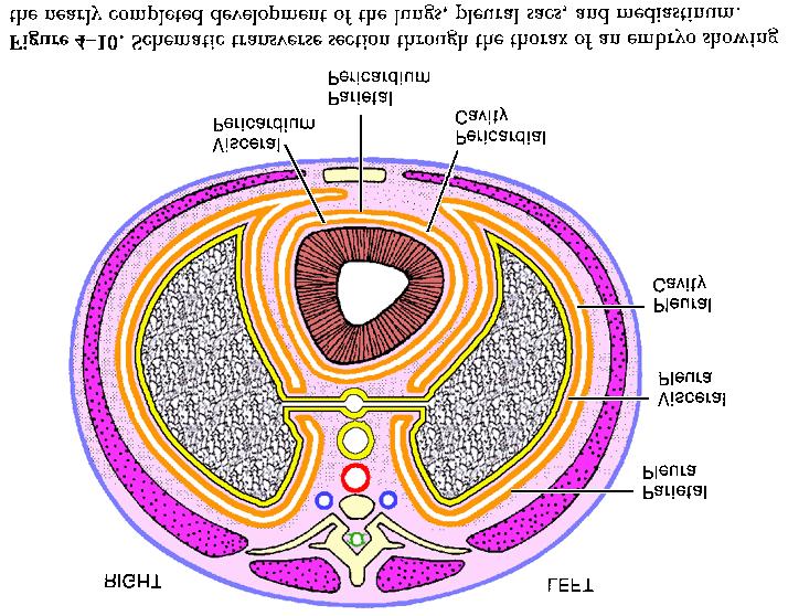
18 pericardial cavity. The part of the pericardial cavity that was ventral to the heart tube before the looping is now ventral, inferior, and even partly dorsal to the heart (see Fig. 4-7B). The dorsal part is called the oblique sinus of the pericardial cavity (the rest has no name). The actual fluid-filled pericardial cavity between visceral and parietal pericardium will be squeezed to a very thin space by the growth of the heart (see Fig. 4-10). The thin layer of fluid between visceral and parietal pericardia is a "lubricant," enabling the heart to beat without encountering friction on its outer wall. Most of the subsequent growth of the heart and its pericardial sac is to the left of the midline. Eventually the parietal pericardium is made thicker by the addition of extra connective tissue to its outer surface. This new layer of connective tissue is said to comprise the fibrous pericardium. The old inner layer of the parietal pericardium that is actually continuous with the visceral pericardium is grouped with it under the name serous pericardium. In the adult, a little pocket of parietal serous pericardium may bulge out through an acquired defect in the fibrous pericardium to produce a so-called pericardial diverticulum. Although uncommon and asymptomatic, pericardial diverticula do alter the cardiac shadow on chest radiographs. Further Development of the Pleural Sacs Just as the pericardial sac and cavity are greatly modified by their relationship to the growing heart, so the pleural sacs and cavities are altered by the development of the lungs. The lungs start as a single tubular outpocketing from the ventral surface of the gut tube where it lies in the neck. This laryngotracheal diverticulum grows down into the chest just anterior to the gut tube (Fig. 4-8). In the chest, the laryngotracheal diverticulum bifurcates, sending one tubular process to the right and one to the left (see Fig. 4-8). These processes are called lung buds, and they will eventually run up against the medial walls of the pleural sacs. That small part of the medial pleural wall that is contacted by the lung bud is called visceral pleura (see Fig. 4-8). All the remainder of the pleural wall is now called parietal pleura. To continue growth, the lung buds must either rupture through the visceral pleura or push it ahead of them. They follow the latter course (Fig. 4-9). Each lung bud begins to branch into the lobar bronchi, segmental bronchi, and so forth, growing in size as it does. The original small spot of visceral pleura grows with the lung bud squeezing pleural fluid out of the way. Eventually, the extensive fluid-filled pleural cavity will be reduced to but a thin fluid-filled space between the visceral and parietal pleurae (Fig. 4-10). To accommodate the growing lungs, each pleural sac expands ventrally around the side of the pericardial sac (see Figs. 4-9, 4-10) toward the sternum. The left pleural sac is impeded in its effort to reach the sternum by the presence of the heart. The pleural sacs also expand inferiorly into the sides and back of the body wall, separating off an inner layer of body wall that is incorporated into the diaphragm (Fig. 4-11). Posteriorly, laterally, and anteriorly, the parietal pleura lies against the inner surfaces of the developing ribs and intercostal muscles. The posterior, lateral, and anterior walls of the pleural sac all grade gently into one another and are said to compose the costal pleura. A thin connective tissue layer called endothoracic fascia will form between this costal pleura, on the one hand, and the epimysium and periosteum of the thoracic wall, on the other. Inferiorly, each pleural sac rests on the upper surface of the developing diaphragm, also separated from it by endothoracic fascia. This inferior wall of the pleural sac is said to form the diaphragmatic pleura. Where the costal pleura meets the diaphragmatic pleura, there is obviously a change in direction of the pleural sac wall (see Fig. 4-11). This change in direction is called the costodiaphragmatic reflection. The part of the pleural cavity just above this reflection is called the costodiaphragmatic recess. 79


19 80

20 81
21 The medial wall of each pleural sac runs a course from front to back (see Fig. 4-10). The central region of the thoracic cavity, trapped between the medial wall of the left pleural sac and the medial wall of the right pleural sac, is called the mediastinum. The pericardial sac and all the organs of the thoracic cavity (except the lungs) are constrained to occupy this central region called mediastinum. The medial wall of each pleural sac is said to constitute the mediastinal pleura. The change in direction where the anterior part of the costal pleura meets the mediastinal pleurae is called the costomediastinal reflection. The part of the pleural cavity just lateral to this reflection is called the costomediastinal recess. Superiorly, each pleural sac ends in blunt apex called the cupola (see Fig. 4-11) The parietal and visceral pleurae are continuous only at the site where the lung bud originally contacted the pleural sac. This site is called the root of the lung. The arteries and nerves that pass from their source among mediastinal structures out to the lungs are constrained to pass through this root, enveloped by a sleeve of pleura. Similarly, the veins and lymphatics that grow from the lungs back to the heart and mediastinal lymph trunks are constrained to pass through the root. Nothing--not the lung, vessels, or nerves--ever enters the pleural cavity. It remains a fluid-filled space; just its shape has changed. Areas of the Mediastinum The imposing presence of the heart and pericardial sac prompts anatomists to give names to areas of the mediastinum. Superior to the heart and pericardial sac is the superior mediastinum. Posterior to the heart and pericardial sac is the posterior mediastinum. Anterior to the heart and pericardial sac is a very small area called anterior mediastinum. The heart and pericardial sac are said to reside in the middle mediastinum. 82 THE HEART (Figs. 4-12, 4-13) The heart is a four-chambered pump composed of a special kind of muscle called cardiac muscle. The muscle of the heart is said to constitute the myocardium. It is overlain by, and adherent to, the epicardium (visceral pericardium). On the inner surface of the myocardium is the endothelial-lined connective tissue that is called endocardium, with which the blood comes into contact. Grooves (sulci) mark the outer surface of the myocardium at the sites where one chamber of the heart meets another. Two of the heart chambers are called atria. They are relatively thin walled, for their only function is to receive blood from organs outside the heart and send it under low pressure to the ventricles. There is a right atrium that receives deoxygenated venous blood from all the organs of the body, and a left atrium that receives oxygenated venous blood from the lungs. The two ventricles have thicker muscular walls than the atria, for they must pump blood through high-resistance capillary networks. The right ventricle sends deoxygenated blood that it has received from the right atrium out to the lungs so that it may be oxygenated. The left ventricle sends oxygenated blood that it has received from the left atrium out to all the tissues of the body. Since the resistance of the pulmonary capillary bed is so much lower than that of the rest of the body, the wall of the right ventricle is not nearly as thick as that of the left ventricle. The difference becomes far less if the right ventricle is forced to pump its blood against high resistance, as occurs in a variety of disease states. Despite their names, the right atrium and ventricle are as much anterior to the left chambers as they are to their right.
22 83
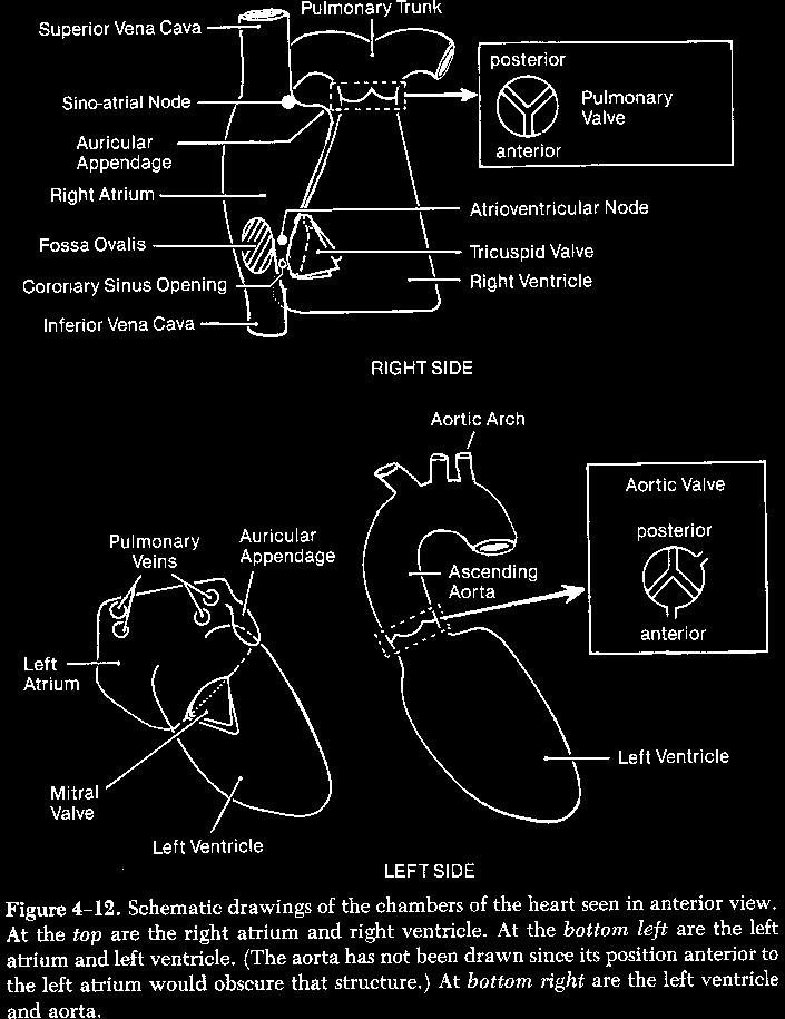
23 84 Right Atrium Deoxygenated venous blood from all parts of the body (except the heart itself) enters the right atrium of the heart via two very large veins--the superior vena cava and inferior vena cava. The superior vena cava, being formed by the junction of the right and left brachiocephalic veins and receiving the azygos vein just before it enters the right atrium, carries deoxygenated venous blood from the body above the diaphragm. The inferior vena cava brings blood from the body below the diaphragm. The bulk of the venous blood from the heart itself is also conveyed to the right atrium, but not by the venae cavae. Such blood runs in the coronary sinus and the anterior cardiac veins, which will be described later in this chapter. Within the wall of the right atrium are the tiny Thebesian veins (venae cordis minimae) that open directly into its cavity. (In fact, all the chambers of the heart have their own Thebesian veins, but these do not carry much blood). The superior vena cava, inferior vena cava, and coronary sinus all enter the smooth-walled posterior part of the right atrium. The superior vena cava opens into its superior end; the inferior vena cava opens into the inferior end; the coronary sinus comes in through the back wall. The smooth-walled posterior part of the right atrium is derived from the embryonic sinus venosus. It lacks the internal muscular ridges (pectinate muscles) that are found in the anterior part of the right atrium, which is derived from the true atrial chamber of the embryonic heart tube. The junction between the posterior smooth-walled part and the anterior rough-walled part is indicated on the outer surface of the right atrium by a vertical groove called the sulcus terminalis. On the inside of the right atrium, this same junction is marked by a vertical muscular ridge called the crista terminalis. From the top of the rough-walled part of the right atrium comes a small medially directed outpocketing. Having resembled an ear to some early anatomist, it is called the auricular appendage. Much of the back wall of the right atrium is fused to the front wall of the left atrium. This common wall is called the interatrial septum, but, of course, it is formed of two leaves. The anterior (or right atrial) leaf has an oval hole (foramen ovale) in it just superior to the opening of inferior vena cava. When you look through this hole you see the posterior (left atrial) leaf of the interatrial septum. This posterior leaf also has a hole (foramen secundum), but it is located superior to the foramen ovale and cannot be seen from inside the right atrium. In fetal life, the fusion of the leaves of the interatrial septum is less complete than in the adult. Consequently blood from the inferior vena cava is able to pass directly up through the foramen ovale and foramen secundum into the left atrium. Such blood bypasses the lungs, which after all are nonfunctional in the fetus. After birth, when fusion of the leaves becomes complete, the foramen ovale is sealed shut by the posterior leaf of the interatrial septum. Thus, the foramen ovale changes its name to the fossa (i.e., depression) ovalis. Even if postnatal fusion of the leaves of the interatrial septum fails to progress normally, the elevated blood pressure within the left atrium presses the posterior leaf against the foramen ovale and produces a functional closure. The coronary sinus enters the right atrium through its back wall, just to the left of the fossa ovalis. The anterior, rough-walled part of the right atrium opens into the bottom of the right ventricle by means of the right atrioventricular orifice. Since the right ventricle lies to the left of, not below, the right atrium, the flow of blood from the atrium to the ventricle is a right-to-left flow, not a top-to-bottom flow. From the circumference of the right atrioventricular orifice, three valvular flaps (cusps) project into the chamber of the right ventricle. They are really located along the anterior, posterior, and inferior margins of the orifice but their nomenclature is confused. True enough, the anterior cusp is called anterior; but the posterior cusp is called septal, because it is nearest the interventricular septum; and the
24 85 inferior cusp is called posterior, because, after all, it is posterior to the anterior cusp. For obvious reasons, the valve is called the tricuspid valve. Right Ventricle The right ventricle is a triangular structure with its base positioned inferiorly and its apex superiorly. Its back wall is the interventricular septum between itself and the left ventricle. The upper part of this septum is formed of tough connective tissue (membranous part of interventricular septum); the lower part is cardiac muscle. The inner surface of the ventricle is marked by numerous muscular ridges called trabeculae carneae. Blood flows from the right atrium into the base of the right ventricle and is pushed out through its apex into the pulmonary trunk. In order to prevent the cusps of the tricuspid valve from being driven through the atrioventricular orifice back into the right atrium whenever right ventricular blood pressure is elevated by this chamber's contraction, the free margins of the valve cusps receive the tendinous insertions of cardiac muscle bundles that arise from the wall of the ventricle. The muscle bundles form little hillocks called papillary muscles, and their tendons are called chordae tendineae. The entrance to the pulmonary trunk is guarded by another valve with three cusps that project upward into the lumen of the trunk. Each cusp has a semicircular attachment (concave upward) to the wall of the pulmonary trunk. When excised, a cusp has the shape of a half-moon and is often called a semilunar cusp. One cusp is attached to the posterior third of the circumference of the pulmonary trunk; one is attached to the left anterior third, and one is attached to the right anterior third (hint: pulmonary begins with a "p", and the pulmonary trunk has one posterior cusp). Blood tending to return to the right ventricle from the pulmonary trunk will flow into the space (valvular sinus) between the cusps and the wall of the pulmonary trunk. This balloons out each cusp so that it contacts its neighbors, closing off the pulmonary orifice. The fact that the attachment site of a semilunar cusp to the aortic wall is arc-shaped effectively prevents it from being driven into the right ventricle. Blood passes up the pulmonary trunk a short distance to the point of its bifurcation, and then passes to the lungs by means of the right and left pulmonary arteries. Left Atrium The left atrium lies at the back of the upper half of the heart. This chamber has the shape of "home plate" in baseball, with the apex pointing inferiorly. Much of the anterior wall of the left atrium participates in formation of the interatrial septum. The depression formed by the sealed foramen secundum can be seen in this wall. The veins draining each lung usually coalesce into two pulmonary veins, which pass through the root of the lung into the mediastinum. The four pulmonary veins (two right and two left) pierce the back wall of the left atrium near its upper border. Originally, there was only one pulmonary vein from each lung. During development, the right and left embryonic pulmonary veins become incorporated into the left atrium. Thus, it is really the two tributaries of each embryonic pulmonary vein that we find entering the adult left atrium. If incorporation of an embryonic pulmonary vein into the left atrium is incomplete, then in the adult the two pulmonary veins from that lung join into a single vessel that enters the left atrium. Virtually the entire left atrium is smooth-walled. This is because virtually the entire left atrium is derived from pulmonary venous tissue that has been incorporated into the heart during embryogenesis.
25 That part of the adult left atrium formed from the original heart tube is really very small. It consists of a small, anteriorly directed, rough-walled outpocketing at the left superior corner of the chamber. This is the auricular appendage of the left atrium, and it serves no useful function. 86 The left atrioventricular orifice lies on the left inferior wall of the atrium near its apex. The flow of blood into the left ventricle is thus downward and to the left. From the circumference of the left atrioventricular orifice are two valvular cusps that project downward into the left ventricle. One attaches to the anterior rim of the orifice, the other to the posterior rim. This bicuspid valve resembles an upside-down Bishop's mitre (hat) and is therefore called the mitral valve. The free edges of the cusps receive the tendinous insertions (i.e., chordae tendineae) of the left ventricular papillary muscles. Left Ventricle This is the thickest of the chambers of the heart because it pumps blood against the highest resistance. Like the right ventricle, its inner surface is also characterized by muscular ridges called trabeculae carneae. The left ventricle is a conical structure with its rounded apex pointing inferiorly and to the left. The base of the cone faces superiorly and to the right. The left atrioventricular orifice is located at the back of this base, and blood flows through it into the left ventricle toward its apex. Contraction of the left ventricle forces blood upward and to the right toward the orifice of the aorta, which is located in the ventricular base anterior to the atrioventricular orifice. Blood heading toward the aortic valve must pass in front of the anterior cusp of the mitral valve. The aortic valve is structured similarly to the pulmonary valve. The semilunar cusps of the aortic valve are anterior, left posterior and right posterior (hint: aorta begins with "a," and there is one anterior cusp). However, this nomenclature is rarely used, because an easier one is made possible by virtue of the fact that the coronary arteries branch off the wall of the aorta at the level of the valvular sinuses. The left coronary artery comes from the left posterior sinus wall; thus, the left posterior cusp is usually called the left coronary cusp. The right coronary artery comes from the anterior sinus wall, and thus the anterior cusp is usually called the right coronary cusp. The right posterior cusp is called the noncoronary cusp, because its sinus wall gives rise to no artery. Conducting System of the Heart The rhythm of the heart is normally controlled by a group of automatically depolarizing specialized cardiac muscle cells called the sinu-atrial node. As its name implies, the sinu-atrial node is located at the junction of the embryonic sinus venosus and atrium. In the adult, it can be found at the upper end of the sulcus terminalis, where the superior vena cava meets the atrium. (Fig. 4-12). The wave of depolarization sweeps down the walls of the atria, stimulating them to contract, and eventually reaches another group of specialized cardiac muscle cells located in the interatrial septum just superior to the opening of the coronary sinus (see Fig. 4-12). This group of cells is called the atrioventricular node. From them emanates the atrioventricular bundle of His (again composed of specialized cardiac muscle cells), which carries the depolarizing current across the insulating barrier between atria and ventricles into the membranous part of the interventricular septum. The bundle of His divides into two branches, which continue down the muscular part of the septum toward its base. Here, the right atrioventricular bundle passes into the muscle of the right ventricle, and the left bundle passes into the muscle of the left ventricle. The two bundles course upward in the ventricular walls, distributing depolarizing current to them.
26 87 Arterial Supply to the Heart (Fig. 4-14) The major coronary arteries are located in the epicardium, lying within the grooves (sulci) between the chambers of the heart. Along its course, each coronary artery sends off branches to the chambers on either side of the groove, and to any portion of a septum that may lie deep to the groove. Right Coronary Artery The right coronary artery leaves the wall of the anterior aortic sinus to enter the right atrioventricular sulcus. It gives off a branch to the sinu-atrial node and then passes inferiorly in the sulcus, giving off branches to the chambers on either side (i.e., right atrium and right ventricle). At the inferior margin of the heart the right coronary artery gives off an acute marginal branch for the lower reaches of the right ventricle and then passes around the inferior border of the heart toward its back surface, where it continues in the atrioventricular sulcus as far as its junction with the posterior interventricular sulcus. In this part of its path the right coronary artery continues its supply of the right atrium and right ventricle. In most individuals, the right coronary artery turns down the posterior interventricular sulcus and assumes the name posterior interventricular (posterior descending) artery. The posterior descending artery supplies the atrioventricular node, adjacent sides of the right and left ventricles, and part of the interventricular septum. Left Coronary (Left Main Coronary) Artery The left main coronary artery arises from the wall of the left posterior aortic sinus and runs toward the left behind the beginning of the pulmonary trunk. It then divides into two branches, the anterior interventricular (left anterior descending, LAD) and the circumflex coronary arteries. The LAD passes inferiorly in the anterior interventricular sulcus, supplying the right and left ventricles and the major part of the interventricular septum. Its branches to the left ventricle are called diagonal
27 88 branches. The LAD turns around the inferior margin of the heart and then heads up the posterior interventricular sulcus a short distance, to anastomose with the posterior descending artery. The circumflex coronary artery enters the left atrioventricular sulcus and winds around the left margin of the heart onto its back surface. It supplies the left atrium and left ventricle. It rather large branches to the left ventricle are called obtuse marginal branches. As the circumflex coronary artery approaches the site where the atrioventricular sulcus intersects the posterior interventricular sulcus, the vessel has usually been reduced to such a small size that it is very difficult to dissect. What remains, however, does anastomose with a small branch of the right coronary artery given off just prior to its turn into the posterior interventricular artery. CORONARY DOMINANCE A heart in which the posterior descending artery is a branch of the right coronary is said to be "right coronary dominant." This is the usual case. However, a common variation in coronary artery anatomy is one in which the circumflex coronary artery, rather than diminishing drastically in size as it approaches the posterior interventricular sulcus, instead stays big and turns into the sulcus to become the posterior descending artery. Obviously, in such cases it is the right coronary artery that becomes diminutive on the back of the heart. Such a heart is said to be "left coronary dominant." Regardless of coronary dominance, the left coronary artery supplies more cardiac tissue, and notably more of the important left ventricle, than does the right coronary artery. While coronary angiographers routinely report which vessel is dominant, it is not so obvious how this information is clinically useful. There is one report that individuals undergoing bypass surgery for left main coronary artery stenosis have a higher perioperative mortality if they are left coronary dominant. Also, the circumflex coronary artery lies closer to the rim (anulus) of the mitral valve in left dominant hearts (maybe because the vessel is bigger) and is more likely to be caught in a suture during mitral valve replacement than if the heart were right coronary dominant. CORONARY ANASTOMOSES Not only are there anastomoses between the LAD and posterior descending arteries within the interventricular sulcus, and between the circumflex coronary and right coronary arteries within the atrioventricular sulcus, but arterioles from each artery anastomose also within the actual muscle of the heart. In cases of slowly developing coronary occlusion, these anastomoses enlarge and enable blood to reach areas that might otherwise be cut off from their arterial supply. The extent of the anastomoses between right and left coronary arterial systems is dramatized by a congenital condition in which the right coronary artery comes off the pulmonary trunk instead of the aorta. At birth, when the blood pressure within the pulmonary system drops (consequent upon expansion of the lungs), blood sent out the left coronary artery under high (aortic) pressure is shunted through anastomotic channels into the low (pulmonary) pressure right coronary artery and thus to the pulmonary trunk.
28 89 Because this blood bypasses the capillary bed of the myocardium, the result may be a cardiac infarct in the newborn. Venous Drainage of the Heart (Fig. 4-15) The veins of the heart run in the epicardium, usually next to the arteries. They drain blood from the heart wall on either side of their paths. A great cardiac vein starts at the inferior margin of the heart and accompanies (but in the opposite direction) the LAD up toward its origin from the left main coronary. At this point, the great cardiac vein turns to follow the circumflex coronary artery around to the back of the heart. Once onto the back of the heart, the great cardiac vein joins with a vein coming from the left atrium, which is called the oblique vein of the left atrium. The latter vessel gets a special name because it is the remnant of a special vein in the embryo (i.e., the left common cardinal vein). Immediately after it receives the oblique vein of the left atrium, the great cardiac vein undergoes a change in histological structure. The smooth muscle in its wall is replaced by cardiac muscle. Such is the case because, from this point on, the great cardiac vein is derived from the sinus venosus. Such a major change in histology and embryonic origin deserves a name change. Thus, the vein with cardiac muscle in its wall is called the coronary sinus. The coronary sinus continues the course of the great cardiac vein within the atrioventricular sulcus, heading toward the back wall of the right atrium (which is also derived from sinus venosus), where it empties just to the left of the fossa ovalis. The coronary sinus picks up two other named tributaries before it opens into the right atrium. It receives a middle cardiac vein that has accompanied the posterior descending artery and a small cardiac vein that ran alongside the acute marginal branch of the right coronary artery.
29 No veins accompany the right coronary artery proximal to its marginal branch. Instead, a few veins from the right ventricle cross transversely over the right coronary artery to reach the right atrium. These are called anterior cardiac veins. It has previously been mentioned that some small veins within the heart muscle itself simply drain directly into the nearest chamber. These are the Thebesian, or smallest, cardiac veins. 90 LUNGS (see Fig. 4-16) The lungs lie within the thoracic cavity, surrounded by visceral pleura and the pleural cavity. Remember, the lungs are outgrowths of a mediastinal tube and they are still connected to mediastinal structures through their roots. The right principal (mainstem) bronchus passes through the root of the right lung. The left mainstem bronchus passes to the left lung through its root. Each mainstem bronchus is accompanied by a branch of the pulmonary artery and some nerves that go to the lung. Leaving the lung through its root and going to the mediastinum are the pulmonary veins and lymphatics. The superior, almost pointed, extremity of the lung is called its apex. The concave surface that rests on the diaphragm is called the base. The outer surface adjacent to the ribs is called the costal surface (which obviously has anterior, lateral, and posterior aspects). The surface facing the mediastinum is the mediastinal surface. The site where structures passing through the root of the lung actually contact pulmonary tissue is called the hilum. Lung parenchyma consists of alveolar sacs to which air is conducted by a series of tubes. The larger of these tubes, which contain cartilage within their walls and seromucous glands in their epithelium, are called bronchi. The smaller tubes, without cartilage or seromucous glands, are called bronchioles. Both bronchi and bronchioles have considerable smooth muscle in their walls. Right Lung The right lung is the larger of the two because the heart does not encroach on its territory. It is divided into three lobes: superior (upper), middle, and inferior (lower). Each lobe has its own bronchial and vascular trees. Soon after entering the lung, the right mainstem bronchus and pulmonary artery give off lobar branches for each of the three lobes. Each lobe also has its own covering of visceral pleura. Where the lobes abut one another, their visceral pleurae are separated by a film of pleural fluid. Not uncommonly, where the upper and middle lobes abut, their visceral pleurae are fused. It is then difficult to separate these two lobes by dissection, but they still maintain separate bronchovascular trees. Within each lobe there are certain blocks of lung tissue separated from other blocks by thin connective tissue septa that normally prevent airflow between the alveoli on either side. Each such block receives its air from a separate branch of the lobar bronchus and each receives its arterial supply from a separate branch of the lobar artery. These blocks of lung tissue are called bronchopulmonary segments. They differ from fused lobes in one major way. The veins draining the bronchopulmonary segments lie within the connective septa between them and, thus, drain adjacent segments. Veins from the lobes do not lie between them and drain only from one lobe.
30 While lacking the independence of lobes, bronchopulmonary segments are sufficiently autonomous that infection, pneumonia, or atelectasis (collapse) may affect one segment while its neighbors remain normal. It is even possible surgically to resect a single bronchopulmonary segment, but this is not commonly done unless the patient has so little healthy lung that leaving in place as much as possible becomes of paramount importance. A segmentectomy is done by deflating the lung, tying off the bronchus and artery to the diseased segment, reinflating the lung, and then removing the tissue that is unfilled by air. Obviously, it is the veins between segments that are at greatest risk during such surgery. 91
31 The upper lobe of the right lung has three bronchopulmonary segments: one at the front (the anterior segment), one at the back (the posterior segment), and one that sits on top of the other two at the apex of the lung (the apical segment). The middle lobe has medial and lateral segments. The lower lobe has one at its top (the superior segment) and four that compose the part of the lower lobe resting on the diaphragm (anterior basal, posterior basal, medial basal, lateral basal segments). Left Lung The left lung has only two lobes. What corresponds to the middle lobe of the right lung is an extension of the upper lobe called the lingula (because it looked tongue-like to some anatomist). The lingula is not separated from the remainder of the upper lobe by visceral pleura and certain veins drain both the lingula and adjacent regions of the rest of the upper lobe. The left lung has an independent lower lobe exactly "homologous" to that of the right side. The nonlingular part of the left upper lobe has the same three segments as the upper lobe of the right lung (i.e., anterior, posterior, and apical). However, since bronchi for the apical and posterior segments come off a common stem, these two are often combined under the name apical-posterior segment. This is more a nomenclatural than a functional grouping. The lingular part of the upper lobe has two segments, a superior and inferior. The lower lobe of the left lung has the same five segments as that of the right lung. However, because the bronchus for the anterior basal segment and that for the medial basal segment share a common stem (for a short distance), these two segments are often nomenclaturally combined under the term anteromedial basal segment. Trachea and Large Bronchi (Fig. 4-16) Within the mediastinum the trachea divides into right and left mainstem bronchi. The cartilaginous wedge that projects upward into the lumen of the trachea at the point of bifurcation is called the carina. It is slightly to the left of the tracheal midline because the diameter of the right mainstem bronchus is greater than that of the left. Presumably this is due to the greater size of the right lung. As a result of the carina being slightly to the left of the tracheal midline, inhaled foreign objects tend to pass into the right bronchus more frequently than into the left. Both bronchi diverge from the trachea at about 45 degrees. However, the left bronchus must travel considerably farther to reach its lung. This occurs primarily because the hilum of the left lung is displaced by the heart, but also because the tracheal bifurcation is usually pushed slightly to the right of the midline by the aortic arch. Thus, shortly after it leaves the trachea at about a 45 degree angle, the left mainstem bronchus turns more laterally to reach its lung. Branches of the Right Principal Bronchus After leaving the trachea, the right mainstem bronchus assumes a position posterior to the right pulmonary artery and enters the root of the lung. Soon after penetrating the hilum, the right mainstem bronchus gives off a branch that runs directly laterally. This is the superior lobe bronchus. After a short course, the superior lobe bronchus trifurcates into the anterior segmental, posterior segmental, and apical segmental bronchi. After the superior lobe bronchus has split off, the continuation of the right mainstem bronchus is called the intermediate bronchus. It will carry air to the middle and lower lobes. After a short course the intermediate bronchus gives off the middle lobe bronchus from its anterior surface. The middle lobe bronchus soon bifurcates into its medial and lateral segmental bronchi. 92
32 After the middle lobe bronchus has split off, the continuation of the intermediate bronchus is in fact the lower lobe bronchus. It is extremely short, giving off the superior segmental bronchus from its back surface virtually at its root. The remainder of the inferior lobe bronchus "quadrifurcates" into the four basal segmental bronchi. Branches of the Left Principal Bronchus The long left mainstem bronchus passes inferior to the aortic arch to reach the posterior surface of the bifurcation of the pulmonary trunk. The bronchus then continues to the left and downward to assume a position inferior to the left pulmonary artery within the root of the lung. Soon after penetrating the hilum, the left mainstem bronchus bifurcates into its superior and inferior lobe bronchi. The superior lobe bronchus passes directly laterally; the inferior lobe bronchus turns downward. The superior lobe bronchus soon bifurcates into a branch that heads down to the lingula and one that heads up to the remainder of the upper lobe. The lingular bronchus splits into its superior and inferior segmental bronchi. The upward-heading branch of the superior lobe bronchus forks into anterior segmental and apical-posterior segmental bronchi. The latter soon bifurcates into apical and posterior segmental bronchi. The inferior lobe bronchus gives rise to a superior segmental bronchus and then trifurcates into an anteromedial basal segmental bronchus, a lateral basal segmental bronchus, and a posterior basal segmental bronchus. The anteromedial basal splits into anterior and medial segmental bronchi. 93 Vasculature of the Lung Arteries The two or three small bronchial arteries from the descending aorta (or sometimes from a posterior intercostal artery near its origin) supply oxygen and nutrients to the bronchial tree probably as far as the smallest bronchiole. If there are two such vessels, one goes to each lung; if there are three bronchial arteries, the left lung gets two of them. The bronchial arteries also supply blood to the nerves, lymphatic tissue, walls of the large vessels, and connective tissue septa of the lungs. The pulmonary arteries run alongside the bronchi but do not supply them. They bring nutrients to the alveolar cells, but these cells get their oxygen directly from the air. The main function of capillaries derived from the pulmonary artery system is to receive oxygen from, and send carbon dioxide to, the air. Veins Venous blood from the larger bronchi enters bronchial veins that empty into the azygos and hemiazygos systems. Venous blood from the smaller bronchi and from the capillary network around the alveoli empties into the pulmonary veins. It has already been mentioned that veins draining segments are intersegmental in location and, therefore, do not run with the segmental bronchi and arteries.
33 94 SUPERIOR AND POSTERIOR MEDIASTINAL STRUCTURES Ascending Aorta and Aortic Arch (see Fig. 4-12) From the site of the aortic valve, the aorta passes superiorly and slightly toward the right. This part is called the ascending aorta. Then the large vessel makes a looping turn backwards and slightly to the left, to reach the left side of the vertebral column. This loop is called the aortic arch. Once it has contacted the vertebral column, the aorta turns inferiorly and during its further course is called the descending aorta (see below). The coronary arteries are branches of the ascending aorta just beyond the aortic orifice, thus from the walls of the valvular sinuses. They have been described previously. Off the arch come the three large arteries that feed the upper limbs, head, and neck. In turn, these are the brachiocephalic, left common carotid, and left subclavian arteries. Their distributions will be discussed in later chapters. Ligamentum Arteriosum Nestled in the concavity is the beginning of the right pulmonary artery. A fibrous cord runs from the very beginning of the left pulmonary artery up to the concave surface of the aortic arch more or less opposite the site of origin of the left subclavian artery. This cord is the remnant of the ductus arteriosus, which, in fetal life, allowed blood to pass from the pulmonary trunk to the aorta, thus bypassing the nonfunctioning lungs. The adult remnant is called the ligamentum arteriosum. Brachiocephalic (Innominate) Veins and Superior Vena Cava Interposed between the beginning of the brachiocephalic artery and the manubrium is another large vessel, the left brachiocephalic (innominate) vein. It is formed deep to the left sternoclavicular joint (between it and the left common carotid artery) by juncture of the internal jugular and subclavian veins of the left side. It passes downward and to the right to join the right brachiocephalic vein, thereby forming the superior vena cava. The superior vena cava passes inferiorly, partly tucked behind the ascending aorta to enter the right atrium. Thymus In the child, a large lymphoid organ, the thymus, lies interposed between the sternum and the great vessels of the heart. At puberty, the thymus begins an involution that reduces it to something that looks like an encapsulated glob of fat. However, within this apparently nondescript structure is a remnant of thymic tissue capable of becoming tumorous and also probably having a relationship to the development of myasthenia gravis. Trachea and Mainstem Bronchi The trachea enters the thorax in the median sagittal plane halfway between the vertebral column and the manubrium. Often one does not appreciate just how deep this is and, when attempting a low tracheostomy, is surprised not to find the trachea directly beneath the skin. At the level of the midpoint of the manubrium, the trachea is just posterior to the origin of the brachiocephalic artery. Below this point it has the aortic arch on its left side and, in fact, is pushed slightly to the right of the midline by the aortic arch. At the level of the sternal angle, the trachea bifurcates. The right mainstem bronchus passes onto the posterior surface to the right pulmonary artery. The left mainstem bronchus passes below the aortic arch onto the posterior surface of the bifurcation of the pulmonary trunk, and then comes to lie inferior to
34 the left pulmonary artery as they go to the lung. The mainstem bronchi enter the hili of the lungs on the same transverse level as the T5/T6 intervertebral disc. Due to the heart, the left pulmonary hilum is further from the midline than is the right. Descending Thoracic Aorta The descending aorta begins on the left anterior side of the T4/5 intervertebral disc. It runs inferiorly on the left anterior surface of the vertebral column (Fig. 4-17), down to the level of the 12 th thoracic vertebra, where it pierces the diaphragm (through a hole called the aortic hiatus) to become the abdominal aorta. From the thoracic part of the descending aorta come the paired 3rd-11th intercostal arteries, the paired subcostal arteries, the paired superior phrenic arteries (to the posterior regions of the diaphragm), two or three bronchial arteries (one or two of which passes through the root of each lung for supply of its nonrespiratory tissues), a couple of esophageal arteries, and some small mediastinal and pericardial branches. 95 Azygos and Hemiazygos Veins The azygos vein enters the thorax from the abdominal cavity by ascending along the ventral surface of the vertebral column just to the right of the descending aorta (see Fig. 4-17). Opposite the 5th thoracic vertebra the azygos vein turns anteriorly and passes just above the root of the right lung to join the superior vena cava immediately before that vessel opens into the right atrium. Throughout its ascent the azygos vein picks up the 5th-11th posterior intercostal veins of the right side. The hemiazygos vein enters the thorax from the abdominal cavity by ascending on the left surfaces of the vertebral bodies behind the descending aorta (see Fig. 4-17). Throughout its ascent the hemiazygos vein picks up the 5th-11th posterior intercostal veins of the left side. It ends superiorly where it receives the left 5th posterior intercostal vein. The hemiazygos vein empties in a peculiar manner. As it
35 passes up the thorax it sends a variable number of anastomotic connections toward the right, across the anterior surface of the vertebral column, to reach the azygos vein. The azygos and hemiazygos veins have come from the abdominal cavity by passing through the aortic hiatus of the diaphragm. While in the abdominal cavity the azygos and hemiazygos veins have anastomotic connections to certain large veins of the abdomen (Chapter 5, p. 131). As mentioned previously (p.71), just as the azygos turns forward to cross above the root of the lung, it receives the right superior intercostal vein, which has formed from the right 2 nd - 4 th posterior intercostal veins. The left superior intercostal vein courses anteriorly, in contact with the left side of the aortic arch, to reach the left brachiocephalic vein deep to the manubrium. Frequently the hemiazygos vein continues superiorly beyond the site where it receives the left 5th posterior intercostal vein. In such cases, the hemiazygos connects to the 4th posterior intercostal vein, allowing venous blood from the left 2nd-4th intercostal spaces to flow either into the left superior intercostal vein or into the hemiazygos vein, depending on the position of the body in space. Thoracic Duct The thoracic duct passes up from the abdomen into the posterior mediastinum through the aortic hiatus of the diaphragm. Here it lies in midline on the anterior surface of T12, posterior to right edge of the aorta. The duct continues upward on the anterior surfaces of vertebral bodies between the aorta and azygos vein, posterior to the esophagus (see Fig. 4-17). At the level of the aortic arch, the thoracic duct shifts from its position on the front of the vertebral column to assume a position on the left surface of the esophagus. It follows the left surface of the esophagus into the neck and then abruptly turns laterally to pass behind the left common carotid artery and reach the beginning of the left brachiocephalic vein. Esophagus The esophagus enters the thorax posterior to the trachea on the anterior surface of the vertebral column, but slightly to the left of the midline (and thus slightly to the left of the trachea). As it descends, the esophagus moves toward the median sagittal plane. However, since the trachea is shifted to the right by the aortic arch, the relative position of the esophagus to the trachea does not change. Thus, the left mainstem bronchus must cross anterior to the esophagus on its way to the root of the lung. Inferior to the tracheal bifurcation the esophagus comes to lie against the posterior surface of the parietal pericardium, separated by it from the left atrium. The esophagus reaches the median sagittal plane opposite T6. At this point it lies on the right side of the descending aorta and anterior to the vertebral column, separated from it by the thoracic duct and azygos vein (Fig. 4-17). As the esophagus continues further downward it moves anteriorly and again to the left. By the time it pierces the diaphragm, at the level of T10 (or the xiphoid process) the esophagus lies anterior to the aorta and about 2 fb left of the midline. 96 During its course in the chest, the esophagus is compressed by two structures. The first of these is the arch of the aorta, which bulges into the left side of the esophagus. The second is the left principal bronchus, which bulges into the anterior surface of the esophagus. The indentations of the esophagus by these structures can usually be identified in a radiograph of a barium swallow. Sometimes only one indentation, the
36 97 so-called broncho-aortic constriction, can be visualized. The broncho-aortic constriction is one of the more common sites of esophageal cancer. The esophagus is not normally indented by the left atrium. However, if the left atrium is enlarged, as in mitral valve disease, it may displace the esophagus posteriorly. This is easily recognized on a radiograph of a barium swallow. Also, microphones may be placed in the esophagus to get ultrasound images of the mitral valve. Phrenic and Vagus Nerves At the root of the neck the vagus nerve lies medial to the internal jugular vein, whereas the phrenic nerve lies lateral to the vein. After entering the thorax the phrenic nerve moves anteriorly and the vagus moves posteriorly. Eventually the phrenic nerves pass from the superior mediastinum into the middle mediastinum. The vagus nerves pass from the superior mediastinum into the posterior mediastinum. The right phrenic continues down on the right sides of the brachiocephalic vein, superior vena cava, pericardium over right atrium, and inferior vena cava to reach the diaphragm. In its course between the mediastinal pleura and the pericardial sac, it passes anterior to the root of the right lung. The right vagus gains the right surface of the trachea. It continues to descend along the side of the trachea toward the posterior surface of the right principal bronchus, where the vagus joins in the formation of the right pulmonary plexus. Those vagal fibers of the posterior pulmonary plexus that are not destined for the lung coalesce again into bundles that pass onto the esophagus, predominantly as the posterior vagal trunk. Because the left brachiocephalic vein passes to the right immediately after it is formed, because no superior vena cava exists on the left side, and because the left side of the trachea is in contact with the left common carotid and subclavian arteries, the left phrenic and vagus nerves must have very different relationships to other structures of the superior mediastinum than do their counterparts on the right side. Passing along the left side of the left common carotid artery, the phrenic nerve crosses the aortic arch where this artery arises. The nerve then crosses anterior to the root of the left lung to reach the interval between pericardium and mediastinal pleura. It follows the left border of the pericardial sac to reach the diaphragm. The left vagus enters the thorax between the left common carotid and left subclavian arteries. It follows them down to the aortic arch, which it crosses heading posteriorly toward the posterior surface of the left principal bronchus. Like the right vagus, it partakes in a pulmonary plexus. Those left vagal fibers not going to the lung coalesce into bundles that pass onto the surface of the esophagus, chiefly as the anterior vagal trunk. As it passes by the concavity of the aortic arch, the left vagus gives off a recurrent laryngeal nerve. This nerve passes toward the right, posterior to the ligamentum arteriosum, and under the aortic arch. When the left recurrent laryngeal nerve hits the interval between the trachea and esophagus, it turns superiorly and runs in this interval all the way up to the neck.
37 98 The position of the left recurrent laryngeal nerve, trapped between the concavity of the aortic arch and the pulmonary bifurcation, makes it susceptible to compression by dilations of either of these two vessels. Also, cancer of the esophagus or cancer in the tracheal nodes may involve the left recurrent laryngeal nerve as it passes up toward the neck on the left side of the interval between esophagus and trachea. The result of damage to the recurrent laryngeal nerve is hoarseness (see Chapter 10, p. 378). Thus, hoarseness may be caused by intrathoracic disease. Inferior Vena Cava The inferior vena cava enters the heart immediately after piercing the abdominal diaphragm. In fact, the anterior wall of the inferior vena cava has no intrathoracic course. Because the diaphragm slopes downward and backward at the site where it is pierced by the inferior vena cava, the posterior wall of the vessel does have a short intrathoracic portion before it enters the right atrium. The posterior wall can usually be visualized on lateral chest radiographs. INNERVATION OF THE INTERNAL ORGANS OF THE THORAX In that the internal organs of the chest are not part of the body wall, they receive no branches from either the ventral or dorsal rami of spinal nerves. As previously mentioned, the postganglionic sympathetic axons for all the internal organs above the diaphragm derive from cells located in the three cervical and upper five (or six) thoracic sympathetic ganglia. The axons do not pass through gray rami, because there is no point in their rejoining the spinal nerve. Instead, they run in bundles that pass from paravertebral ganglia directly to the organ in question. On their way to these organs, the postganglionic sympathetic nerve bundles meet the parasympathetic preganglionic axons from the vagus and interweave with them, forming a number of intrathoracic autonomic plexuses. The preganglionic sympathetic neurons for structures above the diaphragm lie in the upper five (or six) thoracic segments of the spinal cord. These axons use the spinal nerves only as a means of transport to paravertebral ganglia. As stated in Chapter 2, pain fibers from internal organs of the thorax pass toward the spinal nerve in the same bundles that brought sympathetic fibers out to that organ. If you have memorized the sympathetic outflow pathway, then you also know the pathway of inflowing pain. However, it is an important fact that few if any visceral pain fibers emanate from the bronchi or lungs. For this reason, diseases of the lung may progress quite substantially before pain in the chest is a significant complaint. An Example--Innervation of the Heart The preganglionic sympathetic cell bodies concerned with innervation of the heart lie in the intermediolateral column of the spinal gray matter from the 1st-5th thoracic segments of the spinal cord. The preganglionic axons pass out the 1st-5th thoracic ventral roots into the 1st-5th thoracic spinal nerves and then to the 1st-5th thoracic ventral rami. The preganglionic axons leave the ventral rami to enter the 1st-5th thoracic white rami communicantes and are, thus, carried to the 1st-5th thoracic paravertebral ganglia. Some of the axons synapse in these ganglia. Others of the axons turn superiorly and ascend toward cervical ganglia where they will synapse. Bundles of postganglionic axons leave the upper five
38 thoracic ganglia as so-called thoracic sympathetic cardiac nerves. Other bundles of postganglionic sympathetic axons leave the cervical portion of the sympathetic chain and descend into the mediastinum as cervical sympathetic cardiac nerves. Many texts state that each cervical ganglion provides one such bundle. Thus, the authors speak of superior cervical, middle cervical, and inferior cervical sympathetic cardiac nerves. According to Pick 14, these are imaginary, the number and exit site of the cervical sympathetic cardiac nerves being highly variable. Furthermore, Pick observed that those cardiac nerves emanating from the cervical sympathetic chain often join with cardiac branches given off by the vagus. Vagal cardiac nerves carry preganglionic parasympathetic axons. Some arise from the vagus as it descends through the neck; some come from the vagus in the chest; and yet others actually leave its recurrent laryngeal branch. The take-home message is "that the number of variations of the course, anastomoses and distribution of cardiac nerves equals the number of anatomists who dissected these nerves." 3 All the cardiac nerves, be they carrying postganglionic sympathetic axons from the cervical and upper thoracic paravertebral ganglia, or preganglionic parasympathetic fibers from the vagus, or a mixture of both, converge toward the anterior surface of the tracheal bifurcation and (just to its left) the superior surface of the bifurcation of the pulmonary trunk. At this site, the cardiac nerves interweave in what is called the cardiac plexus. Nerve bundles leave the cardiac plexus to surround the pulmonary trunk and ascending aorta, and then travel down along these vessels to reach the epicardium of the heart. In epicardium are located the postganglionic parasympathetic cell bodies. Their axons, and the postganglionic sympathetic axons, distribute to the muscle, conducting system, and coronary vessels. Given the general rule about visceral pain pathways from internal organs of the thorax, we can deduce that pain fibers from the heart travel up the outer surfaces of the pulmonary trunk and ascending aorta into the cardiac plexus and thence via the sympathetic cardiac nerves to the paravertebral ganglia. If a pain fiber enters one of the upper five thoracic paravertebral ganglia, it passes through this into the white ramus and is thus carried to the ventral ramus of one of the upper five thoracic spinal nerves. Running backward in the ventral ramus to the spinal nerve, it enters its dorsal root wherein lies the cell body. The central process of the cell body enters the spinal cord. If a pain fiber enters the cervical sympathetic chain, it turns downward to travel to one of the upper thoracic ganglia. Once there, the pain fiber passes into a white ramus to reach the ventral ramus, and then back to the spinal nerve, its dorsal root, and spinal cord. 99 REFERRED PAIN FROM THE HEART Since the sympathetic supply to the heart originates in the upper five thoracic segments of the spinal cord, the pain fibers (which merely follow the sympathetic nerves backward) end in the upper five thoracic segments of the cord. Referred pain from the heart is felt in the body wall innervated by the upper five thoracic spinal nerves, usually on the left side. This area of referred pain overlies the upper five intercostal spaces and includes the medial surface of the upper limb, which is innervated by T1. (Sometimes the referred pain of heart attack is felt in the neck and radiates up to the jaw. It is not 14 Pick, J: The Autonomic Nervous System. JB Lippincott, Philadelphia, 1970.
39 100 uncommon for referred pain to spread beyond the location we deduce from knowledge of anatomy.) Innervation of Other Internal Thoracic Organs There is not much point in tracing the sympathetic supply to other internal organs of the chest. Their preganglionic neurons all lie somewhere in the upper five (or six) thoracic segments of the spinal cord and all travel out ventral roots to spinal nerves to ventral rami to white rami to the upper five thoracic paravertebral ganglia. Sometimes they ascend to cervical ganglia and synapse, sometimes they synapse in thoracic ganglia. Any postganglionic sympathetic axons leaving cervical ganglia probably course in cardiac nerves, which means that cardiac nerves carry some axons for thoracic organs other than the heart. Connections between the cardiac plexus and other autonomic plexuses of the chest enable these axons to get to the organ for which they are destined. Postganglionic sympathetic axons from thoracic ganglia proceed directly to their respective organs. Those for a lung interweave with each other and with the vagus in a plexus surrounding the mainstem bronchus. This is the pulmonary plexus, most of which is posterior to the bronchus. (It has connections to the cardiac plexus that allow axons that have traveled in cardiac nerves to reach the pulmonary plexus). Those vagal fibers not destined for the lung emerge from the pulmonary plexus and pass inferiorly onto the surface of the esophagus, where they meet sympathetic esophageal nerves, to form the esophageal plexus. It goes without saying that an aortic plexus composed of sympathetic aortic nerves and vagal branches also exists. Most of the vagal axons within the esophageal plexus do not actually supply the esophagus. Instead, they are destined to continue into the abdomen. Many fibers from the right vagus join with a few from the left vagus to form a trunk (more or less embedded in the esophageal plexus) along the right posterior surface of the esophagus. Many fibers of the left vagus join with a few from the right vagus to form a trunk (embedded in the esophageal plexus) along the left anterior surface of the esophagus. These posterior and anterior vagal trunks continue through the esophageal hiatus of the diaphragm to distribute within the abdomen. Pain From the Pericardium and Pleura The wall of a coelomic sac is innervated by the same nerves as the structures to which that wall is applied. Thus, pain from the visceral pericardium is of the same nature and travels the same path as pain from the heart. Because the lung is essentially insensitive to pain, so is the visceral pleura. The costal portion of the parietal pleura, and the parietal pleura on the periphery of the diaphragm, are innervated by intercostal nerves. The parietal pleura on the dome of the diaphragm, the mediastinal pleura, and the parietal pericardium all send their pain fibers up the phrenic nerve. LYMPHATICS OF THE CHEST Nodes That Lie Along Vessels In general throughout the body, lymph nodes are gathered into groups that lie along major blood vessels. Most lymph nodes of the chest follow this rule.
40 101 Axillary Nodes The axillary lymph nodes will be described more completely in Chapter 11 (p. 492). However, some mention of them must be made now because they do receive lymph from the superficial structures of the chest. The axillary nodes lie alongside the axillary vein, and also extend inferiorly around the subscapular and lateral thoracic tributaries of this vessel. Those nodes along the subscapular vein are called subscapular nodes, or also posterior axillary nodes. Those along the lateral thoracic vein are called lateral thoracic nodes, or anterior axillary nodes, or, even more commonly, pectoral nodes. The subscapular and pectoral nodes drain to the nodes around the axillary vein. Internal Thoracic (Internal Mammary, Parasternal) Nodes The internal thoracic (internal mammary, parasternal) nodes lie alongside the internal thoracic vessels. The lowermost members of this group lie on the upper surface of the diaphragm and are often called anterior diaphragmatic nodes. Posterior Mediastinal Nodes The posterior mediastinal nodes lie around the descending thoracic aorta. The lowermost members of this group lie on the upper surface of the diaphragm and are often called posterior diaphragmatic nodes. Intercostal Nodes The intercostal nodes are really lateral extensions of the posterior mediastinal nodes along the first few centimeters of the intercostal vessels. The intercostal nodes drain to the posterior mediastinal nodes (or directly into the thoracic duct). Anterior (Superior) Mediastinal Nodes The anterior mediastinal nodes lie around the great vessels that are deep to the manubrium. They really are anterior nodes of the superior mediastinum. Nodes That Do Not Lie Along Vessels Two groups of thoracic nodes do not lie along blood vessels. Pulmonary, Bronchopulmonary, Tracheobronchial, and Tracheal Nodes Lymph nodes are located within the lung along its segmental and lobar bronchi. In turn, these nodes drain to a continuous chain of nodes that starts around the mainstem bronchus at the hilum of the lung and runs inward along the mainstem bronchus to its junction with the trachea, and then up along the part of the trachea that lies in the chest. Though forming a chain, the nodes draining the lung are often given separate names depending on their precise location. Thus, nodes actually surrounded by lung tissue are called pulmonary nodes; those at the hilum are called bronchopulmonary nodes; those alongside mainstem bronchi are called bronchial nodes; those at the junction of the trachea and mainstem bronchi are called tracheobronchial
41 nodes; those alongside the trachea are called tracheal nodes. As you can see, there is an obvious logic to this nomenclature. 102 SURGICAL NOMENCLATURE OF NODES DRAINING THE LUNGS Surgeons tend to use a nomenclature for lymph nodes draining the lungs that differs from what is found in most anatomy texts, which I have just described. The surgical nomenclature is as follows: peribronchial nodes - lie along bronchi within the substance of a lung hilar nodes - lie around the mainstem bronchus within the root of a lung subcarinal nodes - lie in the inferior angle of the tracheal bifurcation paratracheal nodes - lie alongside the trachea Lateral (Middle) Diaphragmatic Nodes The lateral (or middle) diaphragmatic nodes lie on the upper surface of the diaphragm near the sites where the phrenic nerves enter it. These middle diaphragmatic nodes drain in three directions: back to the posterior diaphragmatic nodes, forward to the anterior diaphragmatic nodes, and upward to the superior mediastinal nodes. Drainage Routes of Thoracic Nodes The foregoing list of thoracic nodes did not mention the drainage of those groups that send their lymph into the bloodstream without intervening nodal groups. 1. The posterior mediastinal nodes empty into the thoracic duct, which lies in the posterior mediastinum (see further on). The thoracic duct empties into the left brachiocephalic vein at the site of its formation. 2. The axillary nodes drain by means of the subclavian trunk. On the right side this trunk empties into the right brachiocephalic vein at its beginning. The left subclavian trunk usually joins the thoracic duct just before that vessel enters the left brachiocephalic vein. 3. The ducts draining the internal thoracic, anterior mediastinal, and para-/pretracheal nodes on each side tend to unite into a single lymphatic vessel called the bronchomediastinal trunk. On the right, this trunk empties into the right brachiocephalic vein at its beginning. The left bronchomediastinal trunk usually joins the thoracic duct just before that vessel enters the left brachiocephalic vein. Lymphatic Drainage of Specific Organs Skin and Superficial Fascia The skin and superficial fascia from the level of the clavicle down to the level of the umbilicus drain almost entirely to the axillary nodes. From the back, lymphatic vessels go first to the subscapular group of axillary nodes. From the side and front of the chest, lymphatic vessels go first to the pectoral nodes. The skin and superficial fascia near the sternum drains to the parasternal nodes.
42 Within the skin and superficial fascia there are communications between lymph vessels going to axillary and those going to the parasternal nodes, and also communications across the midline between vessels going to the right and left parasternal nodes. Both groups of lymphatic vessels communicate with those below the umbilicus draining to the superficial inguinal nodes and with those above the clavicle draining to deep cervical nodes. 103 LYMPHATIC DRAINAGE OF THE BREAST The breast is composed of modified apocrine sweat glands and, as such, its lymphatic drainage is like that of skin. The breast lies on the front of the chest, with a tail projecting into the axilla. About 75% of its lymph drains to the pectoral group of axillary nodes. The remaining 25% of breast lymph, largely from the medial portion of gland, passes to parasternal nodes. If these normal routes of lymph drainage are retarded by tumor, lymph from the breast may pass to deep cervical or even inguinal nodes. Tumor may also spread from one breast to the other via the lymphatic communications that cross the midline. Thus, although metastatic breast cancer should be expected first in the axillary nodes on the same side, one should not be surprised if it ends up elsewhere. Muscle of Chest Wall Lymphatic vessels of the deeper part of the body wall run centrally alongside the arteries that brought blood out to the structure of interest. Structures supplied by branches of the axillary artery send their lymph back to the axillary nodes. Structures supplied by posterior intercostal arteries send their lymph to intercostal nodes. Structures supplied by branches of the internal thoracic artery send their lymph to the parasternal nodes. Internal Organs The standard route of lymphatic drainage from a lung is peribronchial --> hilar --> subcarinal --> paratracheal nodes. The hilar nodes also receive lymphatic vessels that course through the visceral pleura. The paratracheal nodes feed finally to the bronchomediastinal trunk. The right lung drains almost entirely to right paratracheal nodes. The left upper lobe drains almost entirely to left paratracheal nodes. The left lower lobe drains to both right and left sides. Consequently, if there is evidence of left lower lobe cancer, surgeons perform a mediastinoscopy to discover if the cancer has spread to the right paratracheal nodes. Such spread is a counterindication for surgical treatment of the cancer. The structures at the back of the mediastinum (mainly the esophagus and aorta) drain to posterior mediastinal nodes. Where the esophagus pierces the diaphragm it lies further anteriorly and drains to the lateral diaphragmatic nodes. At the upper end of the thorax, where the esophagus lies directly behind the trachea, some of its lymph will go to paratracheal nodes. The heart, pericardium, and great vessels of the superior mediastinum drain to the anterior mediastinal and (to a slight extent) paratracheal nodes.
43 104 SURFACE ANATOMY OF THE CHEST A physician should know where all the organs lie in relation to one another and to the surface of the body. A statement of how you can tell where an organ is located, given only the information that can be seen or felt on the outside of the body, is called surface anatomy. I shall be presenting information on the relationships as if these were immutable. However, the student must never forget that people differ from one another. All that can be memorized is a sort of average person. In order to bring this point home, I shall often use intervertebral discs as landmarks because the very fact that two vertebrae must be mentioned implies a range from the middle of one to the middle of the other. Also, I shall often express distances in terms of fingerbreadths (fb) or handbreadths (hb). Clearly, since different examiners have different-sized fingers and hands, the existence of human variation is emphasized. Finally, I want to stress that all the relationships presented below are in a supine subject. Many internal organs descend under their own weight when a person stands up. Some, like the heart, may drop one or two vertebral levels. Others that are more tightly bound to the body wall drop less, or not at all. Given that a student recognizes the limitations of surface anatomy, it provides an important framework on which to reconstruct what cannot be seen. Surface Landmarks of the Chest Most every important surface landmark of the chest concerns a bone that can be palpated. At the back of the chest one can feel the spines of the vertebra and also the scapula (Fig. 4-18). On the side of the chest one can feel ribs (Fig. 4-19). On the front of the chest one can feel the upper border of the manubrium, the manubriosternal joint (sternal angle, angle of Louis), the xiphisternal joint, and all the ribs and costal cartilages but the first (Fig. 4-20). Bony Landmarks on Back of Chest The highest vertebral spine that can be identified with any degree of certainty is that of the 7th cervical vertebra, because it becomes so prominent when the neck is flexed. One may count the spines of thoracic vertebrae by starting at the 7th cervical and proceeding down. It is well to remember that the spines of thoracic vertebra slant inferiorly from their origins on the vertebral arches (Figs. 4-18, 4-19). Thus, the tip of a thoracic spine is not on the same level as the corresponding vertebral body. The slant is greatest in the middle third of the thoracic column and is less toward the cervical or lumbar regions. In general, the tip of a thoracic vertebral spine is on the same transverse level as the next lower vertebral body but will be a bit lower in the middle third of the thoracic spine and not quite this low toward the ends. Counting vertebral spines is tedious and not so easy. Therefore, one often relies on relationships between the vertebral column and the scapula to identify specific vertebrae (Fig. 4-18). The spine of the scapula intersects its medial (vertebral) border on the same transverse level as the tip of the 3rd thoracic vertebral spine (thus, the 4th thoracic vertebral body). The inferior angle of the scapula is on the same transverse level as the tip of the 7th thoracic vertebral spine (thus, the disc between T8 and T9). The first rib that can be palpated inferior to the scapula is the 8th.
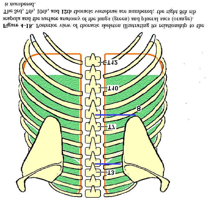

44 105
 lies on the same transverse plane as the disc between T2 and T3 (see Figs. 4-19, 4-20).")
45 106 Bony Landmarks on Front of Chest Placing the manubrium, sternum, and xiphoid in relation to the vertebral column is relatively easy. The superior border of the manubrium (the jugular notch) lies on the same transverse plane as the disc between T2 and T3 (see Figs. 4-19, 4-20). The sternal angle lies on the same transverse plane as the disc between T4 and T5 (see Figs. 4-19, 4-20). Thus the manubrium is in front of the 3rd and 4th thoracic vertebrae. The xiphisternal joint lies on the same transverse plane as the disc between T9 and T10 (see Figs. 4-19, 4-20). Thus the body of the sternum lies in front of 5th-9th thoracic vertebrae. Because the 2nd costal cartilage articulates at the sternal angle, and the latter can usually be palpated by running a finger down the front of the sternum, it is generally possible to identify the 2nd costal cartilage and rib. In fact, the best way to count ribs from the front is by first identifying the 2nd and working down rib by rib. One other landmark often used to position chest organs is a vertical line dropped down from the middle of the clavicle. This midclavicular line passes a centimeter or two medial to the nipple. The vertical line through the nipple is called the mammary line. Heart (Figs to 4-23) The heart has a superior, a right, an inferior, and a left border. The superior border (or base) is formed by the roof of the left atrium and the entrance orifice for the superior vena cava. It runs from side to side behind the sternum at the level of the 2nd intercostal space (specifically it starts 1 fb lateral to the left sternal margin along the lower border of the 2nd costal cartilage and runs slightly downward and to

46 the right to end 1 fb lateral to the right sternal margin at the upper edge of the 3rd costal cartilage). The upper border of the 3rd costal cartilage near the sternum marks the site of entry of the superior vena cava into the right atrium. The right border of the heart is formed by the right atrium. It starts where the superior border ends and virtually parallels the right sternal margin, 1 fb lateral to it, as far down as the xiphisternal joint. 107 The inferior border of the heart starts 1 fb lateral to the right sternal margin at the level of the xiphisternal joint and passes to the left along a line that curves downward to about an inch below the xiphisternal joint and then back upward, to end at the level of the xiphisternal joint 1 hb to the left of the midsternal line in the 5th intercostal space. This is generally at or just medial to where the midclavicular line crosses the fifth interspace. It is clearly medial to the left mammary line, and a heart whose apex is beyond the mammary line is enlarged or displaced. The beginning of the inferior border marks the site of the opening of the inferior vena cava into the right atrium. The last few centimeters represents the apex of the left ventricle. In between these sites, the entire inferior margin of the heart is formed by the right ventricle. The left border of the heart is approximated by a line from the left 5th intercostal space, 1 hb from the midsternal line, up to the lower edge of the left 2nd costal cartilage, 1 fb lateral to the sternal margin. Inferior to the 3rd costal cartilage this border is left ventricle; superior to the 3rd costal cartilage it is left atrium. The right atrioventricular sulcus runs obliquely behind the body of the sternum. The anterior interventricular sulcus runs vertically 2 or so fb from the sternal margin.

47 108 Pulmonary Trunk The pulmonary valve is located deep to the left 3rd sternochondral junction, and the pulmonary trunk extends up from this site to the left 2nd sternochondral junction, where it bifurcates. In its course, the pulmonary trunk lies anterior to the left atrium. Ascending Aorta and Aortic Arch The aortic valve lies deep to the sternum just medial to the left 3rd intercostal space (behind the right ventricle). The ascending aorta lies anterior to the left atrium. The vessel sweeps upward and slightly to the right to end deep to the right 2nd sternochondral junction. The arch of the aorta begins at this point and lies behind the lower half of the manubrium as the vessel passes toward the left side of the vertebral column, where it ends at the same transverse level as it began, i.e., at the level of the 2nd sternochondral junction, but now on the left side. At the back, this level corresponds to the disc between the 4th and 5th thoracic vertebrae. The concave surface of the arch is deep to the sternal angle From the ascending portion of the aortic arch, deep to the midpoint of the manubrium, arises the brachiocephalic artery. The vessel takes an oblique course upward toward the right sternoclavicular joint, deep to which it bifurcates into the right common carotid and right subclavian arteries. The common carotid comes off the anterior surface of the brachiocephalic; the subclavian comes off its posterior surface. Thus, at their origins behind the sternoclavicular joint, the common carotid is anterior to the subclavian. Beyond the origin of the brachiocephalic artery, the left common carotid and left subclavian arteries come off the ascending portion of the aortic arch in rapid succession. Both travel toward the left sternoclavicular joint to which they lie deep, the carotid being anterior to the subclavian, as on the right side.
 vein.")
48 109 Brachiocephalic Veins and Superior Vena Cava Given what has just been said, it is obvious that the first few centimeters of the brachiocephalic, left common carotid, and left subclavian arteries are deep to the upper half of the manubrium. However, interposed between these great vessels and the bone is another large vessel, the left brachiocephalic (innominate) vein. It is formed deep to the left sternoclavicular joint (between it and the left common carotid artery) by juncture of the internal jugular and subclavian veins of the left side. It passes downward and to the right, deep to the upper half of the manubrium, toward the right 1st sternochondral junction. At this site, the left brachiocephalic vein is joined by the right brachiocephalic vein (which forms deep to the right sternoclavicular joint) to form the superior vena cava. The superior vena cava passes inferiorly, partly tucked behind the ascending aorta, down to the superior border of the right 3rd costal cartilage, where it enters the right atrium. Inferior Vena Cava The inferior vena cava enters the heart immediately after piercing the abdominal diaphragm (1 fb lateral to the right sternal margin at the level of the xiphisternal joint). CARDIAC SHADOW On a standard PA radiograph of the chest, the radiodensity that is the cardiac shadow represents not only the heart but the great vessels as well. Thus, the right edge of the cardiac shadow is composed of the superior vena cava and right atrium. The left
The Thoracic wall including the diaphragm. Prof Oluwadiya KS
 The Thoracic wall including the diaphragm Prof Oluwadiya KS www.oluwadiya.com Components of the thoracic wall Skin Superficial fascia Chest wall muscles (see upper limb slides) Skeletal framework Intercostal
CHAPTER 4. Thorax THORACIC CAVITY CHEST WALL VERSUS THORACIC WALL
 59 CHAPTER 4 Thorax THORACIC CAVITY CHEST WALL VERSUS THORACIC WALL THORACIC WALL Skeletal Components Vertebral Bodies Sternum Ribs Muscular Components Intercostal Muscles Structure Function Abdominal
Anatomy of the Thorax
 Anatomy of the Thorax A) THE THORACIC WALL Boundaries Posteriorly by the thoracic part of the vertebral column Anteriorly by the sternum and costal cartilages Laterally by the ribs and intercostal spaces
STERNUM. Lies in the midline of the anterior chest wall It is a flat bone Divides into three parts:
 STERNUM Lies in the midline of the anterior chest wall It is a flat bone Divides into three parts: 1-Manubrium sterni 2-Body of the sternum 3- Xiphoid process The body of the sternum articulates above
THE THORACIC WALL. Boundaries Posteriorly by the thoracic part of the vertebral column. Anteriorly by the sternum and costal cartilages
 THE THORACIC WALL Boundaries Posteriorly by the thoracic part of the vertebral column Anteriorly by the sternum and costal cartilages Laterally by the ribs and intercostal spaces Superiorly by the suprapleural
Anatomy of thoracic wall
 Anatomy of thoracic wall Topographic Anatomy of the Thorax 1 Bones of Thoracic wall ribs 1-7"true" ribs -those which attach directly to the sternum true ribs actually attach to the sternum by means of
thoracic cage inlet and outlet landmarks of the anterior chest wall muscles of the thoracic wall sternum joints ribs intercostal spaces diaphragm
 Thoracic Wall Lecture Objectives Describe the shape and outline of the thoracic cage including inlet and outlet. Describe the anatomical landmarks of the anterior chest wall. List various structures making
Dr. Weyrich G07: Superior and Posterior Mediastina. Reading: 1. Gray s Anatomy for Students, chapter 3
 Dr. Weyrich G07: Superior and Posterior Mediastina Reading: 1. Gray s Anatomy for Students, chapter 3 Objectives: 1. Subdivisions of mediastinum 2. Structures in Superior mediastinum 3. Structures in Posterior
Mediastinum and pericardium
 Mediastinum and pericardium Prof. Abdulameer Al-Nuaimi E-mail: a.al-nuaimi@sheffield.ac.uk E. mail: abdulameerh@yahoo.com The mediastinum: is the central compartment of the thoracic cavity surrounded by
THE DESCENDING THORACIC AORTA
 Intercostal Arteries and Veins Each intercostal space contains a large single posterior intercostal artery and two small anterior intercostal arteries. The anterior intercostal arteries of the lower spaces
DESCRIPTION: This is the part of the trunk, which is located between the root of the neck and the superior border of the abdominal region.
 1 THE THORACIC REGION DESCRIPTION: This is the part of the trunk, which is located between the root of the neck and the superior border of the abdominal region. SHAPE : T It has the shape of a truncated
Note : I put the sheet's info within the slides to easily understand this lecture Done by : Zaid Al-Ghnaneem
 Note : I put the sheet's info within the slides to easily understand this lecture Done by : Zaid Al-Ghnaneem Thoracic Wall Lecture Objectives Describe the shape and outline of the thoracic cage including
Identify the lines used in anatomical surface descriptions of the thorax. median line mid-axillary line mid-clavicular line
 L 14 A B O R A T O R Y Thorax THORACIC WALL Identify the lines used in anatomical surface descriptions of the thorax. median line mid-axillary line mid-clavicular line Identify the surface landmarks of
Lecturer: Ms DS Pillay ROOM 2P24 25 February 2013
 Lecturer: Ms DS Pillay ROOM 2P24 25 February 2013 Thoracic Wall Consists of thoracic cage Muscle Fascia Thoracic Cavity 3 Compartments of the Thorax (Great Vessels) (Heart) Superior thoracic aperture
Large veins of the thorax Brachiocephalic veins
 Large veins of the thorax Brachiocephalic veins Right brachiocephalic vein: formed at the root of the neck by the union of the right subclavian & the right internal jugular veins. Left brachiocephalic
PLEURAE and PLEURAL RECESSES
 PLEURAE and PLEURAL RECESSES By Dr Farooq Aman Ullah Khan PMC 26 th April 2018 Introduction When sectioned transversely, it is apparent that the thoracic cavity is kidney shaped: a transversely ovoid space
Syllabus: 6 pages (Page 6 lists corresponding figures for Grant's Atlas 11 th & 12 th Eds.)
") PLEURAL CAVITY AND LUNGS Dr. Milton M. Sholley SELF STUDY RESOURCES Essential Clinical Anatomy 3 rd ed. (ECA): pp. 70 81 Syllabus: 6 pages (Page 6 lists corresponding figures for Grant's Atlas 11 th &
PLEURAL CAVITY AND LUNGS Dr. Milton M. Sholley SELF STUDY RESOURCES Essential Clinical Anatomy 3 rd ed. (ECA): pp. 70 81 Syllabus: 6 pages (Page 6 lists corresponding figures for Grant's Atlas 11 th &
Salvador Dali - Anthropomorphic Chest of Drawers, 1936
 Salvador Dali - Anthropomorphic Chest of Drawers, 1936 Kaan Yücel M.D., Ph.D. 05.March.2014 the part between the neck and the abdomen Chest X-ray 1.1. REGIONS/T ERMS Thoracic cavity cavity between neck
Right lung. -fissures:
 -Right lung is shorter and wider because it is compressed by the right copula of the diaphragm by the live.. 2 fissure, 3 lobes.. hilum : 2 bronchi ( ep-arterial, hyp-arterial ), one artery mediastinal
Anatomy notes-thorax.
 Anatomy notes-thorax. Thorax: the part extending from the root of the neck to the abdomen. Parts of the thorax: - Thoracic cage (bones). - Thoracic wall. - Thoracic cavity. ** The thoracic cavity is covered
Dana Alrafaiah. - Moayyad Al-Shafei. -Mohammad H. Al-Mohtaseb. 1 P a g e
 - 6 - Dana Alrafaiah - Moayyad Al-Shafei -Mohammad H. Al-Mohtaseb 1 P a g e Quick recap: Both lungs have an apex, base, mediastinal and costal surfaces, anterior and posterior borders. The right lung,
Diaphragm and intercostal muscles. Dr. Heba Kalbouneh Associate Professor of Anatomy and Histology
 Diaphragm and intercostal muscles Dr. Heba Kalbouneh Associate Professor of Anatomy and Histology Skeletal System Adult Human contains 206 Bones 2 parts: Axial skeleton (axis): Skull, Vertebral column,
Anatomy Lecture 8. In the previous lecture we talked about the lungs, and their surface anatomy:
 Anatomy Lecture 8 In the previous lecture we talked about the lungs, and their surface anatomy: 1-Apex:it lies 1 inch above the medial third of clavicle. 2-Anterior border: it starts from apex to the midpoint
Chapter 5: Other mediastinal structures. The Large Arteries. The Aorta. Ascending aorta
 Chapter 5: Other mediastinal structures The Large Arteries The Aorta The aorta is the main arterial trunk of the systemic circulation and in the healthy state its wall contain a large amount of yellow
In the Last Three Lectures We Already Discussed the Importance of the Thoracic Cage.
 -This Lecture Will Revise what we took in the last three lectures and will introduce the concept of the chest cavity ( Thoracic Cavity ) In the Last Three Lectures We Already Discussed the Importance of
10/14/2018 Dr. Shatarat
 2018 Objectives To discuss mediastina and its boundaries To discuss and explain the contents of the superior mediastinum To describe the great veins of the superior mediastinum To describe the Arch of
Surface anatomy of Cardiovascular system
 Surface anatomy of Cardiovascular system Prof. Abdulameer Al-Nuaimi E-mail: a.al-nuaimi@sheffield.ac.uk E. mail: abdulameerh@yahoo.com The lines cover the front, side, and back of the thorax Midsternal
OBJECTIVE: To obtain a fundamental knowledge of the root of the neck with respect to structure and function
 The root of the neck Jeff Dupree, Ph.D. e mail: jldupree@vcu.edu OBJECTIVE: To obtain a fundamental knowledge of the root of the neck with respect to structure and function READING ASSIGNMENT: Moore and
Gateway to the upper limb. An area of transition between the neck and the arm.
 Gateway to the upper limb An area of transition between the neck and the arm. Pyramidal space inferior to shoulder @ junction of arm & thorax Distribution center for the neurovascular structures that serve
slide 23 The lobes in the right and left lungs are divided into segments,which called bronchopulmonary segments
 Done By : Rahmeh Alsukkar Date : 26 /10/2017 slide 23 The lobes in the right and left lungs are divided into segments,which called bronchopulmonary segments Each segmental bronchus passes to a structurally
ANATOMY OF THE PLEURA. Dr Oluwadiya KS
 ANATOMY OF THE PLEURA Dr Oluwadiya KS www.oluwadiya.sitesled.com Introduction The thoracic cavity is divided mainly into: Right pleural cavity Mediastinum Left Pleural cavity Pleural cavity The pleural
Chapter 3: Thorax. Thorax
 Chapter 3: Thorax Thorax Thoracic Cage I. Thoracic Cage Osteology A. Thoracic Vertebrae Basic structure: vertebral body, pedicles, laminae, spinous processes and transverse processes Natural kyphotic shape,
Welcome to the Structure & Development Dissector. Section I
 Welcome to the Structure & Development Dissector The vast majority of questions will be drawn from structures present in the checklist; however, we reserve the right to use a structure or two that is not
3 Mohammad Al-Mohtasib Areej Mosleh
 3 Mohammad Al-Mohtasib Areej Mosleh ***Muscles Connecting the Upper Limb to the Vertebral Column 1.Trapezius Muscle ***The first muscle on the back is trapezius muscle, it s called so according
Conceptual overview 124. Surface anatomy 226. Regional anatomy 139. Clinical cases 235
 Conceptual overview 124 General description 124 Functions 125 Breathing 125 Protection of vital organs 125 Conduit 125 Component parts 125 Thoracic wall 125 Superior thoracic aperture 126 Inferior thoracic
Yara saddam & Dana Qatawneh. Razi kittaneh. Maher hadidi
 1 Yara saddam & Dana Qatawneh Razi kittaneh Maher hadidi LECTURE 10 THORAX The thorax extends from the root of the neck to the abdomen. The thorax has a Thoracic wall Thoracic cavity and it is divided
Bronchioles. Alveoli. Type I alveolar cells are very thin simple squamous epithelial cells and form most of the lining of an alveolus.
 276 Bronchioles Bronchioles continue on to form bronchi. The primary identifying feature is the loss of hyaline cartilage. The epithelium has become simple ciliated columnar, and there is a complete ring
LECTURE -I. Intercostal Spaces & Its Content. BY Dr Farooq Khan Aurakzai. Date:
 LECTURE -I Intercostal Spaces & Its Content BY Dr Farooq Khan Aurakzai Date: 18.04.18 Layers of IC space: Following are the layers of the thoracic region: Skin Subcutaneous CT External IC muscle and membrane
Mediastinum It is a thick movable partition between the two pleural sacs & lungs. It contains all the structures which lie
 Dr Jamila EL medany OBJECTIVES At the end of the lecture, students should be able to: Define the Mediastinum. Differentiate between the divisions of the mediastinum. List the boundaries and contents of
Intercostal Muscles LO4
 Intercostal Muscles LO4 4 List the structures, from superficial to deep, in an intercostal space. Describe their relationships to each other, to the associated neurovascular bundle and to the pleural cavity.
Cardiovascular system:
 Cardiovascular system: Mediastinum: The mediastinum: lies between the right and left pleura and lungs. It extends from the sternum in front to the vertebral column behind, and from the root of the neck
The pectoral region. University of Babylon College of Medicine Dr.HaythemAli Alsayigh M.B.CH.B.-F.I.M.B.S. Surgical Clinical Anatomy
 The pectoral region University of Babylon College of Medicine Dr.HaythemAli Alsayigh M.B.CH.B.-F.I.M.B.S. Surgical Clinical Anatomy Objective Study the Bones and Joints A. Clavicle (collarbone) B. Scapula
BOGOMOLETS NATIONAL MEDICAL UNIVERSITY DEPARTMENT OF HUMAN ANATOMY. Guidelines. Module 2 Topic of the lesson Aorta. Thoracic aorta.
 BOGOMOLETS NATIONAL MEDICAL UNIVERSITY DEPARTMENT OF HUMAN ANATOMY Guidelines Academic discipline HUMAN ANATOMY Module 2 Topic of the lesson Aorta. Thoracic aorta. Course 1 The number of hours 3 1. The
Day 5 Respiratory & Cardiovascular: Respiratory System
 Day 5 Respiratory & Cardiovascular: Respiratory System Be very careful not to damage the heart and lungs while separating the ribs! Analysis Questions-Respiratory & Cardiovascular Log into QUIA using your
Region of upper limb attachment to the trunk Proximal segment of limb overlaps parts of the trunk (thorax and back) and lower lateral neck.
 and lower lateral neck.") Region of upper limb attachment to the trunk Proximal segment of limb overlaps parts of the trunk (thorax and back) and lower lateral neck. includes Pectoral Scapular Deltoid regions of the upper limb
Region of upper limb attachment to the trunk Proximal segment of limb overlaps parts of the trunk (thorax and back) and lower lateral neck. includes Pectoral Scapular Deltoid regions of the upper limb
Anatomy of the thorax
 2018 Anatomy of the thorax Sameh S. Akkila THE THORACIC CAGE The thoracic cage consists of the sternum anteriorly, the twelve thoracic vertebrae and their intervertebral discs posteriorly and the twelve
Posterior Triangle of the Neck By Prof. Dr. Muhammad Imran Qureshi
 Posterior Triangle of the Neck By Prof. Dr. Muhammad Imran Qureshi For the purpose of anatomical description the neck is sub divided into two major triangles, the Anterior and the Posterior by muscle bellies
The posterior abdominal wall. Prof. Oluwadiya KS
 The posterior abdominal wall Prof. Oluwadiya KS www.oluwadiya.sitesled.com Posterior Abdominal Wall Lumbar vertebrae and discs. Muscles opsoas, quadratus lumborum, iliacus, transverse, abdominal wall
Lung & Pleura. The Topics :
 Lung & Pleura The Topics : The Trachea. The Bronchi. The Brochopulmonary Segments. The Lungs. The Hilum. The Pleura. The Surface Anatomy Of The Lung & Pleura. The Root & Hilum. - first of all, the lung
Upper limb Pectoral region & Axilla
 Upper limb Pectoral region & Axilla 黃敏銓 mchuang@ntu.edu.tw 1 Pectoral region Intercostal nerve Anterior branch of lateral cutaneous branch Lateral cutaneous branch Anterior cutaneous branch Anterior cutaneous
Copyright 2010 Pearson Education, Inc.
 E. VERTEBRAL COLUMN 1. The vertebral column extends from the skull to the pelvis and forms the vertical axis of the skeleton. 2. The vertebral column is composed of vertebrae that are separated by intervertebral
Brachial plexuses and axillary lymph nodes
 Brachial plexuses and axillary lymph nodes Introduction about nervous system nervous system central nervous system periphral nervous system brain spinal cord 31 pairs of spinal nerves 12 paris of cranial
Human Anatomy Biology 351
 nnnnn 1 Human Anatomy Biology 351 Exam #2 Please place your name on the back of the last page of this exam. You must answer all questions on this exam. Because statistics demonstrate that, on average,
The Neck the lower margin of the mandible above the suprasternal notch and the upper border of the clavicle
 The Neck is the region of the body that lies between the lower margin of the mandible above and the suprasternal notch and the upper border of the clavicle below Nerves of the neck Cervical Plexus Is formed
11.1 The Aortic Arch General Anatomy of the Ascending Aorta and the Aortic Arch Surgical Anatomy of the Aorta
 456 11 Surgical Anatomy of the Aorta 11.1 The Aortic Arch 11.1.1 General Anatomy of the Ascending Aorta and the Aortic Arch Surgery of the is one of the most challenging areas of cardiac and vascular surgery,
Chest and cardiovascular
 Module 1 Chest and cardiovascular A. Doss and M. J. Bull 1. Regarding the imaging modalities of the chest: High resolution computed tomography (HRCT) uses a slice thickness of 4 6 mm to identify mass lesions
THE HEART OBJECTIVES: LOCATION OF THE HEART IN THE THORACIC CAVITY CARDIOVASCULAR SYSTEM
 BIOLOGY II CARDIOVASCULAR SYSTEM ACTIVITY #3 NAME DATE HOUR THE HEART OBJECTIVES: Describe the anatomy of the heart and identify and give the functions of all parts. (pp. 356 363) Trace the flow of blood
Abdomen: Introduction. Prof. Oluwadiya KS
 Abdomen: Introduction Prof. Oluwadiya KS www.oluwadiya.com Abdominopelvic Cavity Abdominal Cavity Pelvic Cavity Extends from the inferior margin of the thorax to the superior margin of the pelvis and the
Anatomy of the Lungs. Dr. Gondo Gozali Department of anatomy
 Anatomy of the Lungs Dr. Gondo Gozali Department of anatomy 1 Pulmonary Function Ventilation and Respiration Ventilation is the movement of air in and out of the lungs Respiration is the process of gas
THE THORAX THORACIC WALL
 THE THORAX THORACIC WALL BREAST THE THORAX THORACIC WALL BREAST THORACIC WALL THE THORAX THE THORAX THE THORAX - BONES & JOINTS The part of the body between the neck and abdomen. The thoracic cage (rib
THORACIC WALL, ABDOMINAL REGION, MUSCLES OF THE VERTEBRAL COLUMN
 THORACIC WALL, ABDOMINAL REGION, MUSCLES OF THE VERTEBRAL COLUMN 9. 03. 2015 Kaan Yücel M.D., Ph.D. https://fhs122.org Thoracic wall 1. THORAX region between neck 6 abdomen, Chest includes the primary
Bony Thorax. Anatomy and Procedures of the Bony Thorax Edited by M. Rhodes
 Bony Thorax Anatomy and Procedures of the Bony Thorax 10-526-191 Edited by M. Rhodes Anatomy Review Bony Thorax Formed by Sternum 12 pairs of ribs 12 thoracic vertebrae Conical in shape Narrow at top Posterior
The Arm and Cubital Fossa
 The Arm and Cubital Fossa Dr. Andrew Gallagher School of Anatomical Sciences University of the Witwatersrand Introduction The ARM (BRACHIUM) is the most proximal segment of the upper limb musculoskeletal
The External Anatomy of the Lungs. Prof Oluwadiya KS
 The External Anatomy of the Lungs Prof Oluwadiya KS www.oluwadiya.com Introduction The lungs are the vital organs of respiration Their main function is to oxygenate the blood by bringing inspired air into
ABDOMINAL WALL & RECTUS SHEATH
 ABDOMINAL WALL & RECTUS SHEATH Learning Objectives Describe the anatomy, innervation and functions of the muscles of the anterior, lateral and posterior abdominal walls. Discuss their functional relations
Anatomy of the Heart
 Biology 212: Anatomy and Physiology II Anatomy of the Heart References: Saladin, KS: Anatomy and Physiology, The Unity of Form and Function 8 th (2018). Required reading before beginning this lab: Chapter
Sports Medicine Part II : ANATOMY OF THE SPINE, ABDOMEN AND SHOULDER COMPLEX
 Sports Medicine 25 1.1 Part II : ANATOMY OF THE SPINE, ABDOMEN AND SHOULDER COMPLEX c.w.p. Wagner High School, Sports Medicine, A. Morgan, T. Morgan & A. Eastlake, 2008 Muscles of the Upper Limbs In this
HUMAN HEART. Learn the following structures on the heart models.
 HUMAN HEART Learn the following structures on the heart models. The human heart has four chambers that consist of the right atrium, left atrium, right ventricle, and left ventricle. The atria are smaller
LECTURE 5. Anatomy of the heart
 LECTURE 5. Anatomy of the heart Main components of the CVS: Heart Blood circulatory system arterial compartment haemomicrocirculatory (=microvascular) compartment venous compartment Lymphatic circulatory
Thoracolumbar Anatomy Eric Shamus Catherine Patla Objectives
 1 2 Thoracolumbar Anatomy Eric Shamus Catherine Patla Objectives List the muscular and ligamentous attachments of the thoracic and lumbar spine Describe how the muscles affect the spine and upper extremity
Muscle Action Origin Insertion Nerve Innervation Chapter Page. Deltoid. Trapezius. Latissimus Dorsi
 Muscle Action Origin Insertion Nerve Innervation Chapter Page All Fibers Abduct the shoulder (glenohumeral joint) Deltoid Anterior Fibers Flex the shoulder (G/H joint) Horizontally adduct the shoulder
Radiological Anatomy of Thorax. Dr. Jamila Elmedany & Prof. Saeed Abuel Makarem
 Radiological Anatomy of Thorax Dr. Jamila Elmedany & Prof. Saeed Abuel Makarem Indications for Chest x - A chest x-ray may be used to diagnose and plan treatment for various conditions, including: Diseases/Fractures
Theme 30. Structure, topography and function of the lungs and pleura. Mediastinum and its contents. X -ray films digestive and respiratory systems.
 Theme 30. Structure, topography and function of the lungs and pleura. Mediastinum and its contents. X -ray films digestive and respiratory systems. STRUCTURE, TOPOGRAPHY AND FUNCTІON OF LUNGS AND PLEURA.
Spinal nerves and cervical plexus Prof. Abdulameer Al Nuaimi. E mail: a.al E. mail:
 Spinal nerves and cervical plexus Prof. Abdulameer Al Nuaimi E mail: a.al nuaimi@sheffield.ac.uk E. mail: abdulameerh@yahoo.com Branches of ophthalmic artery Muscles of face A spinal nerve Spinal
human anatomy 2016 lecture thirteen Dr meethak ali ahmed neurosurgeon
 Heart The heart is a hollow muscular organ that is somewhat pyramid shaped and lies within the pericardium in the mediastinum. It is connected at its base to the great blood vessels but otherwise lies
Chest cavity, vertebral column and back muscles. Respiratory muscles. Sándor Katz M.D., Ph.D.
 Chest cavity, vertebral column and back muscles. Respiratory muscles. Sándor Katz M.D., Ph.D. Chest cavity - bony structures Chest cavity- bony structures Sternum Ribs True ribs: first seven pairs connect
Anatomy Lecture #19 AN INTRODUCTION TO THE THORAX April 3, 2012
 Page 1 بسم الله الرحمن الرحيم The Thoracic Wall Firstly, when we talk about thorax, we should begin with the thorax wall which means not only bones that construct the thorax but also the muscles which
The arm: *For images refer back to the slides
 The arm: *For images refer back to the slides Muscles of the arm: deltoid, triceps (which is located at the back of the arm), biceps and brachialis (it lies under the biceps), brachioradialis (it lies
The Thoracic Cage. OpenStax College
 OpenStax-CNX module: m46350 1 The Thoracic Cage OpenStax College This work is produced by OpenStax-CNX and licensed under the Creative Commons Attribution License 3.0 By the end of this section, you will
Chapter 20 (1) The Heart
 The Heart") Chapter 20 (1) The Heart Learning Objectives Describe the location and structure of the heart Describe the path of a drop of blood from the superior vena cava or inferior vena cava through the heart out
Chapter 20 (1) The Heart Learning Objectives Describe the location and structure of the heart Describe the path of a drop of blood from the superior vena cava or inferior vena cava through the heart out
Axial Skeleton: Vertebrae and Thorax
 Axial Skeleton: Vertebrae and Thorax Function of the vertebral column (spine or backbone): 1) 2) 3) Composition of Vertebral column The vertebral column is formed by 33 individual vertebrae (some of which
Anatomy of the Shoulder Girdle. Prof Oluwadiya Kehinde FMCS (Orthop)
") Anatomy of the Shoulder Girdle Prof Oluwadiya Kehinde FMCS (Orthop) www.oluwadiya.com Bony Anatomy Shoulder Complex: Sternum(manubrium) Clavicle Scapula Proximal humerus Manubrium Sterni Upper part of
Anatomy of the Shoulder Girdle Prof Oluwadiya Kehinde FMCS (Orthop) www.oluwadiya.com Bony Anatomy Shoulder Complex: Sternum(manubrium) Clavicle Scapula Proximal humerus Manubrium Sterni Upper part of
Mohammad Hisham Al-Mohtaseb. Lina Mansour. Enas Ajarma
 6 Mohammad Hisham Al-Mohtaseb Lina Mansour Enas Ajarma Some recommended videos are attached to this sheet ( if u are studying online click on them, if not u can reach them by typing their names on the
This is not a required assignment but it is recommended.
 SU 12 Name: This is not a required assignment but it is recommended. BIO 116 - Anatomy & Physiology II Practice Assignment 2 - The Respiratory and Cardiovascular Systems 1. The exchange of oxygen and carbon
BRACHIAL PLEXUS. DORSAL SCAPULAR NERVE (C5) supraclavicular branch innervates rhomboids (major and minor) and levator scapulae
 supraclavicular branch innervates rhomboids (major and minor) and levator scapulae") THE BRACHIAL PLEXUS DORSAL SCAPULAR NERVE (C5) supraclavicular branch innervates rhomboids (major and minor) and levator scapulae SCHEMA OF THE BRACHIAL PLEXUS THE BRACHIAL PLEXUS PHRENIC NERVE supraclavicular
THE BRACHIAL PLEXUS DORSAL SCAPULAR NERVE (C5) supraclavicular branch innervates rhomboids (major and minor) and levator scapulae SCHEMA OF THE BRACHIAL PLEXUS THE BRACHIAL PLEXUS PHRENIC NERVE supraclavicular
بسم الله الرحمن الرحيم
 بسم الله الرحمن الرحيم * Last lecture we talked about : thoracic wall sternum ribs (according to their features they are divided into typical and atypical) vertebral column ( which is made of 33 vertebrae
THE VESSELS OF THE HEART
 1 THE VESSELS OF THE HEART The vessels of the heart include the coronary arteries, which supply the heart and the veins and lymph vessels, which drain the heart. THE CORONARY ARTERIES These are the blood
Anatomy Sheet #5. In the previous lecture, we finished discussion about the larynx; now we continue with trachea, lungs and pleura.
 Anatomy Sheet #5 In the previous lecture, we finished discussion about the larynx; now we continue with trachea, lungs and pleura. Trachea and lungs The knowledge about the pleura and lungs is very important
Thorax Lecture 2 Thoracic cavity.
 Thorax Lecture 2 Thoracic cavity. Spring 2016 Dr. Maher Hadidi, University of Jordan 1 Enclosed by the thoracic wall. Extends between (thoracic inlet) & (thoracic outlet). Thoracic inlet At root of the
region of the upper limb between the shoulder and the elbow Superiorly communicates with the axilla.
 1 region of the upper limb between the shoulder and the elbow Superiorly communicates with the axilla. Inferiorly, a number of important structures pass between arm & forearm through cubital fossa. 2 medial
the part between the neck and the abdomen Chest X-ray
 The Two Fridas 1939 by Frida Kahlo Kaan Yücel M.D., Ph.D. 01. 10. 2013 the part between the neck and the abdomen Chest X-ray 1.1. REGIONS/T ERMS Thoracic cavity cavity between neck and abdomen protected
The Anterolateral Abdominal Wall By Prof. Dr. Muhammad Imran Qureshi
 1 P age The Anterolateral Abdominal Wall By Prof. Dr. Muhammad Imran Qureshi Introduction The abdomen is the region of the trunk located between the thorax and the pelvis. It includes the anterolateral
Sheet. April/14 th /2013. Introduction to Anatomy. Dr. Maher Hadidi. Muna Abu Hijleh. 1 P a g e
 Sheet Introduction to Anatomy Dr. Maher Hadidi Muna Abu Hijleh 1 P a g e 29 April/14 th /2013 Superior & Posterior Mediastinum ***Superior mediastinum * is bounded from: -Anterior by manubrium sterni -posterior
Multiple Neurovascular... Pit Baran Chakraborty, Santanu Bhattacharya, Sumita Dutta.
 Multiple Neurovascular... Pit Baran Chakraborty, Santanu Bhattacharya, Sumita Dutta. Fig-3: Showing high formation of Median nerve. Fig-1: Showing atypical formation of cords of Brachial plexus. 1 = Upper
Jordan University Faculty Of Medicine. Breast. Dr. Ahmed Salman. Assistant professor of anatomy & embryology
 Jordan University Faculty Of Medicine Breast Dr. Ahmed Salman Assistant professor of anatomy & embryology The breasts are specialized accessory glands of the skin that secretes milk. They are situated
Chapter 14. The Cardiovascular System
 Chapter 14 The Cardiovascular System Introduction Cardiovascular system - heart, blood and blood vessels Cardiac muscle makes up bulk of heart provides force to pump blood Function - transports blood 2
Key Relationships in the Upper Limb
 Key Relationships in the Upper Limb This list contains some of the key relationships that will help you identify structures in the lab. They are organized by dissection assignment as defined in the syllabus.
3 Circulatory Pathways
 40 Chapter 3 Circulatory Pathways Systemic Arteries -Arteries carry blood away from the heart to the various organs of the body. -The aorta is the longest artery in the body; it branches to give rise to
Human Anatomy, First Edition
 Human Anatomy, First Edition McKinley & O'Loughlin Chapter 22 : Heart 1 Functions of the Heart Center of the cardiovascular system, the heart. Connects to blood vessels that transport blood between the
The Upper Limb III. The Brachial Plexus. Anatomy RHS 241 Lecture 12 Dr. Einas Al-Eisa
 The Upper Limb III The Brachial Plexus Anatomy RHS 241 Lecture 12 Dr. Einas Al-Eisa Brachial plexus Network of nerves supplying the upper limb Compression of the plexus results in motor & sensory changes