Most common fetal cardiac anomalies
|
|
|
- Sandra Ball
- 5 years ago
- Views:
Transcription

1 Most common fetal cardiac anomalies
2 Common congenital heart defects CHD % of cardiac defects Chromosomal Infants Fetuses anomaly (%) 22q11 deletion (%) VSD 30 5~10 20~40 10 PS 9 5 (PA w/ VSD) HLHS 7~ CoA (IAA) TOF 3.5~7 > ~15 c-tga 5~8 5.5 <1 <1 AVSD 5 ~18 40~60 (ASD)
3 Contents 1. Ventricular Septal Defects (VSD) 2. Atrioventricular Septal Defects (AVSD) 3. Pulmonary stenosis (PS) 4. Tetralogy of Fallot (TOF) - perimembranous subaortic VSD - infundibular (infravalvular) PS

4 Ventricular Septal Defects (VSD)

5 Ventricular Septal Defects (VSD) Inlet VSD: In apical 4CV, posterior to TV Muscular VSD: In apical or subcostal 4CV Perimembranous VSD: In 5CV, beneath AV Outlet VSD: In short axis view of great vessels, beneath PV
![PM VSD [ 5CV ] - more than one plane Anechoic area](/docs-images/90/104128458/images/6-0.jpg "in IVS Below AV level above: overlapping MPA Color")

6 PM VSD [ 5CV ] - more than one plane Anechoic area in IVS Below AV level above: overlapping MPA Color Doppler: bidirectional shunting (inconsistent) L-to-R shunting in systole R-to-L shunting in diastole
![Inlet VSD [ Apical 4CV] Echogenic borders of VSD Color](/docs-images/90/104128458/images/7-0.jpg "Doppler: shunting DDx: Echo dropout, Color overlapping,")
7 Inlet VSD [ Apical 4CV] Echogenic borders of VSD Color Doppler: shunting DDx: Echo dropout, Color overlapping, AVSD


8 Muscular VSD [ 4CV: apical or subcostal ] Large: >2-3 mm Echogenic borders of VSD Small at the apex: not visible in gray scale Color Doppler: bidirectional flow


9 DDx Echo dropout : Apical 4CV High color gain: color overlapping over septum Shunting in VSD: low-velocity passive flow


10 VSD: 4CV (subcostal & apical/basal) & 5CV w/ color Doppler
 Endocardial")

11 Atrioventricular Septal Defects (AVSD) Endocardial cushion defect, AV canal defect Best detected in apical 4CV
12 Atrioventricular Septal Defects (AVSD) Endocardial cushion defect, AV canal defect Complete AVSD (97%) : Primum ASD + VSD + common AV valve (linear AV valve) Incomplete AVSD : Primum ASD + cleft in MV and/or TV = Ostium primum ASD (Primum ASD w/o large VSD) - Mitral annulus displaced apically: linear AV valve insertion (+)


13 AVSD In diastole, wide opening within center Color Doppler: single channel of blood In systole, linear common AV valve


, LSVC (dilated")
14 AVSD In diastole, small AVSD : difficult to detect In systole, linear common AV valve Color Doppler: common valve regurgitation DDx: Inlet VSD (AV valve level), LSVC (dilated coronary sinus: Doppler)


15 DDx L R LA L Apical insertion of TV Color Doppler: shunt RA RV R LV cctga w/ VSD
16 Major cardiac defects according to NT Increased NT, TR & abnormal DV flow during 1 st trimester Chromosomal defects (high incidence of AVSD, TOF) Major cardiac defects (AVSD, TOF) with normal karyotype - impairment in cardiac function * Manifested only during 1 st trimester fetal heart compliance: low & cardiac afterload: high (placental resistance)
 [12w3d] NT (4.")
17 AVSD: Increased NT & Aneuploidy (T18) [12w3d] NT (4.77 mm), NB (-)
 [12w3d] 4CV:")
18 AVSD: Increased NT & Aneuploidy (T18) [12w3d] 4CV: R/O AVSD
![[12w3d]](/docs-images/90/104128458/images/19-0.jpg "4CV: R/O")

19 [12w3d] 4CV: R/O AVSD defect in the center
 (TR:>60 cm/s, >50% of the")
20 AVSD: Increased NT & Aneuploidy (T18) [12w3d] TR (-) (TR:>60 cm/s, >50% of the systole)

21 AVSD: Increased NT & Aneuploidy (T18) [12w3d] DV: reversed a wave CVS: 47,XX,+18
![AVSD: Increased NT & Euploidy [12w5d]](/docs-images/90/104128458/images/22-0.jpg "Increased NT (5.")
22 AVSD: Increased NT & Euploidy [12w5d] Increased NT (5.3mm), NB (+) CVS: 46,XY

23 AVSD: Increased NT & Euploidy [15w2d] R/O AVSD CVS 검체 : 22q11 deletion (-)

24 AVSD: Increased NT & Euploidy LV RV LA RA [20w5d] 4CV: R/O complete AVSD (unbalanced)
![AVSD: Increased NT & Euploidy [20w5d]](/docs-images/90/104128458/images/25-0.jpg "Arch view: R/O ctga or DORV w/ subpul")
25 AVSD: Increased NT & Euploidy [20w5d] Arch view: R/O ctga or DORV w/ subpul VSD
 - pressure-induced hypertrophy - shunts in fetal circulation: FO, DA often a/w TR: RA dilatation Infravalvular (infundibular) PS: part of TOF Supravalvular")
26 Valvular PS (m/c) Pulmonary Stenosis (PS) Obstruction of RVOT fusion of valve commissures or dysplastic PV post-stenotic MPA dilatation variable - profound dilatation in mild cases RV variable (dilatation ~ hypoplasia) - pressure-induced hypertrophy - shunts in fetal circulation: FO, DA often a/w TR: RA dilatation Infravalvular (infundibular) PS: part of TOF Supravalvular PS: rare
27 PS [34w6d] Hypertrophic RV wall, Small RV lumen, Post-stenotic dilatation

28 TR, Antegrade flow across PV PS [34w6d]
![[ DA flow ] Retrograde flow](/docs-images/90/104128458/images/29-0.jpg "a/w PA development PS")
29 [ DA flow ] Retrograde flow a/w PA development PS [34w6d]
30 PS [20w1d] R L Heart axis: 90 ; Cardiomegaly: HC/TC = 0.74; RA dilatation
![PS [20w1d]](/docs-images/90/104128458/images/31-0.jpg "LV RV RA RA")

31 PS [20w1d] LV RV RA RA dilatation; TR: holosystolic
![PS [20w1d] Ao RV RVOT Thickened](/docs-images/90/104128458/images/32-0.jpg "PV; Hypoplasia of pulmonary")
32 PS [20w1d] Ao RV RVOT Thickened PV; Hypoplasia of pulmonary trunk
 PS")
33 PSV across PV: high-flow velocity (150 cm/s) PS [20w1d]
![PS [20w1d] PAIVS [23w6d] RA LA LV RV](/docs-images/90/104128458/images/34-0.jpg "Cardiomegaly: HC/TC = 0.83 ( 0.")
34 PS [20w1d] PAIVS [23w6d] RA LA LV RV Cardiomegaly: HC/TC = 0.83 ( 0.74); RA dilatation ; TR
![PS [20w1d] PAIVS [23w6d] Ao MPA RV](/docs-images/90/104128458/images/35-0.jpg "PAIVS: retrograde flow from DA to")
35 PS [20w1d] PAIVS [23w6d] Ao MPA RV PAIVS: retrograde flow from DA to MPA
 - MPA/AAo 0.5~0.8: mild to moderate PS - MPA/AAo <0.")
36 Tetralogy of Fallot (TOF), classic form Subaortic malaligned VSD: perimembranous (m/c) Overriding dilated aortic root (>50% overlies LV) Infravalvular (infundibular) PS - hypoplasia of pulmonary trunk (almost always) - MPA/AAo 0.5~0.8: mild to moderate PS - MPA/AAo <0.5: severe PS - PS may progress to PA RV hypertrophy : typically not present prenatally : shunts in fetal circulation (FO, DA, VSD: R-to-L)
, NB (+) CVS: 46,XX")
37 Increased NT (3.51mm), NB (+) CVS: 46,XX TOF [11w2d]
![TOF [15w2d] LV RV AAo 5CV: overriding](/docs-images/90/104128458/images/38-0.jpg "Ao R/O TOF CVS 검체 : 22q11 deletion")
38 TOF [15w2d] LV RV AAo 5CV: overriding Ao R/O TOF CVS 검체 : 22q11 deletion (-)
![TOF [15w2d] RV LV Arch](/docs-images/90/104128458/images/39-0.jpg "view: overriding Ao R/O")
39 TOF [15w2d] RV LV Arch view: overriding Ao R/O TOF
![TOF [22w5d] LV RV LA RA](/docs-images/90/104128458/images/40-0.jpg "4CV: Axis - leftward")
40 TOF [22w5d] LV RV LA RA 4CV: Axis - leftward deviation
![TOF [22w5d] LV AAo RV](/docs-images/90/104128458/images/41-0.jpg "5CV: subao VSD,")
41 TOF [22w5d] LV AAo RV 5CV: subao VSD, overriding Ao

42 5CV: subao VSD, overriding Ao TOF [22w5d]
![TOF [22w5d] Rt AAo MPA Lt 3VV: AAo dilated,](/docs-images/90/104128458/images/43-0.jpg "Rt anterior deviation; MPA-narrow but")
43 TOF [22w5d] Rt AAo MPA Lt 3VV: AAo dilated, Rt anterior deviation; MPA-narrow but patent
![TOF [22w5d] MPA AAo OT:](/docs-images/90/104128458/images/44-0.jpg "MPA/AAo=0.30/0.52=0.")
44 TOF [22w5d] MPA AAo OT: MPA/AAo=0.30/0.52=0.58 R/O PS

45 SubAo VSD, overriding Ao TOF [28w3d]

46 R/O PS: MPA/Ao=0.37/0.72=0.51 ( 0.58) TOF [28w3d]

47 PS: turbulent antegrade flow w/ color aliasing TOF [28w3d]
 TOF")
48 PS: PSV = 128 cm/s (>90 cm/s at 28wks) TOF [28w3d]
49 Take home message Ventricular Septal Defects (VSD) - 4CV (subcostal & apical/basal) & 5CV - with color Doppler - chromosomal abnormalities: 20~40% Atrioventricular Septal Defects (AVSD) - apical 4CV - common AV valve: linear AV valve insertion - chromosomal abnormalities: 40~60%
50 Take home message Pulmonary stenosis (PS) - Valve leaflets: visible throughout systole & diastole - CD: turbulent antegrade flow with color aliasing - PD: high-flow velocity (> 80~100 cm/s) Tetralogy of Fallot (TOF) - 5CV: perimembranous subaortic VSD, overriding Ao - 3VV: AAo dilated, right anterior deviation - 3VV w/ CD: MPA narrow but patent (MPA/AAo: <0.8)


51
Making Sense of Cardiac Views and Imaging Characteristics for 13 Congenital Heart Defects (CHDs)
") Making Sense of Cardiac Views and Imaging Characteristics for 13 Congenital Heart Defects (CHDs) Manny Gaziano, MD, FACOG obimages.net obimages.net@gmail.com Acknowledgements: Krista Wald, RDMS, sonographer,
Making Sense of Cardiac Views and Imaging Characteristics for 13 Congenital Heart Defects (CHDs) Manny Gaziano, MD, FACOG obimages.net obimages.net@gmail.com Acknowledgements: Krista Wald, RDMS, sonographer,
Giovanni Di Salvo MD, PhD, FESC Second University of Naples Monaldi Hospital
 Giovanni Di Salvo MD, PhD, FESC Second University of Naples Monaldi Hospital VSD is one of the most common congenital cardiac abnormalities in the newborn. It can occur as an isolated finding or in combination
Outflow Tracts Anomalies
 Diagnosis of Outflow Tract Anomalies in the Fetus General Framing D.Paladini Fetal Medicine & Surgery Unit Gasllini Children s Hospital - Genoa dariopaladini@ospedale-gaslini.ge.it Outflow Tracts Anomalies
List of Videos. Video 1.1
 Video 1.1 Video 1.2 Video 1.3 Video 1.4 Video 1.5 Video 1.6 Video 1.7 Video 1.8 The parasternal long-axis view of the left ventricle shows the left ventricular inflow and outflow tract. The left atrium
ECHOCARDIOGRAPHIC APPROACH TO CONGENITAL HEART DISEASE: THE UNOPERATED ADULT
 ECHOCARDIOGRAPHIC APPROACH TO CONGENITAL HEART DISEASE: THE UNOPERATED ADULT Karen Stout, MD, FACC Divisions of Cardiology University of Washington Medical Center Seattle Children s Hospital NO DISCLOSURES
Adult Congenital Heart Disease: What All Echocardiographers Should Know Sharon L. Roble, MD, FACC Echo Hawaii 2016
 1 Adult Congenital Heart Disease: What All Echocardiographers Should Know Sharon L. Roble, MD, FACC Echo Hawaii 2016 DISCLOSURES I have no disclosures relevant to today s talk 2 Why should all echocardiographers
CMR for Congenital Heart Disease
 CMR for Congenital Heart Disease * Second-line tool after TTE * Strengths of CMR : tissue characterisation, comprehensive access and coverage, relatively accurate measurements of biventricular function/
ISUOG Basic Training. Obtaining & Interpreting Heart Views Correctly Alfred Abuhamad, USA. Basic training. Editable text here
 ISUOG Basic Training Obtaining & Interpreting Heart Views Correctly Alfred Abuhamad, USA Learning Objectives 6, 7 & 8 At the end of the lecture you will be able to: describe how to assess cardiac situs
Basic Fetal Cardiac Evaluation
 Basic Fetal Cardiac Evaluation Mert Ozan Bahtiyar, MD Director, Fetal Care Center Division of Maternal Fetal Medicine Department of Obstetrics, Gynecology and Reproductive Sciences S L I D E 1 Background
Congenital Heart Disease An Approach for Simple and Complex Anomalies
 Congenital Heart Disease An Approach for Simple and Complex Anomalies Michael D. Pettersen, MD Director, Echocardiography Rocky Mountain Hospital for Children Denver, CO None Disclosures 1 ASCeXAM Contains
Hypoplastic Left Heart Syndrome: Echocardiographic Assessment
 Hypoplastic Left Heart Syndrome: Echocardiographic Assessment Craig E Fleishman, MD, FACC, FASE Director, Non-invasive Cardiac Imaging The Hear Center at Arnold Palmer Hospital for Children, Orlando SCAI
Anatomy of Atrioventricular Septal Defect (AVSD)
") Surgical challenges in atrio-ventricular septal defect in grown-up congenital heart disease Anatomy of Atrioventricular Septal Defect (AVSD) S. Yen Ho Professor of Cardiac Morphology Royal Brompton and
Surgical challenges in atrio-ventricular septal defect in grown-up congenital heart disease Anatomy of Atrioventricular Septal Defect (AVSD) S. Yen Ho Professor of Cardiac Morphology Royal Brompton and
Congenital Heart Disease: Physiology and Common Defects
 Congenital Heart Disease: Physiology and Common Defects Jamie S. Sutherell, M.D, M.Ed. Associate Professor, Pediatrics Division of Cardiology Director, Medical Student Education in Pediatrics Director,
ADULT CONGENITAL HEART DISEASE. Stuart Lilley
 ADULT CONGENITAL HEART DISEASE Stuart Lilley More adults than children have congenital heart disease Huge variety of congenital lesions from minor to major Heart failure, re-operation and arrhythmia are
Heart and Lungs. LUNG Coronal section demonstrates relationship of pulmonary parenchyma to heart and chest wall.
 Heart and Lungs Normal Sonographic Anatomy THORAX Axial and coronal sections demonstrate integrity of thorax, fetal breathing movements, and overall size and shape. LUNG Coronal section demonstrates relationship
The background of the Cardiac Sonographer Network News masthead is a diagnostic image:
 Number 5 Welcome Number 5 Welcome to the newsletter created just for you: sonographers who perform pediatric echocardiograms in primarily adult echo labs. Each issue features tips on echocardiography of
Fetal Tetralogy of Fallot
 36 Fetal Tetralogy of Fallot E.D. Bespalova, R.M. Gasanova, O.A.Pitirimova National Scientific and Practical Center of Cardiovascular Surgery, Moscow Elena D. Bespalova, MD Professor, Director Rena M,
Cardiac Catheterization Cases Primary Cardiac Diagnoses Facility 12 month period from to PRIMARY DIAGNOSES (one per patient)
") PRIMARY DIAGNOSES (one per patient) Septal Defects ASD (Atrial Septal Defect) PFO (Patent Foramen Ovale) ASD, Secundum ASD, Sinus venosus ASD, Coronary sinus ASD, Common atrium (single atrium) VSD (Ventricular
PRIMARY DIAGNOSES (one per patient) Septal Defects ASD (Atrial Septal Defect) PFO (Patent Foramen Ovale) ASD, Secundum ASD, Sinus venosus ASD, Coronary sinus ASD, Common atrium (single atrium) VSD (Ventricular
Echocardiographic assessment in Adult Patients with Congenital Heart Diseases
 Echocardiographic assessment in Adult Patients with Congenital Heart Diseases Athanasios Koutsakis Cardiologist, Cl. Research Fellow George Giannakoulas Ass. Professor in Cardiology 1st Cardiology Department,
"Lecture Index. 1) Heart Progenitors. 2) Cardiac Tube Formation. 3) Valvulogenesis and Chamber Formation. 4) Epicardium Development.
 Heart Progenitors. 2) Cardiac Tube Formation. 3) Valvulogenesis and Chamber Formation. 4) Epicardium Development.") "Lecture Index 1) Heart Progenitors. 2) Cardiac Tube Formation. 3) Valvulogenesis and Chamber Formation. 4) Epicardium Development. 5) Septation and Maturation. 6) Changes in Blood Flow during Development.
"Lecture Index 1) Heart Progenitors. 2) Cardiac Tube Formation. 3) Valvulogenesis and Chamber Formation. 4) Epicardium Development. 5) Septation and Maturation. 6) Changes in Blood Flow during Development.
Atrioventricular Canal (Septal) Defects. Norman H Silverman MD. D Sc (Med),FACC, FAHA
 Defects. Norman H Silverman MD. D Sc (Med),FACC, FAHA") Atrioventricular Canal (Septal) Defects Norman H Silverman MD. D Sc (Med),FACC, FAHA Embryology of the A-V Canal Looping NHS. Formation of the Atrial Septum Embryology of the A-V Canal NHS. Development
Atrioventricular Canal (Septal) Defects Norman H Silverman MD. D Sc (Med),FACC, FAHA Embryology of the A-V Canal Looping NHS. Formation of the Atrial Septum Embryology of the A-V Canal NHS. Development
Heart and Soul Evaluation of the Fetal Heart
 Heart and Soul Evaluation of the Fetal Heart Ivana M. Vettraino, M.D., M.B.A. Clinical Associate Professor, Michigan State University College of Human Medicine Objectives Review the embryology of the formation
Echocardiography in Adult Congenital Heart Disease
 Echocardiography in Adult Congenital Heart Disease Michael Vogel Kinderherz-Praxis München CHD missed in childhood Subsequent lesions after repaired CHD Follow-up of cyanotic heart disease CHD missed in
By Dickens ATURWANAHO & ORIBA DAN LANGOYA MAKchs, MBchB CONGENTAL HEART DISEASE
 By Dickens ATURWANAHO & ORIBA DAN LANGOYA MAKchs, MBchB CONGENTAL HEART DISEASE Introduction CHDs are abnormalities of the heart or great vessels that are present at birth. Common type of heart disease
September 28-30, 2018
 September 28-30, 2018 Course Director Optimizing Detection of Congenital Heart Disease: Important Anatomic Cardiac Regions The Top 5 Critical Anatomic Regions in Fetal Cardiac Imaging Alfred Abuhamad,
Double outlet right ventricle: navigation of surgeon to chose best treatment strategy
 Double outlet right ventricle: navigation of surgeon to chose best treatment strategy Jan Marek Great Ormond Street Hospital & Institute of Cardiovascular Sciences, University College London Double outlet
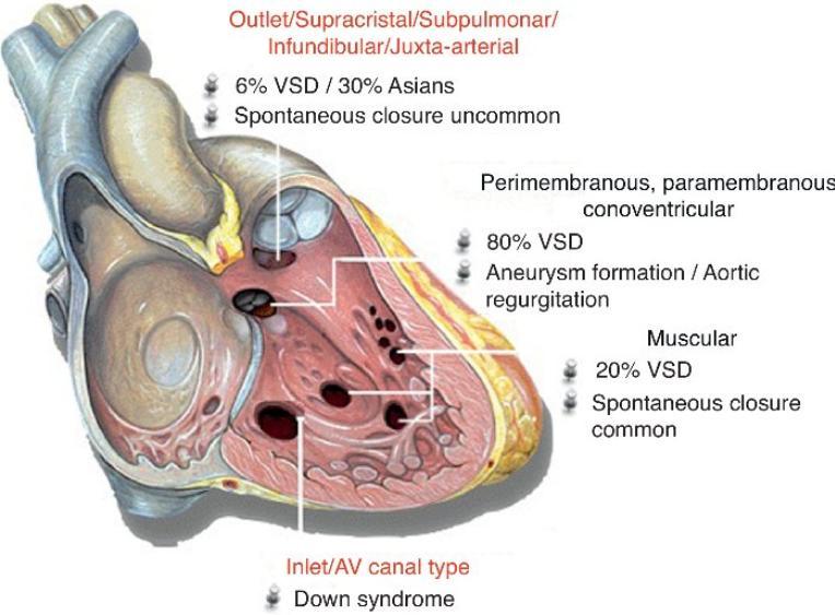
Notes by Sandra Dankwa 2009 HF- Heart Failure DS- Down Syndrome IE- Infective Endocarditis ET- Exercise Tolerance. Small VSD Symptoms -asymptomatic
 Congenital Heart Disease: Notes. Condition Pathology PC Ix Rx Ventricular septal defect (VSD) L R shuntsdefect anywhere in the ventricle, usually perimembranous (next to the tricuspid valve) 30% 1)small
Congenital Heart Defects
 Normal Heart Congenital Heart Defects 1. Patent Ductus Arteriosus The ductus arteriosus connects the main pulmonary artery to the aorta. In utero, it allows the blood leaving the right ventricle to bypass
Atrial Septal Defects
 Supplementary ACHD Echo Acquisition Protocol for Atrial Septal Defects The following protocol for echo in adult patients with atrial septal defects (ASDs) is a guide for performing a comprehensive assessment
ULTRASOUND OF THE FETAL HEART
 ULTRASOUND OF THE FETAL HEART Cameron A. Manbeian, MD Disclosure Statement Today s faculty: Cameron Manbeian, MD does not have any relevant financial relationships with commercial interests or affiliations
Congenital Heart Disease. Mohamed Waheed Elsharief.
 Congenital Heart Disease Mohamed Waheed Elsharief. Objectives l By the end of this lecture you should be able to Fetal Circulation l For the fetus the placenta is the oxygenator so the lungs do little
Congenital Heart Disease
 Congenital Heart Disease Mohammed Alghamdi, MD, FRCPC, FAAP, FACC Associate Professor and Consultant Pediatric Cardiology, Cardiac Science King Fahad Cardiac Centre King Saud University INTRODUCTION CHD
MRI (AND CT) FOR REPAIRED TETRALOGY OF FALLOT
 FOR REPAIRED TETRALOGY OF FALLOT") MRI (AND CT) FOR REPAIRED TETRALOGY OF FALLOT Linda B Haramati MD, MS Departments of Radiology and Medicine Bronx, New York OUTLINE Pathogenesis Variants Initial surgical treatments Basic MR protocols
MRI (AND CT) FOR REPAIRED TETRALOGY OF FALLOT Linda B Haramati MD, MS Departments of Radiology and Medicine Bronx, New York OUTLINE Pathogenesis Variants Initial surgical treatments Basic MR protocols
Anomalous Systemic Venous Connection Systemic venous anomaly
 World Database for Pediatric and Congenital Heart Surgery Appendix B: Diagnosis (International Paediatric and Congenital Cardiac Codes (IPCCC) and definitions) Anomalous Systemic Venous Connection Systemic
Echocardiography in adult congenital heart disease
 S12 Department of Cardiology, Royal Hospital for Sick Children, Glasgow G3 8SJ, UK A Houston S Lilley T Richens University Department of Medicine and Therapeutics, Western Infirmary, Glasgow G11 6NT, UK
5.8 Congenital Heart Disease
 5.8 Congenital Heart Disease Congenital heart diseases (CHD) refer to structural or functional heart diseases, which are present at birth. Some of these lesions may be discovered later. prevalence of Chd
3/14/2011 MANAGEMENT OF NEWBORNS CARDIAC INTENSIVE CARE CONFERENCE FOR HEALTH PROFESSIONALS IRVINE, CA. MARCH 7, 2011 WITH HEART DEFECTS
 CONFERENCE FOR HEALTH PROFESSIONALS IRVINE, CA. MARCH 7, 2011 MANAGEMENT OF NEWBORNS WITH HEART DEFECTS A NTHONY C. CHANG, MD, MBA, MPH M E D I C AL D I RE C T OR, HEART I N S T I T U T E C H I LDRE N
Uptofate Study Summary
 CONGENITAL HEART DISEASE Uptofate Study Summary Acyanotic Atrial septal defect Ventricular septal defect Patent foramen ovale Patent ductus arteriosus Aortic coartation Pulmonary stenosis Cyanotic Tetralogy
Eponymous cardiovascular abnormalities- Imaging review and historical perspectives
 Eponymous cardiovascular abnormalities- Imaging review and historical perspectives Poster No.: C-2567 Congress: ECR 2015 Type: Educational Exhibit Authors: Y. Ahmed, P. Rajiah; Cleveland, Ohio/US Keywords:
2) VSD & PDA - Dr. Aso
 VSD & PDA - Dr. Aso") 2) VSD & PDA - Dr. Aso Ventricular Septal Defect (VSD) Most common cardiac malformation 25-30 % Types of VSD: According to position perimembranous, inlet, muscular. According to size small, medium, large.
2) VSD & PDA - Dr. Aso Ventricular Septal Defect (VSD) Most common cardiac malformation 25-30 % Types of VSD: According to position perimembranous, inlet, muscular. According to size small, medium, large.
Congenital heart disease. By Dr Saima Ali Professor of pediatrics
 Congenital heart disease By Dr Saima Ali Professor of pediatrics What is the most striking clinical finding in this child? Learning objectives By the end of this lecture, final year student should be able
IMAGES. in PAEDIATRIC CARDIOLOGY. Abstract. Case
 IMAGES in PAEDIATRIC CARDIOLOGY Images PMCID: PMC3232604 Isolated subpulmonary membrane causing critical neonatal pulmonary stenosis with concordant atrioventricular and ventriculoarterial connections
Pediatric Echocardiography Examination Content Outline
 Pediatric Echocardiography Examination Content Outline (Outline Summary) # Domain Subdomain Percentage 1 Anatomy and Physiology Normal Anatomy and Physiology 10% 2 Abnormal Pathology and Pathophysiology
HISTORY. Question: What type of heart disease is suggested by this history? CHIEF COMPLAINT: Decreasing exercise tolerance.
 HISTORY 15-year-old male. CHIEF COMPLAINT: Decreasing exercise tolerance. PRESENT ILLNESS: A heart murmur was noted in childhood, but subsequent medical care was sporadic. Easy fatigability and slight
DEVELOPMENT OF THE CIRCULATORY SYSTEM L E C T U R E 5
 DEVELOPMENT OF THE CIRCULATORY SYSTEM L E C T U R E 5 REVIEW OF CARDIAC ANATOMY Heart 4 chambers Base and apex Valves Pericardial sac 3 layers: epi, myo, endo cardium Major blood vessels Aorta and its
HISTORY. Question: What category of heart disease is suggested by the fact that a murmur was heard at birth?
 HISTORY 23-year-old man. CHIEF COMPLAINT: Decreasing exercise tolerance of several years duration. PRESENT ILLNESS: The patient is the product of an uncomplicated term pregnancy. A heart murmur was discovered
What is the Definition of Small Systemic Ventricle. Hong Ryang Kil, MD Department of Pediatrics, College of Medicine, Chungnam National University
 What is the Definition of Small Systemic Ventricle Hong Ryang Kil, MD Department of Pediatrics, College of Medicine, Chungnam National University Contents Introduction Aortic valve stenosis Aortic coarctation
MITRAL STENOSIS. Joanne Cusack
 MITRAL STENOSIS Joanne Cusack BSE Breakdown Recognition of rheumatic mitral stenosis Qualitative description of valve and sub-valve calcification and fibrosis Measurement of orifice area by planimetry
Perimembranous VSD: When Do We Ask For A Surgical Closure? LI Xin. Department of Cardiothoracic Surgery Queen Mary Hospital Hong Kong
 Perimembranous VSD: When Do We Ask For A Surgical Closure? LI Xin Department of Cardiothoracic Surgery Queen Mary Hospital Hong Kong Classification (by Kirklin) I. Subarterial (10%) Outlet, conal, supracristal,
가천의대길병원소아심장과최덕영 PA C IVS THE EVALUATION AND PRINCIPLES OF TREATMENT STRATEGY
 가천의대길병원소아심장과최덕영 PA C IVS THE EVALUATION AND PRINCIPLES OF TREATMENT STRATEGY PA c IVS (not only pulmonary valve disease) Edwards JE. Pathologic Alteration of the right heart. In: Konstam MA, Isner M, eds.
Transposition of the Great Arteries Preoperative Diagnostic Considerations. John Simpson Evelina Children s Hospital London, UK
 Transposition of the Great Arteries Preoperative Diagnostic Considerations John Simpson Evelina Children s Hospital London, UK Areas to be covered Definitions Scope of occurrence of transposition of the
TGA Surgical techniques: tips & tricks (Arterial switch operation)
") TGA Surgical techniques: tips & tricks (Arterial switch operation) Seoul National University Children s Hospital Woong-Han Kim Surgical History 1951 Blalock and Hanlon, atrial septectomy 1954 Mustard et
TGA Surgical techniques: tips & tricks (Arterial switch operation) Seoul National University Children s Hospital Woong-Han Kim Surgical History 1951 Blalock and Hanlon, atrial septectomy 1954 Mustard et
Notes: 1)Membranous part contribute in the formation of small portion in the septal cusp.
Membranous part contribute in the formation of small portion in the septal cusp.") Embryology 9 : Slide 16 : There is a sulcus between primitive ventricular and bulbis cordis that will disappear gradually and lead to the formation of one chamber which is called bulboventricular chamber.
Embryology 9 : Slide 16 : There is a sulcus between primitive ventricular and bulbis cordis that will disappear gradually and lead to the formation of one chamber which is called bulboventricular chamber.
Adel Hasanin Ahmed 1 ASD
 Adel Hasanin Ahmed 1 ASD Atrial septal defect (ASD) is the commonest form of congenital heart disease seen in adults. The commonest form of defect is the secundum ASD, accounting for two thirds of cases,
UPDATE FETAL ECHO REVIEW
 UPDATE 1 FETAL ECHO REVIEW Study Alert for RDCS Candidates D A V I E S P U B L I S H I N G I N C. Fetal Echo Review Study Alert U P D A T E D A U G U S T 1, 2 0 1 2 Nikki Stahl, RT(R)(M)(CT), RDMS, RVT
HISTORY. Question: What category of heart disease is suggested by this history? CHIEF COMPLAINT: Heart murmur present since early infancy.
 HISTORY 18-year-old man. CHIEF COMPLAINT: Heart murmur present since early infancy. PRESENT ILLNESS: Although normal at birth, a heart murmur was heard at the six week check-up and has persisted since
Slide 1. Slide 2. Slide 3 CONGENITAL HEART DISEASE. Papworth Hospital NHS Trust INTRODUCTION. Jakub Kadlec/Catherine Sudarshan INTRODUCTION
 Slide 1 CONGENITAL HEART DISEASE Jakub Kadlec/Catherine Sudarshan NHS Trust Slide 2 INTRODUCTION Most common congenital illness in the newborn Affects about 4 9 / 1000 full-term live births in the UK 1.5
Segmental approach to normal and abnormal situs arrangement - Echocardiography -
 Segmental approach to normal and abnormal situs arrangement - Echocardiography - Jan Marek Great Ormond Street Hospital & Institute of Cardiovascular Sciences, University College London No disclosures
CASE REPORT: DOUBLE ORIFICE MITRAL VALVE WITH CLEFT IN ANTERIOR LEAFLET OF DOMINANT VALVE IN AN AFRO-CARIBBEAN
 CASE REPORT: DOUBLE ORIFICE MITL VAE WITH CLEFT IN ANTERIOR LEAFLET OF DOMINANT VAE IN AN AFRO-CARIBBEAN Disclosure: No potential conflict of interest. Received: 27.08.13 Accepted: 23.06.14 Citation: EMJ
Echocardiography in Congenital Heart Disease
 Chapter 44 Echocardiography in Congenital Heart Disease John L. Cotton and G. William Henry Multiple-plane cardiac imaging by echocardiography can noninvasively define the anatomy of the heart and the
RVOTO adult and post-op
 Right ventricular outflow tract obstruction in the adult: native and post-op Helmut Baumgartner Westfälische Wilhelms-Universität Münster Adult Congenital and Valvular Heart Disease Center University of
Foetal Cardiology: How to predict perinatal problems. Prof. I.Witters Prof.M.Gewillig UZ Leuven
 Foetal Cardiology: How to predict perinatal problems Prof. I.Witters Prof.M.Gewillig UZ Leuven Cardiopathies Incidence : 8-12 / 1000 births ( 1% ) Most frequent - Ventricle Septum Defect 20% - Atrium Septum
Adult Congenital Heart Disease: The New Reality. Disclosures
 Adult Congenital Heart Disease: The New Reality Kathryn Rouine-Rapp, MD Professor of Anesthesia Disclosures I have nothing to disclose 1 Outline Historic perspective Our reality Common lesions Guidelines
Pediatric Board Review Congenital Heart Disease. Steven H. Todman, M.D. Pediatric Cardiologist Louisiana State University
 Pediatric Board Review Congenital Heart Disease Steven H. Todman, M.D. Pediatric Cardiologist Louisiana State University Our Mission To discuss various types of congenital heart disease that are commonly
Chapter 2 Cardiac Interpretation of Pediatric Chest X-Ray
 Chapter 2 Cardiac Interpretation of Pediatric Chest X-Ray Ra-id Abdulla and Douglas M. Luxenberg Key Facts The cardiac silhouette occupies 50 55% of the chest width on an anterior posterior chest X-ray
DORV: The Great Chameleon. Heart Conference October 15, 2016 Tina Kwan, MD
 DORV: The Great Chameleon Heart Conference October 15, 2016 Tina Kwan, MD Kenneth Maehara, Ph.D. May 7, 1942 - August 26, 2013 A.R. A classic case of broken heart 38 week AGA F born at an OSH to
The role of intraoperative TOE in congenital cardiac surgery
 The role of intraoperative TOE in congenital cardiac surgery Justiaan Swanevelder Dept of Anaesthesia Groote Schuur and Red Cross War Memorial Children s Hospitals University of Cape Town, South Africa
How to Assess and Treat Obstructive Lesions
 How to Assess and Treat Obstructive Lesions Erwin Oechslin, MD, FESC, FRCPC, Director, Congenital Cardiac Centre for Adults Peter Munk Cardiac Centre University Health Network/Toronto General Hospital
PROSTHETIC VALVE BOARD REVIEW
 PROSTHETIC VALVE BOARD REVIEW The correct answer D This two chamber view shows a porcine mitral prosthesis with the typical appearance of the struts although the leaflets are not well seen. The valve
Three cross-sectional planes for fetal color Doppler echocardiography
 Ultrasound Obstet Gynecol 2003; 21: 81 93 Published online 11 December 2002 in Wiley InterScience (www.interscience.wiley.com). DOI: 10.1002/uog.5 Three cross-sectional planes for fetal color Doppler echocardiography
Imaging Assessment of the Pulmonary Valve in Stenosis/Atresia and Regurgitation
 Imaging Assessment of the Pulmonary Valve in Stenosis/Atresia and Regurgitation Craig E Fleishman, MD FACC FASE The Heart Center at Arnold Palmer Hospital for Children SCAI Fall Fellows Course 2014 Las
M-Mode Echocardiography Is it still Alive? Itzhak Kronzon, MD,FASE. Sampling Rate M-Mode: 1800 / sec 2D: 30 / sec
 M-Mode Echocardiography Is it still Alive? Itzhak Kronzon, MD,FASE Honoraria: Philips Classical M-mode Echocardiography M-Mode offers better time and image resolution. Sampling Rate M-Mode: 1800 / sec
PATENT DUCTUS ARTERIOSUS (PDA)
") PATENT DUCTUS ARTERIOSUS (PDA) It is a channel that connect the pulmonary artery with the descending aorta (isthumus part). It results from the persistence of patency of the fetal ductus arteriosus after
PATENT DUCTUS ARTERIOSUS (PDA) It is a channel that connect the pulmonary artery with the descending aorta (isthumus part). It results from the persistence of patency of the fetal ductus arteriosus after
Pulmonary Valve Morphology in Patients with Bicuspid Aortic Valves
 https://doi.org/10.1007/s00246-018-1807-x ORIGINAL ARTICLE Pulmonary Valve Morphology in Patients with Bicuspid Aortic Valves Wilke M. C. Koenraadt 1 Margot M. Bartelings 2 Adriana C. Gittenberger de Groot
Transposition of the great arteries
 EuroEcho 2010 - Teaching course on CHD Transposition of the great arteries - Follow-up after the arterial switch Gertjan Tj. Sieswerda, MD PhD Nothing to disclose Interuniversitary Institute for Congenital
Coronary Anomalies & Hemodynamic Identification
 Coronary Anomalies & Hemodynamic Identification David Stultz, MD Cardiology Fellow, PGY 6 May 2, 2006 Anomaly #1 Anomaly #2 Anomaly #3 Figure 18-27 Anomalous origin of the left circumflex artery.
Atrioventricular valve repair: The limits of operability
 Atrioventricular valve repair: The limits of operability Francis Fynn-Thompson, MD Co-Director, Center for Airway Disorders Surgical Director, Pediatric Mechanical Support Program Surgical Director, Heart
Cardiovascular Pathophysiology: Right to Left Shunts aka Cyanotic Lesions
 Cardiovascular Pathophysiology: Right to Left Shunts aka Cyanotic Lesions Ismee A. Williams, MD, MS iib6@columbia.edu Pediatric Cardiology Learning Objectives To discuss the hemodynamic significance of
Cardiovascular Pathophysiology: Right to Left Shunts aka Cyanotic Lesions Ismee A. Williams, MD, MS Pediatric Cardiology
 Cardiovascular Pathophysiology: Right to Left Shunts aka Cyanotic Lesions Ismee A. Williams, MD, MS iib6@columbia.edu Pediatric Cardiology Learning Objectives To discuss the hemodynamic significance of
Absent Pulmonary Valve Syndrome
 Absent Pulmonary Valve Syndrome Fact sheet on Absent Pulmonary Valve Syndrome In this condition, which has some similarities to Fallot's Tetralogy, there is a VSD with narrowing at the pulmonary valve.
Communication of Mitral Valve with Both Ventricles Associated with Double Outlet Right Ventricle
 Communication of Mitral Valve with Both Ventricles Associated with Double Outlet Right Ventricle By RAJENTDRA TANDON, M.D., JAMES H. MOLLR, MD, AND JESSE E. EDWARDS, M.D. SUMMARY A rare case of an infant
Down Syndrome Medical Interest Group Friday, 12 June Cardiac Surgery in patients with Down Syndrome
 Down Syndrome Medical Interest Group Friday, 12 June 2015 Cardiac Surgery in patients with Down Syndrome Mr. Attilio Lotto, FRCS CTh Congenital Cardiac Surgeon Cardiac surgery in patients with Down syndrome
pulmonary valve on, 107 pulmonary valve vegetations on, 113
 INDEX Adriamycin-induced cardiomyopathy, 176 Amyloidosis, 160-161 echocardiographic abnormalities in, 160 intra-mural tumors similar to, 294 myocardial involvement in, 160-161 two-dimensional echocardiography
Before we are Born: Fetal Diagnosis of Congenital Heart Disease
 Before we are Born: Fetal Diagnosis of Congenital Heart Disease Mohamed Sulaiman, MD Pediatric cardiologist Kidsheart: American Fetal & Children's Heart Center Dubai Healthcare City, Dubai-UAE First Pediatric
British Society of Echocardiography
 British Society of Echocardiography Affiliated to the British Cardiac Society A Minimum Dataset for a Standard Adult Transthoracic Echocardiogram From the British Society of Echocardiography Education
CONGENITAL HEART DISEASE (CHD)
") CONGENITAL HEART DISEASE (CHD) DEFINITION It is the result of a structural or functional abnormality of the cardiovascular system at birth GENERAL FEATURES OF CHD Structural defects due to specific disturbance
CONGENITAL HEART DISEASE (CHD) DEFINITION It is the result of a structural or functional abnormality of the cardiovascular system at birth GENERAL FEATURES OF CHD Structural defects due to specific disturbance
Assessment of fetal heart function and rhythm
 Assessment of fetal heart function and rhythm The fetal myocardium Early Gestation Myofibrils 30% of myocytes Less sarcoplasmic reticula Late Gestation Myofibrils 60% of myocytes Increased force per unit
A brief history of valvular surgery
 Cardiac surgery Valvular heart disease University of Pecs, Medical Faculty Heart Institute A brief history of valvular surgery 1925. Souttar closed mitral commissurotomy 1960. McGoon plasty for mitral
Aortic arch anomalies Coarctation of the Aorta Interrupted Aortic Arch Echocardiography
 Aortic arch anomalies Coarctation of the Aorta Interrupted Aortic Arch Echocardiography V.Tomek, J. Marek, J. Škovránek, J. Gilík No disclosures Kardiocentrum, University Hospital Motol, Prague, Czech
Fetal Echocardiography and the Routine Obstetric Sonogram
 JDMS 23:143 149 May/June 2007 143 Fetal Echocardiography and the Routine Obstetric Sonogram SHELLY ZIMBELMAN, RT(R)(CT), RDMS, RDCS ASAD SHEIKH, MD, RDCS Congenital heart disease (CHD) is the most common
Echocardiographic Evaluation of the Cardiomyopathies. Stephanie Coulter, MD, FACC, FASE April, 2016
 Echocardiographic Evaluation of the Cardiomyopathies Stephanie Coulter, MD, FACC, FASE April, 2016 Cardiomyopathies (CMP) primary disease intrinsic to cardiac muscle Dilated CMP Hypertrophic CMP Infiltrative
Cardiac ultrasound protocols
 Cardiac ultrasound protocols IDEXX Telemedicine Consultants Two-dimensional and M-mode imaging planes Right parasternal long axis four chamber Obtained from the right side Displays the relative proportions
Characteristics and Management of Cleft Mitral Valve
 Journal of the American College of Cardiology Vol. 42, No. 11, 2003 2003 by the American College of Cardiology Foundation ISSN 0735-1097/03/$30.00 Published by Elsevier Inc. doi:10.1016/j.jacc.2003.07.019
Case 47 Clinical Presentation
 93 Case 47 C Clinical Presentation 45-year-old man presents with chest pain and new onset of a murmur. Echocardiography shows severe aortic insufficiency. 94 RadCases Cardiac Imaging Imaging Findings C
Congenital heart disease: When to act and what to do?
 Leading Article Congenital heart disease: When to act and what to do? Duminda Samarasinghe 1 Sri Lanka Journal of Child Health, 2010; 39: 39-43 (Key words: Congenital heart disease) Congenital heart disease
When is Risky to Apply Oxygen for Congenital Heart Disease 부천세종병원 소아청소년과최은영
 When is Risky to Apply Oxygen for Congenital Heart Disease 부천세종병원 소아청소년과최은영 The Korean Society of Cardiology COI Disclosure Eun-Young Choi The author have no financial conflicts of interest to disclose
Cases in Adult Congenital Heart Disease
 Cases in Adult Congenital Heart Disease Sabrina Phillips, MD FACC FASE Associate Professor of Medicine The University of Oklahoma Health Sciences Center No Disclosures I Have Palpitations 18 Year old Man
Complete atrioventricular septal defect identified during routine 19 week fetal anomaly ultrasound
 CASE REPORT Complete atrioventricular septal defect identified during routine Mark Hare Royal Brisbane and Women s Hospital, Herston, QLD, Australia doi:10.1002/sono.12014 Introduction Complete atrioventricular
Complex Congenital Heart Disease in Adults
 Complex Congenital Heart Disease in Adults Linda B. Haramati, MD Disclosures Complex Congenital Heart Disease in Adults Linda B. Haramati MD, MS Jeffrey M. Levsky MD, PhD Meir Scheinfeld MD, PhD Department
Surgical options for tetralogy of Fallot
 Surgical options for tetralogy of Fallot Serban Stoica FRCS(CTh) MD ACHD study day, 19 September 2017 Anatomy Physiology Children Adults Complications Follow up Anatomy Etienne Fallot (1850-1911) VSD Overriding