6/19/2018. Acknowledgments. Acknowledgments
|
|
|
- Jody Singleton
- 5 years ago
- Views:
Transcription

1 Sudden cardiac arrest in athletes: Implications of modern epidemiological data on pre participation cardiovascular screening DAVE SIEBERT, MD, CAQSM ASSISTANT PROFESSOR DEPARTMENT OF FAMILY MEDICINE UNIVERSITY OF WASHINGTON UW HUSKY TEAM PHYSICIAN 2018 COXHEALTH SPORTS MEDICINE CONFERENCE SPRINGFIELD, MISSOURI, USA JUNE 23, 2018 Acknowledgments Jonathan A. Drezner, MD Danielle Peterson, MS4 Kimberly G. Harmon, MD Kristen L. Kucera, PhD, MSPH, ATC Leah Cox Thomas, MS, CRC Martha Lopez Anderson Nick of Time Foundation Parent Heart Watch Acknowledgments Shannon L. Woods, MD Ashley Larkin, RNC OB, BSN 1

: A visible event that has a profound impact on an athlete and his or her family, team, and community Generally accepted as the most common")
2 Background The sports physical There is widespread agreement that one of the principle purposes of the pre participation physical exam is to identify conditions that may put the student athlete at unreasonable risk of death or catastrophic injury, with the potential to modify and reduce risk through individualized management. Interassociation Consensus Statement on Cardiovascular Care of College Student Athletes; Hainline et. al. J Am Coll Cardiol Background Disease prevention Van Mechelen. Sports Medicine Background Sudden cardiac arrest Sudden cardiac arrest (SCA): A visible event that has a profound impact on an athlete and his or her family, team, and community Generally accepted as the most common cause of death in exercising athletes Precise epidemiological data remain unclear Historically, estimates of the incidence of SCA have varied widely Most studies retrospective Varying data collection methodologies 2
3 Numerator and denominator Incidence: Number of new cases of a disease / condition per unit time No national reporting system for cases of sudden cardiac arrest ***Continue discussion about controversy of what is frequent and what is rare with regard to SCA being rare as a reason to not screen with ECG*** Epidemiology controversy slide #1 ***Break down argument of rareness being an argument against ECG screening*** Quotes from Maron studies, AHA studies, Minnesota studies 1:200,000 figure that is proposed ***Discuss that if that is the argument, screening by any means would be unwarranted, including the 14 point questionnaire*** ***Transition into difficulty of determining true incidence*** Epidemiology controversy slide #2 ***Graphs of causes of death in NCAA athletes from prior incidence studies*** ***Discuss confirmed versus likely cases, importance of autopsy and including cases of sudden unexplained death*** 3
4 Variable past incidence estimates Study Dates Population SCA or SCD Incidence Van Camp et. al High school and SCD 1:300,000 college athletes Corrado et. al Italian athletes SCD 1:47,000 Maron et. al Athletes SCA + SCD 1:163,934 Harmon et. al High school SCA + SCD 1:63,988 athletes Toresdahl et. al High school SCA + SCD 1:87,719 athletes Roberts et. al High school SCD 1:917,000 athletes Harmon et. al NCAA athletes SCA + SCD 1:43,000 Study purposes 1) To conduct prospective surveillance for new cases of SCA or death (SCA/D) in competitive athletes across the United States 2) To calculate the annual incidence of SCA/D in the competitive athlete population 3) To identify high risk athlete subgroups 4) To determine the relative frequencies of underlying etiologies of SCA/D as well as survival trends 5) To identify the proportion of etiologies that represent electrocardiogram (ECG) detectable conditions Methods and data collection Collaborative effort of active surveillance for new cases of SCA/D, July 1, 2014 through June 30, 2017 The National Center for Catastrophic Sports Injury Research (NCCSIR) Parent Heart Watch (PHW) UW Medicine Center for Sports Cardiology Systematic traditional and social media searches Direct report of cases to each center NCAA resolution lists 4

 When possible, a final diagnosis was determined based on all available information Media reports Direct outreach Autopsy")
5 Methods and data collection Methods and data collection Definition of competitive athlete Age 11 or older Participates in an organized sport requiring regular training or competition Club/select Middle school High school Intercollegiate Professional Former/recent athlete within 1 year Methods and data collection Autopsy reports were obtained and reviewed by a multidisciplinary panel (Sports Medicine, cardiac pathology) When possible, a final diagnosis was determined based on all available information Media reports Direct outreach Autopsy information 5
6 Methods and data collection After review, cases were grouped into one of three categories: 1) Confirmed cardiac in etiology 2) Likely cardiac in etiology based on available details 3) Possibly cardiac in etiology requiring further investigation Over 100 cases excluded as non cardiac deaths/collapses Results Overall 182 total cases identified over 2 year surveillance period 83.0% male athletes Mortality rate of 58.8% Average age 16.7 years old 52.2% Caucasian 30.2% African American Results Level of competition 182 total cases Middle school High school College Professional Former/recent athlete Unknown 6
 after exercise, 3.3% Exercise, 72.")


7 Results Activity level at time of SCA 182 total cases Sleep, 6.0% Unknown, 2.2% Relative rest, 16.5% Soon (<1h) after exercise, 3.3% Exercise, 72.0% Results Primary sport 182 total cases Basketball Football Soccer Track/CC Baseball Swimming Volleyball Lacrosse Other Results Incidence of SCA/D Incidence: Number of new cases per unit time Calculations Numerator: Number of cases grouped into confirmed or likely cardiac in etiology groups Denominator: Annual participation statistics from the NCAA and National Federation of State High School Associations (NFHS) This calculation yields an annual risk in athlete years 7
8 Results Incidence of SCA/D High school athletes, and academic years 1:64,763 athlete years Over a 4 year career 1:44,333 male athlete years 1:11,083 NCAA athletes, and academic years 1:46,942 athlete years 1:32,826 male athlete years Over a 4 year career 1:22,287 African American athlete years 1:1053 1:20,844 football athlete years 1:7,476 male basketball athlete years 1:4,213 male, African American basketball athlete years Results Etiology of SCA/D Autopsy reports, ECG findings, or specific diagnosis available in 116 of 182 (63.7%) cases >17 different etiologies 56.0% of confirmed diagnoses were ECG detectable Cardiomyopathies Wolff Parkinson White Long QT syndrome Results Etiology of SCA/D 1% 1% 1% 10% 2% 3% 3% 3% 4% 4% 4% 5% 6% 6% 15% 7% 11% 13% Coronary artery anomalies (17, 14.8%) Hypertrophic cardiomyopathy (15, 13.0%) Idiopathic LVH/possible HCM (13, 11.3%) Autopsy negative sudden unexplained death (8, 7.0%) Wolff Parkinson White (7, 6.1%) Long QT syndrome (7, 6.1%) Arrhythmogenic cardiomyopathy (6, 5.2%) Dilated cardiomyopathy (5, 4.3%) Aortic dissection (5, 4.3%) Myocarditis (5, 4.3%) Valve disorder (4, 3.5%) Coronary atherosclerosis (4, 3.5%) Commotio cordis (3, 2.6%) Catecholaminergic polymorphic ventricular tachycardia (2, 1.7%) Restrictive cardiomyopathy (1, 0.9%) Left ventricular noncompaction cardiomyopathy (1, 0.9%) Pericarditis (1, 0.9%) Other (11, 9.6%) 8
9 Results ECG detectable conditions All athletes: 56.0% (65 of 116) African American athletes: 62.2% (23 of 37) Basketball athletes: 65.7% (23 of 35) African American basketball athletes: 68.4% (13 of 19) College basketball athletes: 100.0% (4 of 4) Results Mortality data Overall mortality rate: 58.8% Mortality rates Level of competition Middle school: 63.6% High school: 51.5% College: 61.9% (66.7% DIII, 83.3% DII, 50% DI) Results Mortality data Mortality rates Activity level SCA during relative rest, all athletes: 76.7% SCA during exercise (training/competition), all athletes: 51.9% Statistically lower mortality rate during exercise (p=0.0134) NCAA athlete mortality rates SCA during exercise (training/competition): 38.4% SCA during relative rest: 100% Statistically lower mortality rate during exercise (p=0.0185) 9
10 Study limitations 1) Autopsy reports are of variable availability and quality 2) Heavy reliance on traditional and social media reporting 3) Results may be influenced by increased media coverage of more popular sports or higher competition levels Media capture of SCD in the NCAA Level of play NCAA resolution Media reports Division I 87% 87% Division II 83% 61% Division III 89% 44% High school? Harmon KG et. al. Circulation What is the true scope of the problem? Despite barriers to data collection, some trends are clear Present study prospectively confirms high risk groups Risk group (NCAA athletes) Harmon et. al.; (annual risk, retrospective) Present study; (annual risk, prospective) All 1:53,703 1:46,942 Male, all sports 1:37,790 1:32,826 African American (AA), all sports 1:21,491 1:22,287 Football 1:35,951 1:20,844 Men s basketball 1:8,978 1:7,476 AA men s basketball 1:5,348 1:4,213 10
11 Study limitations 1) Autopsy reports are of variable availability and quality 2) Heavy reliance on traditional and social media reporting 3) Results may be influenced by increased media coverage of more popular sports or higher competition levels 87% of cases at NCAA Division I level captured by media 44% of cases at NCAA Division III level captured by media What is the capture rate in high school? Underestimation? Conclusions 1) The incidence of SCA/D in competitive athletes is higher than historical estimates. 2) SCA/D affects certain subgroups disproportionately. 3) The etiology of SCA/D encompasses numerous diseases and cardiac disorders. The majority of confirmed diagnoses in the present study were ECG detectable. 4) Data trends suggest athletes are more likely to survive when they suffer an arrest during exercise, training, or competition. Significance 1) A college athlete has a statistically significantly higher chance of surviving an arrest if it occurs during exercise or competition. 2) Athletes may be more likely to survive an arrest during exercise or competition because of the higher chance of it being a witnessed event and thus triggering the activation of an Emergency Action Plan (EAP) if one exists. 3) However, a 48% high school mortality rate (and, in extension, 38% in college) is still unacceptably high. Better preparedness can lower these rates. 11
12 What s next? Optimization of screening strategies What makes a good screening test? 1968, Wilson and Junger WHO Condition criteria Important health problem Acceptable treatments exist Latent or pre clinical state exists Facilities for treatment exist Chance of poor outcome without screening Test criteria High sensitivity (low false negative) High specificity (low false positive) Easy and reliable Acceptable workup risks/harms Experiences of false positives/misdiagnosed Why screen at all? 2015, AHA, Maron et. al. The central purpose of preparticipation screening of trained competitive athletes is to identify or raise suspicion of those cardiovascular abnormalities and diseases that are potentially responsible for sudden unexpected death on the athletic field. 2016, AMSSM, Drezner et. al. The overall role of the preparticipation physical evaluation is to evaluate the health of the athlete to optimize safe sports participation. Early detection of athletes at risk for sudden cardiac arrest and death is an important objective of the PPE for athletes. 12

13 Optimal screening test Central question: What is the best way to detect silent cardiovascular conditions that may place an asymptomatic athlete at risk of sudden cardiac arrest? Standard history and physical exam? Electrocardiogram The 14 point AHA screening tool Personal history Family history Physical exam Chest pain, discomfort, tightness, pressure related to exercise Unexplained syncope or near syncope Excessive exertional and unexplained dyspnea/fatigue or palpitations, associated with exercise Prior recognition of a heart murmur Elevated systemic blood pressure Prior restriction of participation from sports Prior testing of the heart, ordered by a physician Premature death attributable to heart disease prior to age 50 Disability in relative <50 related to heart disease Family history of congenital heart conditions Heart murmur Femoral pulses Physical stigmata of Marfan syndrome Brachial artery blood pressure Limitations of the 14 point tool Two recent independent studies have shown: Each of 8 college athletes eventually diagnosed with a cardiovascular disorder were done so solely via ECG in an otherwise negative screen, including history and physical exam Not a single athlete with a positive history or physical exam was diagnosed with a cardiovascular disorder 37.2% false positive rate on history questionnaire Fuller C et. al. CJSM Drezner JA et. al. JACC
14 A changing paradigm? Drezner et. al The premise of (cardiovascular) screening in athletes is that early detection of cardiac disorders associated with SCD can reduce morbidity and mortality. Without believing in the benefit of early detection, then screening by any strategy is called into question. If one believes in early detection, screening by history and physical examination alone is inadequate. American Medical Society for Sports Medicine (AMSSM) position statement, 2016: the current (Pre participation physical evaluation), while pragmatic and widely practiced, is limited in its ability to identify athletes with conditions at risk for SCA/D. What s next? We must harness our improving understanding of the epidemiology of SCA/D in athletes to inform screening strategies in hopes of better accomplishing the early detection of potentially lethal cardiovascular disorders. Differential screening protocols? ECG screening in high risk groups? Sport specific protocols? Training of physician infrastructure on ECG interpretation Revisiting the prevention model Van Mechelen. Sports Medicine
15 Revisiting screening test criteria 1) Comparing/contrasting H&P versus ECG 2) False positive / negative discussion Condition criteria Important health problem Acceptable treatments exist Latent or pre clinical state exists Facilities for treatment exist Chance of poor outcome without screening Test criteria High sensitivity (low false negative) High specificity (low false positive) Easy and reliable Acceptable workup risks/harms Experiences of false positives/misdiagnosed Does modifying the criteria come with a cost? Do we sacrifice sensitivity to increase specificity? 30 Performance of ECG Standards False Positive 15 Rate Brosnan no change in sensitivity % sensitivity for SCD associated conditions 11.6 all three criteria identified 98.1% 10.7 of 9.6 athletes 8.1 with established HCM all with 100% sensitivity for the pathological conditions detected Pickham 2014 Sheikh 2014 Riding 2014 Fuller ESC 2010 Stanford Seattle Revised 15
16 Performance of ECG criteria BJSM; 2017 ESC 2010 International Criteria 2017 Specificity 86.9% 95.9% p<0.001 Sensitivity 95.5% 93% p= International Criteria improved the specificity and reduced the number of unnecessary investigations by 69% (from 1:8 athletes to 1:24 athletes) 16

17 Applying the International Criteria for ECG Interpretation in Athletes to a preparticipation screening program DAVE SIEBERT, MD, CAQSM ASSISTANT PROFESSOR DEPARTMENT OF FAMILY MEDICINE UNIVERSITY OF WASHINGTON UW HUSKY TEAM PHYSICIAN 2018 COXHEALTH SPORTS MEDICINE CONFERENCE SPRINGFIELD, MISSOURI, USA JUNE 23, 2018 Background Athlete s heart Increased Vagal Tone Type of Sport Age Gender Size Race/Genetics Enlarged Chamber Size Wall thickness Cavity dimension Sinus bradycardia Sinus arrhythmia Early repolarization 1 AVB Mobitz Type I 2 AVB LVH voltage criteria Incomplete RBBB Ultimate question In the context of a highly trained athlete, which screening ECG changes can be considered normal manifestations of the athlete s heart, and which should be considered pathologic? 1


18 2017 Freely available at: International Criteria Asymptomatic athletes age years Endorsed by 17 international sports medicine and cardiology societies Clear guide to the evaluation of ECG abnormalities Sports medicine and cardiology looking through the same lens Does modifying the criteria come with a cost? Do we sacrifice sensitivity to increase specificity? 2

19 30 Performance of ECG Standards False Positive 15 Rate Brosnan no change in sensitivity % sensitivity for SCD associated conditions 11.6 all three criteria identified 98.1% 10.7 of 9.6 athletes 8.1 with established HCM all with 100% sensitivity for the pathological conditions detected Pickham 2014 Sheikh 2014 Riding 2014 Fuller ESC 2010 Stanford Seattle Revised Performance of ECG criteria BJSM; 2017 ESC 2010 International Criteria 2017 Specificity 86.9% 95.9% p<0.001 Sensitivity 95.5% 93% p= International Criteria improved the specificity and reduced the number of unnecessary investigations by 69% (from 1:8 athletes to 1:24 athletes) International Criteria for ECG Interpretation in Athletes Normal ECG Findings Increased QRS voltage for LVH or RVH Incomplete RBBB Early repolarization/st segment elevation ST elevation followed by T wave inversion V1 V4 in black athletes T wave inversion V1 V3 age 16 years old Sinus bradycardia or arrhythmia Ectopic atrial or junctional rhythm 1 AV block Mobitz Type I 2 AV block Borderline ECG Findings Left axis deviation Left atrial enlargement Right axis deviation Right atrial enlargement Complete RBBB Abnormal ECG Findings T wave inversion ST segment depression Pathologic Q waves Complete LBBB QRS 140 ms duration Epsilon wave Ventricular pre excitation Prolonged QT interval Brugada Type 1 pattern Profound sinus bradycardia < 30 bpm PR interval 400 ms Mobitz Type II 2 AV block 3 AV block 2 PVCs Atrial tachyarrhythmias Ventricular arrhythmias In isolation No further evaluation required in asymptomatic athletes with no family history of inherited cardiac disease or SCD 2 or more Further evaluation required to investigate for pathologic cardiovascular disorders associated with SCD in athletes 3
 Relevant clinical information A. Age, race, and sex of athlete B.")

20 Clinical questions when interpreting ECGs 1) Is the ECG classified as: A. Normal no further evaluation needed B. Abnormal further evaluation needed 2) If the ECG is abnormal : A. What is the specific ECG abnormality? B. What is the appropriate next step in evaluation? 3) Relevant clinical information A. Age, race, and sex of athlete B. Asymptomatic and no family history of inherited cardiac disease or SCD? International Criteria for ECG Interpretation in Athletes Normal ECG Findings Increased QRS voltage for LVH or RVH Incomplete RBBB Early repolarization/st segment elevation ST elevation followed by T wave inversion V1 V4 in black athletes T wave inversion V1 V3 age 16 years old Sinus bradycardia or arrhythmia Ectopic atrial or junctional rhythm 1 AV block Mobitz Type I 2 AV block Borderline ECG Findings Left axis deviation Left atrial enlargement Right axis deviation Right atrial enlargement Complete RBBB Abnormal ECG Findings T wave inversion ST segment depression Pathologic Q waves Complete LBBB QRS 140 ms duration Epsilon wave Ventricular pre excitation Prolonged QT interval Brugada Type 1 pattern Profound sinus bradycardia < 30 bpm PR interval 400 ms Mobitz Type II 2 AV block 3 AV block 2 PVCs Atrial tachyarrhythmias Ventricular arrhythmias In isolation No further evaluation required in asymptomatic athletes with no family history of inherited cardiac disease or SCD 2 or more Further evaluation required to investigate for pathologic cardiovascular disorders associated with SCD in athletes Isolated Increased QRS Voltage >35mm ECG from a 19 year old asymptomatic soccer player demonstrating voltage criteria for LVH (S V1 + R V5 > 35 mm). Note the absence of ST depression, T wave inversion, or pathologic Q waves. Increased QRS amplitude without other ECG abnormalities is a common finding in trained athletes and does not require additional testing. 4

 in II, III, avf, V4 V6 (arrows) and tall, peaked T waves (circles).")

21 Incomplete Right Bundle Branch Block ECG demonstrates incomplete RBBB with rsr pattern in V1 and QRS duration of <120 ms. Incomplete RBBB is a common and normal finding in athletes and does not require additional evaluation. Early Repolarization >35mm ECG from a 29 year old asymptomatic soccer player demonstrating early repolarization (J point and ST elevation) in II, III, avf, V4 V6 (arrows) and tall, peaked T waves (circles). These are common, training related findings in athletes and do not require more evaluation. Black Athlete Repolarization Variant ECG from a 24 year old asymptomatic black/african soccer player demonstrating J point and convex ( domed ) ST elevation followed by T wave inversion in leads V1 V4 (circles). This is a normal repolarization pattern in black/african athletes. 5
")

.")

22 Black Athlete Repolarization Variant: Confined to Leads V1 V4 ECG from a black/african athlete demonstrating voltage criterion for LVH, J point elevation and convex ( domed ) ST segment elevation followed by T wave inversion in V1 V4 (circles). This is a normal repolarization pattern in black athletes. Juvenile T Wave Inversion ECG from a 12 year old asymptomatic Caucasian female soccer player demonstrating the juvenile pattern of T wave inversion in leads V1 V3 (circles). This is a normal finding in athletes <16 years of age. Juvenile T Wave Inversion Age <16 yo; Independent of race; TWI in V1 V3; Does not extend to V4 13 yo Caucasian female 15 yo Asian female No further evaluation needed 6
. Note the constant RR interval between beats.")

 2 AV Block PR 140 ms PR 190 ms PR 200 ms P PR 140 ms PR 190 ms Mobitz Type I (Wenckebach) 2 AV block is")
23 Junctional Escape Rhythm P wave P wave P waves hidden by QRS Complex A 28 year old Caucasian male demonstrating a junctional escape rhythm (red arrows). Note the constant RR interval between beats. 1 Atrioventricular Block PR ECG shows 1 ⁰ AV block (PR interval >200 ms). The PR interval is measured from the beginning of the P wave to the beginning of the QRS complex. In this ECG tracing, the PR interval is constant from beat to beat and measures 300 ms. Mobitz Type I (Wenckebach) 2 AV Block PR 140 ms PR 190 ms PR 200 ms P PR 140 ms PR 190 ms Mobitz Type I (Wenckebach) 2 AV block is demonstrated by progressively longer PR intervals until there is a non conducted P wave and no QRS. The first PR interval after the dropped beat is shorter than the last conducted PR interval prior to the dropped beat 7


24 Mobitz Type I (Wenckebach) 2 AV Block International Criteria for ECG Interpretation in Athletes Normal ECG Findings Increased QRS voltage for LVH or RVH Incomplete RBBB Early repolarization/st segment elevation ST elevation followed by T wave inversion V1 V4 in black athletes T wave inversion V1 V3 age 16 years old Sinus bradycardia or arrhythmia Ectopic atrial or junctional rhythm 1 AV block Mobitz Type I 2 AV block Borderline ECG Findings Left axis deviation Left atrial enlargement Right axis deviation Right atrial enlargement Complete RBBB Abnormal ECG Findings T wave inversion ST segment depression Pathologic Q waves Complete LBBB QRS 140 ms duration Epsilon wave Ventricular pre excitation Prolonged QT interval Brugada Type 1 pattern Profound sinus bradycardia < 30 bpm PR interval 400 ms Mobitz Type II 2 AV block 3 AV block 2 PVCs Atrial tachyarrhythmias Ventricular arrhythmias In isolation No further evaluation required in asymptomatic athletes with no family history of inherited cardiac disease or SCD 2 or more Further evaluation required to investigate for pathologic cardiovascular disorders associated with SCD in athletes 2013 N=2533 athletes: no athlete with isolated left or right axis deviation or atrial enlargement showed evidence of cardiomyopathy N=171 patients with HCM: co existing ECG abnormalities present in 89% with axis deviation or atrial enlargement 8


25 Left Atrial Enlargement ECG demonstrates left atrial enlargement, defined as a prolonged P wave duration of >120 ms in leads I or II with negative portion of the P wave 1 mm in depth and 40 ms in duration in lead V1. Left Axis Deviation ECG demonstrates abnormal left axis deviation defined as frontal plane QRS axis of less than 30. The QRS is positive in lead I and negative in avf and lead II. The QRS axis shown here is about 70. Right Bundle Branch Block R S wave V6 19 yo Caucasian male athlete with complete RBBB. The QRS duration is 120 ms with rsr pattern in V1 and S wave wider than R wave in V6. When found in isolation without other borderline or abnormal findings, and without other clinical markers of concern, complete RBBB does not require more investigation. 9


26 International Criteria for ECG Interpretation in Athletes Normal ECG Findings Increased QRS voltage for LVH or RVH Incomplete RBBB Early repolarization/st segment elevation ST elevation followed by T wave inversion V1 V4 in black athletes T wave inversion V1 V3 age 16 years old Sinus bradycardia or arrhythmia Ectopic atrial or junctional rhythm 1 AV block Mobitz Type I 2 AV block Borderline ECG Findings Left axis deviation Left atrial enlargement Right axis deviation Right atrial enlargement Complete RBBB Abnormal ECG Findings T wave inversion ST segment depression Pathologic Q waves Complete LBBB QRS 140 ms duration Epsilon wave Ventricular pre excitation Prolonged QT interval Brugada Type 1 pattern Profound sinus bradycardia < 30 bpm PR interval 400 ms Mobitz Type II 2 AV block 3 AV block 2 PVCs Atrial tachyarrhythmias Ventricular arrhythmias In isolation No further evaluation required in asymptomatic athletes with no family history of inherited cardiac disease or SCD 2 or more Further evaluation required to investigate for pathologic cardiovascular disorders associated with SCD in athletes Definitions: Abnormal ECG Findings Understand the precise definition of ECG abnormalities Inferolateral T Wave Inversion and ST Depression Abnormal ECG in a patient with hypertrophic cardiomyopathy. Note T wave inversion and ST segment depression in the inferolateral leads (arrows). 10


27 Inferolateral T Wave Inversion and ST Depression Evaluation of inferolateral TWI Additional testing to rule out cardiomyopathy Echo Cardiac MRI Holter + stress testing for grey zone findings Abnormal ECG from a patient with hypertrophic cardiomyopathy. Note T wave inversions in I, avl, and V4 V6 (red arrows), as well as ST segment depression in V4 V5 (black arrows). Lateral T Wave Inversion Markedly abnormal ECG showing TWI 2 mm in V4 V6. Note that the ST segment preceding TWI in V4 6 is flat or downsloping. 11


28 Evaluation of Lateral or Inferolateral TWI Comprehensive evaluation to r/o cardiomyopathy Echocardiogram Cardiac MRI should be a routine diagnostic test for this ECG phenotype 24 hour ECG monitor + stress testing for grey zone findings Apical HCM Lateral T Wave Inversion in V5 Evaluation of lateral TWI in V5 (not V6) Echo required Cardiac MRI for TWI 2 mm, concurrent ST segment depression or other ECG abnormalities, or as indicated from Echo ECG in a 18 yo African American male. TWI extending to V5 is considered abnormal. Only one lead of TWI required in V5 or V6. The Markedly Abnormal ECG ECG from a patient with HCM demonstrating QRS voltage criterion for LVH in association with deep T wave inversion and ST segment depression predominantly in the lateral leads (I, avl, V4 V6), voltage criterion for left and right atrial enlargement, and left axis deviation. 12


 Serial Follow up 19 yo")


29 Long term Follow up of Athletes with Markedly Abnormal ECGs Pelliccia; NEJM 2008 Study Group; Normal Cardiac Imaging 81 9 year Follow up No Symptoms; No CV disease 70 6 Other CV Disease 6% 1 Sudden Death, 5 1 Cardiac Arrest Cardiomyopathies (HCM 3; ARVC 1; DCM 1) Serial Follow up 19 yo African American male, college basketball player A September 2008 Echo and CMR non diagnostic B September 2010 CMR apical hypertrophy 20 mm with +LGE Cardiac MRI Comparison Midventricular Short Axis Views mm mm Sept 2008 Sept 2010 Hypertrophy of interventricular septum over 2 years Yearly repeat of ECG and cardiac imaging indicated for athletes with pathological lateral or inferolateral TWI and initial normal imaging studies. 13


 and pathologic T wave inversion (B).")
. Panel B demonstrates pathologic TWI in V1 V6 with absent J point elevation and a downsloping ST segment (red circles).")
30 Normal or Abnormal? Evaluation of Inferolateral TWI Additional testing to rule out cardiomyopathy Echo Cardiac MRI ECG Holter in a 20 + stress yo black testing athlete for showing grey zone pathological findingsinferolateral TWI in V5 V6, II and If initial avf. TWI studies in V5 V6 are is non diagnostic always considered serial abnormal. (annual) TWI in follow up V3 V4 represents with ECG + Echo (at minimum); the black athlete cardiac repolarization MRI for changes variant in ECG or Echo A. Physiologic Black athlete repolarization variant B. Pathologic T wave inversion Physiologic (A) and pathologic T wave inversion (B). Panel A demonstrates physiologic repolarization in a black athlete with TWI in V1 V4 preceded by J point and convex domed ST segment elevation (green circles). Panel B demonstrates pathologic TWI in V1 V6 with absent J point elevation and a downsloping ST segment (red circles). Anterior T Wave Inversion 21 yo Caucasian male with ECG demonstrating anterior T wave inversion (V1 V4) preceded by a non elevated J point and ST segment. Delayed S wave upstroke in V2 and low voltage (<5 mm) QRS complexes in limb leads I and avl suggest possible ARVC. 14

.")

31 Anterior T Wave Inversion Evaluation of Anterior TWI The extent of investigation may vary based on clinical suspicion for ARVC and results from initial testing. Echo Cardiac MRI Exercise ECG test Minimum 24 hour ECG monitor Signal averaged ECG ECG from a patient with ARVC. Note pathological TWI in V1 V3 (arrows) preceded by a flat or downsloping ST segment and without J point elevation. PVCs also present (circles). Definition 2010 ARVC/D Task Force Criteria: Reproducible low amplitude signal between end of QRS complex to onset of the T wave in the right precordial leads (V1 tov3). Epsilon Wave 2017 Athlete International Criteria : Distinct low amplitude signal (small positive deflection or notch) between the end of the QRS complex and onset of the T wave in leads V1 V3. Platonov et al. Heart Rhythm, 2016 Epsilon Wave Epsilon waves are typically a manifestation of more advanced disease and unlikely to be an isolated ECG finding In patients with ARVC that express an epsilon wave: 89% also manifest TWI in the right precordial leads 100% have a delayed S wave upstroke (prolonged terminal activation duration) 55 ms from the nadir of the S wave to the end of the QRS complex Thus, a suspected epsilon wave should prompt evaluation for other ECG abnormalities suggestive of ARVC (TWI; delayed S wave upstroke; low limb lead voltage). Delayed S wave upstroke 15


32 Inferior T Wave Inversion Normal or Abnormal? 18 yo Caucasian male with TWI in leads III and avf. Lead III is excluded (need 2 contiguous leads). In the absence of symptoms or other clinical markers of concern, this is a normal ECG and no further evaluation is needed. Inferior T Wave Inversion ECG demonstrates TWI in the inferior leads II and avf. This is an abnormal ECG and requires further evaluation (echocardiogram). Pathologic Q Waves New Criteria: Q/R ratio 0.25 or 40 ms in duration 16
, poor R wave progression across the precordial leads with deep S waves in V1 V3, and a single premature ventricular complex (arrow).")
33 Pathologic Q Waves Q/R ratio 0.25 or 40 ms in duration ECG of a young patient with dilated cardiomyopathy. Note inferior Q waves (II and avf), poor R wave progression across the precordial leads with deep S waves in V1 V3, and a single premature ventricular complex (arrow). High degree AV block is also present. Evaluation of Pathologic Q Waves Echocardiogram Consider cardiac MRI (with perfusion study) based on echocardiogram findings and clinical suspicion Coronary artery disease risk factor assessment Consider exercise stress testing, dobutamine echo, or myocardial perfusion scan in athletes >30 yo or if multiple risk factors for CAD are present Repeat ECG for septal (V1 V2) QS pattern pseudo septal infarct pattern from high lead misplacement 17
 If Q waves persist Echo 3) CAD risk assessment Stress testing for")

.")
34 Anterior Q Waves V1 V2 / QS Pattern Evaluation of Anterior Q waves 1) Repeat ECG 2) If Q waves persist Echo 3) CAD risk assessment Stress testing for multiple risk factors or age >30 years ECG in a 22 yo African American male. Anterior Q waves can be from incorrect high lead placement of V1 V2 (ie pseudo septal infarct). Anterior Q Waves? Repeat ECG in same athlete shows RS in V2 (arrows). This suggests QS pattern was lead placement issue. No further evaluation needed. 18
, delta wave (slurred QRS upstroke), and")

, absence of")
35 Ventricular Pre excitation / Wolff Parkinson White Delta wave ECG demonstrating the classic findings of WPW with a short PR interval (<120 ms), delta wave (slurred QRS upstroke), and prolonged QRS (>120 ms). Ventricular Pre excitation / WPW Pattern 17 yo asymptomatic female with negative family history. ECG demonstrates a short PR interval and delta waves (black arrows). Other findings suggestive of WPW include a large Q wave in lead III (red circle), absence of a Q wave in V6 (blue arrowhead), and ST segment depression in V5 V6 and lead II (red arrows). 19

36 Evaluation of Ventricular Pre excitation Exercise ECG test Abrupt cessation of the delta wave (pre excitation) denotes a low risk pathway EP study should be considered if a low risk accessory pathway cannot be confirmed by non invasive testing Consider EP study for moderate to high intensity sports Echocardiogram Association of pre excitation with Ebstein s anomaly and cardiomyopathy Long QT Syndrome? Normal ECG QTc is normal Don t include the U wave in anterior precordial leads! Teach the tangent or Avoid the tail method for manual measurement of the QT interval No further evaluation needed X This figure illustrates the Teach the Tangent or Avoid the Tail method for manual measurement of the QT interval. A straight line is drawn on the downslope of the T wave to the point of intersection with the isoelectric line. The U wave is not included. 20

 Laboratory (electrolyte) testing Family")
37 Abnormal notched T Wave morphology suggests LQT 2 Average QT interval 500 ms QT EVALUATION OF A PROLONGED QTc This alone is NOT a diagnosis of LQTS QTc < 470 ms males QTc < 480 ms females AND No concerning symptoms or family history No further evaluation QTc 470 ms males QTc 480 ms females 1. Repeat resting ECG on separate day 2. Review for QT prolonging medications QTc 470 ms males QTc 480 ms females SYMPTOMS: exercise, emotion, or auditory triggered syncope or seizure FAMILY HX: unexplained syncope, seizures, SCA/D, drowning or MVA QTc 500 ms Possible congenital LQTS Referral to a heart rhythm specialist or sports cardiologist QT interval duration and morphology (notching) Laboratory (electrolyte) testing Family screening (ECGs of firstdegree relatives) Exercise ECG test (paradoxical prolongation of the QTc during the recovery phase) Genetic testing (confirmatory mutation) Abnormal ECG = Temporary Restriction? Temporary restriction from athletic activity should be considered for athletes with abnormal ECGs, especially when there is high clinical suspicion for pathologic cardiac disease, until secondary investigations are completed. Conditional clearance for sports participation pending further evaluation can be considered on a case by case basis. 21
38 Thank you! Questions or clarifications? Please feel free to me: 22
39 INNOVATIONS IN SPORTS MEDICINE TO INFINITY AND BEYOND ERIC GIFFORD MD DISCLOSURE THIS PRESENTATION HIGHLIGHTS TWO CURRENT, EVIDENCE-BASED PROCEDURES AVAILABLE AS TREATMENT MODALITIES AND THEY WILL BE REFERRED TO BY THEIR COMMERCIAL NAMES. BOTH OF THESE PROCEDURES ARE ONE-OF-A-KIND, WITHOUT DIRECT COMPETITORS. ALL EFFORT WILL BE TAKEN TO PRESENT MODALITIES WITHOUT BIAS. DR. ERIC GIFFORD HAS NO FINANCIAL RELATIONSHIPS WITH ANY COMMERCIAL INTERESTS. CHRONIC TENDINOPATHIES MAKE UP A LARGE PERCENTAGE OF CHRONIC PAIN AND DISABILITY SIMILAR TO DEGENERATION OF CARTILAGE OF A JOINT INSTEAD IT IS DEGENERATION OF MUSCLE TENDONS AROUND JOINTS DIFFICULT TO TREAT PHYSICAL THERAPY-SOFT TISSUE WORK, ECCENTRIC EXERCISES, STRETCHING, SCRAPING, ETC REST, BRACING, SPLINTING NSAIDS, INJECTIONS-STEROID, PROLOTHERAPY PRP SURGERY 1


40 CHRONIC TENDINOPATHIES WHAT ELSE CAN BE DONE? TENEX PERCUTANEOUS TENOTOMY ULTRASOUND GUIDED TENDON TREATMENT Problem 1. Conservative measures are often ineffective 2. Surgery is fairly invasive and often still results in prolonged recovery and down time as well as continued pain Solution Replicate surgical approach but through a minimally invasive manner using an ultrasound guided Ultra-sonic needle tip. DISEASE PROCESS Intact Tendon Reactive Inflammatory Tendinopathy Degenerative Chronic Tendinopathy INSULT Neurovascular Mediation Reparative Process? Healed 6 2


41 BURDEN OF DISEASE CHRONIC TENDONOSIS 20 Million Patients per Year in US ELBOW 7MM PATIENTS / YEAR SHOULDER 1.8MM PATIENTS / YEAR PLANTAR FASCIA 3.1MM PATIENTS / YEAR KNEE 2.5MM PATIENTS / YEAR ACHILLES 5.6MM PATIENTS / YEAR * Based on 2013 ICD - 9 diagnosis code UNMET CLINICAL NEED Conservative Minimally Invasive Fully Invasive Rest / Ice OTC Medication Physical Therapy Use of cortisone injection declining due to published reports of damaging effects on tendon TENEX System to cut and remove pain generating tissue Local Anesthesia only Single Treatment Relatively quick recovery and return to activity Open/Arthroscopic Surgery Goal is to cut & remove damaged tissue pain generator General anesthesia Prolonged recovery with rehab protocol/pt When patients fail conservative treatment and experience chronic pain 3 months 6 months > 12 months The TENEX system provides the goals of surgery but with the invasiveness of an injection 8 MICRO-JACKHAMMER TENEX delivers optimized ultrasonic energy to tip of the instrument that precisely cuts diseased tendon tissue while sparing healthy tissue harmonic resonance of diseased tissue (necrotic) is different from healthy tissue (elastic) Cutting of targeted tissue is achieved through longitudinal movement of needle at the speed of sound (ultrasonic) tissue is cut via jack-hammer effect Continuous saline irrigation cools the Microtip to control unwanted heat and also simultaneously removes target tissue 9 3

 pen-like function Pre-assembled")


42 TENEX TX SYSTEM TX1 Console User interface with circuitry for precise & targeted tissue cutting and removal Targeted diseased tissue is removed while sparing healthy tissue with built in safety features TX1 Microtip Percutaneous (18 gauge) pen-like function Pre-assembled w/foot pedal activation Single use entirely disposable PERCUTANEOUS TENOTOMY / FASCIOTOMY 1. VISUALIZE DAMAGED TENDON (DARK REGION) VIA ULTRASOUND DISEASED TENDON TISSUE Ultrasound Imaging used to identify diseased tissue and guide Tenex TX Microtip during procedure TX1 MICROTIP PLACED INTO HYPOECHOIC REGION 2. GUIDE TENEX MICROTIP TO DAMAGED TISSUE WITH ULTRASOUND GUIDANCE CUTS & REMOVES TARGET DAMAGED TISSUE REMOVAL OF HYPOECHOIC REGION 11 TARGETED TISSUE CUTTING AND REMOVAL Harmonic resonance ultrasonic energy only cuts pathology based on density of degenerated tissue Ultrasonic energy optimized for dense degenerated tissue does not affect healthy elastic tissue Healthy Tendon + Ultrasonic Energy 4
 AT THE AFFECTED JOINT AND NOT RESPONSIVE TO CONSERVATIVE MEDICAL TREATMENT (REST, ICE, BRACE, PHYSICAL THERAPY, ETC.")
43 CHRONIC TENDONOSIS TREATMENTS Elbow tennis + golfers elbow 33% 2% Shoulder Over-use workers/athletes Skin Tendon 8% Knee basketball, volleyball Plantar Fascia 25% foot visits Bone 38% 19% Achilles 10% of runners WHO S A CANDIDATE? CHRONIC PAIN (> 3 MONTHS) AT THE AFFECTED JOINT AND NOT RESPONSIVE TO CONSERVATIVE MEDICAL TREATMENT (REST, ICE, BRACE, PHYSICAL THERAPY, ETC.) POINT TENDERNESS POINT OF MAXIMUM PAIN TYPICALLY CORRESPONDS TO THE LOCATION OF THE DAMAGED TISSUE ULTRASOUND CONFIRMATION PLACEMENT OF ULTRASOUND TRANSDUCER ON THE SITE OF MAXIMUM TENDERNESS SHOULD IDENTIFY A REGION OF DEGENERATED TENDON TISSUE VISUALIZED AS A HYPOECHOIC REGION DUE TO IRREGULAR/DISORGANIZED FIBERS AND THICKENED TENDON TISSUE. 5


44 HOWS IT DONE? NO RESTRICTIONS BEFORE PROCEDURE PREP AREA WITH SIMPLE ANTISEPTIC LOCAL ANESTHETIC APPLIED UNDER ULTRASOUND GUIDANCE 4-5MM INCISION MADE DOWN TO THE TARGET TISSUE TENEX NEEDLE IS ADVANCED TO THE TARGET TISSUE AND ULTRASOUND GUIDED TENOTOMY IS PERFORMED TOTAL CUTTING TIME USUALLY LESS THAN 5 MINUTES POST OP PROTOCOL ALLOWED TO GO HOME IMMEDIATELY FOLLOWING PROCEDURE LIGHT ACTIVITY WITH MINIMAL WALKING, LIFTING, PUSHING, PULLING FOR 2 WEEKS FOOT AND ANKLE USUALLY IN A BOOT FOR 2 WEEKS GRADUAL PROGRESSION BACK TO NORMAL ACTIVITY OVER NEXT 4-6 WEEKS PHYSICAL THERAPY TO AUGMENT HEALING PROCESS IN CERTAIN CASES EPICONDYL / ELBOW TENDONOSIS Epicondyl Healthy Tendon Blue strip marks placement of ultrasound transducer Epicondyl Hypoechoic/Damaged Tendon 6
 marks placement of ultrasound transducer on Achilles and (b) shows cross section view to identify mid-substance tendonosis Calcaneus Hypoechoic/Damaged Tendon PLANTAR")
45 PATELLAR TENDONOSIS Patella Healthy Tendon Orange strip marks placement of ultrasound transducer on inferior pole of patella Patella Hypoechoic/Damaged Tendon 54 ACHILLES TENDONOSIS Calcaneus Healthy Tendon Orange strip (a) marks placement of ultrasound transducer on Achilles and (b) shows cross section view to identify mid-substance tendonosis Calcaneus Hypoechoic/Damaged Tendon PLANTAR FASCIITIS/FASCIOSIS Orange strip marks the placement of ultrasound transducer on plantar fascia typically on medial aspect. 7
 Elattrache Operative Techniques In Sports Medicine (Knee) Barnes Operative Techniques In Sports Medicine (Procedure Overview) Khanna Am Academy Of Physical Med & Rehab Poster (Mixed")
46 CLINICAL PUBLICATION SUMMARY In Print/Accepted Koh American Journal Of Sports Medicine (Elbow) Hackel Orthopaedics Today (Procedure Overview / Mixed Tendons) Morrey Techniques In Elbow And Hand Surgery (Elbow) Elattrache Operative Techniques In Sports Medicine (Knee) Barnes Operative Techniques In Sports Medicine (Procedure Overview) Khanna Am Academy Of Physical Med & Rehab Poster (Mixed Tendons) Traistor Am Medical Society Of Sports Medicine (Mixed Tendons) Barnes Journal Of Shoulder And Elbow Surgery (Elbow) Patel Journal Of Orthopedics (Plantar Fascia) Sanders American Journal Of Sports Medicine (Epidemiology Study) Stuhlman Journal of Sports Medicine (Patellar Tendon) Kamineni Journal of Orthopedic Research (Basic Science) Patel Austin Journal of Orthopedics and Rheumatology (Plantar Fibroma) Koh Three Year Clinical Follow-up/American Journal of Sports Medicine (Elbow) Ellis American Journal of American Podiatric Medical Association (Achilles) Submitted/In Preparation Razdan Plantar Fascia Study Moore Tenex Vs Open Surgery (Elbow) Noyes Elbow Study Hackel Tenex vs Endoscopic Surgery (Plantar Fascia) Study revealed 90% 0f patients pain free within weeks of treatment Cost Effective Intervention Less expensive (>$11,000) and quicker recovery time vs surgery Sustained Pain Relief With Long Term Follow-up (6 36 Months) Strong Safety Profile KOH ET AL (SINGAPORE/MAYO) - ELBOW Prospective study of 20 pts with chronic epicondylitis who failed non-surgical treatment (medical, PT, cortisone) Single treatment with TX1 Post-procedure care - no PT, OTC pain control, activity modification for 2 weeks Patient follow-up: 2 weeks, 1, 3, 6,12, 24* and 36* months - Adverse events -Pain score (VAS) - Quality of life (DASH/Disability of the Arm, Shoulder and Hand) - Diagnostic ultrasound at baseline, 3, 6 and 36 months American Journal of Sports Medicine Vol 41, , 2013 * In print, Am Journal of Sports Medicine, 2015 SENG ET AL (3 YR) ELBOW TENDONOSIS & US EXAM 3 mo 6 mo 36 mo Resolution of Tendon Thickness & 13/20 18/20 20/20 Hypoechogenicity Seng, et al; Am Journal of Sports Med,
47 BARNES, BECKLEY, AND SMITH (MAYO) -ELBOW PROSPECTIVE STUDY INVOLVING 19 PATIENTS SYMPTOMATIC FOR AT LEAST 6 MO 7 MEDIAL AND 12 LATERAL TENDONOSIS PATIENTS WHO FAILED CONSERVATIVE TREATMENT (REST, PT, ICE, SINGLE CORTISONE) SINGLE TREATMENT WITH TX1 & NO ADDITIONAL INTERVENTION TOTAL PROCEDURE TIME < 15 MINUTES WITH MEAN ENERGY TIME = 38 SECONDS NO COMPLICATIONS CLINICAL OUTCOMES AT BASELINE, 6 WEEKS, 3 MO, 6 MO AND 12 MO VAS (PAIN) MAYO ELBOW PERFORMANCE SCORE / MEPS (RANGE OF MOTION) DISABILITY OF THE ARM, SHOULDER AND HAND/DASH (QUALITY OF LIFE) Barnes et al, Journal of Shoulder & Elbow Surgery, 2014 MOORE ET AL (IA) TENEX VS SURGERY (ELBOW) PROSPECTIVE STUDY RANDOMIZING TENEX VS OPEN SURGERY FOR ELBOW 45 PATIENTS WITH CHRONIC EPICODYLOSIS NOT RESPONSIVE TO CONSERVATIVE CARE 23 TREATED WITH TX1 22 TREATED WITH OPEN SURGICAL REPAIR OUTCOMES MEASURED AT 1 WEEK, 1 MONTH AND 6 MONTHS POST-TREATMENT TENEX VS. SURGERY EFFICACY/PAIN RELIEF 91% 77% (P<0.01) POST-TREATMENT VISITS (P<0.001) AVG WORK WEEKS MISSED (P<0.001) TOTAL COST/SAVINGS PER PT TENEX WAS $11,753 LESS THAN SURGERY Manuscript submitted for publication ELATTRACHE (KERLAN JOBE) - KNEE PROSPECTIVE STUDY INVOLVING 16 PATIENTS SYMPTOMATIC FOR ATLEAST 6 MO 10 COLLEGIATE-LEVEL ATHLETES FAILED CONSERVATIVE TREATMENT SINGLE TREATMENT WITH TX1 & NO ADDITIONAL INTERVENTION NO COMPLICATIONS CLINICAL OUTCOMES 15/16 (93%) REVEALED RESOLUTION OF SYMPTOMS AT 3 MONTHS SUSTAINED EFFECT AT 12 MONTHS ALL 10 ATHLETES RETURNED TO THEIR PRIOR LEVEL OF COMPETITION Operative Techniques in Orthopedics Vol 23, 2:
48 STUHLMAN, STOWERS & STOWERS (FL STATE) - KNEE PROSPECTIVE STUDY INVOLVING 8 PATIENTS SYMPTOMATIC FOR ATLEAST 6 MO ACTIVITY LIMITING TENDINOPATHY 3 PTS WITH BI-LATERAL PAIN FAILED CONSERVATIVE TREATMENT SINGLE TREATMENT WITH TX1 & NO ADDITIONAL INTERVENTION 12 MONTH FOLLOW-UP NO COMPLICATIONS CLINICAL OUTCOMES 8/8 PATIENTS REPORTED NO PAIN (VAS = 1) IMPROVEMENT IN SYMPTOMS BETWEEN 3 DAYS AND 6 MONTHS AFTER TREATMENT SUSTAINED AT 12 MONTHS Manuscript accepted, Journal of Sports Medicine RAZDAN & VANDERWOUDE (NE) PLANTAR FASCIA PROSPECTIVE STUDY INVOLVING 100 PATIENTS SYMPTOMATIC FOR ATLEAST 6 MO ALL FAILED CONSERVATIVE TREATMENT: PT, ORTHOTICS, OTC MEDICATION SINGLE TREATMENT WITH TX1 & NO ADDITIONAL INTERVENTION 12 MONTH FOLLOW-UP NO COMPLICATIONS CLINICAL OUTCOMES PAIN (VAS) AND DISABILITY INDEX (FADI) MEASURED AT 2 WEEKS, 6 WEEKS, 6 MO AND 12 MO SIGNIFICANT IMPROVEMENT IN PAIN AND DISABILITY INDEX BY 2 WEEKS AND SUSTAINED AT 12 MO 91/100 (91%) PATIENTS PAIN FREE AT 6 MONTHS AND SUSTAINED AT 12 MONTHS Podium Presentation, Society for Interventional Radiology Annual Meeting 2015 Manuscript submitted for publication PATEL (ORTHO INDY) PLANTAR FASCIA PROSPECTIVE STUDY INVOLVING 12 PATIENTS SYMPTOMATIC FOR ATLEAST 6 MO ALL FAILED CONSERVATIVE TREATMENT: PT, ORTHOTICS, EXTRA-CORPOREAL SHOCK WAVE, CORTISONE 4 PTS FAILED OPEN OR ENDOSCOPIC FASCIOTOMY SINGLE TREATMENT WITH TX1 & NO ADDITIONAL INTERVENTION 12 MONTH FOLLOW-UP NO COMPLICATIONS CLINICAL OUTCOMES 11/12 (92%) PATIENTS PAIN FREE AT 3 MONTHS AND SUSTAINED AT 12 MONTHS SIGNIFICANT IMPROVEMENT IN QOL BY 6 MONTHS AND SUSTAINED AT 12 MONTHS MEAN BASELINE AOFAS 30.1 MEAN 12 MO AOFAS 88.1 American Journal of Orthopedics,
49 ELLIS ET AL (AZ) ACHILLES TENDON PROSPECTIVE STUDY OF 26 PATIENTS SYMPTOMATIC FOR AVERAGE OF 18 MONTHS SINGLE TREATMENT WITH TX1 & NO ADDITIONAL INTERVENTION MEAN CUTTING TIME OF 4 MIN 24 SECONDS PTS FOLLOWED UP 1 WEEK, 1 MONTH, 12 MONTHS AND 16 MONTHS POST NO COMPLICATIONS CLINICAL OUTCOMES 23/26 (88%) PATIENTS REVEALED PAIN RELIEF AT 1 MONTH AND SUSTAINED AT 16 MONTHS 24/26 (92%) PATIENTS WOULD HAVE THE PROCEDURE DONE AGAIN Manuscript accepted, Journal of APMA PATEL ET AL (IN) PLANTAR FIBROMA PROSPECTIVE STUDY OF 8 PATIENTS SUFFERING FROM PLANTAR FIBROMA PATIENTS FAILED CONSERVATIVE CARE INCLUSIVE OF ORTHOTICS AVERAGE TIME OF SYMPTOMS 15 MONTHS PERCUTANEOUS CUTTING AND REMOVAL OF LESION WITH SINGLE TREATMENT OF TX1 AVERAGE FOLLOW-UP = 2.5 YEARS CLINICAL OUTCOMES 8/8 PATIENTS PAIN FREE WITH AVERAGE TIME TO RESOLUTION OF SYMPTOMS - 63 DAYS SIGNIFICANT IMPROVEMENT OF AOFAS SCORE FROM PRE-OP OF 30.8 TO 90.1 AT 12 MO NO RECURRENCE OF FIBROMAS AT 2.5 YEARS Austin Journal of Orthopedics and Rheumatology, Vol 2, Issue 2, 2015 COOLIEF COOLED RADIOFREQUENCY TREATMENT 11

50 CHRONIC KNEE AND HIP PAIN DILEMMA RELIEVE PAIN FOR PATIENTS SUFFERING WITH CHRONIC JOINT PAIN UTILIZE INNOVATIVE NEW PROCEDURE FOR TWO CHALLENGING PATIENT POPULATIONS WITH HIP AND KNEE PAIN NON-SURGICAL CANDIDATES OVERWEIGHT AGE TOO YOUNG OR TOO OLD CO-MORBITITIES PATIENTS STILL IN PAIN AFTER JOINT REPLACEMENT PATIENTS NOT RESPONDING TO OTHER CONSERVATIVE MEASURES VARIOUS INJECTIONS BRACING ACTIVITY MODIFICATION MEDICATIONS COOLED RADIOFREQUENCY NEUROTOMY INTERVENTIONAL PAIN SOLUTION COOLED RADIOFREQUENCY PROCEDURE PROVIDES UP TO 2 YEARS IN PAIN RELIEF PROVIDES LARGER LESIONS THAN STANDARD RF ALLOWING FOR TREATMENT OF MORE VARIABLE PERIPHERAL SENSORY NERVES 12
51 RADIOFREQUENCY NEUROTOMY ELECTRODE WITH AN EXPOSED TIP, IS PLACED ONTO A PERIPHERAL NERVE HIGH-FREQUENCY, ELECTRICAL CURRENT CONCENTRATES AROUND THE TIP; HEATS THE IMMEDIATELY SURROUNDING TISSUES; AND COAGULATES THEM, INCLUDING THE TARGET NERVE PAIN RELIEVED BY COAGULATING THE AFFERENT NERVE PREVENTING THE CONDUCTION OF NOCICEPTIVE (PAIN) IMPULSES PAIN RELIEF OCCURS BY ANESTHETIZING THE SOURCE OF PAIN COOLED RF SINGLE LARGER LESION COMPENSATES FOR VARIABLE NERVE COURSE. MORE EFFICIENT AND MORE PREDICTABLE THAN MULTIPLE PASSES WITH SMALLER GAUGE RF PROBE. DISTAL PROJECTION OF LESION PROJECTS HEAT IN AND AROUND DIFFICULT ANATOMY, SCAR TISSUE, SURGICAL HARDWARE ETC. ANATOMIC TECHNIQUES PERFORMED UNDER FLUOROSCOPY RELIES UPON EASILY VISUALIZED LANDMARKS. MAY ALSO USE ULTRASOUND IMAGING FOR NEUROVASCULAR BUNDLE IDENTIFICATION AND NEEDLE TIP PLACEMENT ELECTRICAL STIMULATION CAN BE USED TO VERIFY ABSENCE OF PROXIMITY TO MOTOR NERVE. IMPROVEMENT IN LONG EXISTING TECHNOLOGY, NOW WITH INTERNALLY WATER COOLED ELECTRODES Liver Ablation Cardiac Electrophysiology Chronic spine and peripheral joint pain 13


52 COOLED RF LESION LARGE VOLUME SPHERICAL LESION WITH DISTAL PROJECTION Cooled RF 18Ga, 4mm 40% 10 mm APPLICATIONS OF COOLED RF SACROILIAC PAIN OVERLAPPING AND FORWARD PROJECTING LESIONS ARE MADE TO CAPTURE VARIABLE PATH OF LATERAL BRANCHES ON UNEVEN SACRAL SURFACE THORACIC PAIN LARGE SPHERICAL LESION IS MADE TO ACCOMMODATE VARIABLE NERVE PATH OF MEDIAL BRANCHES ESPECIALLY T5-T7 LUMBAR PAIN FORWARD PROJECTING AND LARGE SPHERICAL LESION ALLOW A PERPENDICULAR APPROACH TO MEDIAL BRANCHES IN ONE NEEDLE PASS AND NOT MULTIPLE PASSES. ADDRESSES DIFFICULT ANATOMY OR PATIENTS WITH HARDWARE FROM PREVIOUS SURGERIES CERVICAL PAIN OFFERING RELIEF IN THE CERVICAL REGION BY DELIVERING LARGE VOLUME LESIONS WHERE ANATOMY AND NERVE PATH ARE VARIABLE. PERIPHERAL NERVE PROCEDURES 14

53 Patients that are not candidates for surgery 29% of patients over the age of 65 with chronic knee pain non responsive to conservative medical management are not candidates for surgery due to contraindications Patients with persistent post-surgical pain 53% of patients with TKA and 38% of patients with THA with chronic pain one year out of surgery COOLIEF COOLED RADIOFREQUENCY NEUROTOMY: A TREATMENT OPTION FOR PATIENTS WITH CHRONIC KNEE AND HIP PAIN PATIENT SELECTION Patients that are not indicated for surgery: THA/TKA contraindications: Age BMI Comorbidities Patients that don t want surgery Patients that still have significant pain following surgery Patients still in pain after surgery due to non-compliance Patients with past TKA/THA IMAGING GUIDANCE Provides up to 2 years pain relief, improved physical function, and reduced drug utilization Larger burn areas takes into account variable nerve course 15
54 CLINICAL DATA FOR COOLED RADIOFREQUENCY 1. Patel et al. Twelve Month Follow up of a Randomized Trial Assessing Cooled Radiofrequency Denervation as a Treatment for Sacroiliac Region Pain. Pain Practice. Jan These 12 month results illustrate the durability of effective CRF/LBN mediated treatment of SI region pain for selected patients 2. Liu et. Al A Cross Sectional Survey on Prevalence and Risk Factors for Persistent Post Surgical Pain 1 Year after Total Hip and Knee Replacement. Regional Anesthesia and Pain Management. Volume 37, Number 4, July August Persistent post surgical pain is common after THR and TKR and is associated with reduced health related quality of life. 3. Franco et. al. Innervation of the Anterior Capsule of the Human Knee: Implications for Radiofrequency Ablation. Reg Anesth Pain Med Jul Aug;40(4): QUESTIONS? 16
55 Syndesmosis Injury Kyle E Arthur, MD Introduction Family Practice Sports Medicine UAMS Fellowship Directed by Dr. Ramon Ylanan Purpose Identify the anatomical structures that comprise the ankle syndesmosis. Recognize the most common mechanism related to syndesmosis injury. List specialized tests used in physical exam to screen for and confirm syndesmosis injury. 1
56 Outline Sprain vs Sprain Anatomy MOI Evaluation Treatment Where is the upper ankle? Joint proximal to the ankle is the knee, right? Low vs High Two distinct injuries Recovery time Long term consequence of arthritis and instability Epidemiology General Athletic Population 10% 20% of all ankle sprains are HAS Specific sports are more prone to HAS Contact (FOOTBALL, rugby, wrestling, lacrosse) Immobilized (Skiing, hockey) 2
57 Recovery Time Lateral Ankle Sprain Improvement in function after 2 weeks. Syndesmotic Sprain Varying reports of recovery time 2.5 weeks in NFL 3 weeks in NCAA FB 8 weeks in West Point Cadet Sequela Without fracture or diastasis: Ankle Pain Ankle Stiffness Recurrent Ankle Sprain Heterotopic Ossification Persistent Disability Prolonged Recovery Bones Ligaments Motion Anatomy 3


58 Bones Tibia Fibula Bones Talus Ankle Mortice 4

59 Articular Surface of Talus Articular Surface of Talus Lateral Complex Medial Complex Syndesmosis Ligaments 5

60 Lateral Complex ATFL PTFL CFL Medial Complex Deltoid Syndesmosis Inferior Anterior TibioFibular Ligament Inferior Posterior TibioFibularLigament Interosseous TibioFibular Ligament Interosseous Membrane 6


61 Anterior TIBFIB Posterior TIBFIB Interosseous Structures 7
62 Motions in 3 Planes Sagittal Dorsiflexion and Plantarflexion Coronal Inversion and Eversion Axial Internal and External Rotation Injury Lateral ankle sprain Inversion injury Planes of motion Coronal +/ Sagittal (plantarflexion) Lateral Complex Compromised High Ankle (Syndesmosis) Syndesmosis Forced External Rotation (Axial) Complicated by Dorsiflexion (Sagittal) Talus widens anteriorly Tighter fit in mortice Less room to accommodate axial rotation Complicated by Eversion (Coronal) Talus acts as wedge to force open ankle mortice 8

 Bimal Fracture")
 Deltoid Ligament Tear")
63 Forced External Rotation Forced Eversion Associated injuries Lateral Malleolus (fibula fracture) Bimal Fracture Massonneuve (proximal fibula fracture) Deltoid Ligament Tear Forced Eversion 9
64 Grade I Stretching of ATibFib Grade II Partial Tear of ATibFib Grade of Injury Grade III Complete Tear of ATibFib Evaluation History and MOI is KEY Acute Shoe off Deformity Neurovasular Check TTP? Bear weight Stand on Toes? Single Leg Heel Raise? Full ROM and Strength can return to play. Evaluation Subacute (Physicians office) Symptoms Pain out of proportion Pain over anterior ankle Physical Exam Tests Combine multiple tests 10


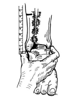
65 Palpation over ATibFib Ligament Dorsiflexion Dorsiflexion With External Rotation 11


66 Squeeze Test Crossed Leg Test Not one perfect test Which Tests to do? Combine Sensitivity and Specificity DFER and Palpation are highly sensitive Squeeze and Crossed Leg are highly specific 12


67 Tenderness Length Plain films Diastasis on Plain Film 13
68 Advanced Imaging CT If no Fx, then doesn t add much MRI Approaches 99% in Sens and Spec US Ninja level skill Treatment Timing Acute Clinical judgement of severity ER, close follow up, splint and crutches. Subacute (PCP office) Evaluation and Imaging NWB Brace in neutral or plantarflexion. Definitive Surgical Therapy RICE Physical Therapy Severity PROM AROM Strengthening Avoiding dorsiflexion entirely initially Balance and stability exercises 14


69 Tape by ATC Brace Bracing and Taping US guided injection AITFL (small space) Injection Therapy CSI vs PRP Study by NFL and NCAA Return approximately 10 days earlier. Surgery 15
70 Conclusion Syndesmosis or High Ankle Sprain Injury mechanism and structures Evaluation Treatment Options Questions? 16
71 TRIGGER POINT DRY NEEDLING Sadie Newman PT DPT Cox Health What is it? 1 A skilled intervention that uses a thin filiform needle to penetrate the skin and stimulate underlying myofascial trigger points, muscular, and connective tissues for the management of neuromuscular pain and movement impairments. How is it different from Acupuncture? 2 Differ in historical, philosophical, indicative, and practical content It is based on western neuroanatomy and modern scientific study of the musculoskeletal and nervous system. Does not use traditional acupuncture theories or terminology. Used by PTs as a modality 1
72 What is a Trigger Point (TrP)? Hyperirritable spots within a taut band of contractured skeletal muscle fibers that produce pain either locally or referred. 1 Active: spontaneously painful 1,2 Latent: Painful when stimulated 1,2 Physiological contractures Local ischemia and hypoxia, lower ph, a chemically altered milieu (active only), altered muscle activation patterns, and referred or local pain 1,3,5 Possible Physiological Theories 1 DN w/rotation Fibroblasts high tension Assumes Lamellar shape Increased Collagen synthesis and cell proliferation. Activated fibroblasts= Release of cytokines and other pro inflammatory mediators= HAPPY MUSCLES! Systemic effect with release of endogenous opioids > Promotes pro inflammatory factors. Mechanotransduction 1 Process by which the body converts mechanical loading into cellular responses. Fibroblast activation with a solid filament has been shown to result in pain neuromodulation 2
73 Types of Dry Needling 1 Deep Inactivates Trigger Points (TrPs) by eliciting local twitch response (LTR) that is modulated by the CNS. Assoc. w/ reduced local and referred pain, improved ROM, decreased TrP irritability both locally and remotely Superficial Activates mechanoreceptors coupled to slow conducting unmyelinated C fiber afferents and indirectly stimulate the anterior cingular cortex. May also be mediated through stimulation of A Delta Fibers or via stretching of fibroblasts in connective tissue. Assoc. w/ reduced local and referred pain Unknown if it has any impact on normalizing the chemical environment or reducing endplate noise assoc. w/trps Indications for Use When Myofascial TrPs are present When there are restrictions in ROM due to contractured muscle fibers, or taut bands, or other soft tissue restrictions (i.e. Fascial adhesions or scar tissues). What can be treated? 1 6 Subscapularis Shoulder Impingement Post CVA Hemiparetic Shoulder Pain Shoulder Impingement with TrPs Chronic RCT (questionable) Neck Pain Scapular retractor pain DeQuervin s Cervical Radiculopathy Patellofemoral Pain Syndrome Knee pain Plantar fascitis TMJ muscular Piriformis Syndrome Plantar heel pain Hip pain Low back pain Lateral and Medial Epicondylitis Plantar Heel Pain And Many Many MORE! 3
74 When is it not recommended? 6 When patient perceives it as threatening (needle phobias) Children younger than 12 y.o. Near damaged skin, thin skin, open wounds, moles, warts, or areas of eczema. Sites with post surgical lymphedema (mastectomy patients should not be needled distal to the site of lymph node removal). Patient with seizure within the last year Precautions 6 Pregnancy Patients with pacemakers receiving electrical dry needling Patients on anticoagulants (INR >3.o should) Patient with seizure (with most recent being >1 year ago). Adverse Reactions 3,6 When performed by a PT was found to have 0.04% adverse reactions as the best reporting and 7% at the worst Pneumothorax (2 cases in 2.2 million sample) and 8.2% adverse events Bruising Bleeding Fainting, Lightheadedness, dizziness, lethargy Reduction in blood sugar levels (caution with diabetics). Recent seizures 4
75 Referrals Referrals must list Trigger Point Dry Needling in physician or provider order for a privileged physical therapist to be able to perform. References 1. (2013, February). Description of Dry Needling In Clinical Practice: An Educational Resource Paper. Retrieved May, 2018, from sourcepaper/ 2. Rise, E. (2015, May). Dry Needling: Getting to the Point. Retrieved May, 2018 from 3. (2016). Copyright Evidence in Motion: Integrated Trigger Point Dry Needling for the Lower Quadrant. 4. Sato, K., Oliveira, A., & Lima, A. (2014). (565) Effectiveness of Dry Needling Reduction Myofascial Trigger Point Pain in the Trapezius Muscle. The Journal of Pain, 15(4). References 5. Calvo Lobo, C., Pacheco Da Costa, S., & Hita Herranz, E. (2017). Efficacy of Deep Dry Needling on Latent Myofascial Trigger Points in Older Adults with Nonspecific Shoulder Pain. Journal of Geriatric Physical Therapy, 40(2), doi: /jpt Continuing Education Course provided by Raymond Butts DPT based on : Copyright of Dry Needling Institute of American Academy of Manipulative Therapy James Dunning, DPT MSc Manip Ther, OCS, MCSP, MAACP (UK), FAAOMPT, MMACP (UK). 5
76 Alpha-Stim Tamra Standage, DPT, COMT Objectives Participants should be able to state underlining principles behind the usage of microcurrent therapy, both with probe application and with cranial electrotherapy stimulation. Participants should be able to state protocols to treat with both the microcurrent probe and cranial electrotherapy stimulation. Participants should be able to state conditions that may respond to alpha stim Cranial Electrotherapy Stimulation 3, 6 History Introduced to the US in 1960s from USSR, Europe as electrosleep Research was completed in US, Russia, and Europe in the 1960s and 1970s to find reliable parameters to induce sleep- none were found However it was found that microcurrent waveforms caused patients: improved relaxation and decreased anxiety 1979 the FDA approved CES for the treatment of insomnia, anxiety, and depression Improved levels of stress, allowed improvement in cognitive function, with an average gain of 12 to 18 points on standardized IQ tests VA is performing studies on veterans to assist with pain, anxiety, depression, insomnia, PTSD 1
 Current of 10-600 microamperes (µa) Pulse width of 500,000 +/- µs Compare to TENS: 2-1000 s Hz Current of approximately 60-100 ma Pulse width of")

 and noradrenergic (NE) systems that project supratentorially.")
77 Cranial Electrotherapy Stimulation 1 Microcurrent: 0.5 Hz typically (up to 100 Hz) Current of microamperes (µa) Pulse width of 500,000 +/- µs Compare to TENS: s Hz Current of approximately ma Pulse width of µs Waveforms of Alpha-Stim and TENS 1, 2 TRADITIONAL TENS Proposed Mechanism of Function of CES 2 Alpha-Stim CES engages the serotonergic (5-HT) raphe nuclei of the brainstem. 5-HT inhibits brainstem cholinergic (ACh) and noradrenergic (NE) systems that project supratentorially. This suppresses thalamocortical activity, arousal, agitation, alters sensory processing and induces EEG alpha rhythm. 5-HT can act directly to modulate pain sensation in the dorsal horn of the spinal cord, and alter pain perception, cognition, and emotionality within the limbic forebrain. 2
 in alpha activity with a simultaneous decrease (Blue) in beta and delta 2. Potential response to CES 2 www.alpha-stim.")

78 EEG changes in brain waves with CES 2,3 qeeg changes in 30 subjects treated with 20 minutes of Alpha Stim CES. There is an increase (Red) in alpha activity with a simultaneous decrease (Blue) in beta and delta 2. Potential response to CES 2 Microcurrent Therapy- Probes 6 Microcurrent has been shown to promote tissue healing Has been shown to assist with bone healing Has been shown to assist with ulcers due to pressure, venous or arterial insufficiency, and diabetes Has been shown to assist with chronic tendonopathy Results of studies demonstrate that microcurrent may assist in and promote tissue healing. More studies are needed to assist with developing guidelines for use and to investigate the underlying process of healing 3


79 Application of Alpha Stim-Probes 2 Contraindications 2 Implanted demand type cardiac pacemakers and implanted defibrillators Do not stimulate directly on the eyes, or press the probes over the carotid sinus Adverse effects: data on approximately 8,800 patients dizziness (6 cases, 0.07%) skin irritation/ electrode burns (6 cases, 0.07%) headaches (9 cases, 0.10%) 4
6/19/2018. Background Athlete s heart. Ultimate question. Applying the International Criteria for ECG
 Applying the International Criteria for ECG Interpretation in Athletes to a preparticipation screening program DAVE SIEBERT, MD, CAQSM ASSISTANT PROFESSOR DEPARTMENT OF FAMILY MEDICINE UNIVERSITY OF WASHINGTON
6/19/2018 INNOVATIONS IN SPORTS MEDICINE DISCLOSURE CHRONIC TENDINOPATHIES
 INNOVATIONS IN SPORTS MEDICINE TO INFINITY AND BEYOND ERIC GIFFORD MD DISCLOSURE THIS PRESENTATION HIGHLIGHTS TWO CURRENT, EVIDENCE-BASED PROCEDURES AVAILABLE AS TREATMENT MODALITIES AND THEY WILL BE REFERRED
Alex Garcia, MD Affinity Orthopedics and Sports Medicine
 Alex Garcia, MD Affinity Orthopedics and Sports Medicine Intact Tendon Reactive Inflammatory Tendinopathy Degenerative Chronic Tendinopathy INSULT Neurovascular Mediation Reparative Process? Healed 3
at least 4 8 hours per week
 ECG IN ATHLETS An athlete is defined as an individual who engages in regular exercise or training for sport or general fitness, typically with a premium on performance, and often engaged in individual
Study methodology for screening candidates to athletes risk
 1. Periodical Evaluations: each 2 years. Study methodology for screening candidates to athletes risk 2. Personal history: Personal history of murmur in childhood; dizziness, syncope, palpitations, intolerance
ECG Underwriting Puzzler Dr. Regina Rosace AVP & Medical Director
 December 2018 ECG Underwriting Puzzler Dr. Regina Rosace AVP & Medical Director To obtain best results Select Slide Show from the ribbon at the top of your PowerPoint screen Select From Beginning on the
EKG screening in athletics
 Use of PPE EKG screening in athletics Stefan Montgomery MD, ATC 4/27/18 The overall role of the preparticipation physical evaluation (PPE) is to evaluate the health of the athlete to optimize safe sports
Current ECG interpretation guidelines in the screening of athletes
 REVIEW ARTICLE 7 How to differentiate physiological adaptation to intensive physical exercise from pathologies Current ECG interpretation guidelines in the screening of athletes Gemma Parry-Williams, Sanjay
DEPARTMENT NAME PRE-PARTICIPATION SCREENING THE SPORTS PHYSICAL
 PRE-PARTICIPATION SCREENING THE SPORTS PHYSICAL Michele Krenek, MSN, RN, FNP-C TCHAPP Conference, Houston, TX April 4, 2019 PRE-PARTICIPATION SPORTS SCREENING According to the AHA the definition of the
Normal ECG And ECHO Findings in Athletes
 Normal ECG And ECHO Findings in Athletes Dr.Yahya Kiwan Consultant Interventional Cardiologist Head Of Departement Of Cardiology Canadian Specialist Hospital Sinus Bradycardia The normal heartbeat is initiated
EVALUATION OF THE ATHLETE. Karen Stout, MD Professor, Medicine and Pediatrics University of Washington
 EVALUATION OF THE 12 ATHLETE Karen Stout, MD Professor, Medicine and Pediatrics University of Washington NO DISCLOSURES OUTLINE Why evaluate athletes? What s the problem? What evaluation should be done?
Interpretation and Consequences of Repolarisation Changes in Athletes
 Interpretation and Consequences of Repolarisation Changes in Athletes Professor Sanjay Sharma E-mail sasharma@sgul.ac.uk @SSharmacardio Disclosures: None Athlete s ECG Vagotonia Sinus bradycardia Sinus
How to Read an Athlete s ECG. Sanjay Sharma BSc (Hons), MD, FRCP, FESC
, MD, FRCP, FESC") How to Read an Athlete s ECG Sanjay Sharma BSc (Hons), MD, FRCP, FESC Athlete s EKG Vagotonia Sinus bradycardia Sinus arrhythmia First degree AVB ST-elevation Tall T waves Increased chamber size Left ventricular
How to Read an Athlete s ECG Sanjay Sharma BSc (Hons), MD, FRCP, FESC Athlete s EKG Vagotonia Sinus bradycardia Sinus arrhythmia First degree AVB ST-elevation Tall T waves Increased chamber size Left ventricular
Sports Cardiology: Matters of the Heart. AMSSM Exchange Lecture AOSSM 2013 Annual Meeting
 Sports Cardiology: Matters of the Heart AMSSM Exchange Lecture AOSSM 2013 Annual Meeting Matthew Gammons, MD Vermont Orthopaedic Clinic Killington Medical Clinic Although sudden cardiac death is a relatively
I have nothing to disclose. Research support from: Cardiac Risk in The Young
 I have nothing to disclose. Research support from: Cardiac Risk in The Young Pre-participation screening of Young Athletes: Current Perspective Professor Sanjay Sharma Disclosures: None SCD in Young Athletes
La valutazione dell atleta: è una strategia salva-vita e costo-efficace?
 La valutazione dell atleta: è una strategia salva-vita e costo-efficace? Primo trattato di Medicina Wilson and Jungner s criteria In the 1960s the World Health Organization adopted the Wilson and Jungner
Dr. Schroeder has no financial relationships to disclose
 Valerie A Schroeder MD MS Assistant Professor University of Kansas Medical Center READING THE WAVES- THE HEART S ELECTRICAL MESSAGE FINANCIAL DISCLOSURE Dr. Schroeder has no financial relationships to
Slide 1. Slide 2. Slide 3. Sudden Cardiac Death In Athletes. Epidemiology. Epidemiology. Shaun McMurtry, MD Primary Care Sports Medicine
 Slide 1 Sudden Cardiac Death In Athletes Shaun McMurtry, MD Primary Care Sports Medicine Slide 2 Epidemiology College and Professional Athletes 500,000 participants each year Competitive Athletics Estimated
The Electrocardiogram part II. Dr. Adelina Vlad, MD PhD
 The Electrocardiogram part II Dr. Adelina Vlad, MD PhD Basic Interpretation of the ECG 1) Evaluate calibration 2) Calculate rate 3) Determine rhythm 4) Determine QRS axis 5) Measure intervals 6) Analyze
2/26/2015.
 Gerry Keenan MMS PA-C Associate Professor -Physician Assistant Studies Arizona School of Health Sciences A T Still University Event Medical Director-USA/Karate- Arizona Clinical Director-MEDfest/Healthy
EVALUATION OF ELECTROCARDIOGRAPHIC FINDINGS IN ATHLETES
 EVALUATION OF ELECTROCARDIOGRAPHIC FINDINGS IN ATHLETES UNIT OF INHERITED CV DISEASES HEART CENTER OF THE YOUNG AND ATHLETES A DPT OF CARDIOLOGY UNIVERSITY OF ATHENS EVALUATION OF ELECTROCARDIOGRAPHIC
Exercise guidelines in athletes with isolated repolarisation abnormalities and structurally normal heart.
 Exercise guidelines in athletes with isolated repolarisation abnormalities and structurally normal heart. Hanne Rasmusen Consultant cardiologist, PhD Dept. of Cardiology Bispebjerg University Hospital
Please check your answers with correct statements in answer pages after the ECG cases.
 ECG Cases ECG Case 1 Springer International Publishing AG, part of Springer Nature 2018 S. Okutucu, A. Oto, Interpreting ECGs in Clinical Practice, In Clinical Practice, https://doi.org/10.1007/978-3-319-90557-0
International recommendations for electrocardiographic interpretation in athletes
 European Heart Journal (2017) 00, 1 19 doi:101093/eurheartj/ehw631 CURRENT OPINION International recommendations for electrocardiographic interpretation in athletes Sanjay Sharma 1 *, Jonathan A Drezner
Paediatric ECG Interpretation
 Paediatric ECG Interpretation Dr Sanj Fernando (thanks to http://lifeinthefastlane.com/ecg-library/paediatric-ecginterpretation/) 3 yo boy complaining of abdominal pain and chest pain Child ECG vs Adult
Sudden Cardiac Arrest in Athletes Capital City Sports Summit June 7, 2012
 Sudden Cardiac Arrest in Athletes Capital City Sports Summit June 7, 2012 John Katopodis, MD, FACC Southern Medical Group Cardiology Tallahassee, Florida Scope of the Problem Relating to Screening and
Ronald J. Kanter, MD Director, Electrophysiology Miami Children s Hospital Professor Emeritus, Duke University Miami, Florida
 S306- Pediatric Electrocardiography: A Potpourri Ronald J. Kanter, MD Director, Electrophysiology Miami Children s Hospital Professor Emeritus, Duke University Miami, Florida Disclosure of Relevant Relationship
François Carré Hôpital Pontchaillou -INSERM UMR1099-Université Rennes 1
 Normal electrocardiogram variants in Athletes François Carré Hôpital Pontchaillou -INSERM UMR1099-Université Rennes 1 Disclosures No disclosure of interest concerning this lecture The cardiovascular sport
Cardiovascular Impacts of long-term endurance exercise: Implications of athlete s heart
 Cardiovascular Impacts of long-term endurance exercise: Implications of athlete s heart Dr. Gary Mak 麥耀光心臟科專科醫生 IPP of HK Associa4on of Sports Medicine and Sports Science Director of Pro-Cardio Heart Disease
Index. cardiacep.theclinics.com. Note: Page numbers of article titles are in boldface type.
 Note: Page numbers of article titles are in boldface type. A AEDs. See Automated external defibrillators (AEDs) AF. See Atrial fibrillation (AF) Age as factor in SD in marathon runners, 45 Antiarrhythmic
Sudden Cardiac Death in Sports: Causes and Current Screening Recommendations
 Sports Cardiology Sudden Cardiac Death in Sports: Causes and Current Screening Recommendations Domenico Corrado, MD, PhD Inherited Arrhytmogenic Cardiomyopathy Unit Department of Cardiac, Thoracic and
Myocardial Infarction. Reading Assignment (p66-78 in Outline )
") Myocardial Infarction Reading Assignment (p66-78 in Outline ) Objectives 1. Why do ST segments go up or down in ischemia? 2. STEMI locations and culprit vessels 3. Why 15-lead ECGs? 4. What s up with avr?
Myocardial Infarction Reading Assignment (p66-78 in Outline ) Objectives 1. Why do ST segments go up or down in ischemia? 2. STEMI locations and culprit vessels 3. Why 15-lead ECGs? 4. What s up with avr?
ECG Interpretation Made Easy
 ECG Interpretation Made Easy Dr. A Tageldien Abdellah, MSc MD EBSC Lecturer of Cardiology- Hull University Hull York Medical School 2007-2008 ECG Interpretation Made Easy Synopsis Benefits Objectives Process
October (Am Journal of Orthopedics, 2014)
") (Am Journal of Orthopedics, 2014) Darryl Barnes, Jim Beckley, and Jay Smith Ultrasonic Percutaneous Tenotomy for Chronic Elbow Tendonosis: A Prospective Study (Journal of Shoulder and Elbow Surgery, 2014).
(Am Journal of Orthopedics, 2014) Darryl Barnes, Jim Beckley, and Jay Smith Ultrasonic Percutaneous Tenotomy for Chronic Elbow Tendonosis: A Prospective Study (Journal of Shoulder and Elbow Surgery, 2014).
Pennsylvania Academy of Family Physicians Foundation & UPMC 43rd Refresher Course in Family Medicine CME Conference March 10-13, 2016
 Pennsylvania Academy of Family Physicians Foundation & UPMC 43rd Refresher Course in Family Medicine CME Conference March 10-13, 2016 Disclosures: EKG Workshop Louis Mancano, MD Speaker has no disclosures
10 ECGs No Practitioner Can Afford to Miss. Objectives
 10 ECGs No Practitioner Can Afford to Miss Mary L. Dohrmann, MD Professor of Clinical Medicine Division of Cardiovascular Medicine University of Missouri School of Medicine No disclosures Objectives 1.
ECG Interpretation Cat Williams, DVM DACVIM (Cardiology)
") ECG Interpretation Cat Williams, DVM DACVIM (Cardiology) Providing the best quality care and service for the patient, the client, and the referring veterinarian. GOAL: Reduce Anxiety about ECGs Back to
ECG Interpretation Cat Williams, DVM DACVIM (Cardiology) Providing the best quality care and service for the patient, the client, and the referring veterinarian. GOAL: Reduce Anxiety about ECGs Back to
Blocks & Dissociations. Reading Assignment (p47-52 in Outline )
") Blocks & Dissociations Reading Assignment (p47-52 in Outline ) Objectives Who are Wenckebach and Mobitz? Review SA and AV Blocks AV Dissociations: learning who s the boss and why 2 nd degree SA Block:
Blocks & Dissociations Reading Assignment (p47-52 in Outline ) Objectives Who are Wenckebach and Mobitz? Review SA and AV Blocks AV Dissociations: learning who s the boss and why 2 nd degree SA Block:
Pre-Participation Cardiac Screening
 Pre-Participation Cardiac Screening Rebecca Martinie MD Assistant Professor Section of Adolescent and Sports Medicine Clinics: CyFair Health Center, Sugarland Health Center & West Campus Goal and Objectives
SUDDEN CARDIAC DEATH IN ATHLETES
 SUDDEN CARDIAC DEATH IN ATHLETES Alix Dufresne, MD, FACP, FACC, FESC Cardiology Division Chief, Interfaith Medical Center Director Cardiology Clinic, Kingsbrook Jewish Center PURPOSE AND OBJECTIVES PURPOSE
December Tenex Health Clinical Publications
 Tenex Health Clinical Publications Barnes Darryl; Beckley, James; and Smith, Jay. Ultrasonic Percutaneous Tenotomy for Chronic Elbow Tendonosis: A Prospective Study, Journal of Shoulder and Elbow Surgery,
REtrive. REpeat. RElearn Design by. Test-Enhanced Learning based ECG practice E-book
 Test-Enhanced Learning Test-Enhanced Learning Test-Enhanced Learning Test-Enhanced Learning based ECG practice E-book REtrive REpeat RElearn Design by S I T T I N U N T H A N G J U I P E E R I Y A W A
HR: 50 bpm (Sinus) PR: 280 ms QRS: 120 ms QT: 490 ms Axis: -70. Sinus bradycardia with one ventricular escape (*)
 PR: 280 ms QRS: 120 ms QT: 490 ms Axis: -70. Sinus bradycardia with one ventricular escape (*)") 1? HR: 50 bpm (Sinus) PR: 280 ms QRS: 120 ms QT: 490 ms Axis: -70 1 Sinus P waves? 2 sinus cycles The pause (2 sinus cycles) suggests that the sinus fired (?) but did not conduct to the atria (i.e., missing
1? HR: 50 bpm (Sinus) PR: 280 ms QRS: 120 ms QT: 490 ms Axis: -70 1 Sinus P waves? 2 sinus cycles The pause (2 sinus cycles) suggests that the sinus fired (?) but did not conduct to the atria (i.e., missing
Office ECG Interpretation
 Office ECG Interpretation Jason Evanchan, DO Assistant Professor of Medicine Division of Cardiovascular Medicine The Ohio State University Wexner Medical Center Outline of topics High risk ischemia T wave
Screening Young Competitive Athletes for Underlying Cardiovascular Disease in British Columbia, Canada A SportsCardiologyBC Study
 Screening Young Competitive Athletes for Underlying Cardiovascular Disease in British Columbia, Canada A SportsCardiologyBC Study Introduction: Following the publication of a 25-year study out of Italy
DR QAZI IMTIAZ RASOOL OBJECTIVES
 PRACTICAL ELECTROCARDIOGRAPHY DR QAZI IMTIAZ RASOOL OBJECTIVES Recording of electrical events in heart Established electrode pattern results in specific tracing pattern Health of heart i. e. Anatomical
Miscellaneous Stuff Keep reading the Outline
 Miscellaneous Stuff Keep reading the Outline Welcome to the 5-Step Method ECG #: Mearurements: Rhythm (s): Conduction: Waveform: Interpretation: A= V= PR= QRS= QT= Axis= 1. Compute the 5 basic measurements:
12-Lead ECG Interpretation. Kathy Kuznar, RN, ANP
 12-Lead ECG Interpretation Kathy Kuznar, RN, ANP The 12-Lead ECG Objectives Identify the normal morphology and features of the 12- lead ECG. Perform systematic analysis of the 12-lead ECG. Recognize abnormalities
ECG Cases and Questions. Ashish Sadhu, MD, FHRS, FACC Electrophysiology/Cardiology
 ECG Cases and Questions Ashish Sadhu, MD, FHRS, FACC Electrophysiology/Cardiology 32 yo female Life Insurance Physical 56 yo male with chest pain Terminology Injury ST elevation Ischemia T wave inversion
SIMPLY ECGs. Dr William Dooley
 SIMPLY ECGs Dr William Dooley Content Basic ECG interpretation pattern Some common (examined) abnormalities Presenting ECGs in context Setting up an ECG Setting up an ECG 1 V1-4 th Right intercostal space
ECG Workshop. Nezar Amir
 ECG Workshop Nezar Amir Myocardial Ischemia ECG Infarct ECG in STEMI is dynamic & evolving Common causes of ST shift Infarct Localisation Left main artery occlusion: o diffuse ST-depression with ST elevation
Sudden cardiac death: Primary and secondary prevention
 Sudden cardiac death: Primary and secondary prevention By Kai Chi Chan Penultimate Year Medical Student St George s University of London at UNic Sheba Medical Centre Definition Sudden cardiac arrest (SCA)
Jonathan Kim MD, FACC
 Jonathan Kim MD, FACC Assistant Professor, Division of Cardiology, Emory University Adjunct Assistant Professor, School of Applied Physiology, Georgia Tech Team Cardiologist, Sports Medicine, Emory University
Return to Basics. ECG Rate and Rhythm. Management of the Hospitalized Patient September 25, 2009
 Management of the Hospitalized Patient September 25, 2009 ECG Refresher and Update 2009 Return to Basics Determine rate and rhythm Determine intervals and axes Define morphology of P-QRS-T-U Compare with
General Introduction to ECG. Reading Assignment (p2-16 in PDF Outline )
") General Introduction to ECG Reading Assignment (p2-16 in PDF Outline ) Objectives 1. Practice the 5-step Method 2. Differential Diagnosis: R & L axis deviation 3. Differential Diagnosis: Poor R-wave progression
General Introduction to ECG Reading Assignment (p2-16 in PDF Outline ) Objectives 1. Practice the 5-step Method 2. Differential Diagnosis: R & L axis deviation 3. Differential Diagnosis: Poor R-wave progression
PAEDIATRIC ECG Dimosthenis Avramidis, MD.
 PAEDIATRIC ECG Dimosthenis Avramidis, MD. Consultant Mitera Children s Hospital Athens Greece S. Associate 1st Cardiology Dpt Evangelismos Hospital Athens Greece 5 y/o with sinus tach Background ECG changes
SABIHA GATI AND SANJAY SHARMA
 9 The athlete s heart SABIHA GATI AND SANJAY SHARMA Pasieka/Science Photo Library In this article, the authors highlight the spectrum, magnitude and determinants of the athlete s heart and provide a practical
Appendix D Output Code and Interpretation of Analysis
 Appendix D Output Code and Interpretation of Analysis 8 Arrhythmia Code No. Description 8002 Marked rhythm irregularity 8110 Sinus rhythm 8102 Sinus arrhythmia 8108 Marked sinus arrhythmia 8120 Sinus tachycardia
2017 EKG Workshop Advanced. Family Medicine Review Course Lou Mancano, MD, FAAFP Reading Health System Family and Community Medicine Reading, PA
 2017 EKG Workshop Advanced Family Medicine Review Course Lou Mancano, MD, FAAFP Reading Health System Family and Community Medicine Reading, PA Part II - Objective Describe a useful approach to interpreting
Return to Basics. Normal Intervals & Axes. ECG Rate and Rhythm
 Return to Basics Management of the Hospitalized Patient October 15, 2010 ECG Refresher and Update 2010 Determine rate and rhythm Determine intervals and axes Define morphology of P-QRS-T-U Compare with
Acute Coronary Syndromes Unstable Angina Non ST segment Elevation MI (NSTEMI) ST segment Elevation MI (STEMI)
 ST segment Elevation MI (STEMI)") Leanna R. Miller, RN, MN, CCRN-CSC, PCCN-CMC, CEN, CNRN, CMSRN, NP Education Specialist LRM Consulting Nashville, TN Objectives Evaluate common abnormalities that mimic myocardial infarction. Identify
Leanna R. Miller, RN, MN, CCRN-CSC, PCCN-CMC, CEN, CNRN, CMSRN, NP Education Specialist LRM Consulting Nashville, TN Objectives Evaluate common abnormalities that mimic myocardial infarction. Identify
UNDERSTANDING YOUR ECG: A REVIEW
 UNDERSTANDING YOUR ECG: A REVIEW Health professionals use the electrocardiograph (ECG) rhythm strip to systematically analyse the cardiac rhythm. Before the systematic process of ECG analysis is described
Reading Assignment (p1-91 in Outline ) Objectives What s in an ECG?
 Objectives What s in an ECG?") Reading Assignment (p1-91 in Outline ) Objectives What s in an ECG? The 5-Step Method ECG #: Mearurements: Rhythm (s): Conduction: Waveform: Interpretation: A= V= PR= QRS= QT= Axis= 1. Compute the 5 basic
Reading Assignment (p1-91 in Outline ) Objectives What s in an ECG? The 5-Step Method ECG #: Mearurements: Rhythm (s): Conduction: Waveform: Interpretation: A= V= PR= QRS= QT= Axis= 1. Compute the 5 basic
Introduction to ECG Gary Martin, M.D.
 Brief review of basic concepts Introduction to ECG Gary Martin, M.D. The electrical activity of the heart is caused by a sequence of rapid ionic movements across cell membranes resulting first in depolarization
Sudden Cardiac Arrest in Athletes: Can We Prevent Sudden Death?
 Sudden Cardiac Arrest in Athletes: Can We Prevent Sudden Death? David Berkson, MD, FAAFP Department of Family, Community, and Preventive Medicine Heart conditions lead to death 16 year-old girl was cross-country
Case 1. Case 2. Case 3
 Case 1 The correct answer is D. Occasionally, the Brugada syndrome can present similar morphologies to A and also change depending on the lead position but in the Brugada pattern the r is wider and ST
A new consensus document on electrocardiographic interpretation in athletes: does it help to prevent sudden cardiac death in athletes?
 Neth Heart J (2018) 26:127 132 https://doi.org/10.1007/s12471-018-1076-6 POINT OF VIEW A new consensus document on electrocardiographic interpretation in athletes: does it help to prevent sudden cardiac
THE PRE-PARTICIPATION EXAM (A.K.A. THE PPE)
") THE PRE-PARTICIPATION EXAM (A.K.A. THE PPE) Peter Hoth, MD Clinical Assistant Professor UI Sports Medicine Department of Family Medicine 45 th Annual Refresher Course for the Family Physician April 5,
THE PRE-PARTICIPATION EXAM (A.K.A. THE PPE) Peter Hoth, MD Clinical Assistant Professor UI Sports Medicine Department of Family Medicine 45 th Annual Refresher Course for the Family Physician April 5,
ECG CONVENTIONS AND INTERVALS
 1 ECG Waveforms and Intervals ECG waveforms labeled alphabetically P wave== represents atrial depolarization QRS complex=ventricular depolarization ST-T-U complex (ST segment, T wave, and U wave)== V repolarization.
FLB s What Are Those Funny-Looking Beats?
 FLB s What Are Those Funny-Looking Beats? Reading Assignment (pages 27-45 in Outline ) The 5-Step Method ECG #: Mearurements: Rhythm (s): Conduction: Waveform: Interpretation: A= V= PR= QRS= QT= Axis=
Abnormal electrocardiographic findings in athletes: recognising changes suggestive of cardiomyopathy
 Scan to access more free content For numbered affiliations see end of article Correspondence to Jonathan A Drezner, Department of Family Medicine, University of Washington, 1959 NE Pacific Street, Box
Degrees AV blocks in athletes
 Degrees AV blocks in athletes Name: BCA Age: 22yo Sex: Male Race: Black Weight: 82 kg Height: 1.96m Biotype: Athletic rofession: professional basketball player Date: 2/09/2001 Clinical diagnosis: athlete
Electrical System Overview Electrocardiograms Action Potentials 12-Lead Positioning Values To Memorize Calculating Rates
 Electrocardiograms Electrical System Overview James Lamberg 2/ 74 Action Potentials 12-Lead Positioning 3/ 74 4/ 74 Values To Memorize Inherent Rates SA: 60 to 100 AV: 40 to 60 Ventricles: 20 to 40 Normal
Sudden Death in Athletes: What is the role of ECG Screening?
 Sudden Death in Athletes: What is the role of ECG Screening? Byron K. Lee MD Professor of Medicine Director of EP Laboratory leeb@medicine.ucsf.edu Division of Cardiology Cardiac Electrophysiology Arrhythmias,
Advances in Ablation Therapy for Ventricular Tachycardia
 Advances in Ablation Therapy for Ventricular Tachycardia Nitish Badhwar, MD, FACC, FHRS Director, Cardiac Electrophysiology Training Program University of California, San Francisco For those of you who
Electrocardiography for Healthcare Professionals. Chapter 14 Basic 12-Lead ECG Interpretation
 Electrocardiography for Healthcare Professionals Chapter 14 Basic 12-Lead ECG Interpretation 2012 The Companies, Inc. All rights reserved. Learning Outcomes 14.1 Discuss the anatomic views seen on a 12-lead
Huseng Vefali MD St. Luke s University Health Network Department of Cardiology
 Huseng Vefali MD St. Luke s University Health Network Department of Cardiology Learning Objectives Establish Consistent Approach to Interpreting ECGs Review Essential Cases for Paramedics and first responders
Emergency Medical Training Services Emergency Medical Technician Paramedic Program Outlines Outline Topic: WPW Revised: 11/2013
 Emergency Medical Training Services Emergency Medical Technician Paramedic Program Outlines Outline Topic: WPW Revised: 11/2013 Wolff-Parkinson-White syndrome (WPW) is a syndrome of pre-excitation of the
December 2018 Tracings
 Tracings Tracing 1 Tracing 4 Tracing 1 Answer Tracing 4 Answer Tracing 2 Tracing 5 Tracing 2 Answer Tracing 5 Answer Tracing 3 Tracing 6 Tracing 3 Answer Tracing 6 Answer Questions? Contact Dr. Nelson
International Criteria for Electrocardiographic Interpretation in Athletes
 International Criteria for Electrocardiographic Interpretation in Athletes Jonathan A. Drezner, MD* 1, Sanjay Sharma, MD* 2, Aaron Baggish, MD 3, Michael Papadakis, MD 2, Mathew G. Wilson, PhD 4, Jordan
Masqueraders of STEMI
 Masqueraders of STEMI Steven M. Costa, M.D. Assistant Professor Department of Medicine Division of Cardiology Scott & White Memorial Hospital and Clinic Texas A&M University Health Science Center Disclosures
ECGs and Arrhythmias: Family Medicine Board Review 2009
 Rate Rhythm Intervals Hypertrophy ECGs and Arrhythmias: Family Medicine Board Review 2009 Axis Jess (Fogler) Waldura, MD University of California, San Francisco walduraj@nccc.ucsf.edu Ischemia Overview
SIMPLY ECGs. Dr William Dooley
 SIMPLY ECGs Dr William Dooley 1 No anatomy just interpretation 2 Setting up an ECG 3 Setting up an ECG 1 V1-4 th Right intercostal space at sternal border 2 V2-4 th Left intercostal space at sternal border
Basic electrocardiography reading. R3 lee wei-chieh
 Basic electrocardiography reading R3 lee wei-chieh The Normal Conduction System Lead Placement avf Limb Leads Precordial Leads Interpretation Rate Rhythm Interval Axis Chamber abnormality QRST change What
Ablative Therapy for Ventricular Tachycardia
 Ablative Therapy for Ventricular Tachycardia Nitish Badhwar, MD, FACC, FHRS 2 nd Annual UC Davis Heart and Vascular Center Cardiovascular Nurse / Technologist Symposium May 5, 2012 Disclosures Research
Cardiac Conditions in Sport & Exercise. Cardiac Conditions in Sport. USA - Sudden Cardiac Death (SCD) Dr Anita Green. Sudden Cardiac Death
 Dr Anita Green. Sudden Cardiac Death") Cardiac Conditions in Sport & Exercise Dr Anita Green Cardiac Conditions in Sport Sudden Cardiac Death USA - Sudden Cardiac Death (SCD)
Cardiac Conditions in Sport & Exercise Dr Anita Green Cardiac Conditions in Sport Sudden Cardiac Death USA - Sudden Cardiac Death (SCD)
Professor Sanjay Sharma St George s University of London
 How to Evaluate an Athlete of Afro- Caribbean Origin. Professor Sanjay Sharma St George s University of London Background: Causes of SCD in Sport Young competitive athlete Personal and family history Physical
Electrocardiography Abnormalities (Arrhythmias) 7. Faisal I. Mohammed, MD, PhD
 7. Faisal I. Mohammed, MD, PhD") Electrocardiography Abnormalities (Arrhythmias) 7 Faisal I. Mohammed, MD, PhD 1 Causes of Cardiac Arrythmias Abnormal rhythmicity of the pacemaker Shift of pacemaker from sinus node Blocks at different
Electrocardiography Abnormalities (Arrhythmias) 7 Faisal I. Mohammed, MD, PhD 1 Causes of Cardiac Arrythmias Abnormal rhythmicity of the pacemaker Shift of pacemaker from sinus node Blocks at different
Cardiology Flash Cards
 Cardiology Flash Cards EKG in a nut shell www.brain101.info Conduction System www.brain101.info 2 Analyzing EKG Step by step Steps in Analyzing ECG'S 1. Rhythm: - Regular _ Sinus, Junctional or Ventricular.
Pre-participation Screening for the Prevention of sudden Cardiac Death in Young Athletes. Thomas W. Allen, DO, MPH
 Pre-participation Screening for the Prevention of sudden Cardiac Death in Young Athletes Thomas W. Allen, DO, MPH Cardiovascular disorders are the leading cause of sudden death in young athletes accounting
Diploma in Electrocardiography
 The Society for Cardiological Science and Technology Diploma in Electrocardiography The Society makes this award to candidates who can demonstrate the ability to accurately record a resting 12-lead electrocardiogram
ECG ABNORMALITIES D R. T AM A R A AL Q U D AH
 ECG ABNORMALITIES D R. T AM A R A AL Q U D AH When we interpret an ECG we compare it instantaneously with the normal ECG and normal variants stored in our memory; these memories are stored visually in
All About STEMIs. Presented By: Brittney Urvand, RN, BSN, CCCC. Essentia Health Fargo Cardiovascular Program Manager.
 All About STEMIs Presented By: Brittney Urvand, RN, BSN, CCCC Essentia Health Fargo Cardiovascular Program Manager Updated 10/2/2018 None Disclosures Objectives Identify signs and symptoms of a heart attack
Supraventricular Arrhythmias. Reading Assignment. Chapter 5 (p17-30)
") Supraventricular Arrhythmias Reading Assignment Chapter 5 (p17-30) The Supraventricular Rhythms In Our Lives Site of Origin Single Events Slow Rates Intermediate Rates Fast Rates (>100 bpm) Sinus Sinus
Supraventricular Arrhythmias Reading Assignment Chapter 5 (p17-30) The Supraventricular Rhythms In Our Lives Site of Origin Single Events Slow Rates Intermediate Rates Fast Rates (>100 bpm) Sinus Sinus
Bundle Branch & Fascicular Blocks. Reading Assignment (p53-58 in Outline )
") Bundle Branch & Fascicular Blocks Reading Assignment (p53-58 in Outline ) Objectives 1. QRS analysis of Right and Left BBB 2. Uncomplicated vs complicated BBB 3. Diagnosis of RBBB with LAFB and LPFB 4.
Bundle Branch & Fascicular Blocks Reading Assignment (p53-58 in Outline ) Objectives 1. QRS analysis of Right and Left BBB 2. Uncomplicated vs complicated BBB 3. Diagnosis of RBBB with LAFB and LPFB 4.
Case-Based Practical ECG Interpretation for the Generalist
 Case-Based Practical ECG Interpretation for the Generalist Paul D. Varosy, MD, FACC, FAHA, FHRS Director of Cardiac Electrophysiology VA Eastern Colorado Health Care System Associate Professor of Medicine
ECG Basics Sonia Samtani 7/2017 UCI Resident Lecture Series
 ECG Basics Sonia Samtani 7/2017 UCI Resident Lecture Series Agenda I. Introduction II.The Conduction System III.ECG Basics IV.Cardiac Emergencies V.Summary The Conduction System Lead Placement avf Precordial
The frontier between normal and abnormal electrocardiogram in athletes
 The frontier between normal and abnormal electrocardiogram in athletes ESC Congress 2011 Paris F. Carré University Rennes 1-Pontchaillou Hospital Inserm U642, Rennes - F-35000 Cardiovascular preparticipation
12 Lead ECG Interpretation
 12 Lead ECG Interpretation Julie Zimmerman, MSN, RN, CNS, CCRN Significant increase in mortality for every 15 minutes of delay! N Engl J Med 2007;357:1631-1638 Who should get a 12-lead ECG? Also include
Sudden Cardiac Death in Youth Athletes. Dublin City Schools Athletic Health Care
 Sudden Cardiac Death in Youth Athletes Dublin City Schools Athletic Health Care Sudden Cardiac Death What is sudden cardiac death in the young athlete? Sudden cardiac death is the result of an unexpected