Mediastinal Paraganglioma: a challenge to the echocardiographic
|
|
|
- Josephine Joseph
- 5 years ago
- Views:
Transcription


1 Case - based learning from ESC Cardiologists of Tomorrow Look for the answer outside the heart Mediastinal Paraganglioma: a challenge to the echocardiographic 1 diagnosis and endovascular treatment 1 On behalf of Sergio Raposeiras Roubín University Clinical Hospital of Santiago de Compostela. SPAIN RM. Agra Bermejo, J. Elices Teja, P. Cabanas Grandio, S. Gestal Romari, E. Pereira Lopez, B. Alvarez Alvarez, C. Gonzalez Cambeiro, R. Fandino Vaquero, A. Martinez Monzonis J.R.G. JUANATEY

 Ortopnea since 2 months J.R.G.")
2 Case Report 59 years old DIABETES MELLITUS ARTERIAL HYPERTENSION OBSTRUCTIVE SLEEP APNEA SYNDROME 2 episodes of SUPRAVENTRICULAR TACHYCARDIA (adenosine --- B-blockers) Ortopnea since 2 months J.R.G. JUANATEY

3 CHEST RADIOGRAPHY

4 TRANSTORACIC ECHOCARDIOGRAPHY


5 TRANSTORACIC ECHOCARDIOGRAPHY
6 TRANSTORACIC ECHOCARDIOGRAPHY

7 TRANSTORACIC ECHOCARDIOGRAPHY

8 TRANSTORACIC ECHOCARDIOGRAPHY

9 TRANSTORACIC ECHOCARDIOGRAPHY
10 TRANSESOPHAGEAL ECHOCARDIOGRAPHY J.R.G. JUANATEY

11 TRANSESOPHAGEAL ECHOCARDIOGRAPHY J.R.G. JUANATEY
12 TRANSESOPHAGEAL ECHOCARDIOGRAPHY J.R.G. JUANATEY


13 COMPUTED TOMOGRAPHY


14 CORONARY ANGIOGRAPHY


15 CORONARY ANGIOGRAPHY


16 CORONARY ANGIOGRAPHY
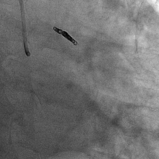

17 CORONARY ANGIOGRAPHY


18 CORONARY ANGIOGRAPHY
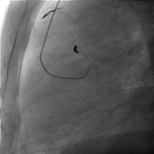

19 CORONARY ANGIOGRAPHY


20 CORONARY ANGIOGRAPHY


21 ECHO-ENDOSCOPY


22 PARAGANGLIOMA Catecho lamines J.R.G. JUANATEY
23 PARAGANGLIOMA 90% chromaffin-cell-originating tumors are located in the adrenal gland and termed pheochromocytomas. The remaining 10% are extra adrenal and are termed paragangliomas or chemodectomas Paragangliomas appear in the abdomen, pelvis, neck and mediastimun. Mediastinal paraganglioma are rare, originating from para-aortic (middle mediastinum) and para-vertebral (posterior mediastinum) sympathetic chain ganglia. Similar to pheochromocytoma, paraganglioma tumors may secrete catecholamines, however in majority of cases they are non-functional. The diagnosis of paraganglioma is based on clinical symptoms, imaging exams (echocardiography, computed tomography or magnetic resonance imaging), and biochemical tests. J.R.G. JUANATEY
24 PARAGANGLIOMA Up to 50% are asymptomatic and the diagnosis is incidental. Clinical symptoms may be related to catecholamine hypersecretion (hypertention / hyperhydrosis) or to a mass effect resulting in complains of hoarseness, dysphagia, shortness of breath and chest pain. Biochemical diagnosis of functional paragangliomas is made by measuring catecholamines and their metabolites in urine. The main imaging methods used for the diagnosis are CT and magnetic resonance imaging, both of which have a sensitivity of nearly 98%. Treatment of choice is complete surgical resection, which is difficult due to hypervascularization and anatomical juxtaposition of the tumor. Combined incision and extracorporeal circulation are often necessary. Due to the large number of small vessels in this area, which contains various major structures, hemostatic clips or a harmonic scalpel could be used in order to guarantee efficient hemostatic control. J.R.G. JUANATEY
25 PARAGANGLIOMA Mediastinal paragangliomas are rare, only approximately 150 cases having been reported in the literature, and two-thirds of these tumors are located in the anterior or middle mediastinum. Mediastinal paragangliomas are derived from the para-aortic and paravertebral ganglion chain. Multicentric tumors are observed in 23% of cases, and there seems to be no specific distribution in the remaining cases. Mediastinal paragangliomas are highly vascularized tumors that adhere to adjacent mediastinal structures, such as the heart, large blood vessels, trachea, and spine, and surgical management is therefore difficult. The differential diagnosis includes Castleman's disease, neuroendocrine tumors, lymphomas, and hemangiomas. J.R.G. JUANATEY
26 IMPLICATIONS TO CLINICAL PRACTICE Primary cardiac tumours are a rare disorder and have an incidence between 0.002% and 0.2% in an autopsy series. They are usually treatable and can often be diagnosed with echocardiography, computed tomography (CT), or magnetic resonance (MR) imaging. Benign tumours account for 75% of this total, being myxomas the most common. In general, 75% of myxomas are located in the left atrium. But cardiologist must note the differential diagnosis with other left atrial masses, as lipomas (16%), fibroelastomas (16%), hemangiomas (6%), fibromas (3%), rhabdomuomas (1%) and rare cases of neurofibromas, teratomas and paragangliomas. The Paragangliomas usually appear as large, echogenic left atrial masses at echocardiography and like a circumscribed, heterogeneous masses with low attenuation at CT. J.R.G. JUANATEY
27 DISCUSSION MEDIASTINAL Echocardiogrphic diagnosis PARAGANGLIOMA Endovascular treatment J.R.G. JUANATEY


28 Thanks for your attention J.R.G. JUANATEY
A case of micturition syncope
 A case of micturition syncope Kimberly Bundick, PA-S S L I D E 1 Agenda Purpose Utilize case to illustrate classic finding of an interesting pathology Agenda Case study Epidemiology, etiology of disease
Retroperitoneal Ganglioneuroma Encasing the Celiac and Superior Mesenteric Arteries
 Case Study TheScientificWorldJOURNAL (2004) 4, 974 977 ISSN 1537-744X; DOI 10.1100/tsw.2004.198 Retroperitoneal Ganglioneuroma Encasing the Celiac and Superior Mesenteric Arteries Justin K. Nelms, Eric
Superior mediastinal paraganglioma associated with von Hippel-Lindau syndrome: report of a case
 Takahashi et al. World Journal of Surgical Oncology 2014, 12:74 WORLD JOURNAL OF SURGICAL ONCOLOGY CASE REPORT Open Access Superior mediastinal paraganglioma associated with von Hippel-Lindau syndrome:
scores in acute coronary syndrome
 ESC CONGRESS 212 Comparing the predictive validity of three contemporary bleeding risk 1 scores in acute coronary syndrome 1 José Ramón González-Juanatey University Clinical Hospital of Santiago de Compostela.
Cardiac Tumors Sharon S. Brouha, MD
 Cardiac Tumors Sharon S. Brouha, MD CARDIAC TUMORS Imaging techniques Sharon Sudarshan Brouha, MD, MPH Assistant Clinical Professor Cardiothoracic Imaging Section University of California San Diego Cardiac
BONE METASTASIS OF MEDIASTINAL PARAGANGLIOMA: CASE REPORT
 BONE METASTASIS OF MEDIASTINAL PARAGANGLIOMA: CASE REPORT Dr. Abhinandan Gupta 1*, Kong Long 1, Prof. Huang Jing Bai 1, Dr. Deepikal Dhakal 1, Dr. Sunil Shrestha 1, Dr. Roshan Kumar Yadav 2 and Dr. Shashi
Cardiac Masses. Dennis A. Tighe, MD, FASE. University of Massachusetts Medical School Worcester, MA
 Cardiac Masses Dennis A. Tighe, MD, FASE University of Massachusetts Medical School Worcester, MA Cardiac Masses: Considerations Definition of the mass Nature Location Benign or malignant Presentation
Cardiac Masses. Cardiac Masses: Considerations. Dennis A. Tighe, MD, FASE. University of Massachusetts Medical School Worcester, MA 4/16/2018
 Cardiac Masses Dennis A. Tighe, MD, FASE University of Massachusetts Medical School Worcester, MA Cardiac Masses: Considerations Definition of the mass Nature Location Benign or malignant Presentation
ADRENAL MEDULLARY DISORDERS: PHAEOCHROMOCYTOMAS AND MORE
 ADRENAL MEDULLARY DISORDERS: PHAEOCHROMOCYTOMAS AND MORE DR ANJU SAHDEV READER AND CONSULTANT RADIOLOGIST QUEEN MARY UNIVERSITY AND ST BARTHOLOMEW S HOSPITAL BARTS HEALTH, LONDON, UK DISCLOSURE OF CONFLICT
THE FACTS YOU NEED TO KNOW
 PHEOCHROMOCYTOMA THE FACTS YOU NEED TO KNOW Pheochromocytoma is a part of the pheochromocytoma and paraganglioma group of syndromes. A pheochromocytoma is a tumor arising in the adrenal gland medulla.
Diagnostic Imaging Prior Review Code List 2 nd Quarter 2018
 Computerized Tomography (CT) Abdomen 6 Abdomen/Pelvis Combination 101 Service 74150 CT abdomen; w/o 74160 CT abdomen; with 74170 CT abdomen; w/o followed by 74176 Computed tomography, abdomen and pelvis;
Paraspinal Venous Malformation Joseph Junewick, MD FACR
 Paraspinal Venous Malformation Joseph Junewick, MD FACR 06/04/2010 History 2 year old with history of fall. Rule out spinal injury. Diagnosis Paraspinal Venous Malformation Additional Clinical CT of the
Index. Note: Page numbers of article titles are in boldface type.
 Index Note: Page numbers of article titles are in boldface type. A Acute coronary syndrome(s), anticoagulant therapy in, 706, 707 antiplatelet therapy in, 702 ß-blockers in, 703 cardiac biomarkers in,
Mediastinal Tumors: Imaging
 Mediastinal Tumors: Imaging References Imaging in Oncology, Husband and Reznek Computed Tomography and Magnetic Resonance of the thorax, Naidich, Zerhouni, Siegelman, Mediastinal compartments Anterior:
Mitral Regurgitation
 UW MEDICINE PATIENT EDUCATION Mitral Regurgitation Causes, symptoms, diagnosis, and treatment This handout describes mitral regurgitation, a disease of the mitral valve. It explains how this disease is
Case 47 Clinical Presentation
 93 Case 47 C Clinical Presentation 45-year-old man presents with chest pain and new onset of a murmur. Echocardiography shows severe aortic insufficiency. 94 RadCases Cardiac Imaging Imaging Findings C
CHAPTER VIII - Primary and Secondary Cardiac Tumours - Marian GASPAR
 CHAPTER VIII - Primary and Secondary Cardiac Tumours - Marian GASPAR 8. 1. Introduction - History Although cardiac tumours have been described on anatomical parts by doctors since the 17th century, their
63-year old female with dyspnea
 Indiana University Pulmonary and Critical Care Fellowship Fellows Case Archive Case #1 63-year old female with dyspnea Gabriel Bosslet, MD; Chadi Hage MD A 63-year-old female presented to pulmonary clinic
Neuroblastoma Joseph Junewick, MD FACR
 Neuroblastoma Joseph Junewick, MD FACR 03/18/2011 History 15 month old with anemia. Diagnosis Neuroblastoma Discussion Neuroblastic tumors derive from primordial neural crest cells destined for sympathetic
Read the following article and answer the questions that follow. Refer to the Keys section to check your answers.
 ENGLISH 183 READING PRACTICE - Pheochromocytoma Read the following article and answer the questions that follow. Refer to the Keys section to check your answers. Pheochromocytoma is a tumor on the medulla
Citation 泌尿器科紀要 (2004), 50(11):
, 50(11):") Title Extra-adrenal pheochromocytoma (par urinary bladder : a case report Author(s) Minagawa, Tomonori; Sato, Tomoya; F Hirabayashi, Naoki; Kato, Haruaki Citation 泌尿器科紀要 (2004), 50(11): 787-790 Issue Date
Title Extra-adrenal pheochromocytoma (par urinary bladder : a case report Author(s) Minagawa, Tomonori; Sato, Tomoya; F Hirabayashi, Naoki; Kato, Haruaki Citation 泌尿器科紀要 (2004), 50(11): 787-790 Issue Date
Pheochromocytoma AMERICAN ASSOCIATION OF CLINICAL ENDOCRINOLOGY ILLINOIS CHAPTER OCTOBER 13, 2018
 Pheochromocytoma AMERICAN ASSOCIATION OF CLINICAL ENDOCRINOLOGY ILLINOIS CHAPTER OCTOBER 13, 2018 Steven A. De Jong, M.D., FACS, FACE Professor and Vice Chair of Surgery Chief, Division of General Surgery
Diagnostic Imaging Utilization Management and Consultation Management Programs Imaging Code Listing for Connecticut, Maine and New Hampshire
 Diagnostic Imaging Utilization Management and Consultation Management Programs Imaging Code Listing for Connecticut, Maine and New Hampshire The grid below contains the CPT * codes that are subject to
AMERICAN IMAGING MANAGEMENT
 2012 CPT Codes Computerized Tomography (CT) CPT Description Abdomen 74150 CT abdomen; w/o 74160 CT abdomen; with 74170 CT abdomen; w/o followed by Chest 71250 CT thorax; w/o 71260 CT thorax; with 71270
AMERICAN IMAGING MANAGEMENT
 2010 BCBS of Georgia CPT Codes With Grouper Numbers Computerized Tomography (CT) CPT Description Abdomen 74150 CT abdomen; w/o contrast 6 74160 CT abdomen; with contrast 74170 CT abdomen; w/o contrast
Cardiac rhabdomyomas in childhood: six cases from a single institution
 The Turkish Journal of Pediatrics 2013; 55: 69-73 Original Cardiac rhabdomyomas in childhood: six cases from a single institution Tezer Kutluk 1, Hacı Ahmet Demir 1, Münevver Büyükpamukçu 1, Süheyla Özkutlu
Looking Outside the Box: Incidental Extracardiac Finding in Echo
 Looking Outside the Box: Incidental Extracardiac Finding in Echo Dr. Aijaz Shah Head of Division, Adult Echocardiography Laboratory Prince Sultan Cardiac Centre Riyadh Case 1 17 year old boy presented
Neckmasses in infancy and childhood: Clinical and radiological classification and imaging approaches M. Mearadji
 Neckmasses in infancy and childhood: Clinical and radiological classification and imaging approaches M. Mearadji International Foundation for Pediatric Imaging Aid Introduction Neck masses are a frequent
Chest X-ray Interpretation
 Chest X-ray Interpretation Introduction Routinely obtained Pulmonary specialist consultation Inherent physical exam limitations Chest x-ray limitations Physical exam and chest x-ray provide compliment
2014 CPT Radiology Codes Requiring Review
 CT Head 1 70480 CT orbit, sella or posterior fossa; w/o contrast 1 CT Head 1 70481 CT orbit, sella or posterior fossa; with CT orbit, sella or posterior fossa; w/o contrast CT Head 1 70482 followed by
ADI Procedure Codes. August 2016 Revised April 2017 Page 1 of 7 ADI Procedure Codes
 Code Description 70450 CT Head without contrast 70460 CT Head with contrast 70470 CT Head with & without contrast 70480 CT Orbit, et al without contrast 70481 CT Orbit, et al with contrast 70482 CT Orbit,
Codes Requiring Authorization from MedSolutions (MSI): Updated 3/2014
: Updated 3/2014") s Requiring Authorization from MedSolutions (): Updated 3/2014 0042T Cerebral Perfusion Analysis using CT with contrast 0159T CAD, including computer algorithm analysis, BREAST MRI 0195T prepare interspace,
s Requiring Authorization from MedSolutions (): Updated 3/2014 0042T Cerebral Perfusion Analysis using CT with contrast 0159T CAD, including computer algorithm analysis, BREAST MRI 0195T prepare interspace,
Diagnostic Imaging Utilization Management and Consultation Management Programs Imaging Code Listing for Connecticut, Maine and New Hampshire
 Diagnostic Imaging Utilization Management and Consultation Management Programs Imaging Code Listing for Connecticut, Maine and New Hampshire The grid below contains the CPT * codes that are subject to
screening; including image post processing CT, heart; without contrast material; with Requires authorization
 0042T Cerebral perfusion analysis using CT; with ; including of parametric maps with determination of cerebral blood flow, cerebral blood volume, and mean transit time 74263 Computed tomographic (CT) colonography,
Index. radiologic.theclinics.com. Note: Page numbers of article titles are in boldface type.
 Index Note: Page numbers of article titles are in boldface type. A ALCAPA. See Anomalous left coronary artery from the pulmonary artery. Angiosarcoma computed tomographic assessment of, 809 811 Anomalous
ADRENAL INCIDENTALOMA. Jamii St. Julien
 ADRENAL INCIDENTALOMA Jamii St. Julien Outline Definition Differential Evaluation Treatment Follow up Questions Case Definition The phenomenon of detecting an otherwise unsuspected adrenal mass on radiologic
A Case Report of Left Atrial Myxoma
 IOSR Journal Of Pharmacy (e)-issn: 2250-3013, (p)-issn: 2319-4219 www.iosrphr.org Volume 4, Issue 6 (June 2014), PP. 64-68 A Case Report of Left Atrial Myxoma 1 Henry Mayala, 2 Tulizo Shem, 3 Kawajika
Double Superior Vena Cava; A Benign Cause of Widened Mediastenum and Implication on Venous Central Access
 ISPUB.COM The Internet Journal of Endovascular Medicine Volume 2 Number 1 Double Superior Vena Cava; A Benign Cause of Widened Mediastenum and Implication on Venous H Enuh, A Patel, A Chaudry, K Diaz,
Aortic arch pathology. Cerebral ischemia following carotid artery stenosis.
 Important: -Subclavian Steal Syndrome -Cerebral ischemia Aortic arch pathology. Cerebral ischemia following carotid artery stenosis. Mina Aubeed & Alba Hernández Pinilla Aortic arch pathology Common arch
Malignant Cardiac Tumors Rad-Path Correlation
 Malignant Cardiac Tumors Rad-Path Correlation Vincent B. Ho, M.D., M.B.A. 1 Jean Jeudy, M.D. 2 Aletta Ann Frazier, M.D. 2 1 Uniformed Services University of the Health Sciences 2 University of Maryland
Wilms Tumor and Neuroblastoma
 Wilms Tumor and Neuroblastoma Wilm s Tumor AKA: Nephroblastoma the most common intra-abdominal cancer in children. peak incidence is 2 to 3 years of age Biology somatic mutations restricted to tumor tissue
Neuroimaging. spine / spinal cord
 Neuroimaging spine / spinal cord Spine & spinal cord imaging methodology Plain x-ray of spine Computed tomography CT - traditional ( normal CT) - reconstructions - myelo-ct Magnetic resonance MR - standard
05/02/ CPT Preauthorization Groupings Effective May 2, Computerized Tomography (CT) Abdomen 6. CPT Description SEGR CT01
 Abdomen 6. CPT Description SEGR CT01") Computerized Tomography (CT) 6 & 101 5 Upper Extremity 11 Lower Extremity 12 Head 3 Orbit 1 Sinus 2 Neck 4 7 Cervical Spine 8 Thoracic Spine 9 Lumbar Spine 10 Colon 13 CPT Preauthorization Groupings CPT
Computerized Tomography (CT) 6 & 101 5 Upper Extremity 11 Lower Extremity 12 Head 3 Orbit 1 Sinus 2 Neck 4 7 Cervical Spine 8 Thoracic Spine 9 Lumbar Spine 10 Colon 13 CPT Preauthorization Groupings CPT
Case Report Complete Resolution of Tumor Burden of Primary Cardiac Non-Hodgkin s Lymphoma
 Volume 2016, Article ID 2124975, 4 pages http://dx.doi.org/10.1155/2016/2124975 Case Report Complete Resolution of Tumor Burden of Primary Cardiac Non-Hodgkin s Lymphoma Rina Mauricio, Ofole Mgbako, Adam
A Patient s Guide to Diffuse Idiopathic Skeletal Hyperostosis (DISH)
") A Patient s Guide to Diffuse Idiopathic Skeletal Hyperostosis (DISH) 6565 Fannin Street Houston, TX 77030 Phone: 713-790-3333 DISCLAIMER: The information in this booklet is compiled from a variety of sources.
A Patient s Guide to Diffuse Idiopathic Skeletal Hyperostosis (DISH) 6565 Fannin Street Houston, TX 77030 Phone: 713-790-3333 DISCLAIMER: The information in this booklet is compiled from a variety of sources.
SUPPLEMENTAL MATERIAL. Supplemental digital content 1, Appendix 1. Ischemic symptoms and electrocardiography
 SUPPLEMENTAL MATERIAL Supplemental digital content 1, Appendix 1. Ischemic symptoms and electrocardiography findings 1. Ischemic symptoms included any of the following: chest discomfort, arm discomfort,
Cigna - Prior Authorization Procedure List: Radiology & Cardiology
 Cigna - Prior Authorization Procedure List: Radiology & Cardiology Product Category CPT Code CPT Code Description Radiology MR 70336 MRI Temporomandibular Joint(s), (TMJ) Radiology CT 70450 CT Head or
Description MRI, TMJ C T Head Without Contrast C T Head With Contrast C T Head Without & With Contrast
 s Requiring Prior Authorization for the Advanced Imaging 70336 MRI, TMJ 70450 C T Head Without Contrast 70460 C T Head With Contrast 70470 C T Head Without & With Contrast 70480 C T Orbit Without Contrast
Cigna - Prior Authorization Procedure List: Radiology & Cardiology
 Cigna - Prior Authorization Procedure List: Radiology & Cardiology Category CPT Code CPT Code Description 93451 Right heart catheterization 93452 Left heart catheterization 93453 Combined right and left
DIAGNOSTIC IMAGING (DMI)
") Diagnostic Imaging (DMI) 1 DIAGNOSTIC IMAGING (DMI) DMI 101 Introduction to Medical Imaging Introduction to Diagnostic Medical Imaging modalities with a special emphasis on Radiologic Technology and Diagnostic
Diagnostic Imaging (DMI) 1 DIAGNOSTIC IMAGING (DMI) DMI 101 Introduction to Medical Imaging Introduction to Diagnostic Medical Imaging modalities with a special emphasis on Radiologic Technology and Diagnostic
2019 Qualified Clinical Data Registry (QCDR) Performance Measures
 Performance Measures") 2019 Qualified Clinical Data Registry (QCDR) Performance Measures Description: This document contains the 18 performance measures approved by CMS for inclusion in the 2019 Qualified Clinical Data Registry
2019 Qualified Clinical Data Registry (QCDR) Performance Measures Description: This document contains the 18 performance measures approved by CMS for inclusion in the 2019 Qualified Clinical Data Registry
Clinical evaluation. Imaging Surgical treatment
 Parapharyngeal Space Khalid adhussain AL-Qahtani a MD,MSc,FRCS(c) Assistant Professor Consultant of Otolaryngology Advance Head & Neck Oncology, Thyroid & Parathyroid,Microvascular Reconstruction, ti and
Hypertensive Crisis During Excision of Retroperitoneal Mass in Patients with Abdominal Aortic Aneurysm - A Case Report -
 경희의학 : 제 31 권제 1 호 증 례 J Kyung Hee Univ Med Cent : Vol. 31, No. 1, 2016 Hypertensive Crisis During Excision of Retroperitoneal Mass in Patients with Abdominal Aortic Aneurysm - A Case Report - Mi Hyeon
MOLINA HEALTHCARE OF MICHIGAN PRIOR AUTHORIZATION / PRE-SERVICE REVIEW GUIDE IMAGING CODES REQUIRING PRIOR AUTHORIZATION EFFECTIVE 1/1/2014
 70336 MRI MRI, temporomandibular joint(s) 70450 CT/CTA CT, head or brain; without contrast material 70460 CT/CTA CT, head or brain; with contrast material(s) 70470 CT/CTA CT, head or brain; without contrast
Sectional Anatomy Quiz II
 Sectional Anatomy II Rashid Hashmi Rural Clinical School, University of New South Wales, Wagga Wagga, New South Wales, Australia A R T I C L E I N F O Article type: Article history: Received: 3 Aug 2017
Retroperitoneal Extra-Adrenal Paraganglioma
 Case Report Retroperitoneal Extra-Adrenal Paraganglioma Damle Rajshri P*, Suryawanshi Kishor H*, Patil Tushar B**,Dravid N. V*, Newadkar D.V*, Gadre A.S* *Department of Pathology, **Department of Surgery,
Heart and Lungs. LUNG Coronal section demonstrates relationship of pulmonary parenchyma to heart and chest wall.
 Heart and Lungs Normal Sonographic Anatomy THORAX Axial and coronal sections demonstrate integrity of thorax, fetal breathing movements, and overall size and shape. LUNG Coronal section demonstrates relationship
Presacral Neuroblastoma Joseph Junewick, MD FACR
 Presacral Neuroblastoma Joseph Junewick, MD FACR 01/12/2010 History 16 month old male with irritability. Diagnosis Presacral Neuroblastoma Additional Clinical Initial US to evaluate for intussusception
ID data. Sex: female Age: 46y/o Birthday: 1955/10/13
 ID data Sex: female Age: 46y/o Birthday: 1955/10/13 Chief Complain Right upper quadrate abdominal tenderness for one month. Present illness (1) This 46 years old female patient was in a healthy condition
MODERN METHODS FOR TREATING ABDOMINAL ANEURYSMS AND THORACIC AORTIC DISEASE
 MODERN METHODS FOR TREATING ABDOMINAL ANEURYSMS AND THORACIC AORTIC DISEASE AAA FACTS 200,000 New Cases Each Year Ruptured AAA = 15,000 Deaths per Year in U.S. 13th Leading Cause of Death 80% Chance of
Intimal angiosarcoma of the thoracic aorta
 CASE IN IMAGES 70 OPEN ACCESS Intimal angiosarcoma of the thoracic aorta Michelle Forman, Michael E Mulligan ABSTRACT Introduction: Sarcomas of the great vessels are uncommon, with aortic being the rarest.
Cavernous Hemangioma of the Right Ventricle: Echocardiographic-Pathologic Correlates
 Cavernous Hemangioma of the Right Ventricle: Echocardiographic-Pathologic Correlates Thomas Cunningham, RDMS, Gerald M. Lawrie, MD, John Stavinoha, Jr., MD, Miguel A. Quiiiones, MD, and William A. Zoghbi,
Sympathetic Nervous System
 Sympathetic Nervous System Lecture Objectives Review the subdivisions of the nervous system. Review the general arrangement and compare the sympathetic and parasympathetic parts. Describe the following
Ambulatory Care Conference
 Ambulatory Care Conference David Stultz, MD August 28, 2002 Case Presentation 50 year old white female presents to ED with substernal chest pain. Pain started while driving, is left substernal in location
Case Presentation. Marianne Ellen Pavel. Charité University Medicine Berlin. ESMO Preceptorship on GI Neuroendocrine Tumors
 Case Presentation Marianne Ellen Pavel Charité University Medicine Berlin ESMO Preceptorship on GI Neuroendocrine Tumors Session 3; Singapore November 2, 2012 06.11.2012 Medical History 46-year-old man
Giant Right Atrial Myxoma: The Importance of Transesophageal Echocardiography during Diagnosis, Evaluation, and Resection
 Elizabeth Ungerman, Wendy Haft CASE REPORT 10.5005/jp-journals-10034-1059 Giant Right Atrial Myxoma: The Importance of Transesophageal Echocardiography during Diagnosis, Evaluation, and Resection 1 Elizabeth
Sectional Anatomy Quiz - III
 Sectional Anatomy - III Rashid Hashmi * Rural Clinical School, University of New South Wales (UNSW), Wagga Wagga, NSW, Australia A R T I C L E I N F O Article type: Article history: Received: 30 Jun 2018
AMSER Case of the Month: January Mediastinal Mass
 AMSER Case of the Month: January 2019 Mediastinal Mass Paige Tannhauser, MS-4, Drexel University College of Medicine Dr. Matthew Hartman MD, Allegheny Health Network Dr. Jeffrey Mueller MD, Allegheny Health
5.8 Congenital Heart Disease
 5.8 Congenital Heart Disease Congenital heart diseases (CHD) refer to structural or functional heart diseases, which are present at birth. Some of these lesions may be discovered later. prevalence of Chd
Mediastinum and pericardium
 Mediastinum and pericardium Prof. Abdulameer Al-Nuaimi E-mail: a.al-nuaimi@sheffield.ac.uk E. mail: abdulameerh@yahoo.com The mediastinum: is the central compartment of the thoracic cavity surrounded by
ADRENAL LESIONS 10/09/2012. Adrenal + lesion. Introduction. Common causes. Anatomy. Financial disclosure. Dr. Boraiah Sreeharsha. Nothing to declare
 ADRENAL LESIONS Financial disclosure Nothing to declare Dr. Boraiah Sreeharsha MBBS;FRCR;FRCPSC Introduction Adrenal + lesion Adrenal lesions are common 9% of the population Increase in the detection rate
HEALTHFIRST 2011 RADIOLOGY PROGRAM CODE LIST
 HEALTHFIRST 2011 RADIOLOGY PROGRAM CODE LIST Outpatient Radiology utilization call Carecore at 1-877-773-6964 Modality CPT CODE Description CT SCANS 70450 CT HEAD/BRAIN W/O CONTRAST CT SCANS 70460 CT HEAD/BRAIN
Cardiac Imaging Tests
 Cardiac Imaging Tests http://www.medpagetoday.com/upload/2010/11/15/23347.jpg Standard imaging tests include echocardiography, chest x-ray, CT, MRI, and various radionuclide techniques. Standard CT and
Adrenal Medulla. Amelyn R. Rafael, M.D.
 Adrenal Medulla Amelyn R. Rafael, M.D. Adrenal Medulla Exodermal in origin Cells derived from the sympathogonia of the primitive neuroectoderm A sympathetic ganglion in which the post-ganglionic cells
A Nervous Breakdown: Multimodality Imaging of Thoracic Neurogenic Tumors
 A Nervous Breakdown: Multimodality Imaging of Thoracic Neurogenic Tumors John P. Lichtenberger III, MD, Maj, USAF, MC Assistant Professor, Dept. or Radiology Uniformed Services University of the Health
Karoline Nowillo, MD. February 1, 2008
 Case Presentation Karoline Nowillo, MD SUNY Downstate t February 1, 2008 Case Presentation Chief complaint enlarging goiter x 8 months History of present illness shortness of breath, heaviness in chest
HIP RADIOLOGY PROGRAM CODE LISTS
 EFFECTIVE OCTOBER 1, 2012 70336 MAGNETIC RESONANCE IMAGING TMJ 70450 COMPUTED TOMOGRAPHY HEAD/BRAIN WITHOUT 70460 COMPUTED TOMOGRAPHY HEAD/BRAIN WITH 70470 COMPUTED TOMOGRAPHY HEAD/BRAIN WITHOUT AND WITH
Autonomic Nervous System DR JAMILA EL MEDANY
 Autonomic Nervous System DR JAMILA EL MEDANY OBJECTIVES At the end of the lecture, students should be able to: Define the autonomic nervous system. Describe the structure of autonomic nervous system Trace
SELF-ASSESSMENT MODULE REFERENCE SPR 2018 Oncologic Imaging Course Adrenal Tumors November 10, :00 12:10 p.m.
 SELF-ASSESSMENT MODULE REFERENCE SPR 2018 Oncologic Imaging Course Adrenal Tumors November 10, 2018 10:00 12:10 p.m. Staging Susan E. Sharp, MD 1. In the International Neuroblastoma Risk Group Staging
Radiology Codes Requiring Authorization*
 70336 Magnetic resonance (eg, proton) imaging, temporomandibular joint(s) 70450 Computed tomography, head or brain; without contrast material 70460 Computed tomography, head or brain; with contrast material(s)
AORTIC ANEURYSM. howmed.net
 AORTIC ANEURYSM howmed.net ANATOMY It is important to understand the anatomy of the aorta Need to know the extent of the aneurysm Need to know the vessels involved This helps with Medical or Surgical management
Contrast Materials Patient Safety: What are contrast materials and how do they work?
 Contrast Materials Patient Safety: What are contrast materials and how do they work? Which imaging exams use contrast materials? How safe are contrast materials? How should I prepare for my imaging procedure
Cardiac Mass and Mass-like Structures
 KSE 2017 Basic Echo Review Course (4) Nov 26, 2017 Cardiac Mass and Mass-like Structures Sun Hwa Lee, MD, PhD Chonbuk National University Hospital & Medical School Introduction Although cardiac tumors
Diagnostic Tests and Investigations: Monthly Data Submission Guidance. Version 5.1
 Diagnostic Tests and Investigations: Monthly Data Submission Guidance Version 5.1 Document Control Version Version 5.1 Date Issued 2 August 211 Document purpose To provide guidance for completion of the
Last Updated: 2/10/2017 Implementation date: 4/3/2017 Radiology & Cardiology Prior Authorization / Utilization Management Procedure List
 Last Updated: 2/10/2017 Implementation date: 4/3/2017 Radiology & Cardiology Prior Authorization / Utilization Management Procedure List Deal Sheet Group Product Category CPT CPT Description 3D Imaging
Cardiovascular Listings. August 25, 2009 Institute of Medicine
 Cardiovascular Listings August 25, 2009 Institute of Medicine Updating the Cardiovascular Listings Laurence Desi, Sr., M.D., M.P.H. Medical Officer Office of Medical Listings Improvement 2 IOM General
Ch 9. The Autonomic Nervous System
 Ch 9 The Autonomic Nervous System SLOs Review the organization of the ANS Describe how neural regulation of smooth and cardiac muscles differs from that of skeletal muscles Describe the structure and innervation
Penetrating Neck Injuries. Jason Levine MD Lutheran Medical Center July 22, 2010
 Penetrating Neck Injuries Jason Levine MD Lutheran Medical Center July 22, 2010 CASE PRESENTATION 19 YO M 3 Stab Wounds Right zone I neck SW 2 SW anterior abdomen Left epigastrium anterior axillary line
Cardiac MRI in ACHD What We. ACHD Patients
 Cardiac MRI in ACHD What We Have Learned to Apply to ACHD Patients Faris Al Mousily, MBChB, FAAC, FACC Consultant, Pediatric Cardiology, KFSH&RC/Jeddah Adjunct Faculty, Division of Pediatric Cardiology
Medical Review Guidelines Magnetic Resonance Angiography
 Medical Review Guidelines Magnetic Resonance Angiography Medical Guideline Number: MRG2001-05 Effective Date: 2/13/01 Revised Date: 2/14/2006 OHCA Reference OAC 317:30-5-24. Radiology. (f) Magnetic Resonance
Supplementary Appendix
 Supplementary Appendix This appendix has been provided by the authors to give readers additional information about their work. Supplement to: Kang D-H, Kim Y-J, Kim S-H, et al. Early surgery versus conventional
IMAGING the AORTA. Mirvat Alasnag FACP, FSCAI, FSCCT, FASE June 1 st, 2011
 IMAGING the AORTA Mirvat Alasnag FACP, FSCAI, FSCCT, FASE June 1 st, 2011 September 11, 2003 Family is asking $67 million in damages from two doctors Is it an aneurysm? Is it a dissection? What type of
Anthem Blue Cross and Blue Shield Virginia Advanced Imaging Procedures Requiring Precertification Revised 02/13/2013
 Anthem Blue Cross and Blue Shield Virginia Advanced Imaging Procedures Requiring Precertification Revised 02/13/2013 Modality and CT Head CTA Head: Cerebrovascular MRI Head MRA Head: Cerebrovascular Functional
Index. cardiology.theclinics.com. Note: Page numbers of article titles are in boldface type.
 Index Note: Page numbers of article titles are in boldface type. A ACHD. See Adult congenital heart disease (ACHD) Adult congenital heart disease (ACHD), 503 512 across life span prevalence of, 504 506
ARRHYTHMIAS AND DEVICE THERAPY
 Topic List A BASICS 1 History of Cardiology 2 Clinical Skills 2.1 History Taking 2.2 Physical Examination 2.3 Electrocardiography 2.99 Clinical Skills - Other B IMAGING 3 Imaging 3.1 Echocardiography 3.2
Chief Complain. For chemotherapy
 Chief Complain For chemotherapy Present Illness 93.12 Progressive weakness of R t arm for 1 year X-ray: peneative lesion over right proximal humorous Bone scan: multiple increased intake Biopsy of distal
SPECIFIC AIMS PROGRAM IMAGING. V year (1st semester) Scientific Field DIAGNOSTIC IMAGING AND RADIOTHERAPY TUTOR ECTS
 Scientific Field DIAGNOSTIC IMAGING AND RADIOTHERAPY TUTOR ECTS") V year (1st semester) Scientific Field DIAGNOSTIC IMAGING AND RADIOTHERAPY TUTOR ECTS SANTONI R. COORDINATOR MED/36 Diagnostic Imaging Santoni Riccardo 1 MED/36 Diagnostic Imaging Manenti Guglielmo 1 MED/36
V year (1st semester) Scientific Field DIAGNOSTIC IMAGING AND RADIOTHERAPY TUTOR ECTS SANTONI R. COORDINATOR MED/36 Diagnostic Imaging Santoni Riccardo 1 MED/36 Diagnostic Imaging Manenti Guglielmo 1 MED/36
Airway Management in a Patient with Klippel-Feil Syndrome Using Extracorporeal Membrane Oxygenator
 Airway Management in a Patient with Klippel-Feil Syndrome Using Extracorporeal Membrane Oxygenator Beckerman Z*, Cohen O, Adler Z, Segal D, Mishali D and Bolotin G Department of Cardiac Surgery, Rambam
CT and MR Imaging Manifestation of Adrenal Hemangioma: a case report
 Chin J Radiol 2004; 29: 371-375 371 CT and MR Imaging Manifestation of Adrenal Hemangioma: a case report MING-TSUNG WANG 1 WEN-SHENG TZENG 2 CHEE-WAI MAK 2 JYH-CHING CHEN 1 JINN-MING CHANG 2 DAVID LU 3
Imaging Of Cystic Paravertebral Masses:
 Imaging Of Cystic Paravertebral Masses: Differential Diagnosis and Key Discriminators John P. Lichtenberger III, MD, Maj, USAF, MC Brent McCarragher, MD, CPT, USA John R. Dryden, MD, LT, USN P. Gabriel