Nuclear cardiology. Zámbó Katalin Department of Nuclear Medicine
|
|
|
- Cameron Craig
- 5 years ago
- Views:
Transcription
1 Nuclear cardiology Zámbó Katalin Department of Nuclear Medicine

2 Imaging techniques Morphology Physiology Metabolism Molecules X-ray / CT MRI NM - SPECT/ PET MR spectroscopy fmri Ultrasound Hybrid imaging: SPECT/CT, PET/CT, (PET/MRI)
3 Radioactivity It is the spontaneous disintegration (decay) of the nucleus of a radioactive atom - in which the number of protons and neutrons are not stable - and various type of radiation (, -, +, ) comes out from the nucleus.
4 Number of protons = elemental identity number Number of protons and neutrons = mass number - Atoms with the same number of protons but differing number of neutrons are called isotopes of that element. -The behaviour of the different radioactive isotopes of an atom is the same as the stable form in every conditions. - Using the radioactive material as a tracer (Hevesy György 1923).
 Electromagnetic ray ( ) Lead Paper")
5 Rays of radioactive decay Corpuscular rays (, -, + ) Electromagnetic ray ( ) Lead Paper Penetration
6 Gamma radiation - really electromagnetic radiation - physically similar to X-rays, but it comes out from the nucleus of the atom - very penetrated and easily pass trough tissue - SO: it can be detected externally well, it can be used for diagnostics - 99meta-technetium (arteficial)

7 Equipments I. Gamma-camera - it sees the whole entire area below the detector
8 Gamma-camera DIGITAL PICTURE ARE PRINTED NaI CRYSTAL
9 Radionuclide studies - are based on the function of an organ or an organ system - are very sensitive, but aspecific methods - are easily performed - need no any premedication - are not associated with any morbidity and complication
10 Nuclear medicine methods Static examinations (scintigraphy): an optimal time-period after the subject administration is delayed and several photos (or SPECT slices) are made of the organ from different directions Dynamic studies: a frame-serie is stored in the computer from the time of the isotope injection during an optimal time-period of the examined organ function
11 Dynamic studies Follow up the physiological or pathophysiological function of an organ or an organ system by radioactive agents. Gamma-camera-computer system ROI (region of interest) technique Time-activity curves, T maximum, T ½
12 Nuclear cardiology 1. First passage examination 2. Radionuclide ventriculography (RNV), multigated analysis (MUGA) 3. Rest myocardial perfusion study 4. Stress/rest myocardial perfusion study (viability, PET/CT)
13 First passage study The radioactive subject: 99mTc-DTPA (rapid movement from the body through the kidneys) Fast circulation through the heart and the lung Bolus of the injection (rapid administration in a small volume) is important Cardio-pulmonary circulation times, cardiac output, stroke volume Indications: cor pulmonale, primer pulmonary hypertony, myocardial infarction, hyperkinetic circulation, intracardial left-to-right shunt

14 The way of the bolus sup. v. cava right ventricle pulm. artery+lungs left ventricle curves ROIs
15 Time-activity curves and circulation times I. Bolus II. Right ventricle III. Left ventricle IV. Lung periphery PCT: Pulmonary circulation time PAT: Pulmonary arterial time PVT: Pulmonary venous time MTT: Mean transit time PSI=MTT/PCT: Pulmonary stagnation index
16 Report

17 Intracardial left-to-right shunt Bolus: Tmax=0.9 s T25%=5.1 s PCT = 20.4 s MTT = 27.5 s PSI = 1.35 PI = 1.14
18 Radionuclide ventriculography (RNV), or multigated analysis (MUGA) The blood-pool of the heart is labelled by isotope (99mTc-pyrophosphate-RBC), and study is performed in equilibrium Gamma-camera-computer-R wave monitor system synchronizes the acquisition to R wave of ECG EF: ED-ES/ED-BG (LAO 30 projection) Wall-motion is analysed by parametric pictures (LAO 30 and LAO 70 projections) Indications: myocardial infarction, cardiomyopathy
19 A representative cycle cardiac cycles are collected within each R-R interval and an average cycle is generated by computer from ED to ED. 16 or 32 frames are made from this cycle.
20 The ejection fraction curve LAO 30 the chambers are separated well EF = ED-ES/ED-BG (%)
21 Parametric pictures Activity picture: the colours represent the activity of the pixels Amplitude picture: the colours represent the amplitude of the change of the activity of the pixels Phase picture: the colours represent the phase of the change of the activity of the pixels


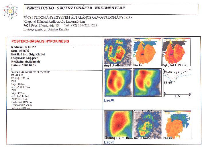
22 VENTRICULO SZCINTIGRÁFIA EREDMÉNYLAP PÉCSI TUDOMÁNYEGYETEM ÁLTALÁNOS ORVOSTUDOMÁNYI KAR Központi Klinikai Radioizotóp Laboratórium 7624 Pécs, Ifjúság útja 13. Tel.: (72) /1229 Intézetvezető: dr. Zámbó Katalin NORMAL FUNCTION OF THE LEFT VENTRICLE Kódszám: KE0351 Szül.: Beküldô int.: Szigetvár Bel. Diagnosis: St.p.inf.myoc. Értékelte: Dr.Schmidt Dátum: : SZIVKAMRA-GÖRBE ELEMZÉSE EF: 64.1 % ES ideje: 398 ms PER ideje: 180 ms seb.: EDV/s PFR ideje: 550 ms seb.: 2.18 EDV/s PFR/PER: 0.87 Ciklusidö: 944 ms Frekvencia: 64/min Infl. pont: 768 ms Lao30 Lao70
23
24
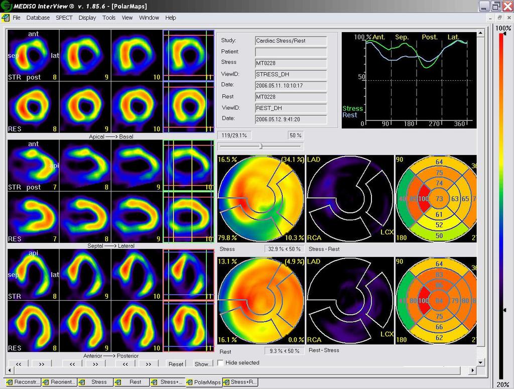
25
 326-222/1229 Intézetvezető: dr. Zámbó Katalin HUGE PARADOX WALL-MOTION ON THE APEX Kódszám: KE0100 Szül.")

26 VENTRICULO SZCINTIGRÁFIA EREDMÉNYLAP PÉCSI TUDOMÁNYEGYETEM ÁLTALÁNOS ORVOSTUDOMÁNYI KAR Központi Klinikai Radioizotóp Laboratórium 7624 Pécs, Ifjúság útja 13. Tel.: (72) /1229 Intézetvezető: dr. Zámbó Katalin HUGE PARADOX WALL-MOTION ON THE APEX Kódszám: KE0100 Szül.: Beküldô int.: Komló Bel. Diagnosis: ISZB Értékelte: Dr.Schmidt Dátum: : SZIVKAMRA-GÖRBE ELEMZÉSE EF: 25.2 % ES ideje: 270 ms PER ideje: 137 ms seb.: EDV/s PFR ideje: 392 ms seb.: 1.26 EDV/s PFR/PER: 0.79 Ciklusidö: 592 ms Frekvencia: 101/min Lao30 Lao70

27 SPECT Equipments II. (Single Photon Emission Computer Tomograph)
28 The principle of the SPECT patient The detectors whirl around the patient and makes pictures from different steps. The transversal, sagittal and coronal slices of the organ are reconstruated and reorientated by computer program.
29 Myocardial perfusion imaging in rest The myocardium is labelled by radioactive agent: - 99mTc-MIBI, 99mTc-tetrofosmin: mitochondria - 201Tl-clorid: Na-K pump Reconstruated and reorientated slices are created from the left ventricle by SPECT or SPECT/CT (attenuation correction) The impairment of the myocardial perfusion is indicated by decreased activity or lack of the activity Indications: myocardial infarction
30 The slices of the myocardium made by SPECT

31 The transversal, sagittal and coronal slices of the myocardium

32 Enlarged left ventricle, increased background

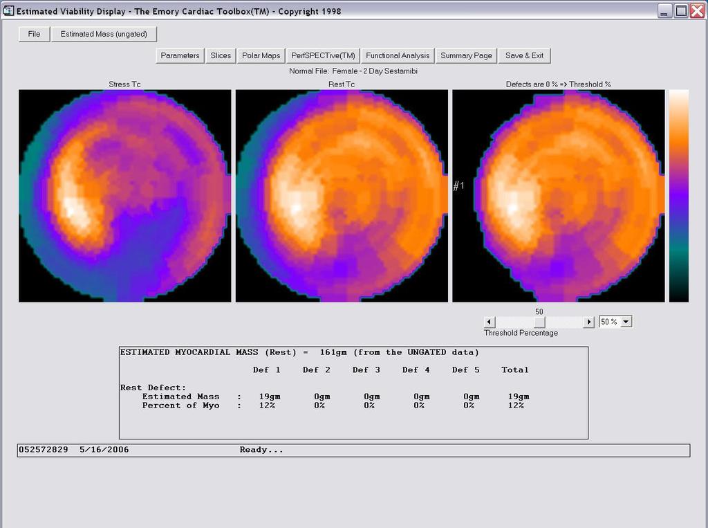
33 Septal-basal hypoperfusion + right ventricle Polar map: short-axis circumferential profiles for quantification of tracer uptake in percentage of the reference zone.

34 Septal + infero-septal + antero-septal hypoperfusion
35 Stress/rest myocardial perfusion study Physical or pharmacological stress (Dipyridamol) is applied The isotope is administered at peak of the stress» SPECT-imaging Rest SPECT-imaging is on the same day (Tl), or one day later (Tc-MIBI) Reversible ischaemy: stress/rest mismatch Fixed abnormality (scar): stress/rest match

36 Normal myocardial perfusion

37 CAD in the infero-lateral wall

38 Apical and antero-apical transient ischaemy

39 Transient ischaemy in the basal part of the septum

40 Infero-basal transient ischaemy

41 Hudge infero-lateral transient ischaemia
42 Polar maps, profil-curves

43 Polar maps of reversibility (Emory toolbox)
")
44 3D (three dimension) imaging
45 Quantitativ evaluation
46 Report

47 Inferior, infero-septal, infero-lateral fix perfusion defect (scar)

48 Inferior, infero-septal, infero-lateral fix perfusion defect (scar)

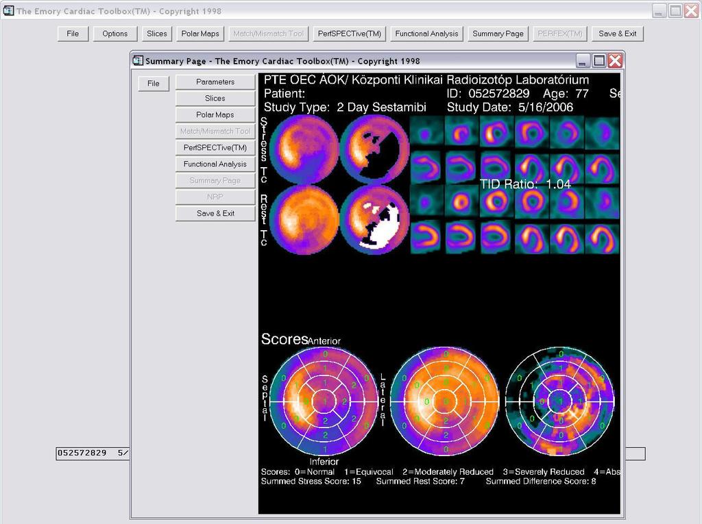
49 Report (Emory toolbox)

50 Reverse perfusion: is impaired in rest DPD - steal mechanism X syndrom, myocardial bridge

51 Gated examination: quantitativ evaluation of thickening, global and regional EF
")
52 Equipments III. - SPECT/CT (Single Photon Emission Computer Tomograph) + CT hybrid: multimodality, fused imaging

53 Myocardium perfusion SPECT/CT study in female

54 Influence of attenuation correction of the anterior wall in female Basic AC Basic AC Basic AC

55 Influence of attenuation correction of the inferior wall in male

56 3D SPECT/CT imaging: stenosis of circumflexa hypoperfusion in the apical part of the left ventricle

57 3D SPECT/CT imaging: stenosis of proximal part of the LAD antero-basalis hypoperfusion


58 4. pts LAD, Cx, RCA sten. s i g n i f i c a n c e? Calcium score: LAD Cx RCA MSCT result questionable : high coron. calcification saccular dilatation of coronaries Kerecsen Gábor


59 4. pts Stress MPS - 3D Stress MPS + MSCT Mild ischemia on inferior wall Answer : the ischemia is mild medical therapy Balogh Ildikó

60 4. pts Balogh I. Kerecsen G.
61 Viability examination of the myocardium When similar fix defect is found in stress and rest situation (scar, hibernated, stunned myocardium) to assess the possibility for succesful revascularization. 201Tl-chlorid has a specific redistribution pattern after 3-4 hours in rest, which depends on the wash-out from the myocytes. After the reinjection the activity of the myocardium depends on primarily the perfusion by the coronary arteries.
62 Function Perfusion Metabolisme Stunned Hibernated Reversibile! Revascularization Necrosis (scar) Irreversibile!

63 Viability examination by 201Tl-clorid: stress-redistribution
64 Viability examination by 201Tl-clorid: redistribution-reinjection

65 Viability examination by 201Tl-clorid stress redistr. reinjection
66 +Beta (positron) radiation - too many protons are in the nucleus - its life is very short, when it slows down, it combines with a normal electron in a process known annihilation, which destroyes both the electron and positron and produces two energetic photons each with 511 kev - they are used for PET examinations - isotopes with ultrashort half-life (11C, 15O, 13N, 18F) - e.g. 18Fluor-FDG to study the metabolic changes of the heart, the brain and the various tumors

67 Equipments III. PET/CT (Positron Emission Tomograph/CT)

68 The principle of the PET Computers Annihilation gamma photons Preprocessing Evaluation, printing Ring detectors


69 PET study MIBI-FDG mismatch Perfusionmetabolic match viability of myocardium scar

70 The coronary angiogram was acquired using CT, while the surface of the myocardium is coloured using the data of the ammonia PET stress perfusion scan done during the PET-CT examination. It can be clearly seen that territories in the distal LAD region are blue, which signifies reduced stress perfusion.

71 SPECT/MRI fused imaging
72 Differential diagnosis Intracardial left-to-right shunt, chronic cor pulmonale: first passage study Acut myocardial infarction, myocardial scar, cardiomyopathy: rest myocardial perfusion study radionuclide ventriculography (MUGA) Pectoral angina, coronary artery disease: stress/rest myocardial perfusion study Viability before revascularization: glucose metabolism by PET/CT

73 Thank you!
Nuclear medicine. Zámbó Katalin Department of Nuclear Medicine
 Nuclear medicine Zámbó Katalin Department of Nuclear Medicine Imaging tehniques Anatomy Physiology Metabolism Molecular X-ray / CT Nuclear medicine / SPECT / PET MRI MR spectroscopy fmri Ultrasound Hybrid
Nuclear pulmonology. Katalin Zámbó Department of Nuclear Medicine
 Nuclear pulmonology Katalin Zámbó Department of Nuclear Medicine Imaging techniques Morphology Physiology Metabolism Molecules X-ray / CT MRI NM - SPECT/ PET MR spectroscopy fmri Ultrasound Hybrid imaging:
Itroduction to the Nuclear Medicine: biophysics and basic principles. Zámbó Katalin Department of Nuclear Medicine
 Itroduction to the Nuclear Medicine: biophysics and basic principles Zámbó Katalin Department of Nuclear Medicine NUCLEAR MEDICINE Application of the radioactive isotopes in the diagnostics and in the
Nuclear medicine studies of the digestiv system. Zámbó Katalin Department of Nuclear Medicine
 Nuclear medicine studies of the digestiv system Zámbó Katalin Department of Nuclear Medicine Imaging tehniques Anatomy Physiology Metabolism Molecular Rtg. / CT PET / SPECT MRI MR spectroscopy fmri Ultrasound
Fundamentals of Nuclear Cardiology. Terrence Ruddy, MD, FRCPC, FACC
 Fundamentals of Nuclear Cardiology Terrence Ruddy, MD, FRCPC, FACC Objectives To understand the Principles of Nuclear Cardiac Imaging Radiotracers Image acquisition and processing Stress protocols To appreciate
SPECT-CT: Τι πρέπει να γνωρίζει ο Καρδιολόγος
 SPECT-CT: Τι πρέπει να γνωρίζει ο Καρδιολόγος Δρ Αναστασία Κίτσιου Διευθύντρια, Καρδιολογική Κλινική, Σισμανόγλειο ΓΝΑ Chair, Education Committee, Section on Nuclear Cardiology & Cardiac CT, EACVI, ESC
NUCLEAR CARDIOLOGY UPDATE
 Nuclear Cardiology David K. Shelton, Jr., MD NUCLEAR CARDIOLOGY UPDATE No Conflicts. No Disclosures. No Smoking. David K. Shelton UCDMC Nuclear Cardiology Nuclear Cardiology Radionuclide Ventriculography
OTHER NON-CARDIAC USES OF Tc-99m CARDIAC AGENTS Tc-99m Sestamibi for parathyroid imaging, breast tumor imaging, and imaging of other malignant tumors.
 DEFINITION OF CARDIAC RADIOPHARMACEUTICAL: A radioactive drug which, when administered for purpose of diagnosis of heart disease, typically elicits no physiological response from the patient. Even though
Pearls & Pitfalls in nuclear cardiology
 Pearls & Pitfalls in nuclear cardiology Maythinee Chantadisai, MD., NM physician Division of Nuclear Medicine, Department of radiology, KCMH Principle of myocardial perfusion imaging (MPI) Radiotracer
Gated blood pool ventriculography: Is there still a role in myocardial viability?
 Gated blood pool ventriculography: Is there still a role in myocardial viability? Oliver C. Alix, MD Adult Clinical and Nuclear Cardiology St. Luke s Medical Centre - Global City Case Presentation A 62-year-old
Rational use of imaging for viability evaluation
 EUROECHO and other imaging modalities 2011 Rational use of imaging for viability evaluation Luc A. Pierard, MD, PhD, FESC, FACC Professor of Medicine Head, Department of Cardiology, CHU Liège, Belgium
SPECT TRACERS Tl-201, Tc-99m Sestamibi, Tc-99m Tetrofosmin
 SPECT TRACERS Tl-201, Tc-99m Sestamibi, Tc-99m Tetrofosmin Elmer Jasper B. Llanes, M.D. Nuclear Cardiology St. Luke s Medical Center Outline Ideal Physiologic Characteristics of MPI radioactive tracers
Cardiovascular nuclear imaging employs non-invasive techniques to assess alterations in coronary artery flow, and ventricular function.
 National Imaging Associates, Inc. Clinical guidelines CARDIOVASCULAR NUCLEAR MEDICINE -MYOCARDIAL PERFUSION IMAGING -MUGA CPT4 Codes: Refer to pages 6-9 LCD ID Number: L33960 J 15 = KY, OH Responsible
1. LV function and remodeling. 2. Contribution of myocardial ischemia due to CAD, and
 1 The clinical syndrome of heart failure in adults is commonly associated with the etiologies of ischemic and non-ischemic dilated cardiomyopathy, hypertrophic cardiomyopathy, hypertensive heart disease,
Rotation: Imaging 2. Nuclear Cardiology (in Imaging 1 and 2)
") Rotation: Imaging 2 Imaging 2 provides addition nuclear cardiology experience and COCATS Level 1 cardiac MRI experience. Fellows administer, process, and read VHVI cardiac nuclear studies with cardiology
Rotation: Imaging 2 Imaging 2 provides addition nuclear cardiology experience and COCATS Level 1 cardiac MRI experience. Fellows administer, process, and read VHVI cardiac nuclear studies with cardiology
Cardiovascular nuclear imaging employs non-invasive techniques to assess alterations in coronary artery flow, and ventricular function.
 National Imaging Associates, Inc. Clinical guidelines CARDIOVASCULAR NUCLEAR MEDICINE -MYOCARDIAL PERFUSION IMAGING -MUGA Original Date: October 2015 Page 1 of 9 FOR CMS (MEDICARE) MEMBERS ONLY CPT4 Codes:
Multiple Gated Acquisition (MUGA) Scanning
 Scanning") Multiple Gated Acquisition (MUGA) Scanning Dmitry Beyder MPA, CNMT Nuclear Medicine, Radiology Barnes-Jewish Hospital / Washington University St. Louis, MO Disclaimers/Relationships Standard of care research
Multiple Gated Acquisition (MUGA) Scanning Dmitry Beyder MPA, CNMT Nuclear Medicine, Radiology Barnes-Jewish Hospital / Washington University St. Louis, MO Disclaimers/Relationships Standard of care research
Radiologic Assessment of Myocardial Viability
 November 2001 Radiologic Assessment of Myocardial Viability Joshua Moss, Harvard Medical School Year III Patient EF 66yo female with a 3-year history of intermittent chest pain previously relieved by sublingual
Click here for Link to References: CMS Website HOPPS CY 2018 Final Rule. CMS Website HOPPS CY2018 Final Rule Updated November 2017.
 Final Compared to 3Q 2017 Rates Medicare Hospital Outpatient Prospective Payment System HOPPS () Nuclear Cardiology Procedures, Radiopharmaceuticals, and Drugs Click here for Link to References: CMS Website
Value of Assessment of Viable and Ischemic Myocardium and Techniques Such as MRI, Radionuclide Imaging
 Chapter 2 Imaging for Viable and Ischemic Myocardium Value of Assessment of Viable and Ischemic Myocardium and Techniques Such as MRI, Radionuclide Imaging Catalin Loghin and K. Lance Gould Introduction
Myocardial viability testing. What we knew and what is new
 Myocardial viability testing. What we knew and what is new Dr B K S Sastry, MD, DM. CARE Hospitals, Hyderabad What is Viability Viability Dysfunctional myocardium subtended by diseased coronary arteries
Medical imaging X-ray, CT, MRI, scintigraphy, SPECT, PET Györgyi Műzes
 Medical imaging X-ray, CT, MRI, scintigraphy, SPECT, PET Györgyi Műzes Semmelweis University, 2nd Dept. of Medicine Medical imaging: definition technical process of creating visual representations about
Tc-99m Sestamibi/Tetrofosmin Stress-Rest Myocardial Perfusion Scintigraphy
 APPROVED BY: Director of Radiology Page 1 of 6 Tc-99m Sestamibi/Tetrofosmin Stress-Rest Myocardial Primary Indications: Evaluation of myocardial perfusion and viability in patients with known or suspected
CHRONIC CAD DIAGNOSIS
 CHRONIC CAD DIAGNOSIS Chest Pain Evaluation 1. Approach to diagnosis of CAD 2. Classification of chest pain 3. Pre-test likelihood CAD 4. Algorithm for chest pain evaluation in women 5. Indications for
Abnormal, Autoquant Adenosine Myocardial Perfusion Heart Imaging. ID: GOLD Date: Age: 46 Sex: M John Doe Phone (310)
") Background: Reason: preoperative assessment of CAD, Shortness of Breath Symptom: atypical chest pain Risk factors: hypertension Under influence: a beta blocker Medications: digoxin Height: 66 in. Weight:
Background: Reason: preoperative assessment of CAD, Shortness of Breath Symptom: atypical chest pain Risk factors: hypertension Under influence: a beta blocker Medications: digoxin Height: 66 in. Weight:
@02-126_Coronary_calcification.ppt. Professor Molecular and Medical Pharmacology
 Assessment of Myocardial Viability Jamshid Maddahi, M.D., FACC, FASNC Professor Molecular and Medical Pharmacology (Nuclear Medicine) and Medicine (Cardiology) David Geffen School of Medicine at UCLA Director,
Cardiac Imaging Tests
 Cardiac Imaging Tests http://www.medpagetoday.com/upload/2010/11/15/23347.jpg Standard imaging tests include echocardiography, chest x-ray, CT, MRI, and various radionuclide techniques. Standard CT and
Fellows on this rotation are expected to attend nuclear conferences and multimodality imaging conference.
 Rotation: Imaging 1 Imaging 1 provides COCATS Level 1 experience for nuclear cardiology (including SPECT and PET) and cardiac CT. Fellows will administer, process, and read cardiac nuclear studies with
Nuclear neurology. Zámbó Katalin Department of Nuclear Medicine
 Nuclear neurology Zámbó Katalin Department of Nuclear Medicine To refresh your memory Brain has a high rate of oxidative metabolism. It has no reserves either of oxygen or of glucose and has a very limited
NUCLEAR CARDIOLOGY AND ADVANCED VASCULAR IMAGING. Joel Kahn, MD, FACC
 NUCLEAR CARDIOLOGY AND ADVANCED VASCULAR IMAGING Joel Kahn, MD, FACC Short History Nuclear Cardiology Hermann blumgart-1927-injected radon to measure circulation time Hal Anger-1952-gamma camera-beginning
Cardiac PET. John Buscombe
 Cardiac PET John Buscombe Why PET? Improved resolution-not really required in cardiology Improved sensitivity this may be important-financially as reduced acquisition time Improved attenuation correction-good
Impaired Regional Myocardial Function Detection Using the Standard Inter-Segmental Integration SINE Wave Curve On Magnetic Resonance Imaging
 Original Article Impaired Regional Myocardial Function Detection Using the Standard Inter-Segmental Integration Ngam-Maung B, RT email : chaothawee@yahoo.com Busakol Ngam-Maung, RT 1 Lertlak Chaothawee,
NUCLEAR MEDICINE OF HEART AND LUNG
 Clinical decision tree in CAD NUCLEAR MEDICINE OF HEART AND LUNG Abnormal pump function Slides of lectures + electronic books: Stress-rest Stress: defect MPI Rest: better or normal Glucose metab? László
Functional aspects of anatomical imaging techniques
 Functional aspects of anatomical imaging techniques Nilendu Purandare Associate Professor & Consultant Radiologist Tata Memorial Centre Functional/metabolic/molecular imaging (radioisotope scanning) PET
Cardiac Imaging. Kimberly Delcour, DO, FACC. Mahi Ashwath, MD, FACC, FASE. Director, Cardiac CT. Director, Cardiac MRI
 Cardiac Imaging Kimberly Delcour, DO, FACC Director, Cardiac CT Mahi Ashwath, MD, FACC, FASE Director, Cardiac MRI Cardiac Imaging Discuss the clinical applications of and indications for: Cardiac CT Nuclear
Conflict of Interest Disclosure
 Comparative Advantages of PET Over SPECT: Is PET Really Better? Timothy M. Bateman M.D. Co-Director, Cardiovascular Radiologic Imaging Mid America Heart Institute Professor of Medicine University of Missouri-Kansas
Prior Authorization for Non-emergency Cardiac Imaging Procedures
 Attention: All Providers Prior Authorization for Non-emergency Cardiac Imaging Procedures The N.C. Medicaid Program is considering implementation of a prior authorization (PA) program for non-emergency
Assessment of Local Myocardial Perfusion in SPECT Images when Bicycle Exercise Test is Noninterpretable
 e 11 Assessment of Local Myocardial Perfusion in SPECT Images when Bicycle Exercise Test is Noninterpretable Ilona Kulakienė, Zigmundas Satkevičius, Juozas Kiudelis, Irena Milvidaitė 1 Kaunas Medical University,
Anthem Blue Cross and Blue Shield Virginia Advanced Imaging Procedures Requiring Precertification Revised 02/13/2013
 Anthem Blue Cross and Blue Shield Virginia Advanced Imaging Procedures Requiring Precertification Revised 02/13/2013 Modality and CT Head CTA Head: Cerebrovascular MRI Head MRA Head: Cerebrovascular Functional
Paradoxical pattern in a patient with previous myocardial infarction F. Mut, M. Kapitan
 Paradoxical pattern in a patient with previous myocardial infarction F. Mut, M. Kapitan Nuclear Medicine Service, Italian Hospital Montevideo, Uruguay Clinical history Woman 66 y.o. Previous MI. Dyspnea
ORIGINAL ARTICLE. Iulia Heinle 1,*, Andre Conradie 2 and Frank Heinle 1
 PJNM 2011, 1:42-48 331691 2011 Pakistan Society of Nuclear Medicine ORIGINAL ARTICLE A clinical and angiographic correlation of myocardial perfusion scintigraphy in the assessment of isolated apical/peri-apical
Reversible defect of 123 I-15-(p-iodophenyl)-9-(R,S)-methylpentadecanoic acid indicates residual viability within infarct-related area
-9-(R,S)-methylpentadecanoic acid indicates residual viability within infarct-related area") ORIGINAL ARTICLE Annals of Nuclear Medicine Vol. 16, No. 3, 183 187, 2002 Reversible defect of 123 I-15-(p-iodophenyl)-9-(R,S)-methylpentadecanoic acid indicates residual viability within infarct-related
ORIGINAL ARTICLE Annals of Nuclear Medicine Vol. 16, No. 3, 183 187, 2002 Reversible defect of 123 I-15-(p-iodophenyl)-9-(R,S)-methylpentadecanoic acid indicates residual viability within infarct-related
Evaluation of myocardial ischaemia
 l2 TOPIC Evaluation of myocardial ischaemia Topic Contents Markers of myocardial injury and infarction 6 Myocardial territories supplied by coronary arteries 8 The 17 segment model 9 Regional assessment
Nuclear Perfusion Imaging of Angina
 January 2002 Nuclear Perfusion Imaging of Angina Davin Quinn HMS III Beth Israel Deaconess Medical Center Radiology Department Goals of Presentation Understand what a perfusion scan is Understand what
Appropriateness of Stress Echocardiography and Nuclear Stress Thallium/Sesta Mibi Testing Methods
 Appropriateness of Stress Echocardiography and Nuclear Stress Thallium/Sesta Mibi Testing Methods Review by Michael Devenport, MS, RDCS Objectives: A basic description and review of appropriate use of
Radionuclides in Medical Imaging. Danielle Wilson
 Radionuclides in Medical Imaging Danielle Wilson Outline Definitions History and development Radionuclide applications & techniques in imaging Conclusion Definition #1 : Radionuclide An unstable nucleus
RADIOTRACERS FOR MYOCARDIAL PERFUSION IMAGING
 RADIOTRACERS FOR MYOCARDIAL PERFUSION IMAGING RAYMOND TAILLEFER, M.D. FRCP(c), ABNM DIRECTOR, DEPARTMENT OF NUCLEAR MEDICINE HOPITAL ST-JEAN-SUR-RICHELIEU Disclosures to Report: Grant Research Support:
EMPHISIS ON PHYSIOLOGY PHYSIOLOGY REQUIRES TIME QUALITATIVE vs. QUANTITATIVE ISOTOPES TO RADIOPHARMACEUTICALS
 1926 Herman Blumgard used solutions of radon gas to measure what he called velocity of the circulation. 1927 Blumberg and Soma Weiss wrote article in (Journal of Clinical Investigation) 1929 Werner Forssmann
Zahid Rahman Khan, MD(USA), MS Diplomate American Board of Nuclear Medicine Consultant t Nuclear Medicine
, MS Diplomate American Board of Nuclear Medicine Consultant t Nuclear Medicine") Zahid Rahman Khan, MD(USA), MS Diplomate American Board of Nuclear Medicine Consultant t Nuclear Medicine i North West Armed Forces Hospital Tabuk, KSA. 1 Introduction Hibernation: Term coined by Rahimtoola
Zahid Rahman Khan, MD(USA), MS Diplomate American Board of Nuclear Medicine Consultant t Nuclear Medicine i North West Armed Forces Hospital Tabuk, KSA. 1 Introduction Hibernation: Term coined by Rahimtoola
PET myocard perfusion & viability Riemer Slart
 PET myocard perfusion & viability Riemer Slart Nuclear Medicine Physician Dept. of Nuclear Medicine and Molecular Imaging University Medical Center Groningen, the Netherlands Professor in Molecular Imaging,
Hospital, 6 Lukon Road, Lukong Town, Changhua Shien, Taiwan 505, Taiwan.
 Volume 1, Issue 1 Image Article Resolution of Inferior Wall Ischemia after Successful Revascularization of LAD Lesion: The Value of Myocardial Perfusion Imaging in Guiding Management of Multi-vessel CAD
The Integral Role of Metabolic and Perfusion Imaging in Assessment of Myocardial Scar: Comparison between 18F-FDG PET and 99Tc-Sestamibi
 Med. J. Cairo Univ., Vol. 82, No. 1, June: 285-289, 2014 www.medicaljournalofcairouniversity.net The Integral Role of Metabolic and Perfusion Imaging in Assessment of Myocardial Scar: Comparison between
The Value of Tc-99m MIBI Washout Rate in Detection of Ischemia compared with standard Myocardial Perfusion Imaging
 Egyptian J. Nucl. Med., Vol. 10, No. 2, Dec 2014 18 Original article, Cardiology The Value of Tc-99m MIBI Washout Rate in Detection of Ischemia compared with standard Myocardial Perfusion Imaging Moustafa,
Cardiovascular Imaging
 Cardiovascular Imaging Cardiovascular Imaging Cardio and Vascular Imaging Vascularization / Angiogenesis Cardiovascular Imaging metabolic imaging of the heart myocardial perfusion imaging Cardiac CT Vascularization
Use of Nuclear Cardiology in Myocardial Viability Assessment and Introduction to PET and PET/CT for Advanced Users
 Use of Nuclear Cardiology in Myocardial Viability Assessment and Introduction to PET and PET/CT for Advanced Users February 1 5, 2011 University of Santo Tomas Hospital Angelo King A-V Auditorium Manila,
Nuclear Cardiology Reimbursement. Todd Lamb, BS, AS, CNMT Clinical Operations Mgr Regions Hospital St. Paul, MN
 Nuclear Cardiology Reimbursement Todd Lamb, BS, AS, CNMT Clinical Operations Mgr Regions Hospital St. Paul, MN Slides are not to be reproduced without the permission of the author Slides are not to be
MPI Overview. Artifacts and Pitfalls in MPI. Acquisition and Processing. Peeyush Bhargava MD, MBA
 Artifacts and Pitfalls in MPI MPI Overview Peeyush Bhargava MD, MBA www.nuclearmd.com ~ 40% of all Nuclear Medicine Procedures ~ 9 million procedures every year in US >95% are SPECT,
J. Schwitter, MD, FESC Section of Cardiology
 J. Schwitter, MD, FESC Section of Cardiology CMR Center of the CHUV University Hospital Lausanne - CHUV Switzerland Centre de RM Cardiaque J. Schwitter, MD, FESC Section of Cardiology CMR Center of the
Cigna - Prior Authorization Procedure List Cardiology
 Cigna - Prior Authorization Procedure List Cardiology Category CPT Code CPT Code Description 33206 Insertion of new or replacement of permanent pacemaker with transvenous electrode(s); atrial 33207 Insertion
Description MRI, TMJ C T Head Without Contrast C T Head With Contrast C T Head Without & With Contrast
 s Requiring Prior Authorization for the Advanced Imaging 70336 MRI, TMJ 70450 C T Head Without Contrast 70460 C T Head With Contrast 70470 C T Head Without & With Contrast 70480 C T Orbit Without Contrast
ASSOCIATION BETWEEN LEFT VENTRICULAR EJECTION FRACTION (LVEF) IN PATIENTS WITH REGIONAL ISCHEMIA AND INFARCTION ON MYOCARDIAL PERFUSION IMAGES
 IN PATIENTS WITH REGIONAL ISCHEMIA AND INFARCTION ON MYOCARDIAL PERFUSION IMAGES") University of New Mexico UNM Digital Repository Biomedical Sciences ETDs Electronic Theses and Dissertations 7-1-2015 ASSOCIATION BETWEEN LEFT VENTRICULAR EJECTION FRACTION (LVEF) IN PATIENTS WITH REGIONAL
University of New Mexico UNM Digital Repository Biomedical Sciences ETDs Electronic Theses and Dissertations 7-1-2015 ASSOCIATION BETWEEN LEFT VENTRICULAR EJECTION FRACTION (LVEF) IN PATIENTS WITH REGIONAL
Qualitative and Quantitative Assessment of Perfusion
 APCDE 2011 Qualitative and Quantitative Assessment of Perfusion Hyun Ju Yoon Chonnam National University Hospital Gwangju, Korea ISCHEMIC CASCADE Blood flow mismatch Perfusion defects on nuclear imaging,
CARDIAC PET PERFUSION IMAGING with RUBIDIUM-82
 CARDIAC PET PERFUSION IMAGING with RUBIDIUM-82 Pr Denis AGOSTINI Président du Groupe de Cardiologie Nucléaire et IRM CHU Caen Bordeaux 2006 Cardiac Perfusion-Metabolism Mismatch with PET Cumulative Survival
Nuclear Cardiology Cardiac Myocardial Perfusion with 82 Rb. Dominique Delbeke, MD, PhD Vanderbilt University Medical Center Nashville, TN
 Nuclear Cardiology Cardiac Myocardial Perfusion with 82 Rb Dominique Delbeke, MD, PhD Vanderbilt University Medical Center Nashville, TN VUMC PET/CT conference 2009 82 Rb Cardiac Perfusion PET 82 Rb is
Nuclear Cardiology Pierre-Yves MARIE Department of Nuclear Medicine CHU-Nancy, FRANCE.
 Nuclear Cardiology Pierre-Yves MARIE Department of Nuclear Medicine CHU-Nancy, FRANCE. Nuclear Cardiology I - A remaining need of a functional information on myocardial perfusion II - The future: - combining
PHYSICS 2: HSC COURSE 2 nd edition (Andriessen et al) CHAPTER 20 Radioactivity as a diagnostic tool (pages 394-5)
 CHAPTER 20 Radioactivity as a diagnostic tool (pages 394-5)") PHYSICS 2: HSC COURSE 2 nd edition (Andriessen et al) CHAPTER 20 Radioactivity as a diagnostic tool (pages 394-5) 1. (a) A radioisotope is an isotope that is unstable and will emit particles from the nucleus
PHYSICS 2: HSC COURSE 2 nd edition (Andriessen et al) CHAPTER 20 Radioactivity as a diagnostic tool (pages 394-5) 1. (a) A radioisotope is an isotope that is unstable and will emit particles from the nucleus
Dual-Tracer Gated Myocardial Scintigraphy
 APPROVED BY: Director of Radiology Page 1 of 7 Dual-Tracer Gated Myocardial Scintigraphy Primary Indications: Evaluation of myocardial perfusion and viability in patients with known or suspected coronary
Nuclear Medicine: Manuals. Nuclear Medicine. Nuclear imaging. Emission imaging: study types. Bone scintigraphy - technique
 Nuclear Medicine - Unsealed radioactive preparations the tracer mixes with the patients body fluids on a molecular level (e.g. after intravenous injection) - 3 main fields: - In vitro : measuring concentrations
Typical chest pain with normal ECG
 Typical chest pain with normal ECG F. Mut, C. Bentancourt, M. Beretta Nuclear Medicine Service, Asociacion Española Montevideo, Uruguay Clinical history Male 41 y.o. Overweight, hypertension, high cholesterol,
Cardiac SECTION 1. Introduction Approach to Cardiac Imaging 4
 SECTION 1 Introduction Approach to Imaging 4 Function and Coronary Artery Disease Left Ventricular Function 6 Myocardial Infarction and Ischemia 10 Myocardial Viability 16 Right-to-Left Shunt 20 Approach
Typical PET Image. Elevated uptake of FDG (related to metabolism) Lung cancer example: But where exactly is it located?
 Lung cancer example: But where exactly is it located?") Typical PET Image Elevated uptake of FDG (related to metabolism) Lung cancer example: But where exactly is it located? PET/CT Oncology Imaging Anatometabolic fusion images are useful in the management
Typical PET Image Elevated uptake of FDG (related to metabolism) Lung cancer example: But where exactly is it located? PET/CT Oncology Imaging Anatometabolic fusion images are useful in the management
A Snapshot on Nuclear Cardiac Imaging
 Editorial A Snapshot on Nuclear Cardiac Imaging Khalil, M. Department of Physics, Faculty of Science, Helwan University. There is no doubt that nuclear medicine scanning devices are essential tool in the
Cigna - Prior Authorization Procedure List: Radiology & Cardiology
 Cigna - Prior Authorization Procedure List: Radiology & Cardiology Product Category CPT Code CPT Code Description Radiology MR 70336 MRI Temporomandibular Joint(s), (TMJ) Radiology CT 70450 CT Head or
AMERICAN IMAGING MANAGEMENT
 2012 CPT Codes Computerized Tomography (CT) CPT Description Abdomen 74150 CT abdomen; w/o 74160 CT abdomen; with 74170 CT abdomen; w/o followed by Chest 71250 CT thorax; w/o 71260 CT thorax; with 71270
AMERICAN IMAGING MANAGEMENT
 2010 BCBS of Georgia CPT Codes With Grouper Numbers Computerized Tomography (CT) CPT Description Abdomen 74150 CT abdomen; w/o contrast 6 74160 CT abdomen; with contrast 74170 CT abdomen; w/o contrast
Imaging and heart failure
 Imaging and heart failure Jeroen J Bax Dept of Cardiology Leiden Univ Medical Center The Netherlands Davos, feb 2013 Research grants: Medtronic, Biotronik, Boston, St Jude, BMS imaging, GE Healthcare,
Myocardial Perfusion: Positron Emission Tomography
 Myocardial Perfusion: Positron Emission Tomography TH. Schindler, MD University Hospitals of Geneva, Cardiovascular Center, Geneva, Switzerland ESC 2010 Stockholm Personal Disclosure Research Grant support
evicore cardiology procedures and services requiring prior authorization
 evicore cardiology procedures and services requiring prior authorization Moda Health Commercial Group and Individual Members* *Check EBT to verify member enrollment in evicore program Radiology Advanced
Le FDG dans l évaluation de la viabilité : Quelle place par rapport aux autres traceurs?
 Le FDG dans l évaluation de la viabilité : Quelle place par rapport aux autres traceurs? Pierre-Yves MARIE, Médecine Nucléaire, CHU de Nancy PET Gated-SPECT MRI FDG imaging: effect of dietary status fasting
Je bénéficie régulièrement de fonds privés, dans le cadre de projets de recherche ou d activités de formation.
 Je bénéficie régulièrement de fonds privés, dans le cadre de projets de recherche ou d activités de formation. Ces fonds proviennent essentiellement d industriels travaillant dans les domaines de l imagerie
Cardiology for the Practitioner Advanced Cardiac Imaging: Worth the pretty pictures?
 Keenan Research Centre Li Ka Shing Knowledge Institute Cardiology for the Practitioner Advanced Cardiac Imaging: Worth the pretty pictures? Howard Leong-Poi, MD, FRCPC Associate Professor of Medicine St.
Nuclear medicine in general practice. Dr Reza Garzan MD, FRACP, FAANMS
 Nuclear medicine in general practice Dr Reza Garzan MD, FRACP, FAANMS Myocardial perfusion study Bone scans in general practice Thyroid scans in general practice Gamma camera Detection of gamma rays Myocardial
MUGA Scan. A Patient s Guide. Copyrighted Material. HeartWise Patient Education
 MUGA Scan Copyrighted Material HeartWise Patient Education 800-747-1606 A Patient s Guide What Is a MUGA Scan? A multiple gated acquisition (or MUGA) scan is a test that uses a radioactive substance, called
Evidence-Based Management of CAD: Last Decade Trials and Updated Guidelines
 Evidence-Based Management of CAD: Last Decade Trials and Updated Guidelines Enrico Ferrari, MD Cardiac Surgery Unit Cardiocentro Ticino Foundation Lugano, Switzerland Conflict of Interests No conflict
CONFUSION IN CARDIAC TESTING. Bilal Aijaz M.D FACC FSCAI
 CONFUSION IN CARDIAC TESTING Bilal Aijaz M.D FACC FSCAI WHY DOES CARDIOLOGY HAVE SO MANY TESTS? to create confusion (of course) to generate more business (maybe?) to accommodate the ever expanding patient
BlueAdvantage SM. & BlueChoice SM Radiology Prior Authorization Program Code List CPT /HCPS
 BlueAdvantage SM & BlueChoice SM Radiology Prior Authorization Program Code List CPT /HCPS 70336 MRI TMJ 70450 CT Head Without Contrast 70460 CT Head With Contrast 70470 CT Head Without & With Contrast
Cardiac Diagnostics Workshop. Lori Savard NP Cardiology Update 2015
 Cardiac Diagnostics Workshop Lori Savard NP Cardiology Update 2015 Disclosure of Commercial Support Potential for conflict(s) of interest: none Objectives Increase understanding regarding stable IHD and
Sung A Chang Department of Internal Medicine, Division of Cardiology, Sungkyunkwan University School of Medicine, Samsung Medical Center
 CMR Perfusion and Viability A STICH Out of Time? Sung A Chang Department of Internal Medicine, Division of Cardiology, Sungkyunkwan University School of Medicine, Samsung Medical Center Can Imaging Improve
45 Hr PET Registry Review Course
 45 HR PET/CT REGISTRY REVIEW COURSE Course Control Document Timothy K. Marshel, MBA, R.T. (R), (N)(CT)(MR)(NCT)(PET)(CNMT) The PET/CT Training Institute, Inc. SNMMI-TS 028600-028632 45hr CEH s Voice Credits
QCVC Committees Scientific Activities Central Hall General Information FAC. SPECT tomography has the advantage of quantifying biventricular volumes.
 QCVC Committees Scientific Activities Central Hall General Information FAC Thematic Units Arrhythmias and Electrophysiology Basic Research Bioengineering and Medical Informatics Cardiac Surgical Intensive
Quantification of Right Ventricular Function in Pulmonary Hypertension using Cardiac PET Images
 Quantification of Right Ventricular Function in Pulmonary Hypertension using Cardiac PET Images By Simisani Takobana A thesis submitted to the Faculty of Graduate and Postdoctoral Affairs in partial fulfillment
Hybrid Imaging Improving Nuclear Cardiology Practice
 Lippincott Williams & Wilkins, - Nuclear Medicine teaching File, 2009 Hybrid Imaging Improving Nuclear Cardiology Practice João V. Vitola, MD, PhD Cardiologist and Nuclear Medicine Physician Quanta Diagnostico
Codes Requiring Authorization from MedSolutions (MSI): Updated 3/2014
: Updated 3/2014") s Requiring Authorization from MedSolutions (): Updated 3/2014 0042T Cerebral Perfusion Analysis using CT with contrast 0159T CAD, including computer algorithm analysis, BREAST MRI 0195T prepare interspace,
s Requiring Authorization from MedSolutions (): Updated 3/2014 0042T Cerebral Perfusion Analysis using CT with contrast 0159T CAD, including computer algorithm analysis, BREAST MRI 0195T prepare interspace,
Previous MI with no intervention
 Previous MI with no intervention F. Mut, M. Beretta Nuclear Medicine Service, Asociacion Española Montevideo, Uruguay Clinical history Woman 68 y.o. Recent acute MI (3 weeks) with no intervention. Discharged
Diagnostic Imaging Utilization Management and Consultation Management Programs Imaging Code Listing for Connecticut, Maine and New Hampshire
 Diagnostic Imaging Utilization Management and Consultation Management Programs Imaging Code Listing for Connecticut, Maine and New Hampshire The grid below contains the CPT * codes that are subject to
Photon Attenuation Correction in Misregistered Cardiac PET/CT
 Photon Attenuation Correction in Misregistered Cardiac PET/CT A. Martinez-Möller 1,2, N. Navab 2, M. Schwaiger 1, S. G. Nekolla 1 1 Nuklearmedizinische Klinik der TU München 2 Computer Assisted Medical
Atypical pain and normal exercise test
 Atypical pain and normal exercise test F. Mut, M. Beretta Nuclear Medicine Service, Asociacion Española Montevideo, Uruguay Clinical history 67-year old male with several coronary risk factors. Atypical
MRI ACS-ben. Tamás Simor MD, PhD, Med Hab. University of Pécs, Heart Institute
 MRI ACS-ben Tamás Simor MD, PhD, Med Hab Time Course of Changes in Infarct Size, Viable Myocardium, and LV Mass After Reperfused and Nonreperfused MI Blue lines denote reperfused myocardial infarction
05/02/ CPT Preauthorization Groupings Effective May 2, Computerized Tomography (CT) Abdomen 6. CPT Description SEGR CT01
 Abdomen 6. CPT Description SEGR CT01") Computerized Tomography (CT) 6 & 101 5 Upper Extremity 11 Lower Extremity 12 Head 3 Orbit 1 Sinus 2 Neck 4 7 Cervical Spine 8 Thoracic Spine 9 Lumbar Spine 10 Colon 13 CPT Preauthorization Groupings CPT
Computerized Tomography (CT) 6 & 101 5 Upper Extremity 11 Lower Extremity 12 Head 3 Orbit 1 Sinus 2 Neck 4 7 Cervical Spine 8 Thoracic Spine 9 Lumbar Spine 10 Colon 13 CPT Preauthorization Groupings CPT
Department of Nuclear Medicine with Positron Emission Tomography
 (PET) Unit [1] Contact information: Registration: +48 41 367 4850 Main office: +48 41 367 4860 Fax: +48 41 367 4887 e-mail: zmnsco@onkol.kielce.pl [2] Head of the Department: Professor Janusz Braziewicz