Nuclear neurology. Zámbó Katalin Department of Nuclear Medicine
|
|
|
- Scot Ross
- 6 years ago
- Views:
Transcription
1 Nuclear neurology Zámbó Katalin Department of Nuclear Medicine
2 To refresh your memory Brain has a high rate of oxidative metabolism. It has no reserves either of oxygen or of glucose and has a very limited capacity for anaerobic metabolism. Therefore normal brain function is critically dependent on maintenance of an adequat blood supply. There is a linear association between glucose metabolism and functional activity of the brain. Normal cerebral blood perfusion: ml/min/100g Cortex: ml/min/100g White matter: ml/min/100g No EEG activity: below 17 ml Failure of Na/K ATP-ase: below 10 ml
3 Methods Brain scintigraphy - damaged blood-brain barrier (BBB) Brain perfusion study - vascular lesions (SPECT/CT) Examination of cerebrospinal fluid (CSF) - (dynamics, liquorrhoea) Receptor studies - gamma or positron emitting isotopes (SPECT/CT, PET/CT) Glucose-, aminoacid metabolism, quantitativ CBF examination (PET/CT)
4 Brain scintigraphy Blood-brain barrier (BBB): lipid-protein double layer - protective function Radiopharmaceutical: MBq 99mTc-DTPA i.v., 40 min. waiting 4 projection - static scans (A, P, right, left) + combined with SPECT or SPECT/CT scan (dual-head SPECT) Abnormalities of permeability indicate disruption of BBB (tumor, vasculare lesion), but CT and MR with contrast show more detailed anatomical correlations Historical role: confirmation of brain death

5 Normal brain scintigraphy RIGHT LATERAL LEFT LATERAL

6 Cerebral metastases in the right hemisphere

7 SPECT (Single Photon Emission Computer Tomograph) The transversal, sagittal and coronal slices of the organ are reconstruated and reorientated by the computer program.

8 Multiple metastatic lesion in the left hemisphere of the brain SPECT scan

9 Multiplex brain metastases in melanoma malignum SPECT imaging

10 Cerebral embolisation in the left hemisphere SPECT scan
11 Cerebral blood flow and perfusion Lipophilic tracer is enriched in the gray matter (800 MBq 99mTc-HM-PAO hexamethylpropylene amine oxime) Advantage of dual-head SPECT = faster acquisition Reconstructed and reorientated transversal, sagittal and coronal slices are made Decreased/absent activity indicates the perfusion abnormalities
12 Indications of CBF SPECT Evaluation of cerebrovascular disease (TIA, stroke, AVM, migraine) Dementias (Alzheimer, Pick) Preoperative detection of seizure focus in epilepsy Suspected brain trauma Substance abuse Inflammations, infections Diagnosis of brain death

13 Transversal section of the human brain 1. Visual cortex 2. Occipital lobe 3. Thalamus 4. Frontal lobe 5. N. caudatus 6. Temporal lobe 7. Lateral ventricle

14 Transversal, sagittal and coronal slices of the normal brain by SPECT
15 SPECT/CT (Multimodality!)


16 Different slices of the brain by SPECT/CT

17 Perfusion abnormalities in different brain disorders

18 Bilateral parieto-occipital hypoperfusion Transversal Sagittal Coronal

19 Bilateral parieto-occipital hypoperfusion CT SPECT SPECT/CT

20 Perfusion defect (stroke) in the right occipital region CT SPECT SPECT/CT

21 Occlusion of the right carotid artery Pre-operative Post-operative

22 Dementia: Pick disease

23 Hyperperfusion Migraine Before treatment After treatment

24 51 y., female patient. Complain: numbness of the left arm. Examination: facio-brachial hypalgesia on the left side, accentuated deep tendon reflexes, mild dysmetry, pronation in Barre position (two columns from left: after DP stress)
25 42 y., female patient. Examination: right-sided hemihypalgesia, latent paresis, accentuated deep tendon reflexes, mild dysmetry and dysdiadochokinesis (two column from left: after DP stress)

26 Epilepsy CT scan Ictal Interictal

27 Therapy of epilepsy in the XII. Century
 into the third ventricle, and then through the mesencephalic duct (cerebral aqueduct) into the fourth")
28 Circulation of the cerebrospinal fluid (CSF) It is produced in the brain by modified ependymal cells in the choroid plexus. It circulates from the choroid plexus through the interventricular foramina (foramen of Monro) into the third ventricle, and then through the mesencephalic duct (cerebral aqueduct) into the fourth ventricle, where it exits through two lateral apertures (foramina of Luschka) and one median aperture (foramen of Magendie). It then flows through the cerebromedullary cistern down the spinal cord and over the cerebral hemispheres.
29 Examination of CSF Radiopharmaceutical: 500 MBq 99mTc-DTPA via intratechal route (lumbal or cisternal) Whole body scan + static lateral scans 1, 3, 6, 24 hours after administration Normal dynamic: cisterna magna-3 hours later, flow to the convexity-6 hours later, predominantly the convexity is shown-24 hours later, no activity in the lateral ventricles! Abnormal: - slow dynamic (spinal tumor) - hydrocephalus: reflux only lateral ventricles


30 Liquor dynamic investigation in a patient with reflux (lumbal administration) 1 hour 3 hrs 6 hrs 24 hrs right left 3 hrs 6 hrs 24 hrs

31 Investigation of liquorrohea (cisternal administration)
.")
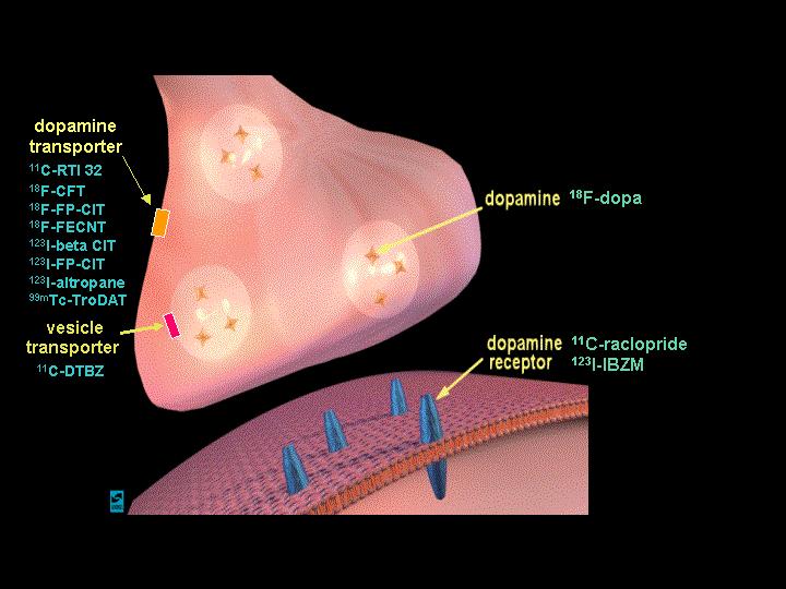
32 Theory of presynaptic dopamin receptor scintigraphy DATSCAN binds to the dopamine transporters (DAT), which are on the neurons in specific areas of the brain. When neuronal degeneration is present, the number of DAT are significantly reduced (for example in patients with Parkinson's disease). By detecting the binding of DATSCAN to the dopamine transporters will be a clear visualisation of dopamine transporter integrity and consequently an accurate diagnosis of disease.
 in a patient with")

33 Dopamin receptor examination (DATSCAN ) in a patient with Parkinson s disease Normal distribution Absence of receptors

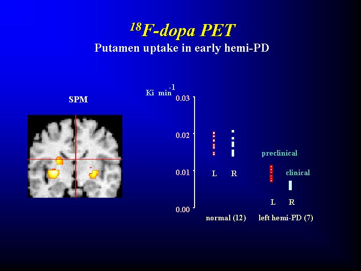
34 Presynaptic dopamin receptor examination in different disorders
 This is the today s")
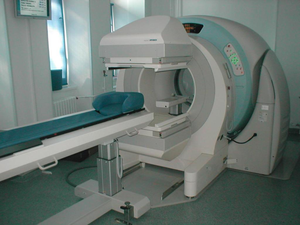
35 PET/CT (positron emission tomograph) This is the today s method
36 How the PET works: - The injected radiopharmaceutical is a positron emitted substance, the equipment is capable to detect the annihilizationrays (two gamma-photons with 511 kev) - Usage of isotopes with ultrashort half-life ( 11 C, 15 O, 13 N, 18 F), which are suitable for measurement of metabolic processes
37 Neuroreceptor studies

38
39
40 Indications of PET/CT Differential diagnosis of Parkinson s disease Dementia Tumors Presurgical localization of epileptic foci Therapy monitoring Neuroreceptor imaging Task-related mapping of the human cortex
41
42
43
 18F-FDG 150-butanol")
44 Brain PET with different ligands (normal uptake and distribution) 18F-FDG 150-butanol 11C-flumazenil

45 Alzheimer disease: decreased bilateral temporal glucose metabolism

46 Epileptic focus: decreased activity 11C-flumazenil 18F-FDG

47 11C-metionin uptake: microadenoma

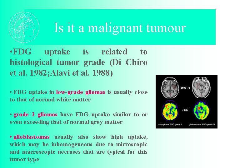
48 18F-FDG uptake after surgery of a recurrent tumor

49 Revealing of recurrence of a parasagittal meningeoma by 18F-FDG

50 18F-FDG PET-MR fused scan of a glioma s recurrence after surgery

51 Evaluation of therapeutic effects by 18F-FDG Stroke after vinpocetin infusion (2weeks later)

52 Mapping of brain function Task-related functional PET investigations

53 Methionin PET and MRI fused imaging: size of recurrent tumor after surgery

54 Thank you for your attention! Loris Marazzi: Your brain is your power
Laura Tormoehlen, M.D. Neurology and EM-Toxicology Indiana University
 Laura Tormoehlen, M.D. Neurology and EM-Toxicology Indiana University Disclosures! No conflicts of interest to disclose Neuroimaging 101! Plain films! Computed tomography " Angiography " Perfusion! Magnetic
Applicable Neuroradiology
 For the Clinical Neurology Clerkship LSU Medical School New Orleans Amy W Voigt, MD Clerkship Director Introduction The field of Radiology first developed following the discovery of X-Rays by Wilhelm Roentgen
Brain Meninges, Ventricles and CSF
 Brain Meninges, Ventricles and CSF Lecture Objectives Describe the arrangement of the meninges and their relationship to brain and spinal cord. Explain the occurrence of epidural, subdural and subarachnoid
Nuclear pulmonology. Katalin Zámbó Department of Nuclear Medicine
 Nuclear pulmonology Katalin Zámbó Department of Nuclear Medicine Imaging techniques Morphology Physiology Metabolism Molecules X-ray / CT MRI NM - SPECT/ PET MR spectroscopy fmri Ultrasound Hybrid imaging:
CEREBRAL BLOOD FLOW AND METABOLISM
 Supported by: HURO/0901/069/2.3.1 HU-RO-DOCS CEREBRAL BLOOD FLOW AND METABOLISM Part 3 Modern imaging methods SPECT, PET, nmri History of Nuclear Medicine Starts with the invention of the X-ray 1946: radioactive
Itroduction to the Nuclear Medicine: biophysics and basic principles. Zámbó Katalin Department of Nuclear Medicine
 Itroduction to the Nuclear Medicine: biophysics and basic principles Zámbó Katalin Department of Nuclear Medicine NUCLEAR MEDICINE Application of the radioactive isotopes in the diagnostics and in the
SCINTIGRAPHY OF THE CENTRAL NERVOUS SYSTEM Part 1: Introduction and BBB studies
 SCINTIGRAPHY OF THE CENTRAL NERVOUS SYSTEM Part 1: Introduction and BBB studies George N. Sfakianakis MD Professor of Radiology and Pediatrics Director, Division of Nuclear Medicine October 2009 FIRST
MRI and CT of the CNS
 MRI and CT of the CNS Dr.Maha ELBeltagy Assistant Professor of Anatomy Faculty of Medicine The University of Jordan 2018 Computed Tomography CT is used for the detection of intracranial lesions. CT relies
Nuclear medicine studies of the digestiv system. Zámbó Katalin Department of Nuclear Medicine
 Nuclear medicine studies of the digestiv system Zámbó Katalin Department of Nuclear Medicine Imaging tehniques Anatomy Physiology Metabolism Molecular Rtg. / CT PET / SPECT MRI MR spectroscopy fmri Ultrasound
Non-Invasive Techniques
 Non-Invasive Techniques Key: Does not hurt the organism Psychology 372 Physiological Psychology Steven E. Meier, Ph.D. Listen to the audio lecture while viewing these slides or view the video presentation
Non-Invasive Techniques
 Many Procedures Non-Invasive Techniques Key: Does not hurt the organism Psychology 372 Physiological Psychology Steven E. Meier, Ph.D. Listen to the audio lecture while viewing these slides or view the
Molecular Imaging and the Brain
 Molecular imaging technologies are playing an important role in neuroimaging, a branch of medical imaging, by providing a window into the living brain. Where CT and conventional MR imaging provide important
Cerebral hemisphere. Parietal Frontal Occipital Temporal
 Cerebral hemisphere Sulcus / Fissure Central Precental gyrus Postcentral gyrus Lateral (cerebral) Parieto-occipital Cerebral cortex Frontal lobe Parietal lobe Temporal lobe Insula Amygdala Hippocampus
Blood Brain Barrier (BBB)
") Cerebral Blood Flow, Cerebral Spinal Fluid, and Brain Metabolism Part Two Guyton Chapter 61 Morgan & Mikhail, 4 th ed, Chapter 25 (or Morgan & Mikhail 5 th ed, Chapter 26) Blood Brain Barrier (BBB) Cerebral
Cerebral Blood Flow, Cerebral Spinal Fluid, and Brain Metabolism Part Two Guyton Chapter 61 Morgan & Mikhail, 4 th ed, Chapter 25 (or Morgan & Mikhail 5 th ed, Chapter 26) Blood Brain Barrier (BBB) Cerebral
Introduction, use of imaging and current guidelines. John O Brien Professor of Old Age Psychiatry University of Cambridge
 Introduction, use of imaging and current guidelines John O Brien Professor of Old Age Psychiatry University of Cambridge Why do we undertake brain imaging in AD and other dementias? Exclude other causes
Round table: Moderator; Fereshteh Sedaghat, MD, PhD Brain Mapping in Dementias and Non-invasive Neurostimulation
 Round table: Moderator; Fereshteh Sedaghat, MD, PhD Brain Mapping in Dementias and Non-invasive Neurostimulation 1. Reflection of Mild Cognitive Impairment (MCI) and Dementias by Molecular Imaging, PET
Department of Nuclear Medicine with Positron Emission Tomography
 (PET) Unit [1] Contact information: Registration: +48 41 367 4850 Main office: +48 41 367 4860 Fax: +48 41 367 4887 e-mail: zmnsco@onkol.kielce.pl [2] Head of the Department: Professor Janusz Braziewicz
Central Nervous System - Brain & Cranial Nerves. Chapter 14 Part A
 Central Nervous System - Brain & Cranial Nerves Chapter 14 Part A Central Nervous System Central nervous system (CNS) is responsible for: Receiving impulses from receptors Integrating information Sending
Chapter 10 The Nervous System: The Brain and Cranial Nerves
 Chapter 10 The Nervous System: The Brain and Cranial Nerves Copyright 2015 Wolters Kluwer Health Lippincott Williams & Wilkins Overview Key Terms aphasia corpus callosum meninges basal nuclei diencephalon
HEAD AND NECK IMAGING. James Chen (MS IV)
") HEAD AND NECK IMAGING James Chen (MS IV) Anatomy Course Johns Hopkins School of Medicine Sept. 27, 2011 OBJECTIVES Introduce cross sectional imaging of head and neck Computed tomography (CT) Review head
HEAD AND NECK IMAGING James Chen (MS IV) Anatomy Course Johns Hopkins School of Medicine Sept. 27, 2011 OBJECTIVES Introduce cross sectional imaging of head and neck Computed tomography (CT) Review head
Principles of Anatomy and Physiology
 Principles of Anatomy and Physiology 14 th Edition CHAPTER 14 The Brain and Cranial Nerves Introduction The purpose of the chapter is to: 1. Understand how the brain is organized, protected, and supplied
Meninges and Ventricles
 Meninges and Ventricles Irene Yu, class of 2019 LEARNING OBJECTIVES Describe the meningeal layers, the dural infolds, and the spaces they create. Name the contents of the subarachnoid space. Describe the
Radionuclides in Medical Imaging. Danielle Wilson
 Radionuclides in Medical Imaging Danielle Wilson Outline Definitions History and development Radionuclide applications & techniques in imaging Conclusion Definition #1 : Radionuclide An unstable nucleus
SPECT IMAGING AND MAIN MEDICAL APPLICATIONS
 SPECT IMAGING AND MAIN MEDICAL APPLICATIONS Teresa Alonso Ubago Raúl Gijón Villanova María Ramos Ontiveros Medical image and instrumentation. UGR Course 2015-2016 Index 1.Introduction: History 2.What is
Brain Perfusion SPECT
 APPROVED BY: Director of Radiology Page 1 of 5 Brain Perfusion SPECT Primary Indications: Brain perfusion SPECT is most commonly performed (1) to aid in identification of the epileptogenic focus in patients
MOLECULAR AND CELLULAR NEUROSCIENCE
 MOLECULAR AND CELLULAR NEUROSCIENCE BMP-218 November 4, 2014 DIVISIONS OF THE NERVOUS SYSTEM The nervous system is composed of two primary divisions: 1. CNS - Central Nervous System (Brain + Spinal Cord)
Nuclear medicine. Zámbó Katalin Department of Nuclear Medicine
 Nuclear medicine Zámbó Katalin Department of Nuclear Medicine Imaging tehniques Anatomy Physiology Metabolism Molecular X-ray / CT Nuclear medicine / SPECT / PET MRI MR spectroscopy fmri Ultrasound Hybrid
Medical imaging X-ray, CT, MRI, scintigraphy, SPECT, PET Györgyi Műzes
 Medical imaging X-ray, CT, MRI, scintigraphy, SPECT, PET Györgyi Műzes Semmelweis University, 2nd Dept. of Medicine Medical imaging: definition technical process of creating visual representations about
Challenges for multivariate and multimodality analyses in "real life" projects: Epilepsy
 Challenges for multivariate and multimodality analyses in "real life" projects: Epilepsy Susanne Mueller M.D. Center for Imaging of Neurodegenerative Diseases Background: Epilepsy What is epilepsy? Recurrent
Nuclear medicine studies of the digestiv system. Zámbó Katalin Department of Nuclear Medicine
 Nuclear medicine studies of the digestiv system Zámbó Katalin Department of Nuclear Medicine Anatomy of the liver Liver scintigraphy The labelled colloid (200 MBq 99mTc-Fyton) is phagocyted by the Kuppfer-cells
Clinical indications for positron emission tomography
 Clinical indications for positron emission tomography Oncology applications Brain and spinal cord Parotid Suspected tumour recurrence when anatomical imaging is difficult or equivocal and management will
Chapter 5: Fetal Central Nervous System 71
 71 Chapter 5 Fetal Central Nervous System Embryology NEURULATION begins with the formation of the neural plate, the neural folds and their ultimate fusion and closure as the NEURAL TUBE. NEURAL PLATE -
ASL Perfusion Imaging: Concepts and Applications
 ASL Perfusion Imaging: Concepts and Applications David C. Alsop, Ph.D. Beth Israel Deaconess Medical Center and Harvard Medical School, Boston USA INTRODUCTION Arterial Spin Labeling (ASL) perfusion imaging
Yin-Hui Siow MD, FRCPC Director of Nuclear Medicine Southlake Regional Health Centre
 Yin-Hui Siow MD, FRCPC Director of Nuclear Medicine Southlake Regional Health Centre Today Introduction to CT Introduction to MRI Introduction to nuclear medicine Imaging the dementias The Brain ~ 1.5
CENTRAL NERVOUS SYSTEM
 Student Name CHAPTER 13 CENTRAL NERVOUS SYSTEM Approximately one hundred billion neurons make up the brain. Everything we are and everything we hope to become are centered in this structure, which is about
Anatomy & Physiology Central Nervous System Worksheet
 1. What are the two parts of the CNS? 2. What are the four functions of the CNS Anatomy & Physiology Central Nervous System Worksheet 3. What are the four functions of the meninges? (p430) 4. Starting
Anatomy and Physiology (Bio 220) The Brain Chapter 14 and select portions of Chapter 16
 The Brain Chapter 14 and select portions of Chapter 16") Anatomy and Physiology (Bio 220) The Brain Chapter 14 and select portions of Chapter 16 I. Introduction A. Appearance 1. physical 2. weight 3. relative weight B. Major parts of the brain 1. cerebrum 2.
Anatomy and Physiology (Bio 220) The Brain Chapter 14 and select portions of Chapter 16 I. Introduction A. Appearance 1. physical 2. weight 3. relative weight B. Major parts of the brain 1. cerebrum 2.
Chapter 18: The Brain & Cranial Nerves. Origin of the Brain
 Chapter 18: The Brain & Cranial Nerves BIO 218 Fall 2015 Origin of the Brain The brain originates from a structure called the neural tube, which arises during a developmental stage called neurulation.
Organization of the nervous system. [See Fig. 48.1]
![Organization of the nervous system. [See Fig. 48.1]](/thumbs/90/103926552.jpg "Organization of the nervous system. [See Fig. 48.1]") Nervous System [Note: This is the text version of this lecture file. To make the lecture notes downloadable over a slow connection (e.g. modem) the figures have been replaced with figure numbers as found
Nervous System [Note: This is the text version of this lecture file. To make the lecture notes downloadable over a slow connection (e.g. modem) the figures have been replaced with figure numbers as found
PET Guidance of Therapy for BNCT and in vivo B-10 imaging
 INFN LNL Legnaro 17-19 Novembre 2009 Principles of Positron Emission Tomography and Radiopharmaceuticals PET Guidance of Therapy for BNCT and in vivo B-10 imaging Luca Menichetti, Ph.D C.N.R. Institute
Test 3. Module 5 & 6
 Test 3 Module 5 & 6 Questions from the GVLS website Define the terms: Muscle- Involuntary- Voluntary- Striated- Smooth- Cardiac- Sarcomere - Actin - Myosin - Myofibril - Muscle Contraction - A-band - I-band
Nuclear Medicine Head and Neck Region. Bán Zsuzsanna, MD University of Pécs, Department of Nuclear Medicine
 Nuclear Medicine Head and Neck Region Bán Zsuzsanna, MD University of Pécs, Department of Nuclear Medicine Thyroid scintigraphy Parathyroid scintigraphy F18-FDG PET examinations in head and neck cancer
The Brain and Cranial Nerves Student Objectives
 The Brain and Cranial Nerves Student Objectives Chapter 14 Textbook and Laboratory Manual Name the major regions of the brain and describe their functions Name the ventricles of the brain and describe
SPECT and PET Imaging: DaT Scan, Cerebral Blood Flow and Epilepsy
 SPECT and PET Imaging: DaT Scan, Cerebral Blood Flow and Epilepsy Dana Mathews Ph.D. M.D. Division of Nuclear Medicine Department of Radiology The University of Texas Southwestern Medical Center Financial
CT - Brain Examination
 CT - Brain Examination Submitted by: Felemban 1 CT - Brain Examination The clinical indication of CT brain are: a) Chronic cases (e.g. headache - tumor - abscess) b) ER cases (e.g. trauma - RTA - child
Announcements. Exam 1. VII. Imaging techniques of the brain. Anatomical/Structural Scans. Structural Scans: CT. Structural Scans: CT 2/17/2014
 Exam 1 None at the moment! Announcements Mean 78.0% Median 80% Mode 86% Min 26% Max 98% Std Dev 12.6% VII. Imaging techniques of the brain A. CT: anatomical B. MRI: anatomical C. fmri: functional D. SPECT
High Resolution Ictal SPECT: Enhanced Epileptic Source Targeting?
 High Resolution Ictal SPECT: Enhanced Epileptic Source Targeting? Marvin A Rossi MD, PhD RUSH Epilepsy Center Research Lab http://www.synapticom.net Chicago, IL USA Medically-Refractory Epilepsy 500,000-800,000
Exam 1. Mean 78.0% Median 80% Mode 86% Min 26% Max 98% Std Dev 12.6%
 Exam 1 Mean 78.0% Median 80% Mode 86% Min 26% Max 98% Std Dev 12.6% None at the moment! Announcements VII. Imaging techniques of the brain A. CT: anatomical B. MRI: anatomical C. fmri: functional D. SPECT
Central Nervous System (CNS) -> brain and spinal cord. Major Divisions of the nervous system:
 -> brain and spinal cord. Major Divisions of the nervous system:") Central Nervous System (CNS) -> brain and spinal cord Major Divisions of the nervous system: Afferent (sensory input) -> cell bodies outside of the central nervous system (CNS), carry info into the CNS
Central Nervous System (CNS) -> brain and spinal cord Major Divisions of the nervous system: Afferent (sensory input) -> cell bodies outside of the central nervous system (CNS), carry info into the CNS
Optimized. clinical pathway. propels high utilization of PET/MR at Pitié-Salpêtrière Hospital
 Optimized propels high utilization of PET/MR at Pitié-Salpêtrière Hospital clinical pathway As one of Europe s largest teaching hospitals, Pitié-Salpêtrière Hospital is renowned for its innovative research
Nuclear cardiology. Zámbó Katalin Department of Nuclear Medicine
 Nuclear cardiology Zámbó Katalin Department of Nuclear Medicine Imaging techniques Morphology Physiology Metabolism Molecules X-ray / CT MRI NM - SPECT/ PET MR spectroscopy fmri Ultrasound Hybrid imaging:
UNIT 5 REVIEW GUIDE - NERVOUS SYSTEM 1) State the 3 functions of the nervous system. 1) 2) 3)
 State the 3 functions of the nervous system. 1) 2) 3)") UNIT 5 REVIEW GUIDE - NERVOUS SYSTEM State the 3 functions of the nervous system. Briefly describe the general function(s) of each of the following neuron types: a) SENSORY NEURONS: b) INTERNEURONS: c)
UNIT 5 REVIEW GUIDE - NERVOUS SYSTEM State the 3 functions of the nervous system. Briefly describe the general function(s) of each of the following neuron types: a) SENSORY NEURONS: b) INTERNEURONS: c)
Imaging in epilepsy: Ictal perfusion SPECT and SISCOM
 Imaging in epilepsy: Ictal perfusion SPECT and SISCOM Patrick Dupont Laboratory for Cognitive Neurology Laboratory for Epilepsy Research Medical Imaging Research Center KU Leuven, Belgium E-mail: Patrick.Dupont@med.kuleuven.be
Brain Tumors. What is a brain tumor?
 Scan for mobile link. Brain Tumors A brain tumor is a collection of abnormal cells that grows in or around the brain. It poses a risk to the healthy brain by either invading or destroying normal brain
CSF. Cerebrospinal Fluid(CSF) System
 System") Cerebrospinal Fluid(CSF) System By the end of the lecture, students must be able to describe Physiological Anatomy of CSF Compartments Composition Formation Circulation Reabsorption CSF Pressure Functions
Cerebrospinal Fluid(CSF) System By the end of the lecture, students must be able to describe Physiological Anatomy of CSF Compartments Composition Formation Circulation Reabsorption CSF Pressure Functions
Announcement. Danny to schedule a time if you are interested.
 Announcement If you need more experiments to participate in, contact Danny Sanchez (dsanchez@ucsd.edu) make sure to tell him that you are from LIGN171, so he will let me know about your credit (1 point).
Introduction to the Course and the Techniques. Jeffry R. Alger, PhD Ahmanson-Lovelace Brain Mapping Center Department of Neurology
 Introduction to the Course and the Techniques Jeffry R. Alger, PhD Ahmanson-Lovelace Brain Mapping Center Department of Neurology (jralger@ucla.edu) CTSI Neuroimaging April 2014 Rationale for the Course
Positron emission tomography Medicare Services Advisory Committee
 Positron emission tomography Medicare Services Advisory Committee Authors' objectives To assess the effectiveness of positron emission tomography (PET), the report addressed the following (truncated) six
Nuclear medicine methods in the urogenital system
 Nuclear medicine methods in the urogenital system Anatomy of the kidneys I. Anatomy of the kidneys II. The types of examinations Static examinations (scintigraphy): 1) the radiopharmaceutical is administered
PET and SPECT in Epilepsy
 PET and SPECT in Epilepsy 12.6.2013 William H Theodore MD Chief, Clinical Epilepsy Section NINDS NIH Bethesda MD American Epilepsy Society Annual Meeting Disclosures Entity DIR NINDS NIH Elsevier Individual
ICP CSF Spinal Cord Anatomy Cord Transection. Alicia A C Waite March 2nd, 2017
 ICP CSF Spinal Cord Anatomy Cord Transection Alicia A C Waite March 2nd, 2017 Monro-Kellie doctrine Intracranial volume = brain volume (85%) + blood volume (10%) + CSF volume (5%) Brain parenchyma Skull
CEREBRO SPINAL FLUID ANALYSIS IN BRAIN TUMOUR
 CEREBRO SPINAL FLUID ANALYSIS IN BRAIN TUMOUR Sankar K 1, Shankar N 2, Anushya 3, ShymalaDevi 4, Purvaja 5 3,4,5 III Biomedical Student, Alpha college of Engineering, Chennai. kssankar10@yahoo.co.in 1,
Principles of nuclear metabolic imaging. Prof. Dr. Alex Maes AZ Groeninge Kortrijk and KULeuven Belgium
 Principles of nuclear metabolic imaging Prof. Dr. Alex Maes AZ Groeninge Kortrijk and KULeuven Belgium I. Molecular imaging probes A. Introduction - Chemical disturbances will precede anatomical abnormalities
Typical PET Image. Elevated uptake of FDG (related to metabolism) Lung cancer example: But where exactly is it located?
 Lung cancer example: But where exactly is it located?") Typical PET Image Elevated uptake of FDG (related to metabolism) Lung cancer example: But where exactly is it located? PET/CT Oncology Imaging Anatometabolic fusion images are useful in the management
Typical PET Image Elevated uptake of FDG (related to metabolism) Lung cancer example: But where exactly is it located? PET/CT Oncology Imaging Anatometabolic fusion images are useful in the management
DISORDERS OF THE NERVOUS SYSTEM
 DISORDERS OF THE NERVOUS SYSTEM Bell Work What s your reaction time? Go to this website and check it out: https://www.justpark.com/creative/reaction-timetest/ Read the following brief article and summarize
CNS Imaging. Dr Amir Monir, MD. Lecturer of radiodiagnosis.
 CNS Imaging Dr Amir Monir, MD Lecturer of radiodiagnosis www.dramir.net Types of radiological examinations you know Plain X ray X ray with contrast GIT : barium (swallow, meal, follow through, enema) ERCP
NEUROIMAGING IN PANS/PANDAS
 NEUROIMAGING IN PANS/PANDAS Harry T. Chugani, M.D. Chief, Pediatric Neurology Nemours A.I. dupont Hospital for Children Wilmington, Delaware, USA Professor of Pediatrics and Neurology Thomas Jefferson
Chapter 14. The Brain Meninges and Cerebral Spinal Fluid
 Chapter 14 The Brain Meninges and Cerebral Spinal Fluid Meninges of the Brain Skull Brain: Blood vessel Pia mater Gray matter White matter Dura mater: Periosteal layer Meningeal layer Arachnoid villus
Slide 1. Slide 2. Slide 3. Tomography vs Topography. Computed Tomography (CT): A simplified Topographical review of the Brain. Learning Objective
: A simplified Topographical review of the Brain. Learning Objective") Slide 1 Computed Tomography (CT): A simplified Topographical review of the Brain Jon Wheiler, ACNP-BC Slide 2 Tomography vs Topography Tomography: A technique for displaying a representation of a cross
Slide 1 Computed Tomography (CT): A simplified Topographical review of the Brain Jon Wheiler, ACNP-BC Slide 2 Tomography vs Topography Tomography: A technique for displaying a representation of a cross
Ventricles, CSF & Meninges. Steven McLoon Department of Neuroscience University of Minnesota
 Ventricles, CSF & Meninges Steven McLoon Department of Neuroscience University of Minnesota 1 Coffee Hour Thursday (Sept 14) 8:30-9:30am Surdyk s Café in Northrop Auditorium Stop by for a minute or an
LYMPHATIC DRAINAGE IN THE HEAD & NECK
 LYMPHATIC DRAINAGE IN THE HEAD & NECK Like other parts of the body, the head and neck contains lymph nodes (commonly called glands). Which form part of the overall Lymphatic Drainage system of the body.
RADIOPHARMACEUTICALS FOR NEUROIMAGING. Prof. Cristina Maria Moriguchi Jeckel Brain Institute of Rio Grande do Sul, PUCRS
 RADIOPHARMACEUTICALS FOR NEUROIMAGING Prof. Cristina Maria Moriguchi Jeckel Brain Institute of Rio Grande do Sul, PUCRS Workshop on Quantitative SPECT and PET Brain Studies BRA6024 PUCRS, Porto Alegre,
FDOPA, C11Choline, C11 Methionine. Dr K.G.Kallur
 FDOPA, C11Choline, C11 Methionine Dr K.G.Kallur Why? 11C Methionine scan Had undergone resection Earlier. Post op recurrent hypercalcemia C11 Methionine Unable to see in Sestamibi scan Brain Tumor After
The Nervous System. PowerPoint Lecture Slides C H A P T E R 7. Prepared by Patty Bostwick-Taylor, Florence-Darlington Technical College
 PowerPoint Lecture Slides Prepared by Patty Bostwick-Taylor, Florence-Darlington Technical College C H A P T E R 7 The Nervous System NERVOUS SYSTEM OVERVIEW Essential Question: What are the primary functions
Fundamentals of Nuclear Medicine Brain Imaging
 Fundamentals of Nuclear Medicine Brain Imaging Nick Gulliver Chief Clinical Technologist, Department of Nuclear Medicine & PET-CT, King s College Hospital NHS Foundation Trust, London, UK EANM Technologist
The central nervous system
 Sectc.qxd 29/06/99 09:42 Page 81 Section C The central nervous system CNS haemorrhage Subarachnoid haemorrhage Cerebral infarction Brain atrophy Ring enhancing lesions MRI of the pituitary Multiple sclerosis
Photon Attenuation Correction in Misregistered Cardiac PET/CT
 Photon Attenuation Correction in Misregistered Cardiac PET/CT A. Martinez-Möller 1,2, N. Navab 2, M. Schwaiger 1, S. G. Nekolla 1 1 Nuklearmedizinische Klinik der TU München 2 Computer Assisted Medical
Statistical parametric mapping analysis of positron emission tomography images for the detection of seizure foci: results in temporal lobe epilepsy
 Yale University EliScholar A Digital Platform for Scholarly Publishing at Yale Yale Medicine Thesis Digital Library School of Medicine 1997 Statistical parametric mapping analysis of positron emission
Austin Radiological Association Nuclear Medicine Procedure BONE MINERAL STUDY (Tc-99m-MDP, Tc-99m-HMDP)
") Austin Radiological Association Nuclear Medicine Procedure BONE MINERAL STUDY (Tc-99m-MDP, Tc-99m-HMDP) Overview The Bone Mineral Study, with either Tc-99m-MDP or Tc-99m-HMDP, depicts the distribution
Austin Radiological Association Nuclear Medicine Procedure BONE MINERAL STUDY (Tc-99m-MDP, Tc-99m-HMDP) Overview The Bone Mineral Study, with either Tc-99m-MDP or Tc-99m-HMDP, depicts the distribution
magnetic resonance (MR) imaging, since both J. L. Kline, R. B. Noto, and M. Glantz
 imaging, since both J. L. Kline, R. B. Noto, and M. Glantz") Single-Photon Emission CT in the Evaluation of Recurrent Brain Tumor in Patients Treated with Gamma Knife Radiosurgery or Conventional Radiation Therapy J. L. Kline, R. B. Noto, and M. Glantz PURPOSE:
Single-Photon Emission CT in the Evaluation of Recurrent Brain Tumor in Patients Treated with Gamma Knife Radiosurgery or Conventional Radiation Therapy J. L. Kline, R. B. Noto, and M. Glantz PURPOSE:
Gross Organization I The Brain. Reading: BCP Chapter 7
 Gross Organization I The Brain Reading: BCP Chapter 7 Layout of the Nervous System Central Nervous System (CNS) Located inside of bone Includes the brain (in the skull) and the spinal cord (in the backbone)
Nuclear imaging of the human brain
 Nuclear imaging of the human brain Steven Laureys Coma Science Group Cyclotron Research Centre & Neurology Dept. University of Liège, Belgium Neuroimaging structure function Neuroimaging: Modalities Structural
Austin Radiological Association Nuclear Medicine Procedure PROSTATE CANCER STUDY (In-111-Capromab Pendetide [ProstaScint ])
![Austin Radiological Association Nuclear Medicine Procedure PROSTATE CANCER STUDY (In-111-Capromab Pendetide [ProstaScint ])](/thumbs/81/82771892.jpg "Austin Radiological Association Nuclear Medicine Procedure PROSTATE CANCER STUDY (In-111-Capromab Pendetide [ProstaScint ])") Austin Radiological Association Nuclear Medicine Procedure PROSTATE CANCER STUDY (In-111-Capromab Pendetide [ProstaScint ]) Overview Indications The Prostate Cancer Study with an indium-111 labeled murine
Austin Radiological Association Nuclear Medicine Procedure PROSTATE CANCER STUDY (In-111-Capromab Pendetide [ProstaScint ]) Overview Indications The Prostate Cancer Study with an indium-111 labeled murine
STRUCTURAL ORGANIZATION OF THE NERVOUS SYSTEM
 STRUCTURAL ORGANIZATION OF THE NERVOUS SYSTEM STRUCTURAL ORGANIZATION OF THE BRAIN The central nervous system (CNS), consisting of the brain and spinal cord, receives input from sensory neurons and directs
version 2.0, approved February 7, 1999 I. Purpose Background Information and Definitions III. Common Indications IV. Procedure
 Society of Nuclear Medicine Procedure Guideline for Brain Perfusion Single Photon Emission Computed Tomography (SPECT) Using Tc-99m Radiopharmaceuticals version 2.0, approved February 7, 1999 A u t h o
Chapter 15. Nervous System and Behavioral Health. Copyright 2018, Elsevier Inc. All Rights Reserved.
 Chapter 15 Nervous System and Behavioral Health Chapter 15 Objective 1: Pronounce organs and anatomic structures of the nervous system. Central Nervous System: Brain and Spinal Cord Simplified View of
III. Studying The Brain and Other Structures
 III. Studying The Brain and Other Structures 1. Accidents (case study) In 1848, a railroad worker named Phineas Gage was involved in an accident that damaged the front part of his brain. Gage s doctor
Blood Supply of the CNS
 Blood Supply of the CNS Lecture Objectives Describe the four arteries supplying the CNS. Follow up each artery to its destination. Describe the circle of Willis and its branches. Discuss the principle
Brain imaging for the diagnosis of people with suspected dementia
 Why do we undertake brain imaging in dementia? Brain imaging for the diagnosis of people with suspected dementia Not just because guidelines tell us to! Exclude other causes for dementia Help confirm diagnosis
Bellringer: The central nervous system is comprised of: What is the name of the outermost layer of the brain? a. Brain. b.
 Bellringer: The central is comprised of: a. Brain b. Spinal cord c. Sensory receptors d. Both a and b What is the name of the outermost layer of the brain? a. Pia mater b. Dura mater c. Arachnoid d. Pons
b. The groove between the two crests is called 2. The neural folds move toward each other & the fuse to create a
 Chapter 13: Brain and Cranial Nerves I. Development of the CNS A. The CNS begins as a flat plate called the B. The process proceeds as: 1. The lateral sides of the become elevated as waves called a. The
What Is an Arteriovenous malformation (AVM)?
?") American Society of Neuroradiology What Is an Arteriovenous malformation (AVM)? From the Cerebrovascular Imaging and Intervention Committee of the American Heart Association Cardiovascular Council Randall
American Society of Neuroradiology What Is an Arteriovenous malformation (AVM)? From the Cerebrovascular Imaging and Intervention Committee of the American Heart Association Cardiovascular Council Randall
MENTAL HOSPITAL PHONE MENU
 If you have low self-esteem, please hang up. Our operators are too busy to talk with you. MENTAL HOSPITAL PHONE MENU Hello and thank you for calling The State Mental Hospital. Please select from the following
Anatomy Lab (1) Theoretical Part. Page (2 A) Page (2B)
 Theoretical Part. Page (2 A) Page (2B)") Anatomy Lab (1) This sheet only includes the extra notes for the lab handout regarding the theoretical part, as for the practical part it includes everything the doctor mentioned. Theoretical Part Page
Anatomy Lab (1) This sheet only includes the extra notes for the lab handout regarding the theoretical part, as for the practical part it includes everything the doctor mentioned. Theoretical Part Page
NeuroGam Software Analysis in Epilepsy Diagnosis Using 99m Tc-ECD Brain Perfusion SPECT Imaging
 e-issn 1643-3750 DOI: 10.12659/MSM.894566 Received: 2015.05.07 Accepted: 2015.06.01 Published: 2015.09.20 NeuroGam Software Analysis in Epilepsy Diagnosis Using 99m Tc-ECD Brain Perfusion SPECT Imaging
SWI including phase and magnitude images
 On-line Table: MRI imaging recommendation and summary of key features Sequence Pathologies Visible Key Features T1 volumetric high-resolution whole-brain reformatted in axial, coronal, and sagittal planes
ISCHEMIC STROKE IMAGING
 ISCHEMIC STROKE IMAGING ผศ.พญ พญ.จ ร ร ตน ธรรมโรจน ภาคว ชาร งส ว ทยา คณะแพทยศาสตร มหาว ทยาล ยขอนแก น A case of acute hemiplegia Which side is the abnormality, right or left? Early Right MCA infarction
Joana Ramalho, MD C. Ryan Miller, MD, PhD
 Joana Ramalho, MD C. Ryan Miller, MD, PhD Case 1 3 month old baby girl Presented with new onset of seizures Newborn. Questionable blurring of the gray-white junction within the right occipital lobe. Findings
Ch 13: Central Nervous System Part 1: The Brain p 374
 Ch 13: Central Nervous System Part 1: The Brain p 374 Discuss the organization of the brain, including the major structures and how they relate to one another! Review the meninges of the spinal cord and
review of existing studies on ASL in dementia Marion Smits, MD PhD
 review of existing studies on ASL in dementia Marion Smits, MD PhD Associate Professor of Neuroradiology Department of Radiology, Erasmus MC, Rotterdam (NL) Alzheimer Centre South-West Netherlands, Rotterdam