UROGENITAL SYSTEM By Dr.Ahmed Salman
|
|
|
- Roberta Brooks
- 5 years ago
- Views:
Transcription

1 The University Of Jordan Faculty Of Medicine Anatomy Department UROGENITAL SYSTEM By Dr.Ahmed Salman Assistance Professor of Anatomy &embryology
2 PELVIS
3 Learning Objectives 1. Bony pelvis, its joints and ligaments 2. Pelvic Diameters 3. Muscles of Pelvic 4. Nerve Supply Of the Pelvis 5. Blood Supply Of pelvis 6. Lymph Drainage of the Pelvis 7. Peritoneum of Pelvis
4 1- Bony pelvis, its joints and ligaments The bony pelvis is formed of 4 bones : Right and left hip bones, the sacrum, and coccyx. They are united by 4 joints: 2 Sacro-iliac joints (plane synovial ) Symphysis pubis and sacrococcygal joints (cartilaginous joints ). The pelvis is supported by 4 Ligament Iliolumbar, lumbosacral, sacrotuberous, and sacrospinous ligaments.

5


6 Normal position of the pelvis In erect posture, the pelvis lies with the anterior superior iliac spine and pubic tubercles in the same coronal plane

7 The pelvic brim An oblique plane extends from the sacral promontory to the upper margin of symphysis pubis.
 Posteroinferior to pelvic brim The term pelvis means the")
8 Pelvis Greater Pelvis Anterosuperior to pelvic brim It is a part of the abdomen. Lesser Pelvis ( True pelvis) Posteroinferior to pelvic brim The term pelvis means the lesser pelvis
9 True Pelvis A-Inlet B- Outlet C- Cavity
10 A- Pelvic inlet (pelvic Brim): Shape: Male :Triangular or heart-shaped Females : Transversely oval Formation: Anteriorly :symphysis pubis Posteriorly :Sacral promontory On either sides : Ala of sacrum, arcuate line, pectineal line, pubic crest
.")
11 Diameters : Anteroposterior diameter: From sacral promontory to upper border of symphysis pubis (4 niches). Oblique diameter: From the sacro-iliac joint to the opposite iliopubic eminence (4.5 inches). Transverse diameter: Between the 2 arcuate lines (5 inches). It is the widest diameter.

12 B- Pelvic Outlet : Formation: Anteriorly: Pubic Arch. Posteriorly : Coccyx. Lateral angles : Ischial tuberosities. Anterolateral sides : Ischiopubic rami Posterolateral sides : Sacrotuberous ligaments
.")
13 Diameters : Anteroposterior diameter: Between the coccyx and lower border symphysis pubis (5 inches). It is the widest diameter at the outlet. Oblique diameter : From the midpoint of the sacrotuberous ligament to junction of the pubic and ischial rami of the opposite side (4.5 inches). Transverse diameter : Between the 2 ischial tuberosities (4 inches).
, Formed of the sacrum and coccyx Lateral walls: Pelvic surface of parts of pubis, ischium and ilium.")
14 C- Pelvic Cavity Anterior wall : Is short (2 inches) Formed by bodies of pubic bones and symphysis pubis. Posterior wall : Is long (6 inches), Formed of the sacrum and coccyx Lateral walls: Pelvic surface of parts of pubis, ischium and ilium. Subdivisions: By pelvic diaphragm (levator ani and coccygeus muscles),it divided into pelvis above and perineum below.

15 Anteroposterior Oblique diameter Transverse diameter diameter Inlet 4 4 1/2 5 Mid-cavity 4 1/2 4 1/2 4 1/2 Outlet 5 4 1/2 4
16 Diagonal conjugate : It is the distance between promontory of sacrum and the lower border of the symphysis pubis. Shorter diagonal conjugate indicates contracted pelvis. Obstetric conjugate: between promontory of sacrum and most bulging point on the back of symphysis pubis. It is less than Diagonal conjugate by cm

17 rd


18 Fetal head stations Bony landmark -2 Above ischial spines -1 0 At ischial spines +1 Below ischial spines (head +2 visible at the introitus). +3

19 Rotation of head during labour Widest diameter of pelvic canal changes from transverse diameter at pelvic inlet to AP diameter at pelvic outlet To obtain best fit of fetal head, the longest diameter of the fetal head passes through the widest diameter of the pelvis. Therefore the head must rotate during labour

20 Sex Differentiation in the Pelvis Female Male 1 Inlet Wider, transversely oval Smaller, heart shaped 2 Cavity Wider, shallower Narrow, deeper 3 Outlet Larger Smaller 4 Subpubic angle Wide Angle Acute angle 5 Ischial tuberosities Are everted externally Are turned in 6 Sacrum Wider, shorter Narrower, longer Side of pubic arch everted externally Not everted

21

22 Types of Female pelvis Gynaecoid pelvis: it is the typical female pelvis previously described. Android pelvis: it is the female pelvis with some male features. Anthropoid pelvis: it simulates the pelvis of apes. It has Small transverse diameter and long anteroposterior diameter. Platypelloid pelvis: it is a flat pelvis in which the inlet has Larger transverse diameter much than the anteroposterior diameter.
23 Fracture Pelvis If the pelvis breaks at any one point, the fracture will be stable and no displacement will occur. If two breaks occur in the pelvis the fracture will be unstable and displacement will occur Coccydynia : is common and is usually caused by direct trauma to the coccyx, as in falling down a flight of concrete steps. Complications of Pelvic Fractures Bleeding from blood vessels injury Injury to Male urethra and urinary bladder Rectum rarely damaged Injury to nerves especially sciatic nerve in fracture include greater sciatic notch
24 2-Joints and ligaments Of The Pelvis

25 1) Pubic Symphysis: It is a secondary cartilaginous joint between the two pubic bones. Ligament: Superiorly : Superior pubic ligament Inferiorly : The arcuate pubic ligament. 2) Sacrococcygeal Joint: Type: it is a secondary cartilaginous joint between sacral apex and coccygeal base.
26 3) Sacroiliac Joint : Type: plane synovial joint between sacral and iliac auricular surfaces. Ligaments : The ventral sacroiliac ligament : it lies anteroinferior to the joint The interosseous sacroiliac ligaments : (the strongest ligament), lies posterior to the joint. The dorsal sacroiliac ligament: lies dorsal to the interosseous ligament. Movements and Functions: It transmits the body weight from lumbar spine to the hip bones. It allows slight rotation around a horizontal axis when the trunk is flexed on the hip joints.

27


28

29
30 Vertebropelvic ligaments: 1) lliolumbar ligament : extends from the tip of the L5 transverse process to iliac crest. 2) Lumbosacral ligament : extends from the inferior aspect of L5 transverse process to the lateral part of the ala of sacrum. 3) Sacrotuberous ligament ; It extends between posterior iliac spines, lower part of the sacrum and coccyx and ischial tuberosity. 4) Sacrospinous ligament: Extends from ischial spine to the lateral margins of sacrum and coccyx. Functions of the Vertebropelvic Ligaments: The iliolumbar and lumbosacral ligaments prevent the anteroinferior displacement of L5 vertebra under effect of body weight. The sacrotuberous and sacrospinous ligaments convert the greater and lesser sciatic notches into foramina. They also prevent the upward tilting of the lower part of sacrum under effect of body weight
31 Relaxation of Pelvic Ligaments and Increased Joint Mobility in Late Pregnancy Increased levels of sex hormones and the presence of the hormone relaxin cause the pelvic ligaments to relax during the last half of pregnancy. This allowing increased movement at the pelvic joints. Relaxation of the sacro-iliac joints and pubic symphysis permits as much as a 10 15% increase in diameters (mostly transverse, including the interspinous distance ) The coccyx is also able to move posteriorly. This is facilitating passage of the fetus through the pelvic canal. ( swayback ) posture Relaxation of sacro-iliac ligaments permitting greater rotation of the pelvis and contributing to this posture

32
33 3- Muscles Of the Pelvis
34 Muscles Of Pelvis Two Muscles in the pelvic wall 1-Piriformis 2-Obturator internus Two Muscles in the pelvic floor 1-Levator ani 2-Coccygeus


35

36 Levator ani and coccygeus (of both sides) form the pelvic diaphragm which forms the pelvic floor The part of the pelvis above levator ani is the pelvic cavity. The part of the pelvis below levator ani is the perineum. The part of obturator internus above origin of levator ani is in the side wall of the pelvic cavity. The part of obturator internus below origin of levator is in the side wall of ischiorectal fossa of the perineum. Anterior borders of the 2 Levator ani muscles are separated by a gap which is filled by puboprostatic ligaments (in male) or pubovesical ligaments (in female).

37 Pelvic Fascia Piriformis fascia: is a part of parietal pelvic fascia Anteriorly related to it internal iliac vessels Posteriorly related to it sacral nerves Obturator fascia It covers the pelvic surface of obturator internus. It fuses with the periosteum at the margins of the muscle except at obturator groove where it passes below obturator nerves and vessels. Between the lower border of pubic body and ischial spine, the fascia thickens to form tendinous arch (white line) which gives origin for levator ani muscle. Below level of levator ani, the fascia lies in the lateral wall of ischiorectal and form the pudendal canal around the internal pudendal A. and pudendal N.
 On its perineal surface : perineal branch (of pudendal N.) Action : 1-Supports and maintains the pelvic viscera in position.")
38 Levator ani Origin : Lower part of back Body of pubis white line of Obturator fascia Pelvis surface of Ischial spine Nerve Supply : On its pelvic surface : fourth sacral N. (sacral plexus) On its perineal surface : perineal branch (of pudendal N.) Action : 1-Supports and maintains the pelvic viscera in position. 2-It resist the rise in intra pelvic pressure during the straining 3-Sphincter action on the anorectal junction, and vagina.
39 Levator Ani Muscle Pubococcygeus Arises from body of pubis and anterior 1/2 of the white line Iliococcygeus Arises posterior 1/2 of the white line and ischial spine Anterior fibres form Levator prostate in males and Sphincter vaginae in females Intermediate fibres Puborectalis Poserior fibres pubococcygeus proper

40 1- Levator prostatae or sphincter vaginae ; It pass horizontally and backwards around the sides of prostate in male or sides of vagina in female to insert into the perineal body It supports the prostate, constrict the vagina and stabilize the perineal body. Perineal Body : is a mass of fibrous tissue, in front of the anal canal. In the male : it lies between anal canal and bulb of the penis. In the female : it lies between anal canal and lower part of vagina.

41 2. The puborectalis It passes inferomedially to become continuous with the opposite ones behind the anorectal junction, so form a U-shaped sling. It is inserted into Anococcygeal body

42 3-Pubococcygeus proper It pass medially to be attached to side of coccyx and anococcygeal Body Anococcygeal body :A small fibrous mass between the tip of the coccyx and the anal canal.

43 Iliococcygeus : It arises from posterior 1/2 of the white line and ischial spine. Its fibres pass medially inferior to the pubococcygeus proper and has the same insertion into side of coccyx and the anococcygeal raphe.

44

45

46 Coccygeus Muscle Origin : Ischial spine Insertion : lower end of the sacrum and into the coccyx Nerve supply: A branch of the 4th and 5th sacral nerves Action: The two muscles assist the levatore ani in supporting the pelvic viscera.
, Alteration in the position of the bladder neck and urethra, leading to stress")
47 Functional Significance of the Pelvic Floor in the Female It helps in head rotation during second stage of labor Injury to the pelvic floor Can happen during a difficult childbirth This leads to loss of support for the pelvic viscera leading to Uterine and vaginal prolapse, Herniation of the bladder (cystocele), Alteration in the position of the bladder neck and urethra, leading to stress incontinence (patient dribbles urine whenever the intra-abdominal pressure is raised, as in coughing). Prolapse of the rectum may also occur.
48 4- Arteries Of The Pelvis

49 This item includes; Internal iliac artery Other arteries: Superior rectal artery (continuation of the inferior mesenteric A) Median sacral artery (from Aorta) The two ovarian arteries (from Aorta). Internal Iliac Artery : Beginning : Opposite the lumbosacral disc Termination : Upper margin of greater sciatic foramen by dividing into anterior and posterior divisions. Superior rectal A Median sacral

50 Internal Iliac artery Posterior Division Anterior Division
51 Anterior Visceral branches Branches of Internal Iliac Artery Anterior Parietal branches Posterior division parietal branches Superior vesical A. Obturator A. Iliolumbar A. Inferior vesical A. (OR vaginal in female) Internal pudendal A Two lateral sacral arteries Middle rectal A. Inferior gluteal A. Superior gluteal A. Uterine A. (has NO corresponding branch in the male)

52
53 Posterior division Branches 1) Iliolumbar A. It passes laterally posterior to psoas major muscle It divides into iliac and lumbar branches. Iliac branch supplies iliacus muscle. Lumbar branch supplies psoas major and quadratus lumborum muscles 2) Two Lateral Sacral Arteries (superior, inferior): These are the arteries of sacral canal. They pass medially and each divides into two branches.(4 branches) They enter the ventral sacral foramina to supply contents of sacral canal and then come through the dorsal sacral foramina to supply the overlying muscles. They anastomose with the median sacral A.

54
55 Anterior division Branches 1)Superior Vesical A. (Obliterated Umbilical A.): It was the umbilical artery in the fetus Its proximal part is patent (= superior vesical A.) and runs between the urinary bladder and side wall of pelvis and gives branches to the bladder, ureter and vas deference. Its distal part is fibrosed form medial umbilical ligament. The medial umbilical ligament produces the medial umbilical fold of peritoneum. 2) Inferior Vesical A. (Vaginal A. in the female): Runs forwards towards base of the bladder. It supplies base of the urinary bladder, seminal vesicle, prostate and gives artery of the vas which runs in the spermatic cord and anastomoses with the testicular artery. In female: The vaginal A. supplies the base of the bladder and gives vaginal branches which anastomose with vaginal branches from uterine A. These anastomoses form 2 median longitudinal vessels called azygos arteries which ascend anterior and posterior to the vagina supplying it.
56 3) Middle Rectal Artery: It passes medially to the rectum. It supplies the musculosa of rectum, seminal vesicles and prostate in the male and vagina in the female. It anastomoses with other rectal arteries. 4) Uterine Artery: It runs medially on the upper surface of the pelvic diaphragm to reach the root of the broad ligament close to the lateral vaginal fornix. It runs a tortuous course along the lateral margin of the uterus. It ends by anastomosing with the ovarian A. It supplies pelvic part of ureter, vagina (azygos arteries), cervix and of uterus, medial part of the uterine tube. 5) Obturator Artery: In the pelvis, it gives a pubic branch which anastomoses with the pubic branch of inferior epigastric Artery on the posterior surface of the body of the pubis

57

58 6)Internal pudendal artery It leaves the pelvis through the greater sciatic foramen and enters the gluteal region below the piriformis muscle. It then enters the perineum by passing through the lesser sciatic foramen and passes forward in the pudendal canal with the pudendal nerve. Branches : In The pudendal canal 1- Inferior rectal. 2- Perineal. Which gives Two scrotal (or iibial) arteries Transverse perineal A In deep perineal pouch 3-Artery of the bulb 4-Urethral artery In the superficial perineal pouch 5-Dorsal artery of the penis 6-Deep artery of the penis
59 Other arteries in the pelvis Superior Rectal Artery The superior rectal artery is a direct continuation of the inferior mesenteric artery at the common iliac artery. It supplies the mucous membrane of the rectum and the upper half of the anal canal. Ovarian Artery The ovarian artery arises from the abdominal part of the aorta at the level L1. It crosses the external iliac artery at the pelvic inlet and enters the suspensory ligament of the ovary. It then passes into the broad ligament and enters the ovary through the mesovarium. Median Sacral Artery The median sacral arises at the bifurcation of the aorta. It descends over the anterior surface of the sacrum and coccyx.
60 5-Venous drainage of the pelvis This is include Internal iliac vein Other veins: superior rectal, median sacral and ovarian veins
61 Internal Iliac Vein: It begins opposite the upper part of greater sciatic foramen It ends at pelvic brim by joining the external iliac vein to form the common iliac vein It receives veins similar to the branches of internal iliac artery except the iliolumbar vein which ends in the common iliac vein. N.B The internal iliac veins drain blood from the pelvic viscera by means of visceral veins and drain blood from the internal vertebral venous plexuses by means of the valveless lateral sacral veins. Increase in the pelvic pressure (e.g. coughing & straining) may cause reflux of blood backwards up to the internal vertebral venous plexuses. In this way, primary tumors in pelvic viscera give rise to secondaries in the lumbar vertebrae


62 Other Veins in The Pelvis: 1.superior rectal vein : arises from the internal rectal venous plexus of rectum and ascends to become inferior mesenteric vein which joins splenic vein. 2.median sacral vein : begins in front of sacrum and ascends to join left common iliac v. 3.Ovarian veins: The right joins I.V.C. and the left joins left renal vein
63 6- Nerves in The Pelvis
64 Nerves of Pelvis Somatic Autonomic Sacral plexus Coccygeal plexus Sympathetic Sacral sympathetic trunks Parasympathetic Pelvic splanchnic nerve arising from S2, S3, S4
65 Sacral plexus is formed by union of lumbosacral trunk (part of L4 and whole L5) and S1, S2, S3 and part of S4 ventral rami The plexus lies between piriformis and its covering parietal pelvic fascia. Vessels Related To The Sacral Plexus: Internal pudendal vessels descends in front of the plexus. The superior gluteal A. passes backwards between the lumbosacral trunk and S 1 ventral ramus. The inferior gluteal A. passes backwards between S 1, S 2 ventral rami The Coccygeal Plexus It is formed by union of the inferior part of S4 and whole S5 and coccygeal ventral rami. It gives Anococcygeal nerves : supplies skin from coccyx to the anus.
66 Branches Of The Sacral Plexus (pelvic branches) 1-To levator ani and coccygeus. 2-Perineal branch of fourth sacral nerve: It supplies external anal sphincter and perianal skin. 3-Pelvic splanchnic nerves (S2, S3, S4): It join inferior hypogastric plexus. 4-Pudendal N, (S2, S3, S4): it is the somatic nerve of the perineum. 5- Perforating cutaneous nerve: Supplies skin of lower medial part of the buttock Pressure from the Fetal Head During the later stages of pregnancy, leads to compression of sacral plexus, the mother often complains of discomfort or aching pain extending down one of the lower limbs.
67
68 Pudendal Nerve The pudendal nerve is a branch of the sacral plexus It leaves the main pelvic cavity through the greater sciatic foramen and enters the perineum through the lesser sciatic foramen Branches and Distribution : 1- Inferior rectal N. (mixed) Distribution : Motor to external canal sphincter and levator ani. Sensory to anal canal below pectinate line, skin around the anus and lower inch of vagina. 2- Perineal nerve (mixed) it gives : Motor fibres to all muscles in the superficial and deep perineal pouches. Sensory : It gives 2 scrotal (labial) nerves. 3- Dorsal nerve of penis or clitoris (sensory)
69

70 Pudendal nerve block In vaginal operations or Episiotomy, the pudendal nerve is infiltrated near the ischial spine by local anaesthetic medication.

71 Autonomic plexuses of pelvis Sacral sympathetic trunk Is continuous above with the abdominal part at Common iliac vessels It runs down behind the rectum and on the front of the sacrum Has 4 or 5 ganglion The two trunks unite in front of the coccyx Branches Grey rami communicantes : Join the sacral, coccygeal nerves for distribution to the blood vessels, sweat glands and erector pilae muscles. Visceral branches : Join inferior hypogastric plexus
72 Pelvic Splanchnic Nerves It forms the parasympathetic part of the autonomic nervous system in the pelvis. The preganglionic fibers arise from the 2nd, 3rd and 4th sacral nerves It synapses in ganglia in the inferior hypogastric plexus or in the walls of the viscera. Some of the parasympathetic fibers supplies large intestine from the left colic flexure to the upper half of the anal canal via inferior mesenteric plexus

73 Superior Hypogastric Plexus Location : It is situated in front of the promontory of the sacrum. It is formed as a continuation of the aortic plexus and from branches of the 3rd and 4th lumbar sympathetic ganglia. It contains sympathetic and sacral parasympathetic nerve fibers and visceral afferent nerve fibers. It divides inferiorly to form the right and left hypogastric nerves

74 Inferior hypogastric plexus Location : Lie medial to internal iliac vessels and lateral to rectum Contributions: 1- Parasympathetic contribution Pelvic splanchnic nerves (S2, S3, S4 ). 2- Sympathetic contribution Is derived mainly from superior hypogastric plexus sacral sympathetic chain Distribution : The branches of the plexus reach the viscera along the branches of internal iliac artery as follows:- A. Branches (parasympatbetic) ascend to join inferior mesenteric plexus to supply hindgut. Superior hypogastric Inferior hypogastric
75 B. Autonomic branches to pelvic viscera and perineal organs: 1. Rectum, anal canal and internal anal sphincter. 2. Urinary bladder and internal urethral sphincter. 3. Male genitalia: Prostate, seminal vesicles, and ejaculatory ducts (for ejaculation). Corpora cavernosa, corpus spongiosum (for erection). N.B- Ejaculation depends upon the sympathetic while erection depends upon the parasympathetic innervation. 4- Female genitalia Uterus, medial part of uterine tube. Vagina, erectile tissue of vestibular bulbs and clitoris
76 Unconscious reflex sensation Visceral afferent fibers superior to the pelvic pain line Inferior to the pelvic pain line Follow the sympathetic fibers follow parasympathetic fibers Inferior thoracic and superior lumbar spinal ganglia. spinal sensory ganglia of S2 S4 Superior to the pelvic pain line : Structures in contact with the peritoneum, except for the distal sigmoid colon and rectum Inferior to the pelvic pain line Structures that do not contact the peritoneum plus the distal sigmoid colon and rectum

77

78
79 7-Lymph Drainage of the Pelvis The lymph drainage of pelvic viscera and deep perineum returns to 3 groups of lymph nodes : 2 main groups : internal and external iliac lymph nodes and one subsidiary : sacral lymph nodes. The final lymph nodes for these 3 groups is the common iliac lymph nodes. Some of pelvic viscera drain directly to the lateral aortic lymph nodes ;ovaries, uterine tubes and fundus of the uterus (see abdomen
80 lateral aortic lymph node Common Iliac lymph node Internal Iliac Lymph node - Pelvic viscera. - Deep structures of the perineum. - Gluteal region of the lower limb External Iliac Lymph node - Inguinal lymph nodes. - Fundus of urinary bladder. - Prostate. - Body and cervix of uterus, upper part of vagina Sacral Lymph node - Posterior pelvic wall. - Rectum. - Neck of urinary bladder. - Parts of uterus and vagina
 External Iliac Lymph Nodes : Site: Close to external iliac vessels. Afferents from; - Inguinal lymph nodes. - Fundus of urinary bladder. - Prostate.")
81 1)Internal Iliac Lymph Nodes: Site: Close to internal iliac vessels. Afferents : - Pelvic viscera. - Deep structures of the perineum. - Gluteal region of the lower limb. Efferent : to common iliac lymph nodes. 2) External Iliac Lymph Nodes : Site: Close to external iliac vessels. Afferents from; - Inguinal lymph nodes. - Fundus of urinary bladder. - Prostate. - Body and cervix of uterus, upper part of vagina. Efferent: to common iliac lymph nodes.
82 3) Sacral Lymph Nodes: Site : Close to lateral sacral vessels in front of sacrum. Afferents : - Posterior pelvic wall. - Rectum. - Neck of urinary bladder. - Parts of uterus and vagina. Efferent : common iliac lymph nodes. 4) Common Iliac Lymph Nodes: Site: Close to common iliac vessels. Afferents : the above mentioned groups. Efferent : lateral aortic lymph nodes.
83 8- Peritoneum of The Pelvis
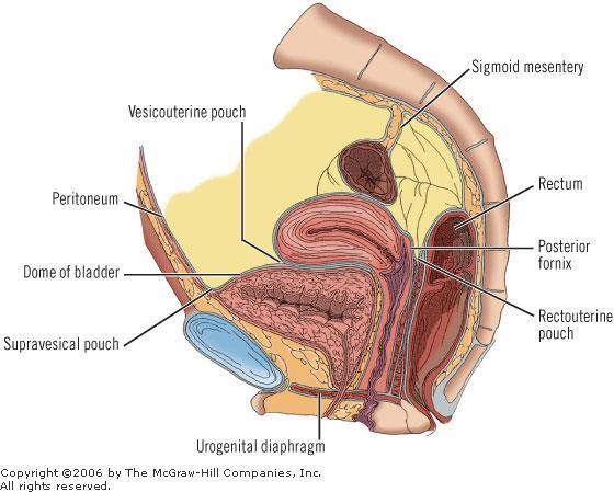
84 1- The peritoneum forms the sigmoid mesocolon, and cover front and, sides of the upper 1/3 of rectum, front only of the middle 1/3 of the rectum. 2- In the male, the peritoneum cover upper part of the fundus and upper surface of urinary bladder and then is reflected on to the anterior abdominal wall. The pouch, so formed, is called rectovesical pouch. 3-In the female, the peritoneum is reflected on to the upper part of posterior vaginal wall, forming recto-vaginal pouch (pouch of Douglas) 4- It covers upper surface of uterus, inferior surface of uterus down to level of internal os, then reflected on to upper surface of the urinary bladder, forming uterovesical pouche,then to anterior abdominal wall.

85

86
87

88
Dana Alrafaiah. - Amani Nofal. - Ahmad Alsalman. 1 P a g e
 - 2 - Dana Alrafaiah - Amani Nofal - Ahmad Alsalman 1 P a g e This lecture will discuss five topics as follows: 1- Arrangement of pelvic viscera. 2- Muscles of Pelvis. 3- Blood Supply of pelvis. 4- Nerve
Inferior Pelvic Border
 Pelvis + Perineum Pelvic Cavity Enclosed by bony, ligamentous and muscular wall Contains the urinary bladder, ureters, pelvic genital organs, rectum, blood vessels, lymphatics and nerves Pelvic inlet (superior
REPRODUCTIVE SYSTEM By Dr.Ahmed Salman
 The University Of Jordan Faculty Of Medicine Anatomy Department REPRODUCTIVE SYSTEM By Dr.Ahmed Salman Assistant Professor of Anatomy &embryology Perineum It is the diamond-shaped lower end of the trunk
Slide Read the tables it is about the difference between male & female pelvis.
 I didn t include the slides, this is only what the doctor read or said because he skipped a lot of things because we took it previously, very important to go back to the slides (*there is an edited version)
NOTES FROM GUTMAN LECTURE 10/26 Use this outline to study from. As you go through Gutman s lecture, fill in the topics.
 NOTES FROM GUTMAN LECTURE 10/26 Use this outline to study from. As you go through Gutman s lecture, fill in the topics. Anatomy above the arcuate line Skin Camper s fascia Scarpa s fascia External oblique
Perineum. done by : zaid al-ghnaneem
 Perineum done by : zaid al-ghnaneem Hello everyone, this sheet will talk about 2 nd Lecture which is Perineum but there are some slides and info from 1 st Lecture. Everything included Slides + Pics Let
Pelvis MCQs. Block 1. B. Reproductive organs. C. The liver. D. Urinary bladder. 1. The pelvic diaphragm includes the following muscles: E.
 Pelvis MCQs Block 1 1. The pelvic diaphragm includes the following muscles: A. The obturator internus B. The levator ani C. The coccygeus D. The external urethral sphincter E. The internal urethral sphincter
Table 2. First Generated List of Expert Responses. Likert-Type Scale. Category or Criterion. Rationale or Comments (1) (2) (3) (4)
 (2) (3) (4)") Table 2. First Generated List of Expert Responses. Likert-Type Scale Category or Criterion Anatomical Structures and Features Skeletal Structures and Features (1) (2) (3) (4) Rationale or Comments 1. Bones
Table 2. First Generated List of Expert Responses. Likert-Type Scale Category or Criterion Anatomical Structures and Features Skeletal Structures and Features (1) (2) (3) (4) Rationale or Comments 1. Bones
Bony ypelvis. Composition: formed by coccyx, and their articulations Two portions
 Pelvis Bony ypelvis Composition: formed by paired hip bones, sacrum, coccyx, and their articulations Two portions Greater pelvis Lesser pelvis Terminal line ( pelvic inlet): formed by promontory of sacrum,
ORIENTING TO BISECTED SPECIMENS ON THE PELVIS PRACTICAL
 ORIENTING TO BISECTED SPECIMENS ON THE PELVIS PRACTICAL The Pelvis is just about as complicated as head and neck and considerably more mysterious. You have to be able to visualize (imagine) the underlying
PELVIS & SACRUM Dr. Jamila El-Medany Dr. Essam Eldin Salama
 PELVIS & SACRUM Dr. Jamila El-Medany Dr. Essam Eldin Salama Learning Objectives At the end of the lecture, the students should be able to : Describe the bony structures of the pelvis. Describe in detail
Pelvis Perineum MCQs. Block 1.1. A. Urinary bladder. B. Rectum. C. Reproductive organs. D. The thigh
 Pelvis Perineum MCQs Block 1.1 1. The pelvic diaphragm includes the following muscles: A. The coccygeus B. The levator ani C. The external urethral sphincter D. The internal urethral sphincter E. The obturator
The posterior abdominal wall. Prof. Oluwadiya KS
 The posterior abdominal wall Prof. Oluwadiya KS www.oluwadiya.sitesled.com Posterior Abdominal Wall Lumbar vertebrae and discs. Muscles opsoas, quadratus lumborum, iliacus, transverse, abdominal wall
THE PELVIS VASCULAR AND NERVOUS SYSTEM SOMATIC AND AUTONOMIC NERVES
 THE PELVIS VASCULAR AND NERVOUS SYSTEM SOMATIC AND AUTONOMIC NERVES THE ABDOMINAL AORTA The abdominal aorta begins at the aor9c hiatus in the diaphragm at the level of the T12 vertebra and ends at the
Skeletal System Module 13: The Pelvic Girdle and Pelvis
 OpenStax-CNX module: m47993 1 Skeletal System Module 13: The Pelvic Girdle and Pelvis Donna Browne Based on The Pelvic Girdle and Pelvis by OpenStax College This work is produced by OpenStax-CNX and licensed
Yes, cranially with ovarian, caudally with vaginal. Yes, with uterine artery (collateral circulation between abdominal +pelvic source)
") Blood supply to internal female genitalia: uterine Internal iliac Sup. large branch: uterus, inf. Small branch: cervix+ sup. Vagina Yes, cranially with ovarian, caudally with vaginal Medially in base of
Blood supply to internal female genitalia: uterine Internal iliac Sup. large branch: uterus, inf. Small branch: cervix+ sup. Vagina Yes, cranially with ovarian, caudally with vaginal Medially in base of
Gross Anatomy of the Urinary System
 Gross Anatomy of the Urinary System Lecture Objectives Overview of the urinary system. Describe the external and internal anatomical structure of the kidney. Describe the anatomical structure of the ureter
-15. -Alaa Albandi. -Dr. Mohammad Almohtasib. 0 P a g e
 -15 -Alaa Albandi - -Dr. Mohammad Almohtasib 0 P a g e In this last lecture, we will talk about the sigmoid colon, rectum, and anal canal. Sigmoid colon It has a mesentery called pelvic mesocolon or sigmoidal
2. List the 8 pelvic spaces: list one procedure or dissection which involves entering that space.
 Name: Anatomy Quiz: Pre / Post 1. In making a pfannensteil incision you would traverse through the following layers: a) Skin, Camper s fascia, Scarpa s fascia, external oblique aponeurosis, internal oblique
Anatomy & Physiology Pelvic Girdles 10.1 General Information
 Anatomy & Physiology Pelvic Girdles 10.1 General Information ICan2Ed, Inc. In human anatomy, the pelvis (plural pelves or pelvises) is the lower part of. The area of the body that is between the abdomen
Urinary Bladder. Prof. Imran Qureshi
 Urinary Bladder Prof. Imran Qureshi Urinary Bladder It develops from the upper end of the urogenital sinus, which is continuous with the allantois. The allantois degenerates and forms a fibrous cord in
Learning objectives. SGD on Functions of Testosterone. Class
 Learning objectives SGD on Functions of Testosterone Class 2016 14-1-2013 Discuss o Process of spermatogenesis. o Sex determination. o Process of maturation of sperms. o Physiology of mature sperms. Discuss
GI module Lecture: 9 د. عصام طارق. Objectives:
 GI module Lecture: 9 د. عصام طارق Objectives: To list structures forming posterior abdominal wall. To follow aorta & its main branches. To describe IVC & its main tributaries. To list nerves of posterior
STRUCTURAL BASIS OF MEDICAL PRACTICE EXAMINATION 3. October 16, 2015
 STRUCTURAL BASIS OF MEDICAL PRACTICE EXAMINATION 3 October 16, 2015 PART l. Answer in the space provided. (12 pts) 1. Identify the structures. (2 pts) A. B. A B C. D. C D 2. Identify the structures. (2
Lumbar Plexus. Ventral rami L1 L4 Supplies: Major nerves.. Abdominal wall External genitalia Anteromedial thigh
 Lower Limb Nerves Lectures Objectives Describe the structure and relationships of the plexuses of the lower limb. Describe the course, relationships and structures supplied for the major nerves of the
Abdomen. Retroperitoneal space
 Abdomen. Retroperitoneal space Abdominal cavity The space bounded by: Anterolateral abdominal wall Posterior abdominal wall Diaphragm Pelvic walls and pelvic floor. Subdivided into: True abdominal cavity
STRUCTURAL BASIS OF MEDICAL PRACTICE EXAMINATION 3. October 17, 2014
 STRUCTURAL BASIS OF MEDICAL PRACTICE EXAMINATION 3 October 17, 2014 PART l. Answer in the space provided. (12 pts) 1. Identify the structures. (2 pts) A. B. A B C. D. C D 2. Identify the structures. (2
Anatomy of the Large Intestine
 Large intestine Anatomy of the Large Intestine 2 Large Intestine Extends from ileocecal valve to anus Length = 1.5-2.5m = 5 feet Regions Cecum = 2.5-3 inch Appendix= 3-5 inch Colon Ascending= 5 inch Transverse=
B) cervix of uterus C) vagina D) rectum. 1. What number illustrates the adnexal area? (Fig. 4-64) A) 4 B) 5 C) 8 D) 9
 cervix of uterus C) vagina D) rectum. 1. What number illustrates the adnexal area? (Fig. 4-64) A) 4 B) 5 C) 8 D) 9") Pelvis Practice Problems 1. What number illustrates the adnexal area? (Fig. 4-64) A) 4 B) 5 C) 8 D) 9 2. What number illustrates the cervix? (Fig. 4-64) A) 4 B) 8 C) 5 D) 6 3. Which of the following is
Pelvis Practice Problems 1. What number illustrates the adnexal area? (Fig. 4-64) A) 4 B) 5 C) 8 D) 9 2. What number illustrates the cervix? (Fig. 4-64) A) 4 B) 8 C) 5 D) 6 3. Which of the following is
Nerves on the Posterior Abdominal Wall
 Nerves on the Posterior Abdominal Wall Lumbar Plexus The lumbar plexus, which is one of the main nervous pathways supplying the lower limb, is formed in the psoasmuscle from the anterior ramiof the upper
Group of students. - Rawan almujabili د. محمد المحتسب - 1 P a g e
 - 14 - Group of students - Rawan almujabili د. محمد المحتسب - 1 P a g e Nerves of the posterior abdominal wall The spinal cord gives off spinal nerves between the vertebrae. In the abdomen, through the
LAB Notes#1. Ahmad Ar'ar. Eslam
 LAB Notes#1 Ahmad Ar'ar Eslam 1 P a g e Anatomy lab Notes Lower limb bones :- Pelvic girdle: It's the connection between the axial skeleton and the lower limb; it's made up of one bone called the HIP BONE
Ureters, Urinary Bladder & Urethra
 Ureters, Urinary Bladder & Urethra Please check our Editing File هذا العمل ال يغني عن المصدر األساسي للمذاكرة Lecture 2 } و م ن ي ت و ك ع ل ا لل ه ف ه و ح س ب ه { Objectives o Describe the course of ureter
THE ABDOMEN SUPRARENAL GLANDS KIDNEY URETERS URINARY BLADDER
 THE ABDOMEN SUPRARENAL GLANDS KIDNEY URETERS URINARY BLADDER THE SUPRARENAL GLANDS The suprarenal (adrenal) glands lie immediately superior and slightly anterior to the upper pole of either kidney. Golden
Pathogenesis of Chronic Pelvic Pain
 Pathogenesis of Chronic Pelvic Pain Yong-Chul Kim Department of anesthesia and pain medicine, Seoul National University College of Medicine 1 Overview Anatomy Nerve innervation CPP by pathology CPP by
Preview from Notesale.co.uk Page 1 of 34
 Abdominal viscera and digestive tract Digestive tract Abdominal viscera comprise majority of the alimentary system o Terminal oesophagus, stomach, pancreas, spleen, liver, gallbladder, kidneys, suprarenal
Copyright 2003 Pearson Education, Inc. publishing as Benjamin Cummings. Dr. Nabil Khouri MD, MSc, Ph.D
 Dr. Nabil Khouri MD, MSc, Ph.D Pelvic Girdle (Hip) Organization of the Lower Limb It is divided into: The Gluteal region The thigh The knee The leg The ankle The foot The thigh and the leg have compartments
The Female and Male External Genitalia. Prof Oluwadiya KS
 The Female and Male External Genitalia Prof Oluwadiya KS www.oluwadiya.com Anatomy of the female external genitalia This consists of : The vulva which is made up of: o The clitoris o Vestibular apparatus
Benha University. Faculty of Medicine. Anatomy Department Course code (MED 0701) Model answer of Anatomy examination. (Abdomen,Pelvis and Thorax)
 Model answer of Anatomy examination. (Abdomen,Pelvis and Thorax)") 1 Benha University Faculty of Medicine Anatomy Department Course code (MED 0701) Model answer of Anatomy examination (Abdomen,Pelvis and Thorax) 1 st year 2 nd term Date :18 /5 /2013 2 I-Short account
1 Benha University Faculty of Medicine Anatomy Department Course code (MED 0701) Model answer of Anatomy examination (Abdomen,Pelvis and Thorax) 1 st year 2 nd term Date :18 /5 /2013 2 I-Short account
[ANATOMY #12] April 28, 2013
![[ANATOMY #12] April 28, 2013](/thumbs/86/93473883.jpg "[ANATOMY #12] April 28, 2013") Sympathetic chain : Sympathetic chain is each of the pair of ganglionated longitudinal cords of the sympathetic nervous system; extend from level of atlas (base of skull) till coccyx. It is paravertebral
Sympathetic chain : Sympathetic chain is each of the pair of ganglionated longitudinal cords of the sympathetic nervous system; extend from level of atlas (base of skull) till coccyx. It is paravertebral
The Thoracic wall including the diaphragm. Prof Oluwadiya KS
 The Thoracic wall including the diaphragm Prof Oluwadiya KS www.oluwadiya.com Components of the thoracic wall Skin Superficial fascia Chest wall muscles (see upper limb slides) Skeletal framework Intercostal
musculoskeletal system anatomy nerves of the lower limb 1 done by: dina sawadha & mohammad abukabeer
 musculoskeletal system anatomy nerves of the lower limb 1 done by: dina sawadha & mohammad abukabeer What is the importance of plexuses? plexuses provides us the advantage of a phenomenon called convergence
The abdominal Esophagus, Stomach and the Duodenum. Prof. Oluwadiya KS
 The abdominal Esophagus, Stomach and the Duodenum Prof. Oluwadiya KS www.oluwadiya.com Viscera of the abdomen Abdominal esophagus: Terminal part of the esophagus The stomach Intestines: Small and Large
Posterior Abdominal wall-
 Structures of posterior abdominal wall: o Bony boundaries: 5 lumber vertebra and their intervertebral disc, iliac fossa and iliac crest. o Muscles: psoas major, quadrates lumborum, transversus abdominis,
The os coxae or hip bone consists of three flat bones, ilium, ischium and pubis, which fuse together to form the acetabulum.
 The os coxae The os coxae or hip bone consists of three flat bones, ilium, ischium and pubis, which fuse together to form the acetabulum. The ilium extends from the acetabulum upwards forming the lateral
Femoral Triangle and Adductor Canal. Dr. Heba Kalbouneh Associate Professor of Anatomy and Histology
 Femoral Triangle and Adductor Canal Dr. Heba Kalbouneh Associate Professor of Anatomy and Histology Femoral Triangle and Adductor Canal Femoral triangle Is a triangular depressed area located in the upper
The Anterolateral Abdominal Wall By Prof. Dr. Muhammad Imran Qureshi
 1 P age The Anterolateral Abdominal Wall By Prof. Dr. Muhammad Imran Qureshi Introduction The abdomen is the region of the trunk located between the thorax and the pelvis. It includes the anterolateral
First practical session. Bones of the gluteal region
 First practical session 2017 Bones of the gluteal region The Hip bone The hip bone is made of: 1 The ilium: superior in position 2 The ischium:postero-inferior in position 3 The pubis: antero-inferior
Perineum. Dept. of Human Anatomy Zhou Hong Ying
 Perineum Dept. of Human Anatomy Zhou Hong Ying OUTLINE Subdivision The Layers Urogenital Diaphragm Main Structures inside Superficial & Deep Perineal Spaces Ischioanal Fossa Perineum A narrow region Urogenital
Gross anatomy of the urinary system. Done by : razan krishan. slide in bold and book in green
 Gross anatomy of the urinary system Done by : razan krishan slide in bold and book in green Kidneys, ureters, urinary bladder & urethra Urine flows from each kidney, down its ureter to the bladder and
Rama Nada. - Ensherah Mokheemer. - Ahmed salman. 1 P a g e
 - 5 - Rama Nada - Ensherah Mokheemer - Ahmed salman 1 P a g e We will continue talking about the urinary bladder The ligaments of the bladder: 1-Median umbilical ligament: Continuous with apex of the bladder
Lecture 10 Arteries and veins of the upper limb
 Lecture 10 Arteries and veins of the upper limb 1. Identify the Subclavian, axillary, brachial (deep and superficial), radial and ulnar arteries and superficial/deep palmar arches 2. Describe the major
ANATYOMY OF The thigh
 ANATYOMY OF The thigh 1- Lateral cutaneous nerve of the thigh Ι) Skin of the thigh Anterior view 2- Femoral branch of the genitofemoral nerve 5- Intermediate cutaneous nerve of the thigh 1, 2 and 3 are
SUBJECTS 2nd year, 1st semester I. 1. Primitive gut - limits, derivatives 2. Foregut -limits, evolution, derivatives 3. Midgut -limits, evolution,
 SUBJECTS 2nd year, 1st semester I. 1. Primitive gut - limits, derivatives 2. Foregut -limits, evolution, derivatives 3. Midgut -limits, evolution, derivatives 4. Hindgut- limits, evolution, derivatives
أحمد رواجبة- محمود الحربي- أحمد السالمان-
 -6 أحمد رواجبة- محمود الحربي- أحمد السالمان- 1 P a g e The Male Reproductive System The male genital system structures are divided into: Internal structures: 1- Prostate 3-Ejaculatory ducts External structures:
Bones of Lower Limb. Dr. Heba Kalbouneh Associate Professor of Anatomy and Histology
 Bones of Lower Limb Dr. Heba Kalbouneh Associate Professor of Anatomy and Histology Bones of the lower limb Hip Bone Made up of 3 bones: 1) Ilium (flat), superior in position 2) Ischium (L), postero-inferior
ischium Ischial tuberosities Sacrotuberous ligament The coccyx
 Perineum General lfeatures Region of below pelvic diaphragm A diamond shape space whose boundaries are those of the pelvic outlet Lower border of symphysis pubis Rami of pubis and ischium Ischial tuberosities
The thigh. Prof. Oluwadiya KS
 The thigh Prof. Oluwadiya KS www.oluwadiya.com The Thigh: Boundaries The thigh is the region of the lower limb that is approximately between the hip and knee joints Anteriorly, it is separated from the
Abdomen: Introduction. Prof. Oluwadiya KS
 Abdomen: Introduction Prof. Oluwadiya KS www.oluwadiya.com Abdominopelvic Cavity Abdominal Cavity Pelvic Cavity Extends from the inferior margin of the thorax to the superior margin of the pelvis and the
THE THORACIC WALL. Boundaries Posteriorly by the thoracic part of the vertebral column. Anteriorly by the sternum and costal cartilages
 THE THORACIC WALL Boundaries Posteriorly by the thoracic part of the vertebral column Anteriorly by the sternum and costal cartilages Laterally by the ribs and intercostal spaces Superiorly by the suprapleural
Lectures of Human Anatomy
 Lectures of Human Anatomy Lower Limb Gluteal Region and Hip Joint By DR. ABDEL-MONEM AWAD HEGAZY M.B. with honor 1983, Dipl."Gynecology and Obstetrics "1989, Master "Anatomy and Embryology" 1994, M.D.
ANATOMY (7). INTRODUCTION TO THE PELVIS. MOHAMMAD ALLOUH. BNB. 1 P a g e
. INTRODUCTION TO THE PELVIS. MOHAMMAD ALLOUH. BNB. 1 P a g e") ANATOMY (7). INTRODUCTION TO THE PELVIS. MOHAMMAD ALLOUH. BNB 1 P a g e Introduction to the Pelvis: Pelvic Wall & Pelvic Cavity pelvis (L, basin, sink ), is The region of the trunk that lies inferoposterior
ANATOMY (7). INTRODUCTION TO THE PELVIS. MOHAMMAD ALLOUH. BNB 1 P a g e Introduction to the Pelvis: Pelvic Wall & Pelvic Cavity pelvis (L, basin, sink ), is The region of the trunk that lies inferoposterior
Omran Saeed. Mohammad Al-muhtaseb. 1 P a g e
 13 Omran Saeed Mohammad Al-muhtaseb 1 P a g e Posterior abdominal wall - The diaphragm separates between thoracic cavity and abdominal cavity. Structures of posterior abdominal wall: (below diaphragm)
Anatomy of the Pelvis
 Anatomy of the Pelvis Please view our Editing File before studying this lecture to check for any changes. Color Code Important Doctors Notes Notes/Extra explanation Objectives At the end of the lecture,
ANATOMY OF PELVICAYCEAL SYSTEM -DR. RAHUL BEVARA
 1 ANATOMY OF PELVICAYCEAL SYSTEM -DR. RAHUL BEVARA 2 KIDNEY:ANATOMY OVERVIEW Kidneys are retroperitoneal, in posterior abdominal region, extending from T12 L3 Bean-shaped Right kidney is lower than left
ABDOMINAL WALL & RECTUS SHEATH
 ABDOMINAL WALL & RECTUS SHEATH Learning Objectives Describe the anatomy, innervation and functions of the muscles of the anterior, lateral and posterior abdominal walls. Discuss their functional relations
Pelvic Angiogram - Male
 Pelvic Angiogram - Male Common iliac artery Internal iliac artery Lateral sacral artery Iliolumbar artery Posterior trunk of internal iliac artery Superior gluteal artery Internal pudendal artery External
PELVIS II: FUNCTION TABOOS (THE VISCERA) Defecation Urination Ejaculation Conception
 Defecation Urination Ejaculation Conception") PELVIS II: FUNCTION TABOOS (THE VISCERA) Defecation Urination Ejaculation Conception REVIEW OF PELVIS I Pelvic brim, inlet Pelvic outlet True pelvis-- --viscera Tilt forward Mid-sagital views-- --how the
PELVIS II: FUNCTION TABOOS (THE VISCERA) Defecation Urination Ejaculation Conception REVIEW OF PELVIS I Pelvic brim, inlet Pelvic outlet True pelvis-- --viscera Tilt forward Mid-sagital views-- --how the
Kaan Yücel M.D., Ph.D. 14.January.2014 Tuesday
 Kaan Yücel M.D., Ph.D. 14.January.2014 Tuesday Sexual differences are related mainly 1. Heavier build and larger muscles of most men 2. Adaptation of the pelvis (particularly the lesser pelvis) in women
TABLE OF CONTENTS A. ORGANISATIONAL COMPONENT B. STUDY COMPONENT. Page. 1. Introduction I. 2. Lecturers I. 3. Timetable I. 4.
 TABLE OF CONTENTS Page A. ORGANISATIONAL COMPONENT 1. Introduction I 2. Lecturers I 3. Timetable I 4. Assessment I 5. Study material II 6. General information II B. STUDY COMPONENT 1. Block XI: Session
TABLE OF CONTENTS. 1. Introduction I. 2. Lecturers I. 3. Timetable I. 4. Assessment I. 5. Study material II. 6. General information II.
 TABLE OF CONTENTS Page A. ORGANISATIONAL COMPONENT 1. Introduction I 2. Lecturers I 3. Timetable I 4. Assessment I 5. Study material II 6. General information II B. STUDY COMPONENT 1. Block XI: Session
rotation of the hip Flexion of the knee Iliac fossa of iliac Lesser trochanter Femoral nerve Flexion of the thigh at the hip shaft of tibia
 Anatomy of the lower limb Anterior & medial compartments of the thigh Dr. Hayder The fascia lata encloses the entire thigh like a sleeve/stocking. Three intramuscular fascial septa (lateral, medial, and
nerve blocks in the diagnosis and therapy of visceral disease
 Visceral Pain nerve blocks in the diagnosis and therapy of visceral disease Guy Hans, MD, PhD Dept. of Anesthesiology, Multidisciplinary Pain Center Visceral Pain? Type of nociceptive pain (although often
Anatomy of thoracic wall
 Anatomy of thoracic wall Topographic Anatomy of the Thorax 1 Bones of Thoracic wall ribs 1-7"true" ribs -those which attach directly to the sternum true ribs actually attach to the sternum by means of
ANATYOMY OF The thigh
 ANATYOMY OF The thigh 1- Lateral cutaneous nerve of the thigh Ι) Skin of the thigh Anterior view 2- Femoral branch of the genitofemoral nerve 5- Intermediate cutaneous nerve of the thigh 1, 2 and 3 are
Organisation of the nervous system
 Chapter1 Organisation of the nervous system 1. Subdivisions of the nervous system The nervous system is divided: i) Structurally The central nervous system (CNS) composed of the brain and spinal cord.
Gluteal region DR. GITANJALI KHORWAL
 Gluteal region DR. GITANJALI KHORWAL Gluteal region The transitional area between the trunk and the lower extremity. The gluteal region includes the rounded, posterior buttocks and the laterally placed
The Lower Limb. Anatomy RHS 241 Lecture 2 Dr. Einas Al-Eisa
 The Lower Limb Anatomy RHS 241 Lecture 2 Dr. Einas Al-Eisa The bony pelvis Protective osseofibrous ring for the pelvic viscera Transfer of forces to: acetabulum & head of femur (when standing) ischial
Anatomy of the renal system. Professor Nawfal K. Al-Hadithi
 Anatomy of the renal system Professor Nawfal K. Al-Hadithi Objectives To describe the posterior abdominal wall To identify the main anatomical landmarks of the kidneys & ureters To describe the suprarenal
OBJECTIVE: To obtain a fundamental knowledge of the root of the neck with respect to structure and function
 The root of the neck Jeff Dupree, Ph.D. e mail: jldupree@vcu.edu OBJECTIVE: To obtain a fundamental knowledge of the root of the neck with respect to structure and function READING ASSIGNMENT: Moore and
Diaphragm and intercostal muscles. Dr. Heba Kalbouneh Associate Professor of Anatomy and Histology
 Diaphragm and intercostal muscles Dr. Heba Kalbouneh Associate Professor of Anatomy and Histology Skeletal System Adult Human contains 206 Bones 2 parts: Axial skeleton (axis): Skull, Vertebral column,
The anatomy of the rectum and anal canal
 The anatomy of the rectum and anal canal Vishy Mahadevan Abstract Diseases of the rectum and anal canal, both benign and malignant, account for a very large part of colorectal surgical practice in the
Basic Body Structure
 Basic Body Structure The Cell All life consists of microscopic living structures called cells. They perform various functions throughout the body. All cells are similar in structure, but not identical.
Anatomy Review File اللهم ال سهل إال ماجعلته سهال وأنت تجعل الحزن
 Anatomy Review File اللهم ال سهل إال ماجعلته سهال وأنت تجعل الحزن إذا شئت سهال This work was done by: Alanoud Abuhaimed Dania Alkelabi Ghada Alothaim Jawaher Abanumy Rawan AlWadee Wejdan Alzeid Mohanad
Spinal nerves and cervical plexus Prof. Abdulameer Al Nuaimi. E mail: a.al E. mail:
 Spinal nerves and cervical plexus Prof. Abdulameer Al Nuaimi E mail: a.al nuaimi@sheffield.ac.uk E. mail: abdulameerh@yahoo.com Branches of ophthalmic artery Muscles of face A spinal nerve Spinal
Lumbar and Sacral Plexuses. Dr. Heba Kalbouneh Associate Professor of Anatomy and Histology
 Lumbar and Sacral Plexuses Dr. Heba Kalbouneh Associate Professor of Anatomy and Histology Structure of Spinal Nerves: Somatic Pathways dorsal root CNS interneuron spinal nerve dorsal ramus somatic sensory
The University Of Jordan Faculty Of Medicine THE LOWER LIMB. Dr.Ahmed Salman Assistant Prof. of Anatomy. The University Of Jordan
 The University Of Jordan Faculty Of Medicine THE LOWER LIMB Dr.Ahmed Salman Assistant Prof. of Anatomy. The University Of Jordan Gluteal Region Cutaneous nerve supply of (Gluteal region) 1. Lateral cutaneous
Human Anatomy Biology 351
 nnnnn 1 Human Anatomy Biology 351 Exam #2 Please place your name on the back of the last page of this exam. You must answer all questions on this exam. Because statistics demonstrate that, on average,
Anatomy of the Thorax
 Anatomy of the Thorax A) THE THORACIC WALL Boundaries Posteriorly by the thoracic part of the vertebral column Anteriorly by the sternum and costal cartilages Laterally by the ribs and intercostal spaces
Lecture 56 Kidney and Urinary System
 Lecture 56 Kidney and Urinary System The adrenal glands are located on the superomedial aspect of the kidney The right diagram shows a picture of the kidney with the abdominal walls and organs removed
Copyright 2003 Pearson Education, Inc. publishing as Benjamin Cummings. Dr. Nabil khouri
 Dr. Nabil khouri Appendicular Skeleton The appendicular skeleton is made up of the bones of the upper and lower limbs and their girdles Two girdles: Pectoral girdles attach the upper limbs to the body
Pancreas & Biliary System. Dr. Vohra & Dr. Jamila
 Pancreas & Biliary System Dr. Vohra & Dr. Jamila 1 Objectives At the end of the lecture, the student should be able to describe the: Location, surface anatomy, parts, relations & peritoneal reflection
Bones of the Lower Limb Bone Structure Description Notes. border of the superior ramus. inferolaterally from the pubic symphysis
 Bones of the Lower Limb Bone Structure Description Notes pubis an angulated bone the forms the anterior part of the pelvis one of three bones that form the os coxae: ilium, ischium, pubis; its forms 1/5
ParasymPathetic Nervous system. Done by : Zaid Al-Ghnaneem
 ParasymPathetic Nervous system Done by : Zaid Al-Ghnaneem In this lecture we are going to discuss Parasympathetic, in the last lecture we took sympathetic and one of the objectives of last lecture was
THE DESCENDING THORACIC AORTA
 Intercostal Arteries and Veins Each intercostal space contains a large single posterior intercostal artery and two small anterior intercostal arteries. The anterior intercostal arteries of the lower spaces
Lower Limb Nerves. Clinical Anatomy
 Lower Limb Nerves Clinical Anatomy Lumbar Plexus Ventral rami L1 L4 Supplies: Abdominal wall External genitalia Anteromedial thigh Major nerves.. Lumbar Plexus Nerves relation to psoas m. : Obturator n.
DISSECTION 8: URINARY AND REPRODUCTIVE SYSTEMS
 8546d_c01_1-42 6/25/02 4:32 PM Page 38 mac48 Mac 48: 420_kec: 38 Cat Dissection DISSECTION 8: URINARY AND REPRODUCTIVE SYSTEMS Typically, the urinary and reproductive systems are studied together, because
Muscles of the lower extremities. Dr. Nabil khouri MD, MSc, Ph.D
 Muscles of the lower extremities Dr. Nabil khouri MD, MSc, Ph.D Posterior leg Popliteal fossa Boundaries Biceps femoris (superior-lateral) Semitendinosis and semimembranosis (superior-medial) Gastrocnemius
Inguinal Canal. It is an oblique passage through the lower part of the anterior abdominal wall. Present in both sexes
 Inguinal canal Inguinal Canal It is an oblique passage through the lower part of the anterior abdominal wall Present in both sexes It allows structures to pass to and from the testis to the abdomen in
The Hip (Iliofemoral) Joint. Presented by: Rob, Rachel, Alina and Lisa
 Joint. Presented by: Rob, Rachel, Alina and Lisa") The Hip (Iliofemoral) Joint Presented by: Rob, Rachel, Alina and Lisa Surface Anatomy: Posterior Surface Anatomy: Anterior Bones: Os Coxae Consists of 3 Portions: Ilium Ischium Pubis Bones: Pubis Portion
The Hip (Iliofemoral) Joint Presented by: Rob, Rachel, Alina and Lisa Surface Anatomy: Posterior Surface Anatomy: Anterior Bones: Os Coxae Consists of 3 Portions: Ilium Ischium Pubis Bones: Pubis Portion