ABSTRACT. RODRIGUEZ, KARINA FLORES. Molecular Mechanisms of Gonadotropin-Induced. Oocyte Maturation. (Under the direction of Dr. Charlotte E.
|
|
|
- Blaze Montgomery
- 5 years ago
- Views:
Transcription
1 ABSTRACT RODRIGUEZ, KARINA FLORES. Molecular Mechanisms of Gonadotropin-Induced Oocyte Maturation. (Under the direction of Dr. Charlotte E. Farin) In vitro maturation of oocytes is routinely utilized for the production of embryos for commercial as well as research purposes. The objectives of the research described in this dissertation were: 1) to examine the molecular mechanism involved in gonadotropin-induced resumption of meiosis in cultured bovine and murine cumulus oocyte complexes (COC); 2) to determine the developmental potential of bovine oocytes maintained in meiotic arrest by inhibition of transcription; and 3) to analyze patterns of gene expression in bovine COC during the onset of gonadotropin-induced oocyte maturation. Murine COC were maintained in meiotic arrest by culture in the presence of either of the transcriptional inhibitors, 5,6-dichloro-1-β-D-ribofuranosylbenzimidazole (DRB) or α- amanitin. For either transcriptional inhibitor to effectively block maturation, FSH but not hcg, was required in the culture medium. Transcriptional inhibition of oocyte maturation was ineffective if denuded oocytes were utilized. Differential activation of Type I and Type II PKA was performed in murine COC. Activation of Type I PKA resulted in the inhibition of maturation whereas activation of Type II did not. Activation of Type II PKA resulted in transcription, which was required for maturation of murine and bovine COC and mimicked the action of FSH. The developmental competency of bovine COC maintained in meiotic arrest for 20 h by DRB was not different from control COC. Comparison of the patterns of mrna for oocytes matured for 0h, 4h or 4h+DRB resulted in the isolation of 4 amplicons that were expressed at 4h but not in 0h or 4h+DRB
2 groups. This result was reconfirmed by semi-quantitative PCR. No homology to known sequences was found suggesting that they may represent novel transcripts. The following model for FSH induced oocyte maturation is proposed: FSH binds to its cumulus cell receptor and increases camp. The elevation of camp results in activation of Type I and Type II PKA. Activation of Type I PKA inhibits oocyte maturation whereas activation of Type II PKA induces gene transcription that subsequently leads to the resumption of meiosis.
3 MOLECULAR MECHANISMS OF GONADOTROPIN-INDUCED OOCYTE MATURATION by KARINA FLORES RODRIGUEZ A dissertation submitted to the Graduate Faculty of North Carolina State University in partial fulfillment of the requirements for the degree of Doctor of Philosophy Physiology Raleigh, NC 2002 APPROVED BY:
4 BIBLIOGRAPHICAL SKETCH Karina Flores Rodriguez Education PhD BS Physiology, 2003, North Carolina State University, Raleigh, NC Animal Science, 1997, North Carolina State University, Raleigh, NC Honors and Awards Nominated for participation in the Graduate Student Professional Development Workshop, for North Carolina State University, College of Agricultural and Life Sciences, October 2000 National Institute of Health Merit Award, September 1997 Magna Cum Laude, May 1997 Professional Affiliations International Embryo Transfer Society Society for the Study of Reproduction Society of Hispanic Professionals ii
5 ACKNOWLEDGMENTS I would like to thank my advisor, Dr. Charlotte Farin, who with her patience and excellent support guided me through my program. I would like to thank Drs. Robert Petters, Gene Eisen and Peter Farin for their advice and guidance. I would also like to thank Jeffrey Sommer for sharing his time and expertise and to Adrienne Crosier, Patrick Blondin, Eric Alexander and Jeremy Miles for their friendship and support. This thesis is dedicated to my husband, Frank, who never stopped supporting me and giving me unconditional love, patience, understanding and the confidence to accomplish my goals and to my daughter Camila, who illuminated my days with her smile. I would to also thank to my parents, Edmundo and Clara who helped us so much, without you I would not have finished, gracias! And my sisters, Andrea and Silvana, who always stood behind me and supported me when I needed it the most. Finally, I would like to thank the rest of my family and friends for their prayers and inspiration. Thank you all. iii
6 TABLE OF CONTENTS Page LIST OF TABLES viii LIST OF FIGURES ix LIST OF ABBREVIATIONS...xi CHAPTER 1: REVIEW OF LITERATURE OOCYTE DEVELOPMENT Oogenesis Folliculogenesis Oocyte-Cumulus Interactions Gap Junctions Gonadotropins and Second Messenger Systems Role of FSH FSH signal transduction..11 Role of LH LH signal transduction OOCYTE MATURATION Nuclear Maturation.15 Structural Aspects of Nuclear Maturation Germinal Vesicle Stage Germinal Vesicle Breakdown Metaphase I Metaphase II.. 16 Biochemical Aspects of Oocyte Maturation 17 Maturation Promoting Factor...17 Mitogen Activated Protein Kinase...20 Cytoplasmic Maturation Mitochondria Cortical Granules Accumulation of mrna 22 MATURATION OF OOCYTES IN VITRO..24 Spontaneous versus Gonadotropin-Induced maturation 26 Manipulation of Oocyte Maturation..28 iv
7 camp 29 Purines..30 Inhibitors of Protein Synthesis.30 Protein Kinase Inhibitors..31 Meiosis Activating Sterol.31 Transcriptional Inhibitors.32 STATEMENT OF THE PROBLEM.34 REFERENCES 35 CHAPTER 2: ROLE OF GENE TRANSCRIPTION AND PKA SUBTYPE ACTIVATION IN MURINE OOCYTE MATURATION 61 ABSTRACT. 61 INTRODUCTION...63 MATERIALS AND METHODS. 64 Reagents and Media.. 64 Role of Transcription in Gonadotropin-Mediated Resumption of Meiosis. 65 Oocyte Recovery and Culture Conditions..65 Effect of Gonadotropins on Inhibition of Meiotic Maturation by DRB or α- Amanitin 66 Effect of Supplementation with Either hcg or FSH on Inhibition of Meiotic Maturation by DRB or α-amanitin..67 Effect of Cumulus Cell Removal on the Inhibition of Oocyte Maturation by DRB or α-amanitin 67 Reversibility of DRB and α-amanitin arrest of GVBD.68 Role of PKA Isozymes in Facilitating Resumption of Meiosis.69 Oocyte Recovery and Culture Conditions..69 Effect of Differential Activation of Type I or Type II PKA on Resumption of Meiosis 69 Role of Transcription in Regulating Meiosis Following Differential Activation of Type I or Type II PKA..70 Statistical Analysis RESULTS 71 Role of Transcription in Gonadotropin-Mediated Resumption of Meiosis.71 v
8 Effect of Gonadotropins on Inhibition of Meiotic Maturation by DRB or α-amanitin.. 71 Effect of hcg or FSH on Inhibition of Meiotic Maturation by DRB or α-amanitin...71 Effect of Cumulus Cell Removal on the Inhibition of Oocyte Maturation by DRB or α-amanitin Reversibility of DRB- and α-amanitin- Mediated Arrest of GVBD. 74 Role of PKA Isozymes in Facilitating Resumption of Meiosis 75 Effect of Activation of Type I or Type II PKA on Resumption of Meiosis: Dose Response Analysis..75 Role of Transcription in Regulating Meiosis Following Differential Activation of Type I or Type II PKA..75 DISCUSSION..78 REFERENCES.82 CHAPTER 3: ACTIVATION OF TYPE II PKA STIMULATES RESUMPTION OF MEIOSIS BY A TRANSCRIPTIONAL MECHANISM IN CULTURED BOVINE COC...86 ABSTRACT 86 INTRODUCTION MATERIALS AND METHODS.89 Reagents..89 Oocyte Recovery and Culture Conditions..89 Assessment of Meiotic Stage..90 Statistical Analysis..90 RESULTS 91 DISCUSSION..93 REFERENCES CHAPTER 4: DEVELOPMENTAL CAPACITY OF BOVINE CUMULUS OOCYTE COMPLEXES AFTER TRANSCRIPTIONAL INHIBITION OF OOCYTE MATURATION.100 ABSTRACT INTRODUCTION.101 MATERIALS AND METHODS Reagents and Media..103 vi
9 Experimental Design, Oocyte Maturation and Embryo Production. 103 Statistical Analysis 106 RESULTS.107 DISCUSSION REFERENCES..112 CHAPTER 5: IDENTIFICATION OF NOVEL TRANSCRIPTS REQUIRED FOR GONADOTROPIN-INDUCED BOVINE OOCYTE MATURATION..116 ABSTRACT.116 INTRODUCTION 117 MATERIALS AND METHODS.118 Experimental Design Reagents and Media..118 Oocyte Recovery and Maturation.119 wcrna Extraction and Reverse Transcription.119 Differential Display PCR..120 Semi-Quantitative PCR and Image Analysis 122 Statistical Analysis 124 RESULTS..125 DISCUSSION REFERENCES CHAPTER 6: GENERAL CONCLUSIONS REFERENCES 146 APPENDICES APPENDIX 1. Time Course Study of the Effect of DRB at Different Time Points During Murine Oocyte Maturation..149 APPENDIX 2. Effect of the Differential Activation of Type I PKA in Bovine Oocyte Maturation 150 APPENDIX 3. List of Publications vii
10 LIST OF TABLES CHAPTER 3: DEVELOPMENTAL CAPACITY OF BOVINE CUMULUS OOCYTE COMPLEXES AFTER TRANSCRIPTIONAL INHIBITION OF OOCYTE MATURATION Table 1. Effect of prolonged meiotic arrest of bovine COC for 20 h by transcriptional inhibition with DRB on post-fertilization development to 72 hpi (cleavage), 168 hpi (day 7) and 216 hpi (day 9) CHAPTER 5: IDENTIFICATION OF NOVEL TRANSCRIPTS REQUIRED FOR GONADOTROPIN-INDUCED BOVINE OOCYTE MATURATION Table 1. List of primers utilized for differential display PCR 121 Table 2. Primer sequences used for semi-quantitative PCR analysis of bovine COC cultured for either 0 h or 4h viii
11 LIST OF FIGURES CHAPTER 1: REVIEW OF LITERATURE Figure 1. Summary for oogenesis and follicular development in cattle Figure 2. camp mediated second messenger system for the gonadotropic hormone FSH..12 Figure 3. Time course of MPF activity related to oocyte maturation, fertilization and early cleavage 19 CHAPTER 2: ROLE OF GENE TRANSCRIPTION AND PKA SUBTYPE ACTIVATION IN MURINE OOCYTE MATURATION Figure 1. Characterization of gonadotropin-mediated GVBD in murine COC.. 73 Figure 2. Effect of activation of Type I or Type II PKA with varying concentrations of camp analogs on the incidence of GVBD in murine COC Figure 3. Role of transcription in GVBD after activation of Type I and Type II PKA CHAPTER 3: ACTIVATION OF TYPE II PKA STIMULATES RESUMPTION OF MEIOSIS BY A TRANSCRIPTIONAL MECHANISM IN CULTURED BOVINE COC Figure 1. Effect of activation of Type II PKA on bovine oocyte maturation.92 CHAPTER 4: DEVELOPMENTAL CAPACITY OF BOVINE CUMULUS OOCYTE COMPLEXES AFTER TRANSCRIPTIONAL INHIBITION OF OOCYTE MATURATION Figure 1. Experimental Design.104 Figure 2. Distribution of meiotic stage of bovine COC after 20 h culture in the presence or absence of the transcriptional inhibitor DRB..108 CHAPTER 5: IDENTIFICATION OF NOVEL TRANSCRIPTS REQUIRED FOR GONADOTROPIN-INDUCED BOVINE OOCYTE MATURATION Figure 1. Representative differential display gel analysis utilized for comparison of patterns of mrna expression during gonadotropin-induced bovine oocyte maturation 123 ix
12 Figure 2. Representative PCR products for the 4 unknown amplicons identified as well as GAPDH Figure 3. Sequences of the 4 differentially expressed amplicons identified in the DD-PCR analysis Figure 4. Sequences for UK#11 products Figure 5. Semi-quantitative assessment of the expression of UK#1 and UK#3 in bovine COC following 0h or 4h from the initiation of culture Figure 6. Semi-quantitative assessment of the expression of UK#1 and UK#3 in bovine COC following 0h or 4h from the initiation of culture CHAPTER 6: GENERAL CONCLUSIONS Figure 1A. Proposed mechanism of meiotic arrest inside the follicle. 144 Figure 1B. Proposed mechanism for spontaneous oocyte maturation. 144 Figure 2A. Proposed mechanism for the inhibitory phase of FSH-induced oocyte maturation.145 Figure 2B. Proposed mechanism for the stimulatory phase of FSH-induced oocyte maturation.145 APPENDICES Figure 1. Time Course Study of the Effect of DRB at Different Time Points During Murine Oocyte Maturation..149 Figure 2. Effect of the Differential Activation of Type I PKA in Bovine Oocyte Maturation 150 x
13 LIST OF ABBREVIATIONS GVBD....Germinal vesicle breakdown MI....Metaphase I MII.. Metaphase II ZP.Zona pellucida protein FGL-α.... Factor in the germ line α FSH Follicle stimulating hormone LH... Luteinizing hormone HCG..Human chorionic gonadotropin GnRH..Gonadotropin-releasing hormone TSH...Thyroid stimulating hormone GDF-9.Differentiation factor 9 BMP-15 Bone morphogenic protein 15 PKA...Protein kinase A mrna...messenger RNA FSHR.. Follicle stimulation hormone receptor LHR..Luteinizing hormone receptor camp.cyclic adenosine monophosphate CREB...cAMP Response Element Binding Protein MAPK.Mitogen-activated protein kinase IP 3.. Inositol-triphosphate MPF Maturation promoting factor DMAP...6-dimethylaminopurine RINGO.. rapid inducer of G2/M transition in oocytes COC.....Cumulus-oocyte complexes GV Germinal vesicle ACE... Adenylation control element EGF. Epidermal growth factor GH Growth hormone ICSI.Intracytoplasmic sperm injection BSA.Bovine serum albumin xi
14 PVA.Polyvinyl alcohol TCM-199..Tissue culture medium 199 msof... Modified synthetic oviductal fluid PVP..Polyvinyl pyrrolidone DRB... 5,6-dichloro-1-β-D-ribofuranosyl-benzimidazole dbcamp.... dibutyryl camp 8BrcAMP...8-Bromo-cAMP MAS. Meiosis activating sterol LDM..Lanosterol 14 α-demethylase AM....α-Amanitin CUM...Cumulus cell N 6... N 6 -monobutyryl-cyclic adenosine monophosphate AHA...Aminohexyl-amino-cyclic adenosine monophosphate PMSG..Pregnant mare serum gonadotropin DMSO Dimethylsulfoxide GLM. General linear models AKAPS. A-kinase anchoring proteins xii
15 REVIEW OF LITERATURE A clear understanding of the process of oocyte maturation is critical for the efficient application of biotechnologies such as in vitro embryo production and mammalian cloning. In the domestic livestock industry, the application of in vitro oocyte maturation technologies has resulted in the production of embryos for both commercial and research purposes. In order to better understand the physiological mechanism that underlies the processes of oocyte maturation, a detailed review of oogenesis from both a developmental and molecular perspective is needed. This will be the major topic of this review. In addition, the impact of in vitro manipulation of oocytes on their subsequent developmental competence will also be reviewed. Although the primary focus will be on the bovine and murine model systems, data from other species will be discussed when appropriate. OOCYTE DEVELOPMENT Oogenesis During early mammalian embryogenesis, primordial germ cells migrate from the yolk sac to the genital ridge (1, 2). On embryonic day 7 the mouse embryo exhibits a few recognizable primordial germ cells in the yolk sack endoderm (3). However, by embryonic day 13 the female mouse embryo contains a differentiated gonad with actively dividing germ cells, now identified as oogonia (1). Oogonia undergo a high frequency of mitotic divisions and possess a distinctive morphology that includes a large size, large nuclear to cytoplasmic ratio, intracytoplasmic vesicles, intracellular bridges that connect adjacent germ cells and the presence of alkaline phosphatase (3, 4, 5). Specific alkaline phosphatase staining is routinely
16 2 utilized for identification of oogonia (5, 6). As a result of the frequent mitotic divisions, the number of oogonia increases to about 20,000 by the time the genital ridge is fully colonized (3). The precise signals that induce oogonia to enter meiosis and transform into oocytes are not known. By embryonic day 17 in mice, all the oogonia have entered meiosis and the murine ovary contains oocytes at different stages of the first meiotic prophase (3, 7). During the pre-leptotene stage following the last mitotic division, DNA replication occurs for the final time. During the zygotene stage, chromosomes form bivalents composed of four chromatids. The oocyte then progresses through pachytene where chiasmata form and genetic recombination occurs as a result of crossing over. Oocytes continue through the diplotene stage of the first meiotic division and become arrested at late diplotene or dictyate stage of prophase I of the first meiotic division (3). At this stage, oocytes are surrounded by a single layer of flattened pre-granulosa cells and are known as primordial follicles (3). This pool of primordial follicles becomes the only source of germ cells throughout an adult females life. This germ cell pool is developed during fetal life in both primates and ruminants and in the early neonatal period in rodents (8). After all oocytes are arrested in prophase I of meiosis and the pool of available oocytes has been established, follicles are recruited to leave this resting pool to begin growth. The physiological signal for recruitment of follicles is unknown. Once a follicle is recruited, it will continue to grow until it is ovulated or becomes atretic (9). Most of the oocytes are lost to atresia (9). In cattle, as in rodents, most of the follicular loss due to atresia occurs near the end of follicular development (10, 11). In humans, follicular atresia is first observed in
17 3 utero at around 6 months of gestation and continues throughout life (12). By the onset of puberty, 95% of all follicles have been lost due to atresia (13). Primordial follicles contain oocytes that are not capable of supporting meiotic progression and embryonic development (14). Acquisition of the capacity to undergo the resumption of meiosis and support embryonic development occurs gradually as the oocyte increases in diameter (15). In mice, meiotic competence is acquired when the oocytes reaches a diameter of 80 µm during the secondary follicle stage (3). In bovine oocytes, acquisition of meiotic competence does not occur until the antral follicle stage when the oocyte diameter is greater than 100 µm (15). As the oocyte grows, it first acquires the capacity to undergo germinal vesicle breakdown (GVBD), then becomes capable of progressing to metaphase I (MI) and subsequently becomes arrested in metaphase II (MII) (15). At a diameter of approximately 110 µm the bovine oocyte exhibits full meiotic competency and can reach MII (15). As the follicular diameter increases to about 2 mm and the oocyte increases in diameter from 110 to 120 um, developmental competency is acquired and the oocyte is capable of supporting fertilization and embryonic development (16). Bovine oocytes from follicles greater than 6 mm in diameter have the greatest developmental competency (17; Figure 1). Oocyte growth involves not only an increase in mass, but also qualitative and quantitative changes in molecules and organelles required for adequate metabolism and acquisition of developmental competency (18). Transcripts are required for synthesis of proteins for oocyte metabolism or for export outside the cell (16). In addition, an increase in Golgi apparatus and mitochondrial reorganization occurs (18). The zona pellucida is a protein layer that appears during oocyte growth, surrounds the oocyte and increases in
18 4
19 5 thickness as the oocyte increases in diameter (3). It is composed of three proteins, zona protein (ZP) 1, ZP 2 and ZP 3, which are synthesized and secreted by the growing oocyte (3). Factor in the germ line α (FGL-α) is a transcription factor that has been recently described which binds to the promoter region of the three mouse zona pellucida genes (19). FGL-α has a role in the coordination of oocyte-specific expression of the zona proteins (19). The zona pellucida contains species-specific sperm receptors that will mediate sperm-oocyte interactions during fertilization. Once a follicle is recruited for growth, a burst of transcription and translation occurs in the oocyte and this continues until the oocyte reaches maximum diameter, depending on the species (18). Folliculogenesis Follicular recruitment is associated with the initiation of oocyte growth (3). In cattle, follicular recruitment likely begins during gestation because antral follicles can be found at birth (20). In rodents, follicular recruitment and growth begins post-natally at approximately 5 days of age (3). Primordial follicles are formed as the oocyte becomes surrounded by a single layer of flattened granulosa cells (21). They populate the ovarian cortex and, as discussed earlier, are the sole source of germ cells throughout adult life. Bovine oocytes measure approximately 30 µm in diameter in primordial follicles (22). These oocytes will increase in their total volume more than 100-fold as they reach a diameter of 120 µm in the tertiary follicle (16, 18). Similarly, murine oocytes experience a 300-fold increase in total volume as they grow from approximately 12 µm in diameter to a final diameter of 80 µm in tertiary follicles (3).
20 6 As the bovine follicle starts to grow, granulosa cells divide by mitosis and become cuboidal (22). A single layer of cuboidal granulosa cells surrounds the oocyte and forms the primary follicle (22). At this time the theca cell layer becomes apparent (23). Secondary follicles are characterized by the presence of the theca cell layer, the presence of more than 2 layers of actively dividing granulosa cells and the formation of the zona pellucida (23). At this stage, the oocyte remains arrested at the dictyate stage of meiosis and, if removed from the follicle, is unable to resume meiosis (15). The hypogonadal mouse, which lacks gonadotropin releasing hormone and therefore gonadotropins, exhibits a significant decrease in the number of growing follicles and no follicular development beyond the early antral stage (24). These observations suggest that gonadotropins have some role in preantral stage growth as well as their recognized role in antrum development (24). Antrum formation depends on FSH stimulation (25), which results in the accumulation of fluid between granulosa cells. Associated with antrum formation is the differentiation of the innermost layer of granulosa cells that surrounds the oocyte. These granulosa cells become columnar in shape and form the layer known as corona radiata. Cells of the corona radiata are intimately associated with the oocyte as a result of gap junctional connections (3). Oocyte Cumulus Interactions Communication between the oocyte and its surrounding somatic cells is critical for folliculogenesis and acquisition of developmental competence (26, 27). This communication occurs through either gap junctions or secreted paracrine signals. Many studies have demonstrated that granulosa cells influence various oocyte functions throughout follicular
21 7 development (28, 29, 30, 31, 32). Conversely, oocytes have a critical role in the regulation of granulosa cells (26, 27) and are required to regulate folliculogenesis in the mouse (27, 33). An oocyte-granulosa cell regulatory loop occurs as physiological input from the oocyte is required for follicular development and follicular signals transmitted to the oocyte are required for normal oogenesis and acquisition of developmental competence (26, 27, 34). Granulosa cells support oocyte growth and development and regulate transcriptional activity of the oocyte genome (35). In turn, specific oocyte-derived factors have been identified and are recognized to influence follicular development. For example, in the mouse growth differentiation factor (GDF)-9, a member of the transforming growth factor β superfamily, is expressed only in oocytes and is required for early folliculogenesis (36). Mice lacking GDF-9 are infertile due to a lack of follicular development past the primaryfollicle stage (36). A second specific oocyte protein, bone morphogenic protein (BMP)-15, also functions in a cooperative manner with GDF-9. Mice lacking BMP-15 have decreased ovulation and fertilization rates (37). Sheep homozygous for the BMP-15 deletion, also exhibit arrest of follicular development at the primary follicle stage (38). Gap Junctions Physical connections between adjacent follicle cells are found at all stages of follicular development (22). Gap junctions allow for metabolic coupling between adjacent granulosa cells and between oocytes and their surrounding cumulus cells (39). These intercellular membrane channels allow for the exchange of ions, second messengers and metabolites less than 1 kda (40). Nucleotides, glucose metabolites and amino acids are known to be transferred into the growing oocyte through gap junctions (41).
22 8 Gap junctions are composed of connexons, which are hexamers of protein subunits called connexins (42). Each connexin consist of four transmembrane domains, 2 extracellular loops, 1 intracellular loop and cytoplasmic N- and C-termini (42). There are nearly 20 published murine and human connexin gene sequences, with more anticipated to be characterized in the future (42, 43). Within the primordial follicle, the oocyte is connected to a single layer of flattened granulosa cells by way of adherent-like junctions (22). Projections of the oolema also penetrate between adjacent granulosa cells (22). In addition, gap junctions can be found between the granulosa cells themselves. Gap junctions between the oocyte and its surrounding cumulus cells do not appear until the secondary follicular stage and this appearance coincides with the acquisition of competence to resume meiosis (22). Oocytegranulosa cell gap junctions are required for the coordination of cytoplasmic and nuclear maturation (34, 44). In turn, gonadotropins modulate the expression of connexins. FSH upregulates the expression of connexin 43 in rat granulosa cell lines and also increases electrical coupling (45). In contrast, LH and hcg have been associated with phosphorylation of connexin 43 (46) which is speculated to account for the closure of the gap junction channels (47). Finally, PKA induces connexin 43 aggregates in porcine granulosa cells (48). These data are consistent with the idea that FSH, through activation of PKA, acts to increase intracellular communication between the cumulus cells and the oocyte whereas following LH exposure, the communication decreases as the gap junctions are closed. Use of connexin knock-out mice has provided insight onto the roles of gap junctions in folliculogenesis and oocyte development (44, 49, 50). Connexin 37 is expressed by oocytes in all stages of folliculogenesis (49). Mice lacking connexin 37 exhibit normal
23 9 development until the late pre-antral stage (44, 49). However, antral follicles never develop and ovulation cannot be induced. Oocyte growth progresses to only 75% of normal mature size and these oocytes do not exhibit full meiotic competence (44). In addition, mice lacking connexin 43 have deficient granulosa cell proliferation and follicular development does not progress past the primary follicle stage (50). Based on these findings, it is clear that gap junctional communication between the oocyte and the granulosa cells is essential for both folliculogenesis and oogenesis (42, 44). Gonadotropins and Second Messenger Systems The gonadotropic hormones, follicle stimulating hormone (FSH) and luteinizing hormone (LH), are part of the glycoprotein hormone family that also includes thyroid stimulating hormone (TSH) and chorionic gonadotropin (synthesized only by the placenta of primates and equines ; 51). Hormones of this class are heterodimers and are composed of an α and β subunit pair. The α subunit is identical for all family members but is conserved within species, conferring species specificity (52). The α subunit combines with one of 4 hormone-specific β subunits forming a biologically active molecule with the explicit characteristics of that hormone (53). The oocyte does not express gonadotropin receptors (54). Thus, the actions of gonadotropins on oocytes are indirect and are mediated by the follicular cells. FSH and LH receptors belong to a sub-class of G-protein associated receptors. Both contain a particularly long extracellular domain (55). Hormone-induced changes in gene expression occur in both granulosa and theca cells (56).
24 10 Role of FSH FSH is synthesized in the gonadotrophs of the anterior pituitary, under the control of gonadotropin-releasing hormone (GnRH). FSH synthesis and secretion can be modulated by inhibin/activin peptides and steroid hormones (reviewed by 57). mrna for the FSH receptor (FSHr) is confined to the gonads and is constitutively expressed by granulosa cells after the primordial follicle stage (58). FSH is clearly required for antrum formation since ablation of the FSH receptor by null mutation in mice resulted in the arrest of follicular development at the preantral stage (25). In primates treated with a GnRH agonist to suppress gonadotroph function, follicular growth proceeds normally in the absence of LH if purified FSH is administered (reviewed by 59). Furthermore, in heifers treated with a GnRH antagonist, follicular growth was restricted to a maximum of 4.5 mm in diameter (60). Administration of FSH to heifers treated with a GnRH antagonist stimulated follicular growth to a level not different from control heifers (60). Exogenous FSH alone is sufficient to produce follicular growth and maturation of developmentally competent oocytes in chronically gonadotropindeficient macaques (59). FSH acts to directly stimulate proliferation of granulosa cells in mice (61), humans (62, 63), rats (64) and cows (65). In addition, treatment with FSH induced DNA synthesis in cultured human granulosa cells (62). Stimulation of cultured granulosa cells with FSH results in the activation of genes required for proliferation and differentiation (63). Specific gene transcription stimulated by FSH includes P 450 cholesterol side chain cleavage complex (66, 67), P 450 aromatase (68, 69, 70), LH receptor (71), IGF and IGF binding proteins (72, 73, 74), tissue plasminogen activator (TPA;75), TPA inhibitor (76), 3β-hydroxysteroid dehydrogenase (77, 78) and heat shock protein -90 (79).
25 11 FSH Signal Transduction FSH receptor (FSHr) is found on the surface of both granulosa and cumulus cells and its expression increases during follicular development (80). Two isoforms of the FSHr have been described in bovine and human granulosa cells (81, 82). In humans, primordial follicles do not express FSHr mrna whereas 33% of primary and 100% of secondary follicles did (58). As noted earlier, FSHr belongs to a subclass of G-protein coupled receptors. The FSHr has a molecular weight of 100 kda and displays a long extracellular domain (57, 83). The actions of FSH are mediated through the camp second messenger system which is induced through the activation of adenylase cyclase following receptor binding (Figure 2; 84). As a result of FSH stimulation in cultured mouse COC, an increase in camp occurs first in the cumulus cells and is then transmitted to the oocyte through gap junctions (85). Activation of protein kinase A (PKA) occurs as a result of the increase in camp (86). The major recognized target for PKA phosphorylation is the nuclear transcription factor camp Response Element Binding Protein (CREB; 87). However, more recently, other transcription factors have been associated with elevated camp such as specificity protein (Sp) 1 and Sp3 (64). FSH, through the action of camp and PKA phosphorylation, also activates the MAP kinase (MAPK) pathway (88) and phosphorylation of histone H1 and H3 (89, 90). The phosphorylation of histone proteins downstream from FSH binding leads to the transcriptional activation of selected genes (90). Role of LH Luteinizing hormone, as well as FSH, is produced by the anterior pituitary in response to GnRH. The actions of LH include signaling for the resumption of meiosis and
26 12
27 13 lutenization. The involvement of LH on oocyte maturation has been clearly demonstrated in vitro. Oocytes within explanted follicles are stimulated to undergo GVBD in response to LH stimulation (91). In addition, LH stimulates theca cell androgenesis and maintains progesterone production by the corpus luteum (57). Oocytes do not express LH receptor (LHr) mrna during folliculogenesis (92). The expression of LHr is restricted to the theca and granulosa cells (92, 93). The concentration of LHr varies according to both the stage of follicular development and the type of follicular cell (77, 94, 95). In bovine follicles, expression of LH receptor mrna was first observed in theca interna cells of follicles shortly after antrum formation (77). The levels of LH receptor mrna in cells of the theca interna of healthy antral follicles increased with follicular size (77). Receptors for FSH, but not for LH, are transcribed in the cumulus and granulosa cells of bovine COC from small and medium size antral follicles (81). In bovine granulosa cells, LH receptor mrna was expressed only in healthy follicles greater than 9 mm in diameter (77). Following the LH surge, a peak in the levels of camp occurs in both granulosa and cumulus cells (96). In the pig and sheep, this increase occurs 1 to 2 h following LH stimulation (96, 97). Interestingly, following LH stimulation in sheep, the concentration of calcium increased first in cumulus cells and then in the oocyte (98). LH Signal Transduction Following LH binding to its receptor, a rise in camp occurs due to activation of adenylate cyclase (99, 100). This increase in camp results in the activation of PKA (99, 100). LH binding also results in the production of inositol-triphosphate (IP 3 ) by turnover of
28 14 phosphoinositol through activation of phospholipase C (99, 100, 101). IP 3 is responsible for the release of calcium from intracellular stores (102). In the absence of gonadotropin stimulation, intracellular calcium levels in cultured sheep COC are stable at around nm (98). Calcium has been proposed as a potential mediator of oocyte maturation in murine (103) and bovine (104, 105) COC. In response to LH stimulation, cultured sheep cumulus cells showed a distinct rise in intracellular calcium levels to approximately 400 nm (98). In addition, a gap-junction dependent increase of calcium in the oocyte was observed in 83% of the COC studied (98). Together these observations suggest the involvement of phospholipase C and IP 3 downstream from LH binding and further suggest that this may result in an influx of calcium into the oocyte itself. A rapid depolarization occurs in response to LH stimulation in cumulus cells, but not in the mural granulosa cells of antral follicles in the sheep (106). Selective activation of either PKA or PKC results in hyperpolarization of cumulus cells due to increased chloride and potassium conductance (106). Simultaneous stimulation of both kinases results in depolarization that may be due to the activation of voltage-gated ion channels (106, 107). Before the LH surge, porcine cumulus cell membrane potential is -50 mv, and the oocyte membrane potential is -30 mv (108). Following the LH surge, the cumulus cell membrane potential increases to -30 mv whereas the oocyte membrane potential remains at -30 mv (108) therefore eliminating the transjunctional potential. Furthermore, preliminary data from Mattioli et al. (107) suggests a transient increase in electrical coupling between the oocyte and the cumulus cell following the LH surge. These observations suggest an increase exchange of molecules occurs between the oocyte and the cumulus cells following LH binding (reviewed in 107).
29 15 OOCYTE MATURATION Oocyte maturation involves cytoplasmic as well as nuclear maturation. Cytoplasmic maturation entails the accumulation of factors in the ooplasm that are required for resumption of meiosis, fertilization and embryonic development. Nuclear maturation includes the events that occur associated with the resumption of meiosis, which results in a haploid oocyte and preparation of the oocyte for fertilization (3). The physiological signal that triggers resumption of meiosis in prophase I arrested oocytes is not well understood. Following follicular recruitment and oocyte growth, the oocyte is capable of undergoing resumption of meiosis and fertilization. Resumption of meiosis in explanted follicles cultured in vitro is induced by gonadotropins (109). In large Graafian follicles, FSH, LH and hcg all triggered resumption of meiosis (110, 111). In rats, oocytes in small antral follicles cultured in vitro resumed meiosis in the presence of FSH and not LH (112). This observation is consistent with the fact that no LH receptors are found in small antral follicles in rodents (113). Nuclear Maturation Structural Aspects of Nuclear Oocyte Maturation Germinal Vesicle Stage COC extracted from follicles are immature and arrested in prophase I of meiosis. Immature oocytes are characterized by the presence of a nuclear envelope that contains decondensed chromosomes (3). Chiasmata and meiotic recombination has occurred before oocytes become arrested at this stage. Oocytes displaying a GV are not able to undergo
30 16 fertilization or zona pellucida reaction. Cumulus cells that surround the oocyte are compacted. Germinal Vesicle Breakdown In vivo, oocytes undergo germinal vesicle breakdown (GVBD) or dissolution of the nuclear membrane in response to the gonadotropin surge. In vitro, oocytes extracted from bovine follicles 2 mm in diameter or from antral follicles from mice undergo GVBD if cultured in either the presence or absence of gonadotropins. Oocytes in this stage display chromosome condensation and the initiation of cumulus cell expansion. Remnants of the nuclear envelope may still be visible. Spindle assembly begins and one kinetochore forms per chromosome rather than one per chromatid. Metaphase I Following GVBD, condensed chromosomes line up forming the metaphase I (MI) plate. Bovine oocytes are found at the MI stage after 12 hours of culture whereas murine oocytes progress to MI after 3 h of culture. The paired chromosomes begin moving to opposite ends (poles) of the cell during anaphase I. Metaphase II Metaphase II (MII) stage oocytes are characterized by the presence of the first polar body and the formation of a nuclear envelope. The oocyte is now haploid but each chromosome has two chromatids. The oocyte is ready to undergo fertilization and support post-fertilization embryonic development.
31 17 Biochemical Aspects of Nuclear Maturation Cell division in eukaryotic cells has been divided into 4 stages which include G1, S, G2 and M. S phase is characterized by DNA replication. G1 and G2 stand for periods of time that separate phases of DNA replication. R is a time point within G1 that marks the irreversible commitment of the cell to division. M phase is characterized by cell division and includes four steps: prophase, metaphase, anaphase and telophase (114). The meiotic cell cycle consists of two consecutive M-phases. Oocytes arrested in prophase I of meiosis are characterized by the presence of a nuclear membrane or germinal vesicle (GV) which contains diffuse chromosomes. Resumption of meiosis is required for fertilization and is characterized by dissolution of the nuclear membrane or germinal vesicle breakdown (GVBD), chromosome condensation, progression through MI and expulsion of the first polar body (115). The oocytes then become arrested at metaphase II (MII) and are ready for fertilization (reviewed by 116). Two kinases are important for the control of oocyte maturation: Maturation Promoting Factor (MPF; 117) and Mitogen-Activated Protein Kinase (MAPK;118, 119). Maturation Promoting Factor (MPF) MPF was first described as a factor in the cytoplasm of matured xenopus oocytes that induced maturation in GV stage oocytes (117). In addition, MPF activity was later identified in somatic cells that were in M phase of mitosis and, because of this, it has also been referred to as M-phase promoting factor. In each case, MPF acts to control the entry and exit of eukaryotic cells into M-phase (120). During the meiotic cell cycle in oocytes, MPF is activated at MI, inactivated between MI and MII and then reactivated at MII (reviewed by 121; Figure 3).
32 18 MPF is a heterodimeric complex composed of cyclin B and a cyclin-dependent kinase (cdk), a 34 kda protein that is homologous to the product of the fission yeast gene, cdc2 (p34 cdc2 ; reviewed by 122). Cdks are catalytically inactive as monomers. Activation of cdk requires that it is bound to a cyclin, phosphorylated on Thr 161 and dephorphorylated on Thr14 and Tyr15 (123). Cdc25, a phosphatase homologous to the protein product of the fission yeast gene cdc25, is the activator of p34 cdc2 (124). The protein product homologous to the fission yeast gene wee1 phosphorylates Tyr15 and Thr14 of p34 cdc2, inactivating it (124). A balance between the activation and the inhibition of MPF regulates the entry into M-phase and resumption of meiosis in oocytes (122). Vanadate, an inhibitor of tyrosine phosphorylation of p34 cdc2 effectively inhibits oocyte maturation in mouse and rats (120, 125). Exit from M-phase occurs as a consequence of cyclin B degradation resulting in the inactivation of MPF (126). The puromycin analog, 6-dimethylaminopurine (DMAP), is a kinase inhibitor that blocks phosphorylation of tyrosine residues on p34 cdc2 (127) and prevents GVBD of starfish (128), mouse (129) and bovine oocytes (130). Chromosome condensation is not prevented by DMAP, suggesting that GVBD and chromosome condensation are independent events (reviewed by 131). Xenopus oocytes are arrested in G2 and are induced to enter M phase of meiosis by progesterone stimulation. They have been used successfully as models for the study of cell cycle regulation in mammals. In the G2-arrested Xenopus oocyte, 10% of the p34 cdc2 is associated with cyclin B and is phosphorylated on Thr161; however it is maintained in an
33 19
34 20 inactive state by phosphorylations on Thr14 and Thy15 (132, 133). As discussed above, binding of p34 cdc2 to cyclin B is required for MPF activation. However, synthesis of cyclin per say is not required for progesterone-induced oocyte maturation in Xenopus leading to the conclusion that other proteins are required for the activation of MPF and initiation of maturation (121). Recently, a novel protein, rapid inducer of G2/M transition in oocytes (RINGO), has also been described in Xenopus oocytes and was required for progesterone induction of maturation (133). RINGO was also identified in immature mouse oocytes and induced oocyte maturation in this species (119). Accumulation of cyclin B1 protein is associated with the resumption of meiosis in bovine cumulus-oocyte complexes (COC; 134). Furthermore, cyclin B1 protein microinjected into cycloheximide-treated bovine oocytes triggered meiotic resumption (134). However, in bovine COC, cyclin B1 mrna levels decrease with increased follicular diameter (135). Taken together, these observations suggest that the decline in cyclin B1 mrna associated with advancing follicular development may be due to translation. This would increase the availability of cyclin B1 protein which is required for activation of MPF and resumption of meiosis (134, 135). Mitogen Activated Protein Kinase The mitogen-activated protein kinase (MAPK) pathway is activated during meiotic maturation in vertebrate oocytes (121) and is involved in oocyte responses to reproductive hormones resulting in the activation of MPF (123). In the mouse, MAPK activity rises as the oocyte enters metaphase I and remains high during oocyte maturation (136). This activity is associated with the assembly of the first meiotic spindle and its migration to the oocyte cortex during MI (116, 137, 138). In immature mice, meiotically incompetent oocytes
35 21 contain MAPK, suggesting that meiotic incompetency does not result from a lack of MAPK (139). Evidence for a role for MAPK in the initiation of GVBD has been described. Chesnel and Eppig (139) suggested that GVBD in murine oocytes required the accumulation of activators of MAPK rather than MAPK itself (reviewed by 123). In the presence of the protein phosphatase inhibitor, okadaic acid, denuded mouse oocytes that were maintained in meiotic arrest by elevated levels of intracellular camp underwent GVBD. In this model, MAPK activation was induced before GVBD occurred (reviewed by 123). Furthermore, in bovine oocytes, activation of MAPK by MOS microinjection resulted in induction of GVBD (140). Microinjection of active MAPK into GV-stage porcine oocytes also induced GVBD (141). Taken together, these observations suggest that, in addition to MPF, MAPK activation is sufficient for the resumption of meiosis. Cytoplasmic Maturation Cytoplasmic maturation involves a variety of metabolic, biochemical and structural modifications required for the progression of meiosis, fertilization and the activation of pathways required for embryonic development (142). Structural modifications include the redistribution of organelles such as cortical granules, mitochondria, Golgi apparatus and endoplasmic reticulum. Biochemical and metabolic modifications include the acquisition of the ability to decondense sperm chromatin (142, 143) and acquisition of stored mrna (18). Mitochondria Mitochondria in primary oocytes are predominantly round and display longitudinal cristae (22). As the oocyte is recruited for growth, mitochondria become elongated, display
36 22 transverse cristae and move to the cortical region (22). Coincident with the acquisition of developmental competence, hooded mitochondria appear within the cytoplasm (22). Cortical Granules Cortical granules are membrane-bound organelles that undergo exocytosis immediately upon fertilization and act to trigger the zona reaction. This contributes to the prevention of polyspermy (3). Formation and accumulation of cortical granules occurs during oocyte growth (3). In the mouse, GV-stage oocytes display cortical granules distributed throughout the cortex (144). Cortical granules in immature bovine oocytes are localized in clusters deep in the sub-cortical region (145). GV-stage oocytes are not capable of undergoing the zona reaction. Instead, this ability is acquired during nuclear maturation (144). Bovine COC from large antral follicles showed a fully dispersed pattern of cortical granule distribution with granules evenly spaced and localized in the oocyte periphery (145). When bovine oocytes aspirated from follicles 2 to 8 mm in diameter are matured in vitro, the distribution of cortical granules within the cortical region of the oolemma changes depending on the presence or absence of cumulus cells (146). After maturation, cortical granules in COC become fully dispersed in the cortical ooplasms, directly under the plasma membrane (146). In contrast, most of the cortical granules in denuded oocytes were localized in clusters deeper in the cortical ooplasm (146). Accumulation of mrna The fully-grown mouse oocyte contains 200 times more RNA than a somatic cell (3, 18). Transcription of these messages occurs according to physiological need in a timely manner (18). Some transcripts required during cytoplasmic maturation, such as genes that code for zona pellucida proteins or normal metabolic activities, are translated during oocyte
37 23 growth whereas other genes such as those involved in ovulation or cell cycle progression are expressed only at specific points in time (18). In addition, an accumulation of mrnas that will be translated during early embryonic development occurs during oocyte growth. These maternal mrnas are required for directing early embryonic development prior to the activation of the embryonic genome (147). The storage of mrna takes place during oocyte growth and the stability of these stored mrnas is associated with the length of their poly-a tail (148). Translational activation is associated with cytoplasmic polyadenylation or lengthening of the poly-a tails (149). In the growing oocyte, after gene transcription and export of the mrnas to the cytoplasm, shortening of the poly-a tails maintains mrnas in a dormant state (150). The principal mechanism involved in time-specific translation of transcripts accumulated during oogenesis is the regulation of polyadenylation (18, 151). Translation of these maternal preformed mrnas occurs following activation by polyadenylation or lengthening of the poly-a tails (150). A highly conserved specific sequence in the 3 - untranslated region signals for polyadenylation (AAUAAA; 23, 152). The dormant maternal mrnas contain a further highly conserved sequence UUUUUAU, which is referred to as the adenylation control element (ACE; 153). This conserved sequence regulates both the polyadenylation and the deadenylation of maternal preformed mrna (23). Inhibition of polyadenylation by codyceptin, an adenosine analogue, prevented both progesterone-induced GVBD and chromatin condensation in Xenopus oocytes (154) and gonadotropin-induced maturation in ovine and bovine COC (151, 155). In bovine oocytes matured in vitro, polyadenylation of mrna that code for proteins required for both GVBD and chromatin condensation occurs within the first 6 h after the
38 24 onset of culture (151). In cultured bovine oocytes, polyadenylation of mrna that encode proteins involved in the activation of MAPK and MPF occurs within 9-12 h of the onset of culture (151). Because in bovine oocytes GVBD occurs between 6-8 hours after the initiation of culture, this observation is consistent with the suggestion that in the bovine oocyte GVBD can occur in the absence of MAPK and MPF activation (151). MATURATION OF OOCYTES IN VITRO Oocytes can resume meiosis if removed from the follicle and cultured in vitro. Bovine follicles from 2 to 8 mm in diameter are commonly used as a source of oocytes for embryo production. Ovaries are usually obtained from abattoir cows that are at different stages of the estrus cycle. In contrast, murine oocytes are typically obtained from superovulated prepubertal mice. In both cases, oocytes are selected based on morphological features that include several layers of compacted cumulus cells. Bovine oocytes are characterized by a dark cytoplasm and determination of meiotic stage involves fixation, staining and microscopic examination. In contrast, murine oocytes exhibit a clear cytoplasm and the GV or polar body is easily visible under microscopic examination. Different media systems are used for in vitro maturation of oocytes. Supplementation of a simple defined medium with different substances has been shown to increase developmental competence following fertilization. Epidermal growth factor (EGF) increased blastocyst development following fertilization and induced changes in protein patterns during maturation (156, 157, 158). Growth hormone (GH) supplementation during maturation enhanced the developmental capacity of bovine oocytes (159). Serum has been shown to increase the
39 25 developmental competence of in vitro matured oocytes (160). In the mouse, immature oocytes undergo nuclear maturation if cultured either in the presence or absence of serum but their developmental competence following fertilization is compromised if matured in the absence of serum because of premature hardening of the zona pellucida (161). Addition of fetuin, a protein component of fetal bovine serum, prevents premature zona hardening and allows in vitro culture of murine oocytes in serum-free medium (113, 161). Recently murine oocytes, matured either in the presence or absence of serum, were compared by use of intracytoplasmic sperm injection (ICSI; 162). Oocyte maturation in the absence of serum was detrimental to fertilizability following ICSI (162). Oocyte maturation in the presence of serum and cumulus cells enhanced both fertilizability and developmental capacity of zygotes produced by ICSI (162). Conventional conditions for the in vitro maturation of bovine oocytes include 10% heat inactivated bovine serum and gonadotropin supplementation (163). Development of a defined maturation system for bovine oocytes is currently being pursued. Replacement of serum by bovine serum albumin (BSA) or polyvinyl alcohol (PVA) resulted in a decreased percentage of zygotes reaching the blastocyst stage (164). However, the developmental competence of COC matured using TCM-199 medium in the absence of serum but in the presence of fetuin was not compromised (164). Culture of bovine COC in modified synthetic oviductal fluid (msof) supplemented with PVA also decreased the percentage of zygotes reaching the morula and blastocyst stage (165). However, development to the morula and blastocysts stage by maturation of COC in msof medium supplemented with polyvinyl pyrrolidone (PVP)-40 was higher than the development of COC matured in msof medium supplemented with serum (165). In contrast, Saeki et al. found that maturation of bovine COC in TCM-199 in the
40 26 absence of hormone or serum supplementation but in the presence of PVP was detrimental for development to the blastocyst stage following fertilization (166). This apparent discrepancy in the developmental competency of oocytes matured in the presence of serum versus maturation in the presence of macromolecules is probably due to the different base media (TCM-199 versus msof) used in each experiment and the possible contribution of unknown substances such as growth factors present in serum that may have rescued oocytes with lower developmental competence. Spontaneous versus Gonadotropin Induced Maturation The kinetics of oocyte maturation in vitro varies between species. Upon release from the follicle, oocytes competent to resume meiosis will spontaneously undergo GVBD within 3 h from the initiation of culture in the mouse and 6 h in the cow (163, 167). Most of the oocytes complete nuclear maturation in approximately 12 h of culture for the mouse and 20 h of culture for the cow. It is widely accepted that spontaneous maturation occurs as a consequence of the removal of the oocyte from the inhibitory environment of the follicle resulting in a subsequent drop in the levels of intra-oocyte camp. This would be consistent with the essentially linear kinetics seen for spontaneously maturing murine (168) and bovine COC (169). Furthermore, spontaneous maturation can be inhibited if the levels of camp within the oocyte are artificially elevated by either camp analogs (168), phosphodiesterase inhibitors (170, 171, 172) or activators of adenylate cyclase (173, 174). Because maturation in vivo requires the exposure of the COC within the follicle to the gonadotropin surge, spontaneous maturation can be considered an artifact of in vitro culture (175). Furthermore, oocytes matured in the presence of gonadotropins, either in vivo or in
41 27 vitro, have higher developmental competence (176). It is clear that two separate pathways are utilized for spontaneous versus gonadotropin-induced maturation. As mentioned above, spontaneous maturation is associated with linear kinetics in both murine (168) and bovine COC (169). These linear kinetics coincide with a decrease in the levels of intra-oocyte camp. FSH-induced maturation likely occurs through FSH binding to its cumulus cell receptors. After binding, an initial increase in the levels of camp occurs (85). This is consistent with the observation that an initial delay occurs before maturation begins when murine (168) and bovine (169) COC are cultured in the presence of FSH. Because gonadotropin receptors are absent from oocytes, FSH must stimulate maturation through an indirect mechanism involving cumulus cells. This is in agreement with the observation that transcriptional inhibitors such as α-amanitin and 5,6-dichloro-1-β-D-ribofuranosylbenzimidazole (DRB) are effective in blocking GVBD only when COC from the pig (177), sheep (155), cow (169, 178) and mouse (179) are cultured in the presence of FSH. Using the mouse as a model, the effect of gonadotropin supplementation during oocyte maturation on developmental competency following fertilization has been examined. No difference in the developmental capacity of murine oocytes obtained from 26 day old ecgprimed females matured in vitro was found when COC were cultured either in the presence or absence of FSH (180). In this study, inclusion of FSH in oocyte maturation medium did not increase the developmental competency of oocytes obtained from ecg-primed mice at 22 days of age, but did increase competency of oocytes obtained from unprimed 22 day old mice (180). In bovine COC, FSH supplementation during in vitro oocyte maturation increased developmental competence (176). Supplementation with LH, FSH and E 2 enhanced fertilizability and development to the blastocyst stage in the presence of serum or PVP (166).
42 28 Supplementation of defined maturation medium with FSH + GH increased the developmental capacity of bovine COC to the blastocyst stage (181). In contrast, other studies have shown no difference in the proportion of oocytes that developed to the blastocyst stage when bovine oocytes were matured either in the presence or absence of FSH (182). This disparity between studies is probably related to the oocyte source and culture conditions. Also, the presence of serum in the medium seems to augment effect of FSH to increase developmental competency. Nevertheless, the use of gonadotropins as medium supplements probably resembles more closely the conditions for in vivo maturation than the use of a gonadotropin-free maturation medium. Manipulation of Oocyte Maturation A variety of systems for in vitro maturation has been developed. The goal of each of these systems is to increase the proportion of oocytes that will develop to the blastocyst stage after fertilization. As previously discussed, the developmental competence of oocytes matured in vivo is greater than the developmental competence of oocytes matured in vitro. This difference implies that probable improvements in vitro culture systems will increase the developmental competence following fertilization. The inhibitory environment of the follicle allows time necessary for growth and for the acquisition of developmental competence through cytoplasmic maturation. The oocytes that are routinely utilized for in vitro maturation, fertilization and culture originate from follicles that in vivo would not be ready to ovulate. Bovine oocytes are generally extracted from follicles that are between 2 8mm in diameter. These oocytes have the potential to benefit from in vitro culture in the presence of a meiosis inhibitor that will, in theory, allow for increased time for the completion of cytoplasmic
43 29 maturation. Also, the manipulation of different factors during the induced meiotic arrest may increase developmental competence (183). It is critical that the meiotic inhibitors utilized for maintaining prolonged meiotic arrest should be fully reversible and not decrease developmental competence. For example, treatment of cycloheximide-arrested oocytes with progesterone increased subsequent development to the blastocyst stage (183). Also, artificially increased levels of camp during cycloheximide arrest slightly improved developmental competence (184). To date, a number of different modulators of maturation has been utilized to maintain the oocyte at the GV stage while cultured in vitro. However, only limited data are available coupling the use of these inhibitors with the improvement of oocyte developmental competency. camp As discussed earlier, the spontaneous maturation of oocytes from a number of species can be prevented by supplementing culture media with different compounds that maintain elevated levels of camp. These compounds include phosphodiesterase inhibitors (185, 186), AMP analogs (187, 188, 189), activators of adenylate cyclase (190) and invasive adenylate cyclase (173). A species-specific response to different camp analogs exists. Murine COC are more sensitive to dibutyryl camp (dbcamp) than to 8-bromo-cAMP (8BrcAMP; 188, 191). In contrast, bovine COC are more sensitive to 8BrcAMP than to dbcamp (189). These observations may be due to the different binding affinities for the regulatory subunit of PKA of each camp isoform and the different kinetics of maturation for each species (175). Culture of murine oocytes with a camp analog that does not activate PKA did not inhibit GVBD (185). Furthermore, murine oocytes maintained at the GV stage by dbcamp and
44 30 then microinjected with an inhibitor of PKA resumed meiosis (185). In contrast, during gonadotropin-induced maturation, camp levels increase, PKA is stimulated and maturation takes place. This apparent contradiction in the action of PKA on oocyte maturation can probably be explained by the presence of different PKA isoenzymes in the oocyte and granulosa cells (192). In the mouse, differential stimulation of Type I PKA inhibits oocyte maturation (179, 192). In contrast, differential activation of Type II PKA stimulates resumption of meiosis (179, 192). Purines Follicular fluid is inhibitory to oocyte maturation in a species independent manner (175). As the follicle matures and increases in diameter, the meiosis-arresting activity of the follicular fluid decreases (193). The observation that follicular fluid was inhibitory to meiotic resumption led to the hypothesis that the oocyte is actively maintained in meiotic arrest by some factor in the follicular fluid. A component of follicular fluid of less than 1,000 daltons was isolated and identified as the purine hypoxanthine (194). Hypoxanthine has been found in follicular fluid of mice (195) and cows (196). When supplemented in culture medium at physiological levels, hypoxanthine inhibited the initiation of spontaneous maturation. Purines act to inhibit the initiation of oocyte maturation by blocking PDE activity, which stops the catabolism of camp, thus maintaining elevated levels of camp within the oocyte (175). Inhibitors of Protein Synthesis Protein synthesis is required for gonadotropin-induced and spontaneous maturation in cultured mammalian oocytes. Ekholm and Magnusson (1979) concluded that short-lived proteins are necessary for resumption of meiosis in rat COC (197). Goat oocytes are also
45 31 sensitive to protein synthesis inhibitors, and the study of protein patterns revealed the appearance of a 27 KDa protein at the onset of GVBD (198). Mouse COC maintained in meiotic arrest by hypoxanthine and then transferred to hypoxanthine-free medium in either the presence or absence of FSH were maintained at the GV stage by culture with cycloheximide (199). Protein synthesis is required for GVBD initiation in bovine (178), porcine (200) and ovine (201) COC. Activation of MAPK, which is needed for normal cell cycle progression, requires protein synthesis in the mouse (129). Bovine oocytes contain small amounts of cyclin B-1 protein (134). This low level of cyclin B-1 seems to be the limiting factor that prevents spontaneous maturation in the presence of protein synthesis inhibitors (134). Protein synthesis inhibitors utilized in the absence of gonadotropins may be blocking the activation of MPF and hence prevent resumption of meiosis, maintaining oocytes at the GV stage. Other studies have found that protein phosphorylation rather than protein synthesis is required for GVBD in the mouse (202) and bovine COC (130). Protein Kinase Inhibitors Because the activity of p34 cdc2 is mediated by phosphorylation, protein kinase inhibitors have been utilized to modulate the activation of MPF and resumption of meiosis. Kinase inhibitors such as butyrolactone (203), 6-dimethylaminopurine (6-DMAP; 204) and roscovitine (205) act by blocking cdk1 kinase by competing with ATP for binding to the catalytic site (206, 207). Meiosis Activating Sterol Meiosis activating sterol (MAS) was first described as a heat-stable substance, secreted by cumulus cells in response to FSH stimulation which induces meiotic maturation
46 32 in murine COC maintained in meiotic arrest by hypoxanthine (208, 209). The ability of cumulus cells to secrete MAS is present even after separation of cumulus cells from their oocytes (209). MAS is present in human follicular fluid (FF-MAS) and stimulated in vitro oocyte maturation in humans (210). MAS is the product of the demethylation of lanosterol by lanosterol 14α-demethylase (LDM; 211). Interestingly, inhibition of LDM, by the highly specific inhibitors azalanstat (212) and ethyldiol (213) failed to confirm a role of MAS in the resumption of meiosis in the rat and mouse (214). Furthermore, LDM expression has been localized mainly to oocytes of primordial and primary follicles and its expression decreases with oocyte growth (212). The localization of LDM to mainly small oocytes is not compatible with a role for MAS in the indirect pathway used by gonadotropins to stimulate maturation of oocytes through cumulus cells of antral follicles (214). MAS clearly has a stimulatory role in hypoxanthine-arrested mouse COC and denuded oocytes (209, 213) but a physiological role in gonadotropin-induced resumption of meiosis is unlikely (214). Transcriptional Inhibitors Bovine (169, 178), porcine (177) and ovine (155) COC cultured in the presence of gonadotropins were maintained at the GV stage by culture in the presence of a transcriptional inhibitor. These observations suggest that de-novo mrna synthesis is required for gonadotropin-induced maturation of bovine COC (169). In hypoxanthine-arrested murine COC, α-amanitin blocked the induction of meiosis by FSH, suggesting a role for transcription in this model system as well (175, 215). In the absence of gonadotropins, transcriptional inhibitors are ineffective in arresting oocyte maturation (169, 216). These data suggest that spontaneous maturation does not require de-novo mrna synthesis.
47 33 In bovine, porcine and ovine COC, transcription required for gonadotropin-induced GVBD occurs within 1 h of the initiation of culture (155, 177, 178). Furthermore, cumulus cells are required for the inhibitory action of the transcriptional inhibitors on meiotic progression (155, 169). Finally, microinjection of α-amanitin directly into the ooplasm had no effect on the progression of maturation (177). These observations suggest that during gonadotropin-induced oocyte maturation, required transcription takes place in cumulus cells and results in a meiosis-inducing signal that is then transmitted to the oocyte.
48 34 STATEMENT OF THE PROBLEM Oocytes matured in vitro exhibit a lower developmental competence than oocytes matured in vivo (217). Furthermore, oocytes that have been exposed to gonadotropins either in vivo or in vitro are more developmentally competent than oocytes matured in the absence of gonadotropins (176, 217). Understanding the mechanisms involved in gonadotropininduced oocyte maturation is critical for the development and improvement of efficient in vitro culture systems in that it would allow the development of in vitro systems that more closely resemble these events. Unfortunately, the molecular mechanism involved in gonadotropin-induced maturation is unknown. Therefore, the objectives of the research described in this dissertation were: first, to examine molecular mechanisms involved in gonadotropin-induced resumption of meiosis in cultured bovine and murine cumulus oocyte complexes; second, to determine the developmental potential of bovine oocytes maintained in meiotic arrest by inhibition of transcription; and third, to analyze patterns of gene expression in bovine cumulus oocyte complexes during the onset of gonadotropin-induced oocyte maturation.
49 35 REFERENCES 1. Clark JM, Eddy EM. Fine structural observations on the origin and associations of primordial germ cells of the mouse. Dev Biol 1975; 47: Eddy EM, Clark JM, Gong D, Fenderson B. Origen and migration of primordial germ cells in mammals. Gamete Res 1981; 4: Wasserman PM, Albertini DF In: The Physiology of Reproduction. E. Knobil and J. D. Neill (eds). New York: Raven Press Baker TG, Franchi LL. The fine structure of oogonia and oocytes in human ovaries. J Cell Sci 1967; 2: Lavoir MC, Basrur PK, Betteridge KJ. Isolation and identification of germ cells from fetal bovine ovaries. Mol Reprod Dev 1994; 37: Richards AJ, Enders GC, Resnick JL. Differentiation of murine premigratory primordial germ cells in culture. Biol Reprod 1999; 61: Bachvarova R, Burns JP, Spiegelman I, Choy J, Chaganti RS. Morphology and transcriptional activity of mouse oocyte chromosomes. Chromosoma 1982; 86: Marion GB, Gier HT. Ovarian and uterine embryogenesis and morphology of the nonpregnant female mammal. J Anim Sci 1971; 32: Fortune JE. Ovarian follicular growth and development in mammals. Biol Reprod 1994; 50: Lussier JG, Matton P, Dufour JJ. Growth rates of follicles in the ovary of the cow. J Reprod Fertil 1987; 81:
50 Hirshfield AN. Development of follicles in the mammalian ovary. Int Rev Cytol 1991; 124: Himelstein-Braw R, Byskov AG, Peters H, Faber M. Follicular atresia in the infant human ovary. J Reprod Fertil 1976; 46: Hurwitz A, Ruutiainen-Altman K, Marzella L, Botero L, Dushnik M, Adashi EY. Follicular atresia as an apoptotic process: atresia-associated increase in the ovarian expression of the putative apoptotic marker sulfated glycoprotein-2. J Soc Gynecol Investig 1996; 3: Sorensen RA, Wassarman PM. Relationship between growth and meiotic maturation of the mouse oocyte. Dev Biol 1976; 50: Fair T, Hyttel P, Greve T. Bovine oocyte diameter in relation to maturational competence and transcriptional activity. Mol Reprod Dev 1995; 42: Hyttel P, Fair T, Callesen H, Greve T. Oocyte growth, capacitation and final maturation in cattle. Theriogenology 1997; 47: Lonergan P, Monaghan P, Rizos D, Boland MP, Gordon I. Effect of follicle size on bovine oocyte quality and developmental competence following maturation, fertilization, and culture in vitro. Mol Reprod Dev 1994; 37: Gosden RG. Oogenesis as a foundation for embryogenesis. Mol Cell Endocrinol 2002; 186: Liang L, Soyal SM, Dean J. FIG alpha, a germ cell specific transcription factor involved in the coordinate expression of the zona pellucida genes. Development 1997; 124:
51 Erickson BH. Development and senescence of the postnatal bovine ovary. J Anim Sci 1966; 25: Gosden R, Bownes M In: Gametes - The Oocyte. J. G. Grudzinskas and J. L. Yovich (eds). New York: Cambridge University Press Fair T, Hulshof SC, Hyttel P, Greve T, Boland M. Oocyte ultrastructure in bovine primordial to early tertiary follicles. Anat Embryol (Berl) 1997; 195: Picton H, Briggs D, Gosden R. The molecular basis of oocyte growth and development. Mol Cell Endocrinol 1998; 145: Halpin DM, Jones A, Fink G, Charlton HM. Postnatal ovarian follicle development in hypogonadal (hpg) and normal mice and associated changes in the hypothalamicpituitary ovarian axis. J Reprod Fertil 1986; 77: Abel MH, Wootton AN, Wilkins V, Huhtaniemi I, Knight PG, Charlton HM. The effect of a null mutation in the follicle-stimulating hormone receptor gene on mouse reproduction. Endocrinology 2000; 141: Matzuk MM, Burns KH, Viveiros MM, Eppig JJ. Intercellular communication in the mammalian ovary: oocytes carry the conversation. Science 2002; 296: Eppig JJ, Wigglesworth K, Pendola FL. The mammalian oocyte orchestrates the rate of ovarian follicular development. Proc Natl Acad Sci U S A 2002; 99: Canipari R. Cell-cell interactions and oocyte growth. Zygote 1994; 2: Buccione R, Schroeder AC, Eppig JJ. Interactions between somatic cells and germ cells throughout mammalian oogenesis. Biol Reprod 1990; 43:
52 Cecconi S, D'Aurizio R, Colonna R. Role of antral follicle development and cumulus cells on in vitro fertilization of mouse oocytes. J Reprod Fertil 1996; 107: Cecconi S, Rossi G, De Felici M, Colonna R. Mammalian oocyte growth in vitro is stimulated by soluble factor(s) produced by preantral granulosa cells and by Sertoli cells. Mol Reprod Dev 1996; 44: Driancourt MA, Thuel B. Control of oocyte growth and maturation by follicular cells and molecules present in follicular fluid. A review. Reprod Nutr Dev 1998; 38: Dean J. Oocyte-specific genes regulate follicle formation, fertility and early mouse development. J Reprod Immunol 2002; 53: Vozzi C, Formenton A, Chanson A, Senn A, Sahli R, Shaw P, Nicod P, Germond M, Haefliger JA. Involvement of connexin 43 in meiotic maturation of bovine oocytes. Reproduction 2001; 122: De La Fuente R, Eppig JJ. Transcriptional activity of the mouse oocyte genome: companion granulosa cells modulate transcription and chromatin remodeling. Dev Biol 2001; 229: Dong J, Albertini DF, Nishimori K, Kumar TR, Lu N, Matzuk MM. Growth differentiation factor-9 is required during early ovarian folliculogenesis. Nature 1996; 383: Yan C, Wang P, DeMayo J, DeMayo FJ, Elvin JA, Carino C, Prasad SV, Skinner SS, Dunbar BS, Dube JL, Celeste AJ, Matzuk MM. Synergistic roles of bone morphogenetic protein 15 and growth differentiation factor 9 in ovarian function. Mol Endocrinol 2001; 15:
53 Galloway SM, McNatty KP, Cambridge LM, Laitinen MP, Juengel JL, Jokiranta TS, McLaren RJ, Luiro K, Dodds KG, Montgomery GW, Beattie AE, Davis GH, Ritvos O. Mutations in an oocyte-derived growth factor gene (BMP15) cause increased ovulation rate and infertility in a dosage-sensitive manner. Nat Genet 2000; 25: Anderson E, Albertini DF. Gap junctions between the oocyte and companion follicle cells in the mammalian ovary. J Cell Biol 1976; 71: Bruzzone R, White TW, Paul DL. Connections with connexins: the molecular basis of direct intercellular signaling. Eur J Biochem 1996; 238: Eppig JJ. Intercommunication between mammalian oocytes and companion somatic cells. Bioessays 1991; 13: Kidder GM, Mhawi AA. Gap junctions and ovarian folliculogenesis. Reproduction 2002; 123: Eiberger J, Degen J, Romualdi A, Deutsch U, Willecke K, Sohl G. Connexin genes in the mouse and human genome. Cell Adhes Commun 2001; 8: Carabatsos MJ, Sellitto C, Goodenough DA, Albertini DF. Oocyte-granulosa cell heterologous gap junctions are required for the coordination of nuclear and cytoplasmic meiotic competence. Dev Biol 2000; 226: Sommersberg B, Bulling A, Salzer U, Frohlich U, Garfield RE, Amsterdam A, Mayerhofer A. Gap junction communication and connexin 43 gene expression in a rat granulosa cell line: regulation by follicle-stimulating hormone. Biol Reprod 2000; 63: Granot I, Dekel N. Phosphorylation and expression of connexin-43 ovarian gap junction protein are regulated by luteinizing hormone. J Biol Chem 1994; 269:
54 Granot I, Dekel N. The ovarian gap junction protein connexin43: regulation by gonadotropins. Trends Endocrinol Metab 2002; 13: Godwin AJ, Green LM, Walsh MP, McDonald JR, Walsh DA, Fletcher WH. In situ regulation of cell-cell communication by the camp-dependent protein kinase and protein kinase C. Mol Cell Biochem 1993; : Simon AM, Goodenough DA, Li E, Paul DL. Female infertility in mice lacking connexin 37. Nature 1997; 385: Juneja SC, Barr KJ, Enders GC, Kidder GM. Defects in the germ line and gonads of mice lacking connexin43. Biol Reprod 1999; 60: Pierce JG, Parsons TF. Glycoprotein hormones: structure and function. Annu Rev Biochem 1981; 50: Fiddes JC, Talmadge K. Structure, expression, and evolution of the genes for the human glycoprotein hormones. Recent Prog Horm Res 1984; 40: Boime I, Ben-Menahem D. Glycoprotein hormone structure-function and analog design. Recent Prog Horm Res 1999; 54: Hillier SG. Current concepts of the roles of follicle stimulating hormone and luteinizing hormone in folliculogenesis. Hum Reprod 1994; 9: Simoni M, Gromoll J, Hoppner W, Nieschlag E. Molecular pathophysiology of the pituitary-gonadal axis. Adv Exp Med Biol 1997; 424: Richards JS, Fitzpatrick SL, Clemens JW, Morris JK, Alliston T, Sirois J. Ovarian cell differentiation: a cascade of multiple hormones, cellular signals, and regulated genes. Recent Prog Horm Res 1995; 50:
55 Huhtaniemi IT, Aittomaki K. Mutations of follicle-stimulating hormone and its receptor: effects on gonadal function. Eur J Endocrinol 1998; 138: Oktay K, Briggs D, Gosden RG. Ontogeny of follicle-stimulating hormone receptor gene expression in isolated human ovarian follicles. J Clin Endocrinol Metab 1997; 82: Christenson LK, Stouffer RL. Follicle-stimulating hormone and luteinizing hormone/chorionic gonadotropin stimulation of vascular endothelial growth factor production by macaque granulosa cells from pre- and periovulatory follicles. J Clin Endocrinol Metab 1997; 82: Garverick HA, Baxter G, Gong J, Armstrong DG, Campbell BK, Gutierrez CG, Webb R. Regulation of expression of ovarian mrna encoding steroidogenic enzymes and gonadotrophin receptors by FSH and GH in hypogonadotrophic cattle. Reproduction 2002; 123: Robker RL, Richards JS. Hormone-induced proliferation and differentiation of granulosa cells: a coordinated balance of the cell cycle regulators cyclin D2 and p27kip ; 62. Yong EL, Turner M, Baird DT, Hillier SG. Molecular basis of gonadotrophin action on human granulosa cell function. Ann Acad Med Singapore 1992; 21: Hillier SG. Gonadotropic control of ovarian follicular growth and development. Mol Cell Endocrinol 2001; 179: Alliston TN, Maiyar AC, Buse P, Firestone GL, Richards JS. Follicle stimulating hormone-regulated expression of serum/glucocorticoid-inducible kinase in rat ovarian granulosa cells: a functional role for the Sp1 family in promoter activity. Mol Endocrinol 1997; 11:
56 Langhout DJ, Spicer LJ, Geisert RD. Development of a culture system for bovine granulosa cells: effects of growth hormone, estradiol, and gonadotropins on cell proliferation, steroidogenesis, and protein synthesis. J Anim Sci 1991; 69: Goldring NB, Durica JM, Lifka J, Hedin L, Ratoosh SL, Miller WL, Orly J, Richards JS. Cholesterol side-chain cleavage P450 messenger ribonucleic acid: evidence for hormonal regulation in rat ovarian follicles and constitutive expression in corpora lutea. Endocrinology 1987; 120: Richards JS, Hickey GJ, Chen SA, Shively JE, Hall PF, Gaddy-Kurten D, Kurten R. Hormonal regulation of estradiol biosynthesis, aromatase activity, and aromatase mrna in rat ovarian follicles and corpora lutea. Steroids 1987; 50: Steinkampf MP, Mendelson CR, Simpson ER. Regulation by follicle-stimulating hormone of the synthesis of aromatase cytochrome P-450 in human granulosa cells. Mol Endocrinol 1987; 1: Xu Z, Garverick HA, Smith GW, Smith MF, Hamilton SA, Youngquist RS. Expression of messenger ribonucleic acid encoding cytochrome P450 side- chain cleavage, cytochrome p alpha-hydroxylase, and cytochrome P450 aromatase in bovine follicles during the first follicular wave. Endocrinology 1995; 136: Fitzpatrick SL, Richards JS. Regulation of cytochrome P450 aromatase messenger ribonucleic acid and activity by steroids and gonadotropins in rat granulosa cells. Endocrinology 1991; 129: Segaloff DL, Wang HY, Richards JS. Hormonal regulation of luteinizing hormone/chorionic gonadotropin receptor mrna in rat ovarian cells during follicular development and luteinization. Mol Endocrinol 1990; 4:
57 Oliver JE, Aitman TJ, Powell JF, Wilson CA, Clayton RN. Insulin-like growth factor I gene expression in the rat ovary is confined to the granulosa cells of developing follicles. Endocrinology 1989; 124: Adashi EY, Resnick CE, Hurwitz A, Ricciarellie E, Hernandez ER, Roberts CT, Leroith D, Rosenfeld R. The intra-ovarian IGF system. Growth Regul 1992; 2: Voutilainen R, Miller WL. Coordinate tropic hormone regulation of mrnas for insulin-like growth factor II and the cholesterol side-chain-cleavage enzyme, P450scc [corrected], in human steroidogenic tissues. Proc Natl Acad Sci U S A 1987; 84: O'Connell ML, Canipari R, Strickland S. Hormonal regulation of tissue plasminogen activator secretion and mrna levels in rat granulosa cells. J Biol Chem 1987; 262: Ny T, Bjersing L, Hsueh AJ, Loskutoff DJ. Cultured granulosa cells produce two plasminogen activators and an antiactivator, each regulated differently by gonadotropins. Endocrinology 1985; 116: Xu Z, Garverick HA, Smith GW, Smith MF, Hamilton SA, Youngquist RS. Expression of follicle-stimulating hormone and luteinizing hormone receptor messenger ribonucleic acids in bovine follicles during the first follicular wave. Biol Reprod 1995; 53: Miro F, Smyth CD, Whitelaw PF, Milne M, Hillier SG. Regulation of 3 betahydroxysteroid dehydrogenase delta 5/delta 4- isomerase and cholesterol side-chain cleavage cytochrome P450 by activin in rat granulosa cells. Endocrinology 1995; 136: Ben-Ze'ev A, Amsterdam A. Regulation of heat shock protein synthesis by gonadotropins in cultured granulosa cells. Endocrinology 1989; 124:
58 Camp TA, Rahal JO, Mayo KE. Cellular localization and hormonal regulation of follicle-stimulating hormone and luteinizing hormone receptor messenger RNAs in the rat ovary. Mol Endocrinol 1991; 5: van Tol HT, van Eijk MJ, Mummery CL, van den Hurk R, Bevers MM. Influence of FSH and hcg on the resumption of meiosis of bovine oocytes surrounded by cumulus cells connected to membrana granulosa. 1996; 82. Reinholz MM, Zschunke MA, Roche PC. Loss of alternately spliced messenger RNA of the luteinizing hormone receptor and stability of the follicle-stimulating hormone receptor messenger RNA in granulosa cell tumors of the human ovary. Gynecol Oncol 2000; 79: Segaloff DL, Ascoli M. The gonadotrophin receptors: insights from the cloning of their cdnas. Oxf Rev Reprod Biol 1992; 14: Bousfield GR, Perry WM, Ward N In: The Physiology of Reproduction. E. Knobil and J. D. Neill (eds). New York: Raven Press Webb RJ, Marshall F, Swann K, Carroll J. Follicle-stimulating hormone induces a gap junction-dependent dynamic change in [camp] and protein kinase a in mammalian oocytes. Dev Biol 2002; 246: Walsh DA, Perkins JP, Krebs EG. An adenosine 3',5'-monophosphate-dependant protein kinase from rabbit skeletal muscle. J Biol Chem 1968; 243: Habener JF, Miller CP, Vallejo M. camp-dependent regulation of gene transcription by camp response element-binding protein and camp response element modulator. Vitam Horm 1995; 51: Maizels ET, Cottom J, Jones JC, Hunzicker-Dunn M. Follicle stimulating hormone (FSH) activates the p38 mitogen-activated protein kinase pathway, inducing small heat
59 45 shock protein phosphorylation and cell rounding in immature rat ovarian granulosa cells. Endocrinology 1998; 139: DeManno DA, Cottom JE, Kline MP, Peters CA, Maizels ET, Hunzicker-Dunn M. Follicle-stimulating hormone promotes histone H3 phosphorylation on serine-10. Mol Endocrinol 1999; 13: Salvador LM, Park Y, Cottom J, Maizels ET, Jones JC, Schillace RV, Carr DW, Cheung P, Allis CD, Jameson JL, Hunzicker-Dunn M. Follicle-stimulating hormone stimulates protein kinase A-mediated histone H3 phosphorylation and acetylation leading to select gene activation in ovarian granulosa cells. J Biol Chem 2001; 276: Moor RM, Osborn JC, Cran DG, Walters DE. Selective effect of gonadotrophins on cell coupling, nuclear maturation and protein synthesis in mammalian oocytes. J Embryol Exp Morphol 1981; 61: Peng XR, Hsueh AJ, LaPolt PS, Bjersing L, Ny T. Localization of luteinizing hormone receptor messenger ribonucleic acid expression in ovarian cell types during follicle development and ovulation. Endocrinology 1991; 129: Mattioli M. Transduction mechanisms for gonadotrophin-induced oocyte maturation in mammals. Zygote 1994; 2: Eppig JJ. Maintenance of meiotic arrest and the induction of oocyte maturation in mouse oocyte-granulosa cell complexes developed in vitro from preantral follicles. Biol Reprod 1991; 45: Baltar AE, Oliveira MA, Catanho MT. Bovine cumulus/oocyte complex: quantification of LH/hCG receptors. Mol Reprod Dev 2000; 55:
60 Moor RM, Heslop JP. Cyclic AMP in mammalian follicle cells and oocytes during maturation. J Exp Zool 1981; 216: Mattioli M, Galeati G, Barboni B, Seren E. Concentration of cyclic AMP during the maturation of pig oocytes in vivo and in vitro. J Reprod Fertil 1994; 100: Mattioli M, Gioia L, Barboni B. Calcium elevation in sheep cumulus-oocyte complexes after luteinising hormone stimulation. Mol Reprod Dev 1998; 50: Davis JS, Weakland LL, West LA, Farese RV. Luteinizing hormone stimulates the formation of inositol trisphosphate and cyclic AMP in rat granulosa cells. Evidence for phospholipase C generated second messengers in the action of luteinizing hormone. Biochem J 1986; 238: Gudermann T, Birnbaumer M, Birnbaumer L. Evidence for dual coupling of the murine luteinizing hormone receptor to adenylyl cyclase and phosphoinositide breakdown and Ca2+ mobilization. Studies with the cloned murine luteinizing hormone receptor expressed in L cells. J Biol Chem 1992; 267: Dimino MJ, Snitzer J, Brown KM. Inositol phosphates accumulation in ovarian granulosa after stimulation by luteinizing hormone. Biol Reprod 1987; 37: Berridge MJ. Inositol trisphosphate and calcium signalling. Nature 1993; 361: Coticchio G, Fleming S. Inhibition of phosphoinositide metabolism or chelation of intracellular calcium blocks FSH-induced but not spontaneous meiotic resumption in mouse oocytes. Dev Biol 1998; 203: He CL, Damiani P, Parys JB, Fissore RA. Calcium, calcium release receptors, and meiotic resumption in bovine oocytes. Biol Reprod 1997; 57:
61 Boni R, Cuomo A, Tosti E. Developmental potential in bovine oocytes is related to cumulus-oocyte complex grade, calcium current activity, and calcium stores. Biol Reprod 2002; 66: Mattioli M, Barboni B, Gioia L. Activation of protein kinase A and protein kinase C mediates the depolarising effect of LH in ovine cumulus-corona cells. J Endocrinol 1996; 150: Mattioli M, Barboni B. Signal transduction mechanism for LH in the cumulus-oocyte complex. Mol Cell Endocrinol 2000; 161: Mattioli M, Barboni B, Bacci ML, Seren E. Maturation of pig oocytes: observations on membrane potential. Biol Reprod 1990; 43: Tsafriri A, Dekel N. Molecular Mechanisms in Ovulation. In: Findley JK, editor. Molecular Biology of the Female Reproductive System. California 1994; Academic Press: p Hillensjo T. Oocyte maturation and glycolysis in isolated pre-ovulatory follicles of PMS-injected immature rats. Acta Endocrinol (Copenh) 1976; 82: Tsafriri A, Lieberman ME, Koch Y, Bauminger S, Chobsieng P, Zor U, Lindner HR. Capacity of immunologically purified FSH to stimulate cyclic AMP accumulation and steroidogenesis in Graafian follicles and to induce ovum maturation and ovulation in the rat. Endocrinology 1976; 98: Dekel N, Galiani D, Beers WH. Induction of maturation in follicle-enclosed oocytes: the response to gonadotropins at different stages of follicular development. Biol Reprod 1988; 38:
62 Eppig JJ, Wigglesworth K, Pendola F, Hirao Y. Murine oocytes suppress expression of luteinizing hormone receptor messenger ribonucleic acid by granulosa cells. Biol Reprod 1997; 56: Takizawa CG, Morgan DO. Control of mitosis by changes in the subcellular location of cyclin-b1- Cdk1 and Cdc25C. Curr Opin Cell Biol 2000; 12: Motlik J, Fulka J. Breakdown of the germinal vesicle in pig oocytes in vivo and in vitro. J Exp Zool 1976; 198: Dekel N. Protein phosphorylation/dephosphorylation in the meiotic cell cycle of mammalian oocytes. Rev Reprod 1996; 1: Masui Y, Markert CL. Cytoplasmic control of nuclear behavior during meiotic maturation of frog oocytes. J Exp Zool 1971; 177: Boulton TG, Yancopoulos GD, Gregory JS, Slaughter C, Moomaw C, Hsu J, Cobb MH. An insulin-stimulated protein kinase similar to yeast kinases involved in cell cycle control. Science 1990; 249: Terret ME, Ferby I, Nebreda AR, Verlhac MH. RINGO efficiently triggers meiosis resumption in mouse oocytes and induces cell cycle arrest in embryos. Biol Cell 2001; 93: Choi T, Aoki F, Mori M, Yamashita M, Nagahama Y, Kohmoto K. Activation of p34cdc2 protein kinase activity in meiotic and mitotic cell cycles in mouse oocytes and embryos. Development 1991; 113: Nebreda AR, Ferby I. Regulation of the meiotic cell cycle in oocytes. Curr Opin Cell Biol 2000; 12:
63 Kishimoto T. Activation of MPF at meiosis reinitiation in starfish oocytes. Dev Biol 1999; 214: Sun QY, Breitbart H, Schatten H. Role of the MAPK cascade in mammalian germ cells. Reprod Fertil Dev 1999; 11: Parker LL, Atherton-Fessler S, Piwnica-Worms H. p107wee1 is a dual-specificity kinase that phosphorylates p34cdc2 on tyrosine 15. Proc Natl Acad Sci U S A 1992; 89: Goren S, Dekel N. Maintenance of meiotic arrest by a phosphorylated p34cdc2 is independent of cyclic adenosine 3',5'-monophosphate. Biol Reprod 1994; 51: Polanski Z, Ledan E, Brunet S, Louvet S, Verlhac MH, Kubiak JZ, Maro B. Cyclin synthesis controls the progression of meiotic maturation in mouse oocytes. Development 1998; 125: Jessus C, Rime H, Haccard O, Van Lint J, Goris J, Merlevede W, Ozon R. Tyrosine phosphorylation of p34cdc2 and p42 during meiotic maturation of Xenopus oocyte. Antagonistic action of okadaic acid and 6-DMAP. Development 1991; 111: Neant I, Guerrier P. 6-Dimethylaminopurine blocks starfish oocyte maturation by inhibiting a relevant protein kinase activity. Exp Cell Res 1988; 176: Motlik J, Rimkevicova Z. Combined effects of protein synthesis and phosphorylation inhibitors on maturation of mouse oocytes in vitro. Mol Reprod Dev 1990; 27: Fulka J, Jr., Leibfried-Rutledge ML, First NL. Effect of 6-dimethylaminopurine on germinal vesicle breakdown of bovine oocytes. Mol Reprod Dev 1991; 29: Sirard MA, Richard F, Mayes M. Controlling meiotic resumption in bovine oocytes: a review. Theriogenology 1998; 49:
64 Karaiskou A, Perez LH, Ferby I, Ozon R, Jessus C, Nebreda AR. Differential regulation of Cdc2 and Cdk2 by RINGO and cyclins. J Biol Chem 2001; 276: Ferby I, Blazquez M, Palmer A, Eritja R, Nebreda AR. A novel p34(cdc2)-binding and activating protein that is necessary and sufficient to trigger G(2)/M progression in Xenopus oocytes. Genes Dev 1999; 13: Levesque JT, Sirard MA. Resumption of meiosis is initiated by the accumulation of cyclin B in bovine oocytes. Biol Reprod 1996; 55: Robert C, Barnes FL, Hue I, Sirard MA. Subtractive hybridization used to identify mrna associated with the maturation of bovine oocytes. Mol Reprod Dev 2000; 57: Sobajima T, Aoki F, Kohmoto K. Activation of mitogen-activated protein kinase during meiotic maturation in mouse oocytes. J Reprod Fertil 1993; 97: Verlhac MH, Kubiak JZ, Clarke HJ, Maro B. Microtubule and chromatin behavior follow MAP kinase activity but not MPF activity during meiosis in mouse oocytes. Development 1994; 120: Verlhac MH, Lefebvre C, Guillaud P, Rassinier P, Maro B. Asymmetric division in mouse oocytes: with or without Mos. Curr Biol 2000; 10: Chesnel F, Eppig JJ. Induction of precocious germinal vesicle breakdown (GVB) by GVB- incompetent mouse oocytes: possible role of mitogen-activated protein kinases rather than p34cdc2 kinase. Biol Reprod 1995; 52: Fissore RA, He CL, Vande Woude GF. Potential role of mitogen-activated protein kinase during meiosis resumption in bovine oocytes. Biol Reprod 1996; 55:
65 Inoue M, Naito K, Nakayama T, Sato E. Mitogen-activated protein kinase translocates into the germinal vesicle and induces germinal vesicle breakdown in porcine oocytes. Biol Reprod 1998; 58: Izadyar F, Hage WJ, Colenbrander B, Bevers MM. The promotory effect of growth hormone on the developmental competence of in vitro matured bovine oocytes is due to improved cytoplasmic maturation. Mol Reprod Dev 1998; 49: Ducibella T, Kurasawa S, Duffy P, Kopf GS, Schultz RM. Regulation of the polyspermy block in the mouse egg: maturation- dependent differences in cortical granule exocytosis and zona pellucida modifications induced by inositol 1,4,5- trisphosphate and an activator of protein kinase C. Biol Reprod 1993; 48: Ducibella T, Duffy P, Reindollar R, Su B. Changes in the distribution of mouse oocyte cortical granules and ability to undergo the cortical reaction during gonadotropinstimulated meiotic maturation and aging in vivo. Biol Reprod 1990; 43: Fair T, Hulshof SC, Hyttel P, Greve T, Boland M. Nucleus ultrastructure and transcriptional activity of bovine oocytes in preantral and early antral follicles. Mol Reprod Dev 1997; 46: Hosoe M, Shioya Y. Distribution of cortical granules in bovine oocytes classified by cumulus complex. Zygote 1997; 5: Telford NA, Watson AJ, Schultz GA. Transition from maternal to embryonic control in early mammalian development: a comparison of several species. Mol Reprod Dev 1990; 26: Wormington M. Poly(A) and translation: development control. Curr Opin Cell Biol 1993; 5:
66 Bachvarova RF. A maternal tail of poly(a): the long and the short of it. Cell 1992; 69: Huarte J, Stutz A, O'Connell ML, Gubler P, Belin D, Darrow AL, Strickland S, Vassalli JD. Transient translational silencing by reversible mrna deadenylation. Cell 1992; 69: Krischek C, Meinecke B. In vitro maturation of bovine oocytes requires polyadenylation of mrnas coding proteins for chromatin condensation, spindle assembly, MPF and MAP kinase activation. Anim Reprod Sci 2002; 73: Zarkower D, Stephenson P, Sheets M, Wickens M. The AAUAAA sequence is required both for cleavage and for polyadenylation of simian virus 40 pre-mrna in vitro. Mol Cell Biol 1986; 6: Fox CA, Sheets MD, Wickens MP. Poly(A) addition during maturation of frog oocytes: distinct nuclear and cytoplasmic activities and regulation by the sequence UUUUUAU. Genes Dev 1989; 3: Kuge H, Inoue A. Maturation of Xenopus laevis oocyte by progesterone requires poly(a) tail elongation of mrna. Exp Cell Res 1992; 202: Osborn JC, Moor RM. Time-dependent effects of α-amanitin on nuclear maturation and protein synthesis in mammalian oocytes. J Embryol Exp Morphol 1983; 73: Watson AJ, De Sousa P, Caveney A, Barcroft LC, Natale D, Urquhart J, Westhusin ME. Impact of bovine oocyte maturation media on oocyte transcript levels, blastocyst development, cell number, and apoptosis. Biol Reprod 2000; 62:
67 Ali A, Sirard MA. The effects of 17beta-estradiol and protein supplement on the response to purified and recombinant follicle stimulating hormone in bovine oocytes. Zygote 2002; 10: Goff AK, Yang Z, Cortvrindt R, Smitz J, Miron P. Protein synthesis during maturation of bovine oocytes, effect of epidermal growth factor. Reprod Domest Anim 2001; 36: Moreira F, Paula-Lopes FF, Hansen PJ, Badinga L, Thatcher WW. Effects of growth hormone and insulin-like growth factor-i on development of in vitro derived bovine embryos. Theriogenology 2002; 57: Choi TS, Mori M, Kohmoto K, Shoda Y. Beneficial effect of serum on the fertilizability of mouse oocytes matured in vitro. J Reprod Fertil 1987; 79: Eppig JJ, Wigglesowrth K, O'Brien MJ. Comparison of embryonic developmental competence of mouse oocytes grown with and without serum. Mol Reprod Dev 1992; 32: Yamazaki Y, Wakayama T, Yanagimachi R. Contribution of cumulus cells and serum to the maturation of oocyte cytoplasm as revealed by intracytoplasmic sperm injection (ICSI). Zygote 2001; 9: Sirard MA, Florman HM, Leibfried-Rutledge ML, Barnes FL, Sims ML, First NL. Timing of nuclear progression and protein synthesis necessary for meiotic maturation of bovine oocytes. Biol Reprod 1989; 40: Landim-Alvarenga FC, Boyazoglu SE, Carvalho LR, Choi YH, Squires EL, Seidel GE, Jr. Effects of fetuin on zona pellucida hardening, fertilization and embryo development in cattle. Anim Reprod Sci 2002; 71:
68 Ali A, Sirard MA. Effect of the absence or presence of various protein supplements on further development of bovine oocytes during in vitro maturation. Biol Reprod 2002; 66: Saeki K, Hoshi M, Leibfried-Rutledge ML, First NL. In vitro fertilization and development of bovine oocytes matured in serum-free medium. Biol Reprod 1991; 44: Donahue RP. Maturation of the mouse oocyte in vitro. I. Sequence and timing of nuclear progression. J Exp Zool 1968; 169: Downs SM, Eppig JJ. Cyclic adenosine monophosphate and ovarian follicular fluid act synergistically to inhibit mouse oocyte maturation. Endocrinology 1984; 114: Farin CE, Yang L. Inhibition of germinal vesicle breakdown in bovine oocytes by 5,6- dichloro-1-β-d-ribofuranosylbenzimidazole (DRB). Mol Reprod Dev 1994; 37: Sirard MA, First NL. In vitro inhibition of oocyte nuclear maturation in the bovine. Biol Reprod 1988; 39: Homa ST. Neomycin, an inhibitor of phosphoinositide hydrolysis, inhibits the resumption of bovine oocyte spontaneous meiotic maturation. J Exp Zool 1991; 258: Thomas RE, Armstrong DT, Gilchrist RB. Differential effects of specific phosphodiesterase isoenzyme inhibitors on bovine oocyte meiotic maturation. Dev Biol 2002; 244: Aberdam E, Hanski E, Dekel N. Maintenance of meiotic arrest in isolated rat oocytes by the invasive adenylate cyclase of Bordetella pertussis. Biol Reprod 1987; 36:
69 Bilodeau S, Fortier MA, Sirard MA. Effect of adenylate cyclase stimulation on meiotic resumption and cyclic AMP content of zona-free and cumulus-enclosed bovine oocytes in vitro. J Reprod Fertil 1993; 97: Downs SM In: Gametes - The Oocyte. J. G. Grudzinskas and J. L. Yovich (eds). New York: Cambridge University Press Izadyar F, Zeinstra E, Bevers MM. Follicle-stimulating hormone and growth hormone act differently on nuclear maturation while both enhance developmental competence of in vitro matured bovine oocytes. Mol Reprod Dev 1998; 51: Meinecke B, Meinecke-Tillmann S. Effects of α-amanitin on nuclear maturation of porcine oocytes in vitro. J Reprod Fertil 1993; 98: Kastrop PM, Hulshof SC, Bevers MM, Destree OH, Kruip TA. The effects of α- amanitin and cycloheximide on nuclear progression, protein synthesis, and phosphorylation during bovine oocyte maturation in vitro. Mol Reprod Dev 1991; 28: Rodriguez KF, Petters RM, Crosier AE, Farin CE. Roles of gene transcription and PKA subtype activation in maturation of murine oocytes. Reproduction 2002; 123: Eppig JJ, Schroeder AC, O'Brien MJ. Developmental capacity of mouse oocytes matured in vitro: effects of gonadotrophic stimulation, follicular origin and oocyte size. J Reprod Fertil 1992; 95: Harper KM, Brackett BG. Bovine blastocyst development after in vitro maturation in a defined medium with epidermal growth factor and low concentrations of gonadotropins. Biol Reprod 1993; 48: Choi YH, Carnevale EM, Seidel GE, Jr., Squire EL. Effects of gonadotropins on bovine oocytes matured in TCM-199. Theriogenology 2001; 56:
70 Sirard MA. Resumption of meiosis: mechanism involved in meiotic progression and its relation with developmental competence. Theriogenology 2001; 55: Luciano AM, Pocar P, Milanesi E, Modina S, Rieger D, Lauria A, Gandolfi F. Effect of different levels of intracellular camp on the in vitro maturation of cattle oocytes and their subsequent development following in vitro fertilization. Mol Reprod Dev 1999; 54: Bornslaeger EA, Mattei P, Schultz RM. Involvement of camp-dependent protein kinase and protein phosphorylation in regulation of mouse oocyte maturation. Dev Biol 1986; 114: Tsafriri A, Chun SY, Zhang R, Hsueh AJ, Conti M. Oocyte maturation involves compartmentalization and opposing changes of camp levels in follicular somatic and germ cells: studies using selective phosphodiesterase inhibitors. Dev Biol 1996; 178: Sato E, Wood HN, Lynn DG, Koide SS. Modulation of oocyte maturation by cyclic adenosine 3', 5'- pyrophosphate. Cell Differ 1985; 17: Eppig JJ. The participation of cyclic adenosine monophosphate (camp) in the regulation of meiotic maturation of oocytes in the laboratory mouse. J Reprod Fertil Suppl 1989; 38: Homa ST. Effects of cyclic AMP on the spontaneous meiotic maturation of cumulusfree bovine oocytes cultured in chemically defined medium. J Exp Zool 1988; 248: Ekholm C, Hillensjo T, Magnusson C, Rosberg S. Stimulation and inhibition of rat oocyte meiosis by forskolin. Biol Reprod 1984; 30:
71 Tornell J, Brannstrom M, Hillensjo T. Different effects of cyclic nucleotide derivatives upon the rat oocyte- cumulus complex in vitro. Acta Physiol Scand 1984; 122: Downs SM, Hunzicker-Dunn M. Differential regulation of oocyte maturation and cumulus expansion in the mouse oocyte-cumulus cell complex by site-selective analogs of cyclic adenosine monophosphate. Dev Biol 1995; 172: Channing CP, Liu CQ, Jones GS, Jones H. Decline of follicular oocyte maturation inhibitor coincident with maturation and achievement of fertilizability of oocytes recovered at midcycle of gonadotropin-treated women. Proc Natl Acad Sci U S A 1983; 80: Downs SM, Coleman DL, Ward-Bailey PF, Eppig JJ. Hypoxanthine is the principal inhibitor of murine oocyte maturation in a low molecular weight fraction of porcine follicular fluid. Proc Natl Acad Sci U S A 1985; 82: Eppig JJ, Ward-Bailey PF, Coleman DL. Hypoxanthine and adenosine in murine ovarian follicular fluid: concentrations and activity in maintaining oocyte meiotic arrest. Biol Reprod 1985; 33: Kadam AL, Koide SS. Identification of hypoxanthine in bovine follicular fluid. J Pharm Sci 1990; 79: Ekholm C, Magnusson C. Rat oocyte maturation: effects of protein synthesis inhibitors. Biol Reprod 1979; 21: Le Gal F, Gall L, De Smedt V. Changes in protein synthesis pattern during in vitro maturation of goat oocytes. Mol Reprod Dev 1992; 32: Downs SM. Protein synthesis inhibitors prevent both spontaneous and hormonedependent maturation of isolated mouse oocytes. Mol Reprod Dev 1990; 27:
72 Fulka J, Jr., Motlik J, Fulka J, Jilek F. Effect of cycloheximide on nuclear maturation of pig and mouse oocytes [published erratum appears in J Reprod Fertil 1986 Sep;78(1):preceding i]. J Reprod Fertil 1986; 77: Moor RM, Crosby IM. Protein requirements for germinal vesicle breakdown in ovine oocytes. J Embryol Exp Morphol 1986; 94: Rime H, Neant I, Guerrier P, Ozon R. 6-Dimethylaminopurine (6-DMAP), a reversible inhibitor of the transition to metaphase during the first meiotic cell division of the mouse oocyte. Dev Biol 1989; 133: Kubelka M, Motlik J, Schultz RM, Pavlok A. Butyrolactone I reversibly inhibits meiotic maturation of bovine oocytes,without influencing chromosome condensation activity. Biol Reprod 2000; 62: Kalous J, Kubelka M, Rimkevicova Z, Guerrier P, Motlik J. Okadaic acid accelerates germinal vesicle breakdown and overcomes cycloheximide- and 6- dimethylaminopurine block in cattle and pig oocytes. Dev Biol 1993; 157: Mermillod P, Tomanek M, Marchal R, Meijer L. High developmental competence of cattle oocytes maintained at the germinal vesicle stage for 24 hours in culture by specific inhibition of MPF kinase activity. Mol Reprod Dev 2000; 55: De Azevedo WF, Leclerc S, Meijer L, Havlicek L, Strnad M, Kim SH. Inhibition of cyclin-dependent kinases by purine analogues: crystal structure of human cdk2 complexed with roscovitine. Eur J Biochem 1997; 243: Meijer L, Kim SH. Chemical inhibitors of cyclin-dependent kinases. Methods Enzymol 1997; 283:
73 Byskov AG, Andersen CY, Nordholm L, Thogersen H, Xia G, Wassmann O, Andersen JV, Guddal E, Roed T. Chemical structure of sterols that activate oocyte meiosis. Nature 1995; 374: Byskov AG, Yding Andersen C, Hossaini A, Guoliang X. Cumulus cells of oocytecumulus complexes secrete a meiosis-activating substance when stimulated with FSH. Mol Reprod Dev 1997; 46: Grondahl C, Hansen TH, Marky-Nielsen K, Ottesen JL, Hyttel P. Human oocyte maturation in vitro is stimulated by meiosis-activating sterol. Hum Reprod 2000; 15 Suppl 5: Aoyama Y, Yoshida Y. Evidence for the contribution of a sterol 14-reductase to the 14 alpha- demethylation of lanosterol by yeast. Biochem Biophys Res Commun 1986; 134: Vaknin KM, Lazar S, Popliker M, Tsafriri A. Role of meiosis-activating sterols in rat oocyte maturation: effects of specific inhibitors and changes in the expression of lanosterol 14alpha- demethylase during the preovulatory period. Biol Reprod 2001; 64: Downs SM, Ruan B, Schroepfer GJ, Jr. Meiosis-activating sterol and the maturation of isolated mouse oocytes. Biol Reprod 2001; 64: Tsafriri A, Cao X, Vaknin KM, Popliker M. Is meiosis activating sterol (MAS) an obligatory mediator of meiotic resumption in mammals. Mol Cell Endocrinol 2002; 187: Downs SM, Humpherson PG, Martin KL, Leese HJ. Glucose utilization during gonadotropin-induced meiotic maturation in cumulus cell-enclosed mouse oocytes. Mol Reprod Dev 1996; 44:
74 Wolf CJ, Farin CE. Effect of gonadotropins on the ability of 5,6-dichloro-1-β-Dribofuranosylbenzamidazole (DRB) to inhibit germinal vesicle breakdown in bovine oocytes. Theriogenology 1996; 46: de la Pena EC, Takahashi Y, Katagiri S, Atabay EC, Nagano M. Birth of pups after transfer of mouse embryos derived from vitrified preantral follicles. Reproduction 2002; 123:
75 61 ROLE OF GENE TRANSCRIPTION AND PKA SUBTYPE ACTIVATION IN MURINE OOCYTE MATURATION ABSTRACT The objectives of this study were to examine the role of transcription and the coincident involvement of Type I and Type II PKA in the resumption of meiosis in murine cumulus oocyte complexes. The transcriptional inhibitors 5,6-dichloro-1-β-D-ribofuranosylbenzimidazole (DRB) or α-amanitin (AM) were used for this purpose. The first series of experiments were designed to characterize the role of transcription in gonadotropin-mediated and spontaneous murine oocyte maturation, to examine the roles of specific gonadotropins (FSH vs. hcg) and cumulus cells in transcriptionally mediated oocyte maturation and, to determine the reversibility of the transcriptional arrest of meiosis. In the presence of FSH, transcriptional inhibitors arrested germinal vesicle breakdown (GVBD; DRB: 2.4± 2%, Control: 75.7±2% and AM: 4.4±4%, Control: 69.9±4%). Furthermore, cumulus cells (CUM) were shown to be required for transcriptional inhibitors to arrest GVBD (DRB+CUM: 0±15%, DRB-CUM: 94.3±13% and AM+CUM: 14.7±2%, AM-CUM: 98.8±2%). Thus, in the mouse, FSH-mediated GVBD utilizes a transcriptional mechanism, likely occurring within the cumulus cell compartment. In a second experimental series, the role of transcription in mediating the resumption of meiosis after activation of either Type I or Type II PKA was examined. Activation of Type I PKA in murine COC resulted in the arrest of This chapter is published as: Role of Gene Transcription and PKA Subtype Activation in Murine Oocyte Maturation. K.F. Rodriguez, R.M. Petters, A.E. Crosier, C.E. Farin. Reproduction 2002; 123:
76 62 GVBD that was independent of a transcription (+DRB: 7.3±9% GVBD, -DRB: 11.4±9% GVBD). In contrast, activation of Type II PKA resulted in the resumption of meiosis, which required the occurrence of gene transcription (+DRB: 12.4±9% GVBD, -DRB: 80.1±9% GVBD). Because FSH binding to cumulus cells is known to activate the PKA second messenger system, our results suggest that in cultured murine COC, FSH binding to cumulus cells results in the activation of Type II PKA, which in turn, mediates downstream transcription required for the initiation of GVBD.
77 63 INTRODUCTION The resumption of meiosis in oocytes is characterized by chromosome condensation, germinal vesicle breakdown (GVBD) and progression into meiosis I. In vivo, this occurs in response to the preovulatory surge of gonadotropins (1). In vitro, maturation occurs spontaneously in the absence of gonadotropins when the oocyte is removed from the intrafollicular environment (2). Because maturation can be prevented by elevation of camp in isolated cumulus oocyte complexes (COC) and denuded oocytes (3, 4, 5, 6), spontaneous maturation is believed to result from a fall in levels of intra-oocyte camp. Specific inhibitors of RNA polymerase II, such as α-amanitin or 5,6-dichloro-1-β-Dribofuranosylbenzimidazole (DRB), have been used to demonstrate that gonadotropinmediated resumption of meiosis in the pig (7), sheep (8), and cow (9, 10) requires an initial transcription that occurs in the cumulus cell compartment of COC. In bovine COC, transcriptions initiated by FSH did not occur when COC resumed meiosis spontaneously or in the presence of hcg (11). In the mouse, it has been suggested that GVBD is not mediated by transcription (12). However, in hypoxanthine-arrested murine COC, α-amanitin blocked the induction of meiosis by FSH, suggesting a role for transcription in this model system (13, 14). Signal transduction by FSH occurs following binding to its receptor, which results in stimulation of adenylyl cyclase and activation of cyclic AMP-dependent protein kinase A (PKA; 15). Protein kinase A is a tetrameric molecule consisting of a regulatory subunit dimer. Each regulatory subunit is bound to one inactive catalytic subunit. Two isozymes of PKA exist and have been designated as Type I and Type II based on the order of elution of their
78 64 regulatory subunits (RI and RII, respectively) from anion exchange resins (16). There are two camp binding sites found on each of these two regulatory subunits. Within each regulatory subunit, the two camp binding sites differ based on their rate of exchange and affinity for [ 3 H]- camp (17, 18). By using combinations of camp analogs that selectively bind to each site on the regulatory subunits, one can differentially activate either Type I or Type II PKA (19). Differential activation of PKA isozymes has opposing effects on murine oocyte maturation. Oocytes were arrested at the germinal vesicle (GV) stage after activation of Type I PKA, whereas activation of Type II PKA stimulated GVBD in oocytes arrested by IBMX (20). The role of de-novo gene transcription following specific gonadotropin stimulation and PKA activation has not been characterized for murine oocyte maturation. The objectives of this study were to examine the role of transcription and the coincident involvement of Type I and Type II PKA in the resumption of meiosis in murine cumulus oocyte complexes. MATERIALS AND METHODS Reagents and Media 5,6-Dicholoro-1-β-D-ribofuranosylbenzimidazole (DRB, Lot #027H4086) and the camp analogs, N 6 -monobutyryl-cyclic adenosine monophosphate (N 6 ), aminohexyl-aminocyclic adenosine monophosphate (AHA), and 8-bromo-cyclic adenosine monophosphate (8 - Br) were purchased from Sigma Chemical Co. (St. Louis, MO). α-amanitin (AM) was purchased from Boehringer Mannheim (Indianapolis, IN). Human chorionic gonadotropin
79 65 (hcg; 75 IU/L, Sigma Chemical Co., St. Louis, MO) and FSH (0.5 µg/ml USDA ofsh-18) were used as media supplements. Unless otherwise noted, chemicals for preparation of media were from Sigma Chemical Co. (St. Louis, MO) and were of tissue culture grade. PMSG used for superovulation was obtained from Sigma Chemical Co (St. Louis, MO). Waymouth medium supplemented with 5% fetal bovine serum, 0.23 mm pyruvate, 7 mm taurine, 50 mg/l streptomyocin sulfate, and 75 mg/l penicillin G was used for all cultures. For experiments using α-amanitin, the drug was dissolved in medium and used immediately at a concentration of 10 µg/ml. For experiments using DRB, a 20 mg/ml stock solution was prepared in DMSO and used at a final concentration of 120 µm. This dose was chosen based on an evaluation of doses ranging from 30 µm to 150 µm DRB to maintain murine COC at the germinal vesicle stage for a 3 hour culture period (data not shown). Differential activation of PKA subunits was performed by supplementing media with different combinations of camp analogs. For the activation of Type I PKA, a combination of N 6 and AHA was used (20). For activation of Type II PKA, a combination of 8-Br and N 6 was used (20). The analogs were dissolved in DMSO and used immediately. An equimolar concentration of each analog was used to achieve the final concentration indicated for specific experimental group. Role of Transcription in Gonadotropin-Mediated Resumption of Meiosis Oocyte Recovery and Culture Conditions. Prepubertal Swiss Albino mice (20-23 days old; Taconic, Germantown, NY) were maintained on a 12L:12D light cycle with access to food and water ad libitum. For all
80 66 experiments, a minimum of six mice was used in each replicate. For each experimental replication, mice were given an intraperitoneal injection of 5 IU PMSG and killed by cervical dislocation 48 hours later. Ovaries were removed from the body cavity and placed into culture medium where they were extracted from the bursa, cleaned of adhering fat, bisected, and distributed to dishes containing appropriate treatment media. These demi-ovaries were then bisected a second time and distributed such that each mouse contributed ovarian tissue to all four treatment groups included in each experiment. Antral follicles on each quarterovary were punctured with a sterile needle and COC were collected. COC were washed once in fresh treatment medium and then cultured in multiwell dishes containing 0.5 ml treatment medium. Use of this procedure assured that COC were collected directly into treatment media and eliminated the need for maturation inhibitors such as IBMX or hypoxanthine to be included in the experimental protocol. All cultures were conducted at 37 C in an atmosphere of 5% CO 2 in air with 100% humidity. At the termination of culture, oocytes were denuded of their cumulus cells by manual pipetting and visually assessed for the resumption of meiosis at a magnification of 60X using a stereomicroscope. Oocytes not demonstrating an intact GV were classified as having undergone GVBD. Oocytes demonstrated a dark or fragmented ooplasm or dark cumulus cells were classified as degenerated. All procedures involving animals were carried out in accordance with NIH guidelines for the care and use of laboratory animals under the approval of the North Carolina State University Institutional Animal Care and Use Committee. Effect of Gonadotropins on Inhibition of Meiotic Maturation by DRB or α-amanitin. Two experiments were conducted. For both Experiments IA and IB, COC were distributed to one of four treatments in a 2 x 2 factorial arrangement. In Experiment IA, the
81 67 main factors were transcriptional inhibitor (DRB vs. vehicle control) and gonadotropin treatment (FSH+hCG vs. medium control). In Experiment IB, the main factors were transcriptional inhibitor (α-amanitin vs. medium control) and gonadotropin treatment (FSH+hCG vs. medium control). For both experiments, COC were cultured 3h after which time oocytes were stripped of their cumulus by manual pipetting and assessed for GVBD. Each experiment (IA and IB) was replicated four times and each included an average of 145 ± 28 COC and 143±27 COC per replicate respectively. Effect of Supplementation with Either hcg or FSH on Inhibition of Meiotic Maturation by DRB or α-amanitin Two experiments were conducted. Each experiment included treatments in a 2 x 2 factorial arrangement with the major factors being presence or absence of specific gonadotropins (FSH, hcg). In Experiment IIA, all media were supplemented with 120 µm DRB. In Experiment IIB, all media were supplemented with 10 µg/ml α-amanitin. For both experiments, COC were cultured for 3h after which time oocytes were denuded by manual pipetting and assessed for incidence of GVBD. Experiments IIA and IIB were each replicated four times and included an average of 129 ± 18 and 136 ± 30 COC per replicate, respectively. Effect of Cumulus Cell Removal on the Inhibition of Oocyte Maturation by DRB or α- Amanitin. For these two experiments, all media were supplemented with 0.5 µg/ml FSH. When appropriate, COC were denuded by manual pipetting in treatment medium prior to the
82 68 initiation of culture. In Experiment IIIA treatment groups were in a 2 x 2 factorial arrangement with the major factors being presence or absence of transcriptional inhibitor (DRB) and presence or absence of cumulus cells surrounding the oocyte. In Experiment IIIB, the major treatment factors were transcriptional inhibitor (α-amanitin) and the presence or absence of cumulus cells (COC). For both experiments cultures continued for 3h. After culture, all COC were denuded by manual pipetting and groups were assessed for GVBD. Experiments IIIA and IIIB were replicated four times and included and average of 127 ± 25 and 135 ± 24 COC per replicate, respectively. Reversibility of DRB- and α-amanitin-mediated arrest of GVBD All media for Experiments IVA and IVB were supplemented with 0.5 µg/ml FSH. In Experiment IVA, COC were distributed to one of five treatment groups: DRB 3h (culture for 3h in control medium); +DRB 3h (culture for 3h in medium supplemented with DRB); +DRB reverse (culture for 3h in medium supplemented by DRB followed by transfer to inhibitor-free medium and culture for an additional 3h); DRB reverse (culture for 3h in control medium, transfer to fresh control medium, culture for an additional 3h); +DRB 6h (culture for 3h in DRB-supplemented medium, transfer to DRB-supplemented medium, culture for an additional 3h). At the termination of treatment, COC from each group were denuded and assessed for the occurrence of GVBD. In Experiment IVB the treatment groups were the same as in Experiment IVA with the exception that α-amanitin was used as the transcriptional inhibitor in place of DRB. Experiments IVA and IVB were each replicated four times and included and average of 164 ± 50 and 82 ± 13 COC per replicate respectively.
83 69 Role of PKA Isozymes in Facilitating Resumption of Meiosis Oocyte Recovery and Culture Conditions Prepubertal Swiss Albino mice (20-23 days old) were housed and superovulated as previously described. For all experiments, a minimum of 12 mice was used in each replicate. Ovaries were collected as previously described and then bisected. Each demi-ovary was distributed randomly to dishes containing treatment media so no mouse would contribute more than one demi-ovary to the same treatment. Cultures were performed in the absence of gonadotropin stimulation unless specifically noted. COC were cultured for 3h or 4h after which the COC were stripped by manual pipetting and assessed for GVBD. Effect of Differential Activation of Type I or Type II PKA on Resumption of Meiosis Analogs utilized for the activation of Type I PKA were N 6 - monobutyryl-camp and 8-aminohexylamino-cAMP, whereas analogs utilized for the activation of Type II PKA were N 6 - monobutyryl-camp and 8-bromo-cAMP (20). In the first two replicates of this experiment, analogs used for activation of either Type I or Type II PKA were used at either 0, 50 or 100 µm. In the second two replicates two additional concentrations (5 µm and 10 µm) were also included. That is, dosages included in the final two replicates were 0, 5, 10, 50, 100 µm. Cultures were continued for 3h after which time oocytes were stripped from their cumulus cells and assessed for GVBD. The experiment was replicated four times and included an average of 212 ± 25 COC per replicate.
84 70 Role of Transcription in Regulating Meiosis Following Differential Activation of Type I or Type II PKA Based on the previous analysis, two effective concentrations for activation of Type I or Type II PKA where chosen for further study. COC were distributed to one of 12 treatment groups and cultured for 4h. The 12 treatment groups were arranged in 3 subgroups: Control groups: n=4 groups that included all combinations of ± FSH and ± DRB; Type I activation groups: n=4 groups that included 10 µm or 50 µm Type I camp analogs in the presence or absence of DRB; and, Type II activation groups: n=4 groups that included 5 µm or 10 µm Type II camp analogs in the presence or absence of DRB. Following termination of culture, COC were stripped of their cumulus cells by manual pipetting and assessed for GVBD. The experiment was replicated four times with an average of 201±94 COC per replicate. Statistical Analysis Data on the percentage of oocytes undergoing GVBD were analyzed as raw data and arcsin transformed data before being analyzed by analysis of variance using General Linear Model Procedures (SAS, 1986). All models included treatment groups and replicate. When a significant F-statistic was found, means were separated using Duncan s New Multiple Range Test (SAS, 1988). All data are reported as least squares means ± standard error and the significance differences were based on the analysis of the transformed data. The number of oocytes per replicate is expressed as mean ± standard deviation. A probability of p < 0.05 was considered statistically significant.
85 71 RESULTS Role of Transcription in Gonadotropin-Mediated Resumption of Meiosis Effect of Gonadotropins on Inhibition of Meiotic Maturation by DRB or α-amanitin Meiotic maturation occurring in the presence of gonadotropins was sensitive to transcriptional inhibition by either DRB or α- amanitin (Figure 1, Panels IA, IB). In contrast, neither inhibitor was effective in arresting GVBD when COC were cultured in the absence of gonadotropins. Based on these observations, nascent gene transcription was required for gonadotropin-mediated resumption of meiosis. In contrast, resumption of meiosis in the absence of gonadotropins (spontaneous maturation) did not require transcription. To determine if transcriptional activation occurred in response to a specific gonadotropin (hcg or FSH) a subsequent experiment was conducted. Effect of hcg or FSH on inhibition of meiotic maturation by DRB or α-amanitin Germinal vesicle breakdown initiated in the presence of FSH and hcg or FSH alone was effectively blocked by treatment with either DRB or α-amanitin (Figure 1, Panels IIA, IIB). GVBD initiated in the presence of hcg alone or in the complete absence of gonadotropins was insensitive to transcriptional inhibition. Thus, FSH-mediated resumption of meiosis occurred through a transcriptional pathway that was not utilized during either spontaneous maturation or hcg-mediated resumption of meiosis.
86 72 Figure 1. Characterization of gonadotropin-mediated GVBD in murine COC. Panel IA, B: Effect of gonadotropins on inhibition of meiotic maturation by DRB or α-amanitin (AM; GN: FSH + hcg). Panels II A, B: Effect of hcg or FSH on inhibition of meiotic maturation by DRB or AM. All treatments depicted in Panel IIA included 120 µm DRB. All treatments represented in Panel IIB 10 µg/ml AM. Panels III A, B: Effect of cumulus removal on the inhibition of oocyte maturation by DRB or AM (CUM: cumulus cell; all treatment groups included 0.5 µg/ml FSH). Panel IV A, B: Reversibility of DRB- and AM-mediated arrest of GVBD (all treatments included 0.5 µg/ml FSH; for additional details of experimental treatments see text). a, b, c, P<0.001; *,**,*** P <0.001 within each panel.
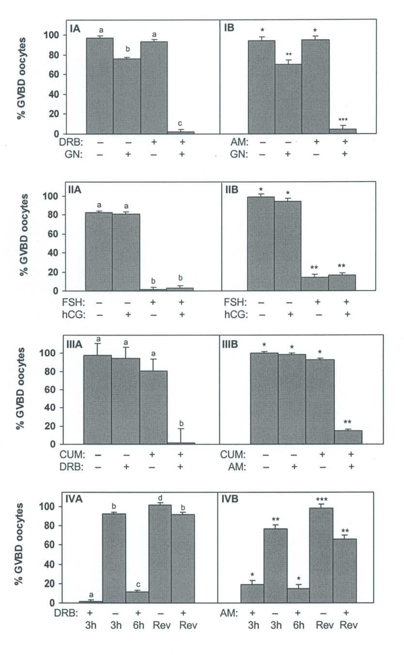
87 73
88 74 Effect of Cumulus Removal on the Inhibition of Oocyte Maturation by DRB or α- Amanitin In the presence of cumulus cells both DRB and α-amanitin effectively inhibited FSH-mediated GVBD (Figure 1, Panels IIIA, IIIB). However, when cumulus cells were removed, neither transcriptional inhibitor was effective in preventing GVBD. These data suggest that transcription initiated during FSH mediated GVBD likely occurred in the cumulus cell compartment. Reversibility of DRB- and α-amanitin-mediated Arrest of GVBD Because α-amanitin has been associated with oocyte and embryo degeneration (21, 22), the capacity of murine COC to continue maturation after culture in the presence of either transcriptional inhibitor was examined. Transcriptional inhibition of FSH-mediated GVBD was reversible and was not associated with oocyte degeneration (Figure 1, Panel IVA, IVB). Ninety-eight percent of COC cultured for 3h in the presence of DRB and 81% of COC cultured in the presence of α-amanitin remained at the GV stage (+DRB 3h and +AM 3h, respectively). After 6h of culture in the presence of DRB, 89% of COC remained at the GV stage, and after 6h of culture in the presence of α-amanitin 85% remained at the GV stage (+DRB 6h or +AM 6h respectively). When COC were exposed to DRB or α-amanitin and then transferred to inhibitor-free medium and cultured for an additional 3h, approximately 92% of COC in the case of DRB and 66% in the case of α-amanitin underwent GVBD (+DRB Rev and +AM Rev, respectively). Compared to treatments with DRB, differences in reversibility associated with α-amanitin were not due to an increased incidence of oocyte
89 75 degeneration since less that 1.5% (28/2009) of oocytes were degenerate after treatment with α-amanitin. Role of PKA Isoenzymes in Facilitating Resumption of Meiosis Effect of activation of Type I or Type II PKA on meiotic resumption: dose-response analysis Increasing concentrations of Type I PKA activators resulted in a dose-dependent decrease in the incidence of GVBD in cultured murine COC (Figure 2). Because activation of Type II PKA was reported to initiate resumption of meiosis in dbcamp-arrested COC (20), the most effective concentrations for activation of Type II PKA were defined as the lowest concentration used that did not interfere with oocyte maturation. These concentrations were 5 µm and 10 µm (84.1± 14.3% GVBD and 88.5 ± 14.3% GVBD, respectively; Figure. 2). Role of transcription in regulating meiosis following differential activation of Type I or Type II PKA In the absence of FSH, treatment of murine COC with Type I PKA activators at 10µM or 50µM significantly inhibited the occurrence of GVBD (p< 0.001). There was no effect of treatment with DRB on the percentage of oocytes remaining in GV arrest following activation of Type I PKA. These observations suggest that inhibition of GVBD by activation of Type I PKA does not require transcription. In contrast, activation of Type II PKA resulted in resumption of meiosis requiring a transcription. When DRB was added to media in the presence of activators of Type II PKA, GVBD was inhibited (Figure 3). In addition, this degree of inhibition was not different from that found when DRB was used in the presence of FSH (20.7 ± 8.4% and 12.5 ± 8.4% GVBD, respectively).
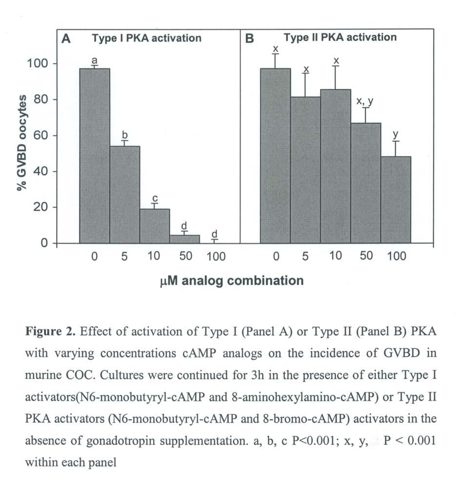
90 76
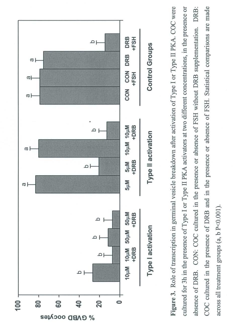
91 77
92 78 DISCUSSION Data presented in this study demonstrate that in cultured murine COC, FSH-mediated GVBD requires gene transcription. The use of two inhibitors, each of which prevents transcription by different mechanisms, led to similar conclusions. DRB, an analog of the nucleoside adenosine, acts by preventing the formation of a stable transcription initiation complex (23). In contrast, α-amanitin acts by specifically inhibiting nucleoplasmic RNA polymerase II (24). Treatment with either inhibitor did not induce oocyte degeneration during the short-term cultures used throughout these experiments since only 85 oocytes of a total of 4282 oocytes (2%) were classified as degenerated following culture. In addition, COC were not required to be maintained in a hypoxanthine-induced meiotic arrest (13, 14) in order for either transcriptional inhibitor to prevent gonadotropin-mediated GVBD. When gonadotropins were included in the culture medium, GVBD was initially inhibited in murine (25, 26) and bovine (10) COC. This transient inhibition was followed by an acceleration in the rate of GVBD compared to that seen during spontaneous maturation (10, 27). The initial inhibitory phase has been hypothesized to result from an increase in camp occurring in response to hormone binding (28, 29). In bovine, porcine, and ovine COC, transcription required for gonadotropin-induced GVBD occurs within 1 hour of the initiation of culture (7, 8, 9). Because the kinetics of maturation in murine oocytes are rapid (4, 30), it is likely that this transcriptional event occurs within minutes of the initiation of culture. In cattle and sheep, gonadotropin-mediated maturation required the presence of cumulus cells (8, 22, 31). In the present study, removal of cumulus cells rendered both DRB and α-amanitin ineffective in blocking murine GVBD.
93 79 These data are consistent with a model in which FSH induces GVBD through transcription that occurs in the cumulus cells. It is unlikely that the lack of effectiveness of the transcriptional inhibitors to block GVBD in denuded oocytes resulted from a decreased access of the inhibitor to the oocyte. Treatment of bovine COC with DRB resulted in greater than a 94% reduction in [ 3 H]-uridine incorporation into oocyte mrna regardless of the presence or absence of gonadotropins in the culture medium (10). In addition, injection of α-amanitin directly into denuded porcine oocytes was ineffective in blocking GVBD (7). Neither DRB nor α-amanitin was effective in preventing spontaneous maturation or hcg-mediated maturation of COC. In contrast, in the presence of FSH both transcriptional inhibitors were effective in preventing meiotic resumption. These observations are consistent with those for bovine COC (11) and suggest that GVBD initiated in the presence of FSH involves a transcriptional step that is not required when GVBD occurs spontaneously or under hcg stimulation. It should be noted that COC from these PMSG-stimulated follicles may have few, if any, receptors for LH (32, 33, 34). Therefore, these COC may have been non-responsive to hcg treatment and, as a result, even in the presence of hcg they may have undergone spontaneous GVBD. Based on data from both the present study and the literature, a general consensus can be reached regarding mechanisms of mammalian oocyte maturation for in vitro cultured COC. In this regard, oocytes can be said to undergo either spontaneous or gonadotropinmediated maturation. Spontaneous maturation occurs as a result of a decline in intraoocyte camp levels after removal of the oocyte or COC from an inhibitory intrafollicular environment. This would be consistent with the essentially linear kinetics seen for
94 80 spontaneously maturing murine (5) and bovine COC (10). In contrast, gonadotropinmediated maturation likely occurs through a mechanism involving FSH binding to cumulus cells, initiating an increase in intracellular camp and activation of both Type I and Type II PKAs. Activation of Type I PKA results in a temporary inhibition of oocyte maturation by an intracellular mechanism not involving transcription. Simultaneously, activation of Type II PKA would initiate a new gene transcription event that subsequently leads to the resumption of meiosis. This hypothesized mechanism would be consistent with observations in the present study and well as with observations of Downs and Eppig (5) and Farin and Yang (10) that culture of COC in the presence of gonadotropins first inhibited and then accelerated the rate of GVBD in mice and cattle, respectively. Based on data in the present study, it becomes apparent why oocytes did not revert to spontaneous maturation when arrested at the GV stage by transcriptional inhibitors in the presence of FSH. In this case, it is likely that FSH-induced increases in camp continued to stimulate Type I PKA, which then maintained oocytes in GV arrest. However, the simultaneous activation of transcription that is mediated by stimulation of Type II PKA, and is required for GVBD, was blocked by the actions of DRB or α-amanitin. Based on this model, it would follow that neither transcriptional nor translational inhibitors would be effective in the absence of an FSH stimulus in any species. This is consistent with reports for the mouse (27), rat (35), pig (7, 36), and cow (10). Conversely, in the presence of an FSH stimulus it would be anticipated that both transcriptional and translational inhibitors would interrupt GVBD. This has been clearly established in the cow and pig (7, 9, 10, 22, 36). Furthermore, murine COC that were meiotically arrested with hypoxanthine and then cultured in the presence of FSH were also inhibited from undergoing
95 81 GVBD by treatment with cycloheximide (14) or α-amanitin (13, 14). Data in the present study demonstrate that in the mouse, transcriptional inhibition by either DRB or α-amanitin occurred only in the presence of FSH, could be mimicked by activation of Type II PKA, and did not require prior hypoxanthine-induced meiotic arrest. Taken together, these observations strongly support the conclusion that transcriptional and translational regulation of GVBD in both murine and livestock COC is remarkably similar, particularly when assessed under analogous culture conditions. In summary, mechanisms for FSH-mediated GVBD in cultured murine COC are similar to those described for in vitro matured COC of other species, including cattle. Thus, in cultured murine COC FSH binding to cumulus cells results in the activation of Type II PKA which, in turn, mediates transcription required for the initiation of germinal vesicle breakdown.
96 82 REFERENCES 1. Tsafriri A, Lieberman ME, Koch Y, Bauminger S, Chobsieng P, Zor U, Lindner HR. Capacity of immunologically purified FSH to stimulate cyclic AMP accumulation and steroidogenesis in GrAMfian follicles and to induce ovum maturation and ovulation in the rat. Endocrinology 1976; 98: Pincus G, Enzmann EV. The comparative behaviour of mammalian eggs in vivo and in vitro. Cell Biol 1935; 109: Dekel N, Lawrence TS, Gilula NB, Beers WH. Modulation of cell-to-cell communication in the cumulus-oocyte complex and the regulation of oocyte maturation by LH. Dev Biol 1981; 86: Schultz RM, Montgomery RR, Belanoff JR. Regulation of mouse oocyte meiotic maturation: implication of a decrease in oocyte camp and protein dephosphorylation in commitment to resume meiosis. Dev Biol 1983; 97: Downs SM, Eppig JJ. Cyclic adenosine monophosphate and ovarian follicular fluid act synergistically to inhibit mouse oocyte maturation. Endocrinology 1984; 114: Eppig JJ. The participation of cyclic adenosine monophosphate (camp) in the regulation of meiotic maturation of oocytes in the laboratory mouse. J Reprod Fertil Suppl 1989; 38: Meinecke B, Meinecke-Tillmann S. Effects of α-amanitin on nuclear maturation of porcine oocytes in vitro. J Reprod Fertil 1993; 98: Osborn JC, Moor RM. Time-dependent effects of α-amanitin on nuclear maturation and protein synthesis in mammalian oocytes. J Embryol Exp Morphol 1983; 73: Kastrop PM, Hulshof SC, Bevers MM, Destree OH, Kruip TA. The effects of α-amanitin and cycloheximide on nuclear progression, protein synthesis, and phosphorylation during bovine oocyte maturation in vitro. Mol Reprod Dev 1991; 28:
97 Farin CE, Yang L. Inhibition of germinal vesicle breakdown in bovine oocytes by 5,6- dichloro-1-β-d-ribofuranosylbenzimidazole (DRB). Mol Reprod Dev 1994; 37: Wolf CJ, Farin CE. Effect of gonadotropins on the ability of 5,6-dichloro-1-β-Dribofuranosylbenzamidazole (DRB) to inhibit germinal vesicle breakdown in bovine oocytes. Theriogenology 1996; 46: Crozet N, Szollosi D. Effect of actinomyocin D and alpha-amanitin on the nuclear ultrastructure of mouse oocytes. Biol. Cell 1980; 1980: Downs SM In: Gametes - The Oocyte. J. G. Grudzinskas and J. L. Yovich (eds). New York: Cambridge University Press Downs SM, Humpherson PG, Martin KL, Leese HJ. Glucose utilization during gonadotropin-induced meiotic maturation in cumulus cell-enclosed mouse oocytes. Mol Reprod Dev 1996; 44: Hunzicker-Dunn M, Jungmann RA. Rabbit ovarian protein kinases. III. Gonadotrophininduced activation of soluble adenosine 3',5'-monophosphate-dependent protein kinases. Endocrinology 1978; 103: Corbin JD, Keely SL, Soderling TR, Park CR. Hormonal regulation of adenosine 3',5'- monophosphate-dependent protein kinase. Adv Cyclic Nucleotide Res 1975; 5: Potter RL, Taylor SS. The structural domains of camp-dependent protein kinase I. Characterization of two sites of proteolytic cleavage and homologies to camp-dependent protein kinase II. J Biol Chem 1980; 255: Doskeland SO, Ogreid D. Characterization of the interchain and intrachain interactions between the binding sites of the free regulatory moiety of protein kinase I. J Biol Chem 1984; 259:
98 Ogreid D, Ekanger R, Suva RH, Miller JP, Doskeland SO. Comparison of the two classes of binding sites (A and B) of type I and type II cyclic-amp-dependent protein kinases by using cyclic nucleotide analogs. Eur J Biochem 1989; 181: Downs SM, Hunzicker-Dunn M. Differential regulation of oocyte maturation and cumulus expansion in the mouse oocyte-cumulus cell complex by site-selective analogs of cyclic adenosine monophosphate. Dev Biol 1995; 172: Kidder GM, Green AF, McLachlin JR. On the use of α-amanitin as a transcriptional blocking agent in mouse embryos: a cautionary note. J Exp Zool 1985; 233: Hunter AG, Moor RM. Stage-dependent effects of inhibiting ribonucleic acids and protein synthesis on meiotic maturation of bovine oocytes in vitro. J Dairy Sci 1987; 70: Zandomeni R, Bunick D, Ackerman S, Mittleman B, Weinmann R. Mechanism of action of DRB. III. Effect on specific in vitro initiation of transcription. J Mol Biol 1983; 167: Zubay G Biochemistry. New York: Addison-Wesley Publishing Co. 25. Schultz RM, Montgomery RR, Ward-Bailey PF, Eppig JJ. Regulation of oocyte maturation in the mouse: possible roles of intercellular communication, camp, and testosterone. Dev Biol 1983; 95: Eppig JJ, Freter RR, Ward-Bailey PF, Schultz RM. Inhibition of oocyte maturation in the mouse: participation of camp, steroid hormones, and a putative maturation-inhibitory factor. Dev Biol 1983; 100: Downs SM. Protein synthesis inhibitors prevent both spontaneous and hormonedependent maturation of isolated mouse oocytes. Mol Reprod Dev 1990; 27:
99 Eppig JJ, Downs SM. Gonadotropin-induced murine oocyte maturation in vivo is not associated with decreased cyclic adenosine monophosphate in the oocyte-cumulus cell complex. Gamete Res 1988; 20: Downs S. The maintenance of meiotic arrest in mamalian oocytes. In: Bavister BD, Cummins J, Roldan ER (eds.): "Fertilization in mammals". Massachusetts: Serono Symposia 1990; pp Downs SM, Daniel SA, Eppig JJ. Induction of maturation in cumulus cell-enclosed mouse oocytes by follicle-stimulating hormone and epidermal growth factor: evidence for a positive stimulus of somatic cell origin. J Exp Zool 1988; 245: Fagbohun CF, Downs SM. Metabolic coupling and ligand-stimulated meiotic maturation in the mouse oocyte-cumulus cell complex. Biol Reprod 1991; 45: Peng XR, Hsueh AJ, LaPolt PS, Bjersing L, Ny T. Localization of luteinizing hormone receptor messenger ribonucleic acid expression in ovarian cell types during follicle development and ovulation. Endocrinology 1991; 129: Eppig JJ, Wigglesworth K, Pendola F, Hirao Y. Murine oocytes suppress expression of luteinizing hormone receptor messenger ribonucleic acid by granulosa cells. Biol Reprod 1997; 56: Eppig JJ, Pendola FL, Wigglesworth K. Mouse oocytes suppress camp-induced expression of LH receptor mrna by granulosa cells in vitro. Mol Reprod Dev 1998; 49: Ekholm C, Magnusson C. Rat oocyte maturation: effects of protein synthesis inhibitors. Biol Reprod 1979; 21: Fulka J, Jr., Motlik J, Fulka J, Jilek F. Effect of cycloheximide on nuclear maturation of pig and mouse oocytes [published erratum appears in J Reprod Fertil 1986 Sep; 78(1):preceding i]. J Reprod Fertil 1986; 77:
100 86 ACTIVATION OF TYPE II PKA STIMULATES RESUMPTION OF MEIOSIS BY A TRANSCIPTIONAL MECHANISM IN CULTURED BOVINE COC ABSTRACT The objective of this study was to assess the role of transcription after activation of Type II PKA in bovine cumulus oocyte complexes (COC) matured in vitro. Differential activation of Type II PKA was performed by culture of COC in the presence of an isomolar concentration of a combination of N 6 -monobutyryl-cyclic adenosine monophosphate (N 6 ) and 8-bromo-cyclic adenosine monophosphate (8-Br). The role of transcription was assessed by culture of COC in the presence or absence of the transcriptional inhibitor 5,6-dichloro-1- β-d-ribofuranosylbenzimidazole (DRB). Selected COC were distributed to one of 8 treatment groups arranged into 2 subgroups: Control groups: n=4 which included all combinations of ± FSH and ± DRB; and, Type II PKA activation groups: n=4 which included 0.5 mm or 1.0 mm Type II camp analogs in the presence or absence or DRB. The proportion of oocytes that underwent germinal vesicle breakdown (GVBD) was assessed after 20 h of culture. As expected, FSH-induced maturation was inhibited by culture in the presence of the transcriptional inhibitor (FSH: 97 ± 4 % GVBD; FSH+DRB: 19 ± 4% GVBD; P<0.001). When COC were cultured with either 0.5 mm or 1.0 mm Type II PKA activators in the presence of DRB, GVBD was also inhibited (0.5 mm: 95 ± 6%, 0.5mM + DRB: 37 ± 6% GVBD, P<0.01; 1.0 mm: 85 ± 4%, 1.0 mm + DRB: 36 ± 4% GVBD; P<0.01). In summary, activation of Type II PKA in cultured bovine COC stimulated maturation by initiating transcription required for the onset of GVBD. This chapter has been submitted for publication as: Activation of Type II PKA Stimulates Resumption of Meiosis by a Transcriptional Mechanism in Cultured Bovine COC. K.F. Rodriguez and C.E. Farin. Dom Anim Endo.
101 87 INTRODUCTION During embryonic development, the oocyte becomes arrested at the dictyate stage of prophase I of meiosis. Throughout the life of the female, oocytes remain in dictyate arrest within their follicles until either the preovulatory surge of gonadotropins signals the resumption of meiosis or atresia ensues (1). Oocytes removed from bovine follicles 2 mm in diameter and cultured in vitro can resume meiosis either in the presence or absence of gonadotropins (2). Spontaneous maturation is believed to result from a fall in levels of intraoocyte camp, since maturation is prevented by elevation of camp in isolated cumulus oocyte complexes (COC) and denuded oocytes (3, 4, 5, 6). In the presence of gonadotropins maturation is mediated by the cumulus cells and, in murine COC, is associated with an initial increase in the levels of camp (7). Specific inhibitors of RNA polymerase II, such as α-amanitin or 5,6-dichloro-1-β-Dribofuranosylbenzimidazole (DRB), have been used to demonstrate that gonadotropinmediated resumption of meiosis in the pig, (8) sheep, (9) cow (10, 11) and mouse (12) requires initial transcription, which occurs in the cumulus cell compartment of COC. In bovine and murine COC, transcription initiated by FSH did not occur when COC resumed meiosis spontaneously or in the presence of hcg (12, 13). Following binding to its receptor, FSH stimulates an elevation of camp levels by activation of adenylyl cyclase. This elevated level of camp results in activation of cyclic AMP-dependent protein kinase A (PKA; 14). Protein kinase A is a tetrameric molecule consisting of paired regulatory subunit dimers each bound to one inactive catalytic subunit. Two isozymes of PKA exist and have been designated as Type I and Type II based on the order of elution of their regulatory subunits (RI and RII, respectively) from anion exchange resins
102 88 (15). There are two camp binding sites found on each of the two regulatory subunits. Within each regulatory subunit, the two camp binding sites differ based on their rate of exchange and affinity for [ 3 H]-cAMP (16, 17). By using combinations of camp analogs that selectively bind to each site on the regulatory subunits, one can differentially activate either Type I or Type II PKA (18). Differential activation of PKA isoenzymes has opposing effects on maturation of cultured murine oocytes. Oocytes were arrested at the germinal vesicle (GV) stage after activation of Type I PKA (12, 19). In this case, activation of Type I PKA appears to maintain meiotic arrest by a non-transcriptionally mediated mechanism (12). In contrast, activation of Type II PKA stimulated GVBD in oocytes arrested by IBMX (19). Furthermore, stimulation of Type II PKA resulted in resumption of meiosis in culture murine COC that required nascent gene transcription and mimicked the mechanism utilized by FSH to initiate GVBD (12). Bovine COC can be maintained at the GV stage by the increased levels of camp that occur following the use of invasive adenylate cyclase (iac; 20). Furthermore, inhibition of maturation by culture of bovine COC with IBMX or forskolin also maintained COC arrest at the GV stage (21, 22). These inhibitors are dependent upon activation of PKA (20, 23) but the specific role of PKA in the maturation of bovine COC has not been described. The objective of the present experiment was to assess the potential role of transcription following Type II PKA activation in regulating bovine oocyte maturation.
103 89 MATERIALS AND METHODS Reagents 5,6-Dicholoro-1-β-D-ribofuranosylbenzimidazole (DRB, Lot #027H4086) and the camp analogs, N 6 -monobutyryl-cyclic adenosine monophosphate (N 6 ) and 8-bromo-cyclic adenosine monophosphate (8 - Br) were purchased from Sigma Chemical Co. (St. Louis, MO). Unless otherwise noted, chemicals for preparation of media were from Sigma Chemical Co. (St. Louis, MO) and were of tissue culture grade. A 20 mg/ml stock solution of DRB was prepared in DMSO and used at a final concentration of 120 µm. Differential activation of Type II PKA was performed by supplementing media with a combination of the camp analogs 8-Br and N 6 (12, 19). The analogs were dissolved in DMSO and used immediately. An equimolar concentration of each analog was used to achieve the final concentration indicated for each experimental group. Oocyte Recovery and Culture Conditions Ovaries collected at a local abattoir were transported to the laboratory in saline with 0.75 µg/ml penicillin. Follicles from 2- to 8-mm in diameter were aspirated using an 18 ga needle and 10 cc syringe. COC with several layers of cumulus cells were collected, washed 5 times in modified Tyrode s medium (TL-Hepes) and matured for 20 h in 1 ml of TCM-199 supplemented with 10% heat-inactivated estrus cow serum (ECS), 1 µg/ml estradiol, 200 µm pyruvate, and 50 µg/ml gentamicin. Bovine COC were distributed to one of 8 treatment groups and cultured for 20 h. The 8 treatment groups were arranged in 2 subgroups: control groups (n=4) that included all combinations of ± DRB (120 µm in 0.2% DMSO) and ± FSH
104 90 (5 µg/ml); and Type II activation groups: (n=4) that included 0.5 mm and 1.0 mm activators of Type II PKA in the presence or absence of DRB. The experiment was replicated 12 times with an average of 62 ± 19 COC per replicate. All cultures were maintained in an atmosphere of 5% CO 2 in air with 100% humidity. Control and treatment media were changed at 4 h intervals throughout the 20 h culture period. Assessment of Meiotic Stage At the end of the culture period, oocytes were assessed for stage of meiotic maturation as previously described (11). Briefly, COC were manually denuded, placed on a microscope slide and gently covered with a cover slip on which Vaseline was placed on two opposing edges. COC were fixed in ethanol-acetic acid (3:1; v/v) for approximately 20 h, stained with 1% orcein in 25% acetic acid and de-stained with ethanol-acetic acid. Oocytes were evaluated for stage of meiotic maturation using criteria established by Motlik et al. (24) using differential interference contrast microscopy at a magnification of 200X. Statistical Analysis Data were arcsin transformed and analyzed by one-way ANOVA with separation of means using Duncan s Multiple Range Test. P < 0.05 was considered statistically significant. All data are reported as least squares means ± standard error.
105 91 RESULTS Consistent with previous data (11, 13), GVBD initiated in the presence of FSH was effectively blocked by treatment with DRB (Control + FSH: 97 ± 4% GVBD, DRB + FSH: 19 ± 4% GVBD, respectively; P<0.001; Figure 1). Compared to controls, GVBD initiated in the absence of FSH was inhibited by approximately 13% when COC were cultured with DRB alone (Control ± FSH: 97 ± 4% GVBD, DRB FSH: 84 ± 4% GVBD, respectively; P<0.01; Figure 1). No differences were found in the proportion of oocytes that underwent GVBD in the presence of either 0.5 or 1.0 mm Type II PKA activators in the absence of DRB (0.5mM: 95 ± 6%, 1.0 mm: 85 ± 4% GVBD; respectively; Figure 1). Compared to 0.5 mm and 1.0 mm Type II PKA controls matured in the absence of DRB, GVBD initiated in the presence of Type II PKA activators was effectively blocked by culture with DRB (0.5 mm + DRB: 36.5 ± 6% GVBD, 1.0mM + DRB: 35.9 ± 4% GVBD; p< 0.01; Figure 1). The inhibition of GVBD achieved when Type II PKA activators were used in combination with DRB was not as effective as when DRB was utilized in FSH-supplemented medium (DRB + FSH: 19 ± 4% GVBD, 0.5 mm + DRB: 36.5 ± 6% or 1.0 mm + DRB: 35.9 ± 4% GVBD; P< 0.001, Figure 1).
106 92
107 93 DISCUSSION Consistent with previously published data in bovine (11) and murine COC (12) the transcriptional inhibitor DRB effectively blocked FSH-induced but not spontaneous maturation. These data imply that FSH-induction of maturation required transcription that does not occur during spontaneous maturation. Signal transduction by FSH occurs following stimulation of adenylate cyclase and activation of cyclic AMP-dependent protein kinase A (PKA; (14). Data in the present study demonstrated that activation of Type II PKA in cultured bovine COC also results in transcription required for GVBD. These findings are in agreement with those reported for murine COC (12). In contrast with our findings in the mouse, treatment with DRB alone decreased the proportion of COC that underwent spontaneous GVBD compared to controls. These data suggest that 13% of the spontaneously maturing COC appear to be DRB-sensitive and require gene transcription for GVBD to occur. We suspect that sensitivity to DRB in this small proportion of spontaneously maturing COC may reflect the inherent heterogeneity of the bovine COC obtained from abattoir ovaries. Unlike homogeneous populations of murine COC that are obtained from gonadotropin-treated prepubertal females, bovine COC are aspirated from abattoir ovaries obtained at random stages of the estrous cycle. Thus, it is probable that a proportion of selected bovine COC might have been exposed to endogenous gonadotropins prior to their collection. In these COC, the process of gonadotropin-induced maturation may have already begun. Thus, these COC would be sensitive to DRB treatment and would not undergo GVBD, even when cultured in the absence of FSH.
108 94 It is clear that the mechanism of maturation occurring in the presence of Type II PKA activators differs from spontaneous maturation because they can be differentiated by their sensitivity to DRB. Activation of Type II PKA in the absence of FSH seems to commit oocytes to a gonadotropin-induced maturation pathway because culture in presence of the DRB inhibits GVBD and mimics the effect of FSH supplementation. Furthermore, oocytes that are cultured in the presence of either FSH or Type II PKA activators remain arrested at the GV stage by DRB and do not revert to a spontaneous pathway. Our previous results (12) as well as those of Downs and Hunzinker-Dunn (19) suggest that in the mouse, activation of Type I PKA results in inhibition of GVBD. An unsuccessful attempt was made to activate Type I PKA in bovine COC (Rodriguez and Farin, unpublished). We suspect that the inability to consistently activate Type I PKA in bovine COC also reflects the differences between use of the superovulated-prepubertal mouse model, which results in a homogeneous population of COC, compared to the abattoir collected bovine ovary model which results in a more heterogeneous population of COC. Subcellular compartmentalization of Type II PKA, through its interaction with A- kinase anchoring proteins (AKAPs), may increase its specificity in regulating meiosis (25). FSH promotes redistribution of Type II PKA holoenzyme in rat granulosa cells through induction of AKAPs (26). It has been proposed that the initial delay in maturation that occurs following binding of FSH to its receptor may be a result of activation of Type I PKA (12, 19). This initial delay would allow time for the induction of AKAPs and redistribution and activation of Type II PKA. Based on data presented in this study, activation of Type II PKA results in the initiation of gene transcription and the production of required mrna
109 95 signals for GVBD. This may occur through PKA-dependent phosphorylation of histone H3 as has been shown for FSH-mediated transcription in rat granulosa cells (27). In summary, activation of Type II PKA promotes GVBD by a transcriptional mechanism that mimics the action of FSH in stimulating GVBD in cultured bovine COC. Identification of the transcriptional signal required for the initiation of GVBD in COC cultured in the presence of FSH is critical for understanding the molecular mechanisms underlying gonadotropin-induced meiotic maturation.
110 96 REFERENCES 1. Wasserman PM, Albertini DF In: The Physiology of Reproduction. E. Knobil and J. D. Neill (eds). New York: Raven Press Pincus G, Enzmann EV. The comparative behaviour of mammalian eggs in vivo and in vitro. Cell Biol 1935; 109: Dekel N, Lawrence TS, Gilula NB, Beers WH. Modulation of cell-to-cell communication in the cumulus-oocyte complex and the regulation of oocyte maturation by LH. Dev Biol 1981; 86: Schultz RM, Montgomery RR, Belanoff JR. Regulation of mouse oocyte meiotic maturation: implication of a decrease in oocyte camp and protein dephosphorylation in commitment to resume meiosis. Dev Biol 1983; 97: Downs SM, Eppig JJ. Cyclic adenosine monophosphate and ovarian follicular fluid act synergistically to inhibit mouse oocyte maturation. Endocrinology 1984; 114: Eppig JJ. The participation of cyclic adenosine monophosphate (camp) in the regulation of meiotic maturation of oocytes in the laboratory mouse. J Reprod Fertil Suppl 1989; 38: Eppig JJ, Downs SM. Gonadotropin-induced murine oocyte maturation in vivo is not associated with decreased cyclic adenosine monophosphate in the oocyte-cumulus cell complex. Gamete Res 1988; 20: Meinecke B, Meinecke-Tillmann S. Effects of α-amanitin on nuclear maturation of porcine oocytes in vitro. J Reprod Fertil 1993; 98:
111 97 9. Osborn JC, Moor RM. Time-dependent effects of α-amanitin on nuclear maturation and protein synthesis in mammalian oocytes. J Embryol Exp Morphol 1983; 73: Kastrop PM, Bevers MM, Destree OH, Kruip TA. Protein synthesis and phosphorylation patterns of bovine oocytes maturing in vivo. Mol Reprod Dev 1991; 29: Farin CE, Yang L. Inhibition of germinal vesicle breakdown in bovine oocytes by 5,6- dichloro-1-β-d-ribofuranosylbenzimidazole (DRB). Mol Reprod Dev 1994; 37: Rodriguez KF, Petters RM, Crosier AE, Farin CE. Roles of gene transcription and PKA subtype activation in maturation of murine oocytes. Reproduction 2002; 123: Wolf CJ, Farin CE. Effect of gonadotropins on the ability of 5,6-dichloro-1-β-Dribofuranosylbenzamidazole (DRB) to inhibit germinal vesicle breakdown in bovine oocytes. Theriogenology 1996; 46: Hunzicker-Dunn M, Jungmann RA. Rabbit ovarian protein kinases. III. Gonadotrophininduced activation of soluble adenosine 3',5'-monophosphate-dependent protein kinases. Endocrinology 1978; 103: Corbin JD, Keely SL, Soderling TR, Park CR. Hormonal regulation of adenosine 3',5'- monophosphate-dependent protein kinase. Adv Cyclic Nucleotide Res 1975; 5: Potter RL, Taylor SS. The structural domains of camp-dependent protein kinase I. Characterization of two sites of proteolytic cleavage and homologies to campdependent protein kinase II. J Biol Chem 1980; 255:
112 Doskeland SO, Ogreid D. Characterization of the interchain and intrachain interactions between the binding sites of the free regulatory moiety of protein kinase I. J Biol Chem 1984; 259: Ogreid D, Ekanger R, Suva RH, Miller JP, Doskeland SO. Comparison of the two classes of binding sites (A and B) of type I and type II cyclic-amp-dependent protein kinases by using cyclic nucleotide analogs. Eur J Biochem 1989; 181: Downs SM, Hunzicker-Dunn M. Differential regulation of oocyte maturation and cumulus expansion in the mouse oocyte-cumulus cell complex by site-selective analogs of cyclic adenosine monophosphate. Dev Biol 1995; 172: Aktas H, Wheeler MB, First NL, Leibfried-Rutledge ML. Maintenance of meiotic arrest by increasing [camp]i may have physiological relevance in bovine oocytes. J Reprod Fertil 1995; 105: Sirard MA, First NL. In vitro inhibition of oocyte nuclear maturation in the bovine. Biol Reprod 1988; 39: Bilodeau S, Fortier MA, Sirard MA. Effect of adenylate cyclase stimulation on meiotic resumption and cyclic AMP content of zona-free and cumulus-enclosed bovine oocytes in vitro. J Reprod Fertil 1993; 97: Rose-Hellekant TA, Bavister BD. Roles of protein kinase A and C in spontaneous maturation and in forskolin or 3-isobutyl-1-methylxanthine maintained meiotic arrest of bovine oocytes. Mol Reprod Dev 1996; 44: Motlik J, Koefoed-Johnsen HH, Fulka J. Breakdown of the germinal vesicle in bovine oocytes cultivated in vitro. J Exp Zool 1978; 205:
113 Kovo M, Schillace RV, Galiani D, Josefsberg LB, Carr DW, Dekel N. Expression and modification of PKA and AKAPs during meiosis in rat oocytes. Mol Cell Endocrinol 2002; 192: Carr DW, DeManno DA, Atwood A, Hunzicker-Dunn M, Scott JD. Follicle-stimulating hormone regulation of A-kinase anchoring proteins in granulosa cells. J Biol Chem 1993; 268: Salvador LM, Park Y, Cottom J, Maizels ET, Jones JC, Schillace RV, Carr DW, Cheung P, Allis CD, Jameson JL, Hunzicker-Dunn M. Follicle-stimulating hormone stimulates protein kinase A-mediated histone H3 phosphorylation and acetylation leading to select gene activation in ovarian granulosa cells. J Biol Chem 2001; 276:
114 100 DEVELOPMENTAL CAPACITY OF BOVINE COC AFTER TRANSCRIPTIONAL INHIBITION OF GERMINAL VESICLE BREAKDOWN ABSTRACT Oocytes cultured in the presence of FSH with the transcriptional inhibitor DRB remain in meiotic arrest at the germinal vesicle stage (GV). The objective of this study was to assess the developmental capacity of bovine cumulus oocyte complexes (COC) following prolonged meiotic arrest by DRB. COC were matured for 20 h in Tissue Culture Medium (TCM)-199 supplemented with 10% estrous cow serum (ECS), 10 µg/ml LH, and 5 µg/ml FSH in the presence or absence of 120 µm DRB. COC in both groups were fertilized and then randomly assigned to one of two development media: TCM % ECS or msof+0.6% fatty acid-free BSA. Development was assessed at 72 hpi, 168 hpi (day 7) and 216 hpi (day 9). As expected, culture with DRB arrested oocyte maturation at the GV stage (DRB, 91 ± 3% GV; Control, 1 ± 3% GV; p<0.001). However, no significant differences were found for inhibitor treatment, development medium or their interaction on cleavage of zygotes at 72 hpi (TCM-199, 88 ± 4% vs. msof, 85 ± 4%; DRB, 82 ± 4% vs. Control, 91 ± 4%). Similarly, no effect of inhibitor, development medium or their interaction was found on development to the blastocyst stage on either day 7 (TCM-199; Control, 31 ± 6% vs. DRB, 21 ± 6%; msof; Control, 21 ± 6% vs. DRB, 14.4 ± 6%) or on day 9 (TCM-199; Control, 41 ± 9% vs. DRB, 35 ± 9%; msof: Control 43 ± 8% vs. DRB: 21 ± 8%). In summary, bovine COC maintained in meiotic arrest for 20 h by culture in the presence of DRB retain their capacity to develop to the blastocyst stage after fertilization in vitro. This chapter has been submitted for publication as: Developmental Capacity of Bovine COC after Transcriptional Inhibition of GVBD. Rodriguez, KF and Farin CE. Theriogenology.
115 101 INTRODUCTION Throughout follicular development oocytes remain arrested at prophase I of meiosis. During this meiotic arrest, oocyte growth continues with increases in oocyte diameter and accumulation of maternal RNA transcripts required for acquisition of developmental competence following fertilization (1, 2, 3). Both cytoplasmic and nuclear maturation must occur for the oocyte to become competent to support subsequent post-fertilization development. Cytoplasmic maturation includes cellular changes including alignment of cortical granules, increased lipid accumulation (4) and accumulation of gluthatione (5, 6). Nuclear maturation refers to acquisition of the ability to undergo dissolution of the germinal vesicle (nuclear membrane), condensation of the chromosomes, release of the first polar body and subsequent arrest at metaphase II (MII; 7, 8). Resumption of meiosis in vivo is triggered by the preovulatory gonadotropin surge (9). However, when bovine cumulus oocyte complexes (COC) are aspirated from antral follicles greater than 2 mm in diameter and cultured in vitro, they resume meiosis either spontaneously or in the presence of gonadotropins (10, 11). De-novo transcription is required for gonadotropin-mediated, but not spontaneous, oocyte maturation (12). Specific inhibitors of transcription, such as α-amanitin or 5,6-dichloro-1-β-D-ribofuranosylbenzimidazole (DRB), have been used to arrest gonadotropin-mediated resumption of meiosis in the pig (13), sheep (14), cow (12, 15) and mouse (16). For transcriptional inhibitors to effectively arrest maturation at the GV stage several layers of cumulus cells must surround the oocytes and gonadotropins must be present in the maturation medium (12, 16). These observations support the conclusion that gonadotropin stimulation results
116 102 transcription, which occurs in the cumulus cells and is required for the initiation of GVBD (12, 16). Only 30 to 40% of bovine oocytes that undergo maturation and fertilization in vitro reach the blastocyst stage (17, 18, 19). This is in contrast to the observation that approximately 80% of oocytes matured and fertilized in vivo reach the blastocyst stage (20). It has been suggested that the developmental competence of in vitro matured oocytes might be increased if oocytes could be maintained in meiotic arrest allowing an additional opportunity for cytoplasmic maturation to occur (3, 21). Roscovitine, a purine that specifically inhibits maturation promoting factor (MPF) kinase activity and effectively blocks cell cycle progression, has been used to maintain bovine oocytes at the germinal vesicle stage without compromising their subsequent developmental potential (21). In contrast, the developmental capacity of bovine COC maintained in transcriptional arrest of meiosis with α-amanitin is severely reduced (22). It is unclear if the reduction in developmental capacity after fertilization noted in de Witt and Kruip (22) was a generalized effect following transcriptionally-mediated meiotic arrest or an effect of the specific transcriptional inhibitor used. Therefore, the objective of this study was to assess the developmental capacity of bovine COC after prolonged meiotic arrest in the presence of the transcriptional inhibitor DRB.
117 103 MATERIALS AND METHODS Reagents and Media Tissue culture medium (TCM-199 with Earl s salts) was purchased from Gibco BRL (Grand Island, NY). Equine pituitary LH (11.5 NIH LH-S1 U/mg) and porcine pituitary FSH (50 mg/vial Armour FSH standard) preparations were obtained from Sigma Chemical Co. (St. Louis, MO). The transcriptional inhibitor 5,6-dichloro-1-β-D-ribofuranosylbenzimidazole (DRB) was obtained from Sigma Chemical Co. (St. Louis, MO). All other reagents and medium supplements were of tissue culture grade and purchased from Sigma Chemical Co. Experimental Design, Oocyte Maturation and Embryo Production The overall experimental design is illustrated in Figure 1. Ovaries collected at a local abattoir were transported to the laboratory in saline with 0.75 µg/ml penicillin. Follicles 2- to 8-mm in diameter were aspirated using an 18 ga needle and 10 cc syringe. COC were collected, washed 5 times in modified Tyrode s medium (TL-Hepes), and matured for 20 h in 1 ml of TCM-199 with 10% heat-inactivated estrus cow serum (ECS), 10 µg/ml LH, 5 µg/ml FSH, 1 µg/ml estradiol, 200 µm pyruvate and 50 µg/ml gentamicin, either in the presence of DRB (120 µm in 0.2% DMSO) or vehicle control (0.2% DMSO). All cultures were maintained in an atmosphere of 5% CO 2 in air with 100% humidity. Both control and treatment media were changed at 4 h intervals throughout the 20 h inhibitor treatment period.
118 104
119 105 At the end of the 20 h inhibitor treatment period, a subset of COC from the DRB-treatment group and all COC from the control group were assessed for stage of meiotic maturation as previously described (12). Briefly, COC were manually denuded, placed on a microscope slide and a three-dimensional mount was prepared using a cover slip on which Vaseline had been placed on two opposing edges. COC were fixed in ethanol-glacial acetic acid (3:1) for approximately 20 h, then stained with 1% orcein in 25% acetic acid and de-stained with ethanol-acetic acid. Oocytes were evaluated for stage of meiotic maturation using criteria established by Motlik et al. (23) using differential interference contrast microscopy at a magnification of 200X. The remaining COC from the DRB treatment group were washed 5 times in maturation medium and then transferred to inhibitor-free medium for an additional 20 h of oocyte maturation. This 20 h maturation period was chosen based on data from a preliminary experiment demonstrating that after release from the inhibitor, COC follow normal kinetics of maturation (data not shown). A new group of untreated COC were collected, washed and matured simultaneously for use as a control group (see Figure 1). After maturation, all COC were washed once and placed into 0.75 ml fertilization medium consisting of Tyrodealbumin-lactate-pyruvate medium supplemented with 6 mg/ml fatty acid-free BSA and 10 µg/ml heparin (24). Motile spermatozoa were collected using a swim-up procedure (24) and fertilization was conducted for h using a final concentration of 1 x 10 6 spermatozoa/ml. Thawed frozen semen from the same sire was used for the production of all embryos. Presumptive zygotes were washed 6 times in TL-Hepes and transferred into either 1 ml TCM-199 with 10% ECS and 50 µg/ml gentamicin or into 1 ml modified Synthetic Oviductal Fluid (msof) supplemented with 0.6% fatty acid free BSA, 1% (v/v) minimal
120 106 essential medium non-essential amino acids and 50 µg/ml gentamicin (25). Cultures in TCM-199-based development medium were performed in an atmosphere of 5% CO 2 in air with 100% humidity. Medium was changed at 48 h intervals throughout culture. Cultures in msof were performed in an atmosphere of 90%N:5%O 2 :5%CO 2 and the medium was undisturbed throughout the entire development period. At 72 hours post insemination (hpi) a subset of zygotes from each treatment group was assessed to determine the percent of zygotes cleaved within each experimental replicate (n=61 ± 9 zygotes per replicate; 4 replicates). The remaining undisturbed zygotes were continued in culture and assessed for stage of development at 168 hpi (day 7) and 216 hpi (day 9; n=65 ± 10 COC per replicate; 4 replicates). Statistical Analysis Data for the effect of treatment on the percentage of oocytes within specific meiotic stages were arcsin transformed and analyzed using Student s t-tests. Data for the effect of treatment on preimplantation embryo development were arcsin transformed and analyzed by two-way ANOVA (26). The statistical model included the effects of replicate, medium, inhibitor and the interaction of medium and inhibitor. Means were considered statistically different at P<0.05. Data are reported as least squares means ± standard error.
121 107 RESULTS Consistent with previous reports (12, 27, 28), bovine COC matured for 20 h in the presence of the transcriptional inhibitor DRB remained at the GV stage (91 ± 2% GV) compared to control COC (0 ± 2 % GV; P<0. 01, Figure 2). No significant difference was found in the proportion of COC remaining at the MI stage between control and DRB-arrested groups (3 ± 3 % vs. 4 ± 3%, respectively). The majority of COC in the control group reached metaphase II (95 ± 4% vs. 4 ± 4% for control and DRB-treated COC, respectively; P< 0.01, Figure 2). At 72 hpi, the percentage of zygotes cleaving to the 2-cell stage or beyond was not different between control and DRB-arrested COC regardless of development medium (TCM- 199 Control, 95 ± 4% cleaved; TCM-199 DRB-arrested, 82 ± 4% cleaved; msof Control, 87 ± 4% cleaved; msof DRB-arrested, 82 ± 4% cleaved). The percentage of COC that developed to the blastocyst stage after 168 hpi (day 7) and 216 hpi (day 9) did not differ between control and DRB-arrested groups when either TCM-199 or msof was used for culture. This was true whether data were expressed as a percent of COC matured or as a percent of oocytes cleaved (Table 1).
122 108
123 109
124 110 DISCUSSION Prolonged transcriptional inhibition of oocyte maturation by DRB does not appear to significantly compromise subsequent development to the blastocyst stage. Our results contrast with those of de Wit and Kruip (22) who found that arrest of meiosis by prolonged transcriptional inhibition using α-amanitin was severely detrimental to oocyte developmental competence. In that study, no COC held in prolonged meiotic arrest for 24 h by α-amanitin developed to the blastocyst stage after fertilization in vitro. Differences in the mechanism of action of the transcriptional inhibitor used in de Wit and Kruip (22) compared to the inhibitor used in the present study likely accounts for the observed differences in developmental capacities of the arrested oocytes. α-amanitin acts by specifically inhibiting nucleoplasmic RNA synthesis by permanently binding to RNA polymerase II (29). In contrast, DRB specifically inhibits transcription by preventing the formation of a stable transcription initiation complex by binding to DRB-sensitive transcription factors present within the cell (30). Binding of DRB to these sensitive factors is highly reversible (30). In the present study, oocytes held in meiotic arrest by DRB resume meiosis with normal kinetics after removal of the inhibitor from the culture medium (Rodriguez and Farin, unpublished). Therefore, it appears that use of a general transcriptional arrest mechanism to prolong maturation prior to fertilization is not itself detrimental to oocyte developmental competency. A greater proportion of in vivo matured oocytes reach the blastocyst stage than in vitro matured oocytes (20, 31, 32, 33). The observation that in vivo matured COC have different developmental competencies than in vitro matured COC supports the premise that in vitro maturation systems can be improved. The manipulation of factors that may affect developmental competence while COC are maintained in meiotic arrest needs to be explored
125 111 (3). Progesterone treatment, which stimulates transcription of de-novo mrnas through a receptor-mediated mechanism (reviewed in 34) had a significant positive effect on subsequent oocyte developmental competence when administered during a 6 h period of meiotic arrest maintained by cycloheximide (3). Similarly, analogs of camp have been shown to stimulate progesterone receptor-mediated transcription in the absence of progesterone (reviewed in 34) and increase oocyte developmental competence during a 6 h cycloheximide arrest (35). These observations suggest that the bovine oocyte can respond to the manipulation of factors during meiotic arrest. Based on studies of [ 3 H]-uridine incorporation, the bovine oocyte is transcriptionally active during folliculogenesis (10, 36). Transcriptional activity decreases during antrum formation but a low level of activity is detected in fully-grown mouse (37) and bovine oocytes (10, 36). This low level of transcriptional activity may represent the activation of genes that influence the acquisition of developmental competence as well as the activation of genes that signal for the resumption of meiosis. Unfortunately, culture of bovine COC in the presence of DRB would inhibit all transcription initiation, including the generation of mrnas needed for both of these physiological functions. Because subsequent developmental capacity of COC maintained in meiotic arrest by culture in the presence of DRB appears not to be compromised, the DRB-arrest model represents a useful physiological approach for the identification of gene products that regulate the onset of GVBD. In summary, bovine COC maintained in meiotic arrest for 20 h by culture in the presence of the transcriptional inhibitor DRB maintain their capacity to develop to the blastocyst stage.
126 112 REFERENCES 1. Blondin P, Coenen K, Guibault L, Sirard M. In vitro competence of bovine embryos:developmental competence is acquired before maturation. Theriogenology 1997; 47: Hyttel P, Viuff D, Fair T, Laurincik J, Thomsen PD, Callesen H, Vos PL, Hendriksen PJ, Dieleman SJ, Schellander K, Besenfelder U, Greve T. Ribosomal RNA gene expression and chromosome aberrations in bovine oocytes and preimplantation embryos. Reproduction 2001; 122: Sirard MA. Resumption of meiosis: mechanism involved in meiotic progression and its relation with developmental competence. Theriogenology 2001; 55: Fair T, Hulshof SC, Hyttel P, Greve T, Boland M. Oocyte ultrastructure in bovine primordial to early tertiary follicles. Anat Embryol (Berl) 1997; 195: de Matos DG, Furnus CC, Moses DF, Baldassarre H. Effect of cysteamine on glutathione level and developmental capacity of bovine oocyte matured in vitro. Mol Reprod Dev 1995; 42: Furnus CC, de Matos DG, Moses DF. Cumulus expansion during in vitro maturation of bovine oocytes: relationship with intracellular glutathione level and its role on subsequent embryo development. Mol Reprod Dev 1998; 51: Brackett BG. In vitro oocyte maturation and fertilization. J Anim Sci 1985; 61: Fulka J, Jr., First NL, Moor RM. Nuclear and cytoplasmic determinants involved in the regulation of mammalian oocyte maturation. Mol Hum Reprod 1998; 4: Tsafriri A, Lieberman ME, Koch Y, Bauminger S, Chobsieng P, Zor U, Lindner HR. Capacity of immunologically purified FSH to stimulate cyclic AMP accumulation and steroidogenesis in Graafian follicles and to induce ovum maturation and ovulation in the rat. Endocrinology 1976; 98:
127 Fair T, Hyttel P, Greve T. Bovine oocyte diameter in relation to maturational competence and transcriptional activity. Mol Reprod Dev 1995; 42: Arlotto T, Schwartz JL, First NL, Leibfried-Rutledge ML. Aspects of follicle and oocyte stage that affect in vitro maturation and development of bovine oocytes. Theriogenology 1996; 45: Farin CE, Yang L. Inhibition of germinal vesicle breakdown in bovine oocytes by 5,6- dichloro-1-β-d-ribofuranosylbenzimidazole (DRB). Mol Reprod Dev 1994; 37: Meinecke B, Meinecke-Tillmann S. Effects of α-amanitin on nuclear maturation of porcine oocytes in vitro. J Reprod Fertil 1993; 98: Osborn JC, Moor RM. Time-dependent effects of α-amanitin on nuclear maturation and protein synthesis in mammalian oocytes. J Embryol Exp Morphol 1983; 73: Kastrop PM, Hulshof SC, Bevers MM, Destree OH, Kruip TA. The effects of α-amanitin and cycloheximide on nuclear progression, protein synthesis, and phosphorylation during bovine oocyte maturation in vitro. Mol Reprod Dev 1991; 28: Rodriguez KF, Petters RM, Crosier AE, Farin CE. Roles of gene transcription and PKA subtype activation in maturation of murine oocytes. Reproduction 2002; 123: Wiemer KE, Watson AJ, Polanski V, McKenna AI, Fick GH, Schultz GA. Effects of maturation and co-culture treatments on the developmental capacity of early bovine embryos. Mol Reprod Dev 1991; 30: Izadyar F, Colenbrander B, Bevers MM. In vitro maturation of bovine oocytes in the presence of growth hormone accelerates nuclear maturation and promotes subsequent embryonic development. Mol Reprod Dev 1996; 45: Ward F, Enright B, Rizos D, Boland M, Lonergan P. Optimization of in vitro bovine embryo production: effect of duration of maturation, length of gamete co-incubation, sperm concentration and sire. Theriogenology 2002; 57:
128 Rizos D, Ward F, Duffy P, Boland MP, Lonergan P. Consequences of bovine oocyte maturation, fertilization or early embryo development in vitro versus in vivo: implications for blastocyst yield and blastocyst quality. Mol Reprod Dev 2002; 61: Mermillod P, Tomanek M, Marchal R, Meijer L. High developmental competence of cattle oocytes maintained at the germinal vesicle stage for 24 hours in culture by specific inhibition of MPF kinase activity. Mol Reprod Dev 2000; 55: de Wit AA, Kruip TA. Bovine cumulus-oocyte-complex-quality is reflected in sensitivity for alpha-amanitin, oocyte-diameter and developmental capacity. Anim Reprod Sci 2001; 65: Motlik J, Koefoed-Johnsen HH, Fulka J. Breakdown of the germinal vesicle in bovine oocytes cultivated in vitro. J Exp Zool 1978; 205: Parrish JJ, Susko-Parrish JL, Leibfried-Rutledge ML, Critser ES, Eyestone WH, First NL. Bovine in vitro fertilization with frozen-thawed semen. Theriogenology 1986; 25: Crosier AE, Farin PW, Dykstra MJ, Alexander JE, Farin CE. Ultrastructural morphometry of bovine blastocysts produced in vivo or in vitro. Biol Reprod 2001; 64: SAS. SAS User's Guide: Statistics, release 6.03 ed. Cary, NC: Statistical Analysis System Institute 1988; 27. Martus NS, Farin CE. Effectiveness of DRB for inhibiting germinal vesicle breakdown in bovine oocytes. Theriogenology 1994; 42: Wolf CJ, Farin CE. Effect of gonadotropins on the ability of 5,6-dichloro-1-β-Dribofuranosylbenzamidazole (DRB) to inhibit germinal vesicle breakdown in bovine oocytes. Theriogenology 1996; 46: Zubay G. Biochemistry. 1983: Addison-Wesley Publishing Co., 1983.
129 Zandomeni R, Bunick D, Ackerman S, Mittleman B, Weinmann R. Mechanism of action of DRB. III. Effect on specific in vitro initiation of transcription. J Mol Biol 1983; 167: Dieleman SJ, Hendriksen PJ, Viuff D, Thomsen PD, Hyttel P, Knijn HM, Wrenzycki C, Kruip TA, Niemann H, Gadella BM, Bevers MM, Vos PL. Effects of in vivo prematuration and in vivo final maturation on developmental capacity and quality of preimplantation embryos. Theriogenology 2002; 57: Hendriksen PJ, Vos PL, Steenweg WN, Bevers MM, Dieleman SJ. Bovine follicular development and its effect on the in vitro competence of oocytes. Theriogenology 2000; 53: van de Leemput EE, Vos PL, Zeinstra EC, Bevers MM, van der Weijden GC, Dieleman SJ. Improved in vitro embryo development using in vivo matured oocytes from heifers superovulated with a controlled preovulatory LH surge. Theriogenology 1999; 52: Mahesh VB, Brann DW, Hendry LB. Diverse modes of action of progesterone and its metabolites. J Steroid Biochem Mol Biol 1996; 56: Guixue Z, Luciano AM, Coenen K, Gandolfi F, Sirard MA. The influence of camp before or during bovine oocyte maturation on embryonic developmental competence. Theriogenology 2001; 55: Memili E, First NL. Developmental changes in RNA polymerase II in bovine oocytes, early embryos, and effect of alpha-amanitin on embryo development. Mol Reprod Dev 1998; 51: Wassarman PM, Letourneau GE. RNA synthesis in fully-grown mouse oocytes. Nature 1976; 261: 73-4.
130 116 IDENTIFICATION OF NOVEL TRANSCRIPTS REQUIRED FOR GONANDOTROPIN-INDUCED BOVINE OOCYTE MATURATION ABSTRACT Bovine oocyte maturation can occur spontaneously or in the presence of gonadotropins. De-novo transcription is required for the initiation of gonadotropin-induced but not spontaneous maturation. The purpose of this experiment was to identify mrnas whose expression is associated with the initiation of GVBD. Bovine ovaries were collected from an abattoir and 2 8 mm follicles were aspirated to obtain cumulus oocyte complexes (COC). COC were selected, washed and either immediately snap frozen in liquid nitrogen (0h) or snap frozen after culture for 4h. The medium used for COC culture was TCM-199 with 10% estrus cow serum, 5 µg/ml FSH, 10 µg/ml LH, 1µg/ml estradiol, 200 µm pyruvate and 50 µg/ml gentamicin in the absence or presence of the transcriptional inhibitor DRB (4h and 4h+DRB, respectively). COC were stored at -80ºC prior to the extraction of whole cell RNA. Differential display was performed using 4 anchor primers and 24 arbitrary primers. Differentially expressed bands were confirmed by replication and then isolated. Amplicon specific primers and independent pools of cdna from COC frozen at either 0h or 4h were used to reconfirm differential expression using semi-quantitative PCR. Expression of GAPDH was utilized as a housekeeping control. A total of 4 amplicons was found to be differentially expressed in cdna from COC from the 4h culture group compared to the 0h or 4h+DRB groups. No differences were found in the expression of GAPDH between treatment groups. Homology searches conducted for the 4 amplicons using the BLAST algorithm did not identify similarities to known sequences suggesting that these amplicons represent novel transcripts.
131 117 INTRODUCTION Mammalian oocytes become arrested in the dictyate stage of prophase I and normally resume meiosis in response to the pre-ovulatory surge of gonadotropins (1). When bovine or murine oocytes are extracted from the follicle and cultured in the presence of gonadotropins, they resume meiosis in a transcriptionally dependent manner (2, 3, 4). This transcriptional event occurs in the cumulus cell since inhibition of transcription during maturation of denuded bovine (2) and murine (4) oocytes is ineffective in arresting GVBD. Furthermore, microinjection of the transcriptional inhibitor α-amanitin does not block GVBD in denuded porcine COC (5). Together, these observations suggest that gonadotropin binding to cumulus cells results in transcription which is required for GVBD (4). Following FSH-binding to its receptor on the cumulus cell, activation of protein kinase A (PKA) occurs. In murine COC, activation of Type I PKA is associated with an inhibition of maturation and activation of Type II PKA is associated with an induction of maturation (4, 6). Activation of Type II PKA requires transcription for the initiation of GVBD in murine (4) and bovine COC (Chapter 3). In bovine COC, addition of a transcriptional inhibitor to maturation medium at different times after the initiation of culture indicated that the transcriptional event required for the gonadotropin-induced GVBD occurs between 1-2 h of the initiation of culture (3). Furthermore, addition of the protein synthesis inhibitor cycloheximide blocks FSH-induced oocyte maturation if added within 6h after the initiation of culture (3). The transcripts required for GVBD are likely present in the ooplasm between 2 and 6 h of the initiation of culture. However, the identity of these gonadotropin-induced mrnas required for the resumption of meiosis and GVBD are not known. The objective of this study was to compare the transcriptional profiles of bovine COC cultured for either 0h, 4 h after the
132 118 initiation of culture (4h) and for 4 h after the initiation of culture in the presence of the transcriptional inhibitor DRB (4h+DRB) to initially identify potential transcripts whose expression is associated with the initiation of GVBD. MATERIALS AND METHODS Experimental Design Experiments were performed to assess and compare the effect of transcriptional inhibition on mrna expression profiles of bovine COC before the initiation of maturation (0 h), after 4 h of culture in the presence of gonadotropins (4h) and after 4 h of culture in the presence of gonadotropins and DRB (4h+DRB). In addition, semi-quantitative PCR analysis to verify differentially expressed amplicons was performed using independent samples of cdna of bovine COC cultured for 0 h or 4 h in the presence of gonadotropins. Reagents and Media Tissue culture medium (TCM-199 with Earl s salts) was purchased from Gibco BRL (Grand Island, NY). Equine pituitary LH (11.5 NIH LH-S1 U/mg) and porcine pituitary FSH (50 mg/vial Armour FSH standard) preparations were obtained from Sigma Chemical Co. (St. Louis, MO). The transcriptional inhibitor 5,6-dichloro-1-β-D-ribofuranosylbenzimidazole (DRB) was obtained from Sigma Chemical Co. (St. Louis MO). All other reagents and medium supplements were of tissue culture grade and purchased from Sigma Chemical Co. Tri-reagent was purchased from Molecular Research Center Inc., (Cincinnati, OH). DNase I and Taq Polymerase were purchased from Roche Molecular Biochemicals
133 119 (Mannheim, Germany). Superscript II reverse transcriptase and dntps were purchased from Invitrogen (Carlsbad, CA). Random hexamers were purchased from Promega Co. (Madison, WI). Gene-Clean II purification kit was purchased from Bio 101 (Carlsbad, CA). QIAquick PCR purification kit was purchased from Qiagen Inc. (Valencia, CA). All primers for PCR and differential display were synthesized by Qiagen Operon (Alameda, CA). Oocyte Recovery and Maturation Ovaries were collected from a local abattoir and transported to the laboratory in 0.9% sterile saline supplemented with 0.75 µg/ml penicillin. Follicles 2- to 8-mm in diameter were aspirated using an 18 ga needle and 10 cc syringe. COC were collected, washed 3 times in modified Tyrode s medium (TL-Hepes) and either snap frozen in liquid nitrogen (0h) or snap frozen after culture for 4 h in the presence of DRB (120 µm in 0.2% DMSO; 4h+DRB) or vehicle control (0.2% DMSO; 4h). All cultures were conducted in 1 ml of TCM-199 supplemented with 10% heat-inactivated estrus cow serum (ECS), 10 µg/ml LH, 5 µg/ml FSH, 1 µg/ml estradiol, 200 µm pyruvate, and 50 µg/ml gentamicin. Cultures were maintained in an atmosphere of 5% CO 2 in air with 100% humidity. After snap freezing in liquid nitrogen all COC stored at -80ºC prior to extraction of whole cell (wc) RNA. wcrna Extraction and Reverse Transcription Tri-reagent was added to frozen COC at a ratio of 800 µl to 120 COC. Extraction was performed according to manufacturer s instructions and the resulting wcrna was resuspended in diethyl pyrocarbonate-treated water at a concentration of 250 ng/ml. Quality of the extracted wcrna was assessed by visualization of 18S and 28S rrna bands on 1%
134 120 ethidium bromide-stained agarose gels. Aliquots (1 µg) of wcrna were snap-frozen in liquid nitrogen and stored at -80ºC prior to analysis. Individual aliquots were thawed on ice and for each cdna synthesis reaction 0.5 µg of each sample was treated with 15U of DNase I for 20 min at 37ºC. DNA-free wcrna was reversed transcribed using Superscript II RNase H - for 1h at 37ºC. The final 50 µl reactions included 0.5 µg of sample mrna, 500 ng of 3 primer, 0.8 mm dntp mix, 1X first strand buffer (50 mm Tris-HCL, 75 mm KCL, 3 mm MgCl 2 ), 0.01 mm dithiothreitol and 400 units of Superscript II reverse transcriptase. For each treatment, aliquots of COC cdna for DD-PCR analysis were synthesized using each of the 4 anchor primers: dt8va, dt8vg, dt8vc and dt8gc. cdna used for semiquantitative PCR analysis was synthesized with 1.5 µg random hexamer primers. A negative control reaction that included all reagents but no wcrna was included. All cdna samples were then cleaned to remove reverse transcription reagents using either Gene Clean II (BIO 101) or PCR clean-up Kit (Qiagen) as recommended by the manufacturer. cdna was then resuspended in diethyl pyrocarbonate-treated water at a concentration of 5 ng/µl. Differential Display PCR (DD-PCR) DD-PCR was performed according to the methods of Liang and Pardee (1992; 7). All reactions were performed in duplicate. For the DD-PCR analysis, a total of 24 arbitrary primers was used in combination with each anchor primer (n=96 reactions, Table 1; 8). Each PCR reaction was run in a final volume of 20 µl and included 5 µl of cdna sample (25 ng cdna), 200 ng of the anchor primer, 40 ng of the arbitrary primer, 16 µm dntp mix, 10 µci of [ 35 S]-dATP (Amersham Pharmacia Biotech), 1X PCR buffer (10 mm Tris-HCL, 1.5 mm MgCl 2, 50mM KCl) and 2.5 U of Taq polymerase. A negative control that included all
135 121
136 122 reagents in the final PCR mix and no cdna was included. All PCR reactions were assembled on ice, overlaid with mineral oil and briefly spun before placing into a PTC-100 thermal cycler (MJ Research, Watertown, MA). PCR was performed for 40 cycles under the following conditions: denaturation for 30 sec at 94ºC, annealing for 30 sec at 45ºC and primer extension for 60 sec at 72ºC. A final extension for 5 min at 72ºC was performed. PCR products were electrophoresed on 6% denaturing polyacrylamide gels in 0.6X TBE buffer at 60mA until the xylene cyanol loading dye reached the bottom of the gel (approximately 3h). Gels were dried for 4h at 60-80ºC and exposed to X OMAT-AR film (Kodak) for 48 h at room temperature. Differentially expressed fragments were located by superimposing the radiograph on the gel using preset alignment markings. Amplicon bands present in the 4h group but not in the 0h or 4h+DRB groups were selected for further analysis. When a band of interest was identified, PCR reactions were repeated with the same cdna pool. If positive, PCR reactions were then performed on independent 0h, 4h and 4h+DRB cdna pools. If still positive, then original fragments of interest were excised with a sterile blade. Amplicon DNA was released from the gel fragment by boiling for 10 min. The resuspended DNA was then subcloned (TA Cloning Kit, Promega, Madison, WI) and sequenced (Duke University Sequencing Facility, Durham, NC). Semi-Quantitative PCR and Image Analysis Primers for semi-quantitative PCR were designed using the Gene Amplify 1.2 (Madison, WI) and Oligo primer analysis software (Plymouth, MA). In addition, primers for bovine glyceraldehyde-3-phosphate dehydrogenase (GAPDH) were synthesized based on previously published sequences (9; Table 2). GAPDH was utilized as a housekeeping standard. Independent pools of cdna from 0h and 4h groups were utilized for

 and 3 arbitrary primers (RP# 13, 14, 16).")
137 123 A dt8gc RP #13 RP #14 RP# D 0 4 4D 0 4 4D B 0h 4h 4h+DRB Figure 1. Representative differential display gel analysis utilized for comparison of patterns of mrna expression during gonadotropin-induced bovine oocyte maturation. A: Gel depicts DD-PCR products from the amplification of 0h, 4h and 4h+DRB COC cdna for one anchor primer (dt8gc) and 3 arbitrary primers (RP# 13, 14, 16). All reactions were run in duplicate. B: Magnified image of boxed portion of gel shown in A. The arrow denotes a band of interest demonstrating differential expression, present in duplicate reactions at 4 h but not at 0h or 4h+DRB.
138 124 PCR. Assays for unknown (UK) #3, UK#6 and UK#11 were replicated 3 times utilizing independent pools of 0h and 4h COC cdna. Assays for UK#1 were replicated twice utilizing independent pools of 0h and 4h COC cdna. For each PCR assay, the individual 20 µl reactions included 200 ng of the appropriate forward and reverse primers, 16 µm dntp mix, 1X buffer (10 mm Tris-HCL, 1.5 mm MgCl 2, 50mM KCl) and 2.5 U Taq polymerase. Reaction mixes for UK#3, UK#6 and UK#11 also included 4 µl of Q solution (Qiagen). The reaction mixes for UK#3 and UK#6 included 75 ng of sample cdna. The reaction mixes for UK#11 included 100 ng of sample cdna and for UK#1, included 125 ng of sample cdna. All reactions were assembled on ice, overlaid with oil and briefly spun at 4ºC before placing into a PTC-100 thermal cycler for 2 min at 94 ºC. The standard PCR program included denaturation for 10 sec at 94 ºC, the appropriate annealing temperature for 10 sec (Table 2) and 10 sec at 72º C for primer extension. The final cycle included a final period for primer extension at 72 ºC for 5 min. The band intensity of each PCR product was determined using computer-assisted video densitometry (Optimus 6.1; Media Cybernetics, Bothell, WA). The ratio of the signal intensities for the specific amplicon products was expressed relative to the signal intensity of GAPDH. Statistical Analysis All data were analyzed using General Linear Model procedures (10). All means were considered statistically different at P<0.05. The statistical model used to analyze signal intensities included the main effects of reverse transcription reaction, treatment and the interaction of cycle number by treatment.
139 125 RESULTS Comparison of the mrna profiles expressed in bovine COC matured for 0h, 4h or 4h+DRB resulted in the initial identification of 11 differentially expressed amplicons. These amplicons were designated unknown (UK) #1 through 11 and were expressed only in the 4h treatment group but not in the 0h or the 4h+DRB treatment groups. Differential expression was confirmed by repeatability of the bands within duplicates, by repeated PCR reactions on the same cdna and by repeated PCR using new cdna pools. In Figure 1 is illustrated a representative differential display gel depicting one of the differentially expressed bands identified. Subcloned inserts were sequenced following band isolation and subcloning. Following initial band identification, reconfirmation and semi-quantitative RT-PCR analysis, four differentially expressed mrna amplicons were identified (Figure 2). The original primers utilized for differential display and the nested primers designed for each amplicon as well as their expected product lengths and annealing temperature are presented in Table 2. The nucleotide sequences of these amplicons and the nested primers within each amplicon are presented in Figure 3. PCR of independent pools of 0h and 4h COC cdna using nested primers for UK#1, UK#3 and UK#6 resulted in amplification products that matched the expected product lengths (Table 2). The amplification product from 0h and 4h COC cdna pools using nested primers for UK#11 was expected to be 319 bp in length; however, the amplified product was found to be 510 bp in length. Sequence analysis of this product revealed that amplification occurred from only one primer of the nested primer pair (Figure 4). Comparison of the product sequence revealed it to be dissimilar to the original sequence of the UK#11 amplicon.
140 126
141 127 0h 4h UK#1 277 bp UK#3 198 bp UK#6 289 bp UK# bp GAPDH 120 bp Figure 2. Representative PCR products for the 4 UK amplicons identified as well as GAPDH. Amplification of the PCR products was performed utilizing nested primers generated from the sequences of the differentially expressed amplicons (Table 2). Each PCR reaction was performed in duplicate on cdna from COC cultured for either 0h or 4h in the presence of gonadotropins. The gel for UK#1 depict the products after 25 cycles of amplification. Gels for UK#3, UK#6 and UK#11 depict the products after 30 cycles of amplification
142 128 UK#1 TTACAACGAGGACATAGAAGTTGACAGACTATCAAATTATCAGGCATATAAAACTGATACTGCT GGACGTTACAAAGAAAATCATCTTGCTGTTTCATCAGCACCAGATCCAAACCTGATTCAACCCAGT GCAGAAAAGACAAAAATGCAAGATGCAGCAATTCAGACAATCCCCTCCTGTGGCAGTTTTGATGG GGATCAGCAAGATCATAATTTGTCTGACGTCAAAGTTGATGAAAGTGTGCAAACTTCAAGTAACA ACAAATCAACTCAACACTTATCTTTAAGCCCACAAGATTCTATAGATATCTCAGGAAAATTCAGG AGTAAGGGCCCCCTCGTTGTA UK#3 TACAACGAGGGTGATGTCAAATAGCAATTTCCCACAACTGATTAAAAACTGAGTTGGAAGTCAGG AGTTCTTTCTGTCGCCCTGTTTGTCCTCTGTTCAATGGTTATTTGGTAATTATATTTGAGGTAAGT AGAAAGGTAATTTTGGACCAAAGCCTTTACGGTATATCCACAGATGTCAAATTGCTGAGACTAACT CCTTCAAATTCCCTCTCTCTGAAATTCTTCCCTGAGTTCCACCTTCTTGATAAATCTATCAGTGCTT CAGTGGCGATGAAATGTTTGCTGAGGGACATCTGGGTGTATCAAGTGGCAGAGGACCAATCTTCT GTATCGCCTCGTTGTA UK#6 TACAACGAGGCATAGGTATCACTCCCATTTTCAACACTGGGAAACCTGAGGGTTTAGAGGCTTA GGTTTAGACACTTGCCTAAAAGAGGCCAAGCTAGTGAATGGCAGGAAGCTATGGTGAGAACTCGG GCCTGGCTGAGCCCAGAACTGGGTCTTCATGACCTTTAGTACCATCTGTGATCTGATGGTGCTTGA AGCGGAGCTGGGTTAAACAGACAGCTCTGGCAGTCTAAGAGGGCTCAGATTTTCCTCTAGAGTGG GGCCGGTTCCCTTGAGACCTGACTTGGGGGTCCCCTTGGCCTTGCGTGCTGCTTTCCTCCTGCCGT CTGTTGCCGCATCCCTCGTTGTA UK#11 GATCCAGTACCCACAAGTAAATGTCTGATCCAGTACCATACAAAAGAGTTTATTGAACTGTAGC AAGTTCAAAGGAGAATGACCATATCATTCAAAGTGTCTTCTTTAAGCCTTTATTTCCATTTTATGTT CAGAACAGCTTTATTCTTACTAATTTACCTTATAATAAACGTTATAAACATTATTTAAAATAATGTT TGAACTTACAGAAGAAATCTTAAGTAAATTTTAAATATAAATTACTTAATTACACTCACACACATG TAAAAATATAAAATTGTCTATGTTTTATGTATATTTGGAGATAGATAGGTAATCTCCTATAGCTA GTACTGGATC Figure 3. Sequences of the 4 differentially expressed amplicons identified in the DD-PCR analysis. The sequence and position of the nested primers utilized for semi-quantitative PCR analysis are shown underlined in bold type.
143 129 UK#11 insert sequence GATCCAGTACCCACAAGTAAATGTCTGATCCAGTACCATACAAAAGAGTTTATTGAACTGTAGC AAGTTCAAAGGAGAATGACCATATCATTCAAAGTGTCTTCTTTAAGCCTTTATTTCCATTTTATGTT CAGAACAGCTTTATTCTTACTAATTTACCTTATAATAAACGTTATAAACATTATTTAAAATAATGTT TGAACTTACAGAAGAAATCTTAAGTAAATTTTAAATATAAATTACTTAATTACACTCACACACATG TAAAAATATAAAATTGTCTATGTTTTATGTATATTTGGAGATAGATAGGTAATCTCCTATAGCTA GTACTGGATC UK#11 product sequence (F) TACGACTCACTATAGGGCGAATTGAATTTAGCGGCCGCGAATTCGCCCTTGATCCAGTACCCACA AGTAATAGGCTTTTTTATGTTTCCATGGGTGGACTTCCTTAGGGGTGAGTCTTTCTGAAGCTTATCC TACCCAGAACGGTTGCAACCTGAGAGCCAGGCATCCCCGGGTCAGGGTGCAGATCCCCAGGCAGG TTGAGGCGTGACAGCCTCCTAGAGATCGAATCCCCGGGCAGGTCAAGGCGTGACGGCCTCCTGAG GATTGGGTCCCTGGGCAGGTCGAGGTGTGATGACCTCCTGGGACCCCTGGGACTAGAGGATCCCC GAGCAGGTTGAGACGTGACAACCTTTTGAAGACCGATCCCTAGGCAGGTCAAGGCGTGATGACCT CCTGGGACCCCCGGACTGGAGTTTGGGCATCCCCAAGATCTCCAGTGTGACCAGTATTGGGTAGG ATTAGGGGGTTGGATATTATAGAGTATTTCCCTCCCAAGGCCAACTATTTTCTGGNTGNCTNTNCA NAANATNGNANAGGCAAACTNGNGGGTNCTGGATCAAGGGC UK#11 product sequence (R) TTACGCCAGCTCAGNAATTAACCCTCACTAAAGGGACTAGTCCTGCAGGTTTAAACGAATTCGCCC TTGATCCAGTACCCACAAGTTTGCCTCTACAATCTTCTGGAGAGACAACCAGTAAAATAGTTGGC CTTGGGAGGGAAATACTCTATAATATCCAACCCCCTAATCCTACCCAATACTGGTCACACTGGAGA TCTTGGGGATGCCCAAACTCCAGTCCGGGGGTCCCAGGAGGTCATCACGCCTTGACCTGCCTAGG GATCGGTCTTCAAAAGGTTGTCACGTCTCAACCTGCTCGGGGATCCTCTAGTCCCAGGGGTCCCAG GAGGTCATCACACCTCGACCTGCCCAGGGACCCAATCCTCAGGAGGCCGTCACGCCTTGACCTGC CCGGGGATTCGATCTCTAGGAGGCTGTCACGCCTCAACCTGCCTGGGGATCTGCACCCTGACCCGG GGATGCCTGGCTCTCAGGTTGCAACCGTTCTGGGTAGGATAAGCTTCANAAAGACTCACCCCTAA GGAAGTCCACCCATGGAAACATAAAAAAGCCTATTACTTGNGGGTACTGGATCAAGGGCGAATTC NNGGNCGNTAAATNCAATTNNCCCTATAGNGAGNCNN Figure 4. Sequences for UK#11 products. The original amplicon sequence is shown at the top with the nested primers in underlined bold type. The forward (F) and reverse (R) sequences of the PCR product obtained from utilizing the nested primers are also shown. The products were obtained from the amplification from only one primer. The sequences of the PCR products have no homology with the original insert sequence.
144 130 Comparison of the signal intensities of the PCR products from COC cdna samples from 0h and 4h treatment groups using semi-quantitative PCR demonstrated that the expression of each of the four unknown mrnas was more abundant after 4h of in vitro culture compared to 0h (Figure 2, 5 and 6). The expression of mrna for GAPDH was not different between treatments. When signals intensities of the PCR products representing the four differentially expressed amplicons were expressed as a ratio of the signal intensities of products representing mrna for GAPDH, statistical differences were maintained (Figure 2, 5 and 6). Homology searches conducted for the 4 amplicons using the BLAST algorithm did not identify any similarities to known sequences. DISCUSSION Differential display-pcr is a technique that allows for the identification of differentially expressed genes (7, 8, 11). The use of defined combinations of primers for reverse transcription PCR generates a series of mrnas that correspond to a fraction of the mrna present in the extract (7). The major disadvantage of this method is that, by design, the cdna amplicons generated represent sequences primarily from the 3 untranslated region (UTR) and not more upstream in the coding sequences or 5 - UTR of mrnas. As a result, the correct identification of the amplicon sequences generated becomes more difficult because 3 -UTRs of mammalian mrnas are highly divergent. Thus, further characterization of mrna sequences represented by differentially expressed amplicons frequently requires analysis using rapid amplification of cdna ends (RACE) techniques.
145 131
146 132
147 133 In the present study, use of DD-PCR resulted in the initial identification of eleven differentially expressed amplicons. As expected, the differential expression of four of the initial 11 amplicons was independently reconfirmed by semi-quantitative PCR. The expression of COC mrna represented by all 4 of these amplicons was greater after 4h of culture in the presence of gonadotropins compared to 0h culture controls. Based on selection criteria, these mrnas were also not expressed in the 4h+DRB treatment group. For all of these amplicons, no identity to known sequences was found using the BLAST algorithm. This observation suggests that the 4 amplicons represented novel mrnas. This result may not be surprising since 25% of mrnas identified in human oocytes by SAGE analysis were also found to be novel sequences (12, 13). In bovine COC, addition of α-amanitin (3) or DRB (Farin, unpublished) no later than 1 2 h after the initiation of culture was required for these transcriptional inhibitors to block oocyte maturation. In rabbit COC, α-amanitin had to be present in the culture medium within 2 h of the initiation of culture to effectively arrest oocyte maturation (14). Similarly, ovine COC were maintained at the GV stage only if α-amanitin was present in the maturation medium within 1 2 h of the initiation of culture (15). Together, these observations indicate that the transcription induced by gonadotropin stimulation occurs within a 2h window after the start of culture in a number of mammalian species. In sheep, cattle and pigs protein synthesis inhibitors such as cycloheximide must be present between 2 and 6h after the initiation of culture to inhibit GVBD (3,16, 17, 18). Thus, in these species it is likely that mrna synthesis begins after 1-2 h from the initiation of culture and continues to the translated up to 6 h after the initiation of culture. In this study, the 4 h culture time was chosen because it provided the highest probability of finding detectable levels of mrna.
148 134 Following gonadotropin stimulation, an increase in the levels of camp occurs as a result of the activation of adenylase cyclase and leads to activation of type I and type II PKA. In the mouse, activation of Type I PKA resulted in the inhibition of maturation whereas activation of Type II PKA stimulated maturation (6). Activation of Type II PKA in murine COC results in transcription required for the initiation of GVBD (4). The inhibitory effects of the activation of Type I PKA coincide with the initial inhibition of oocyte maturation that follows gonadotropin-stimulation (2, 19). Interestingly, in rat granulosa cells, FSH stimulates phosphorylation of histone H3 through the activation of PKA (20, 21). Furthermore, also in rat granulosa cells, phosphorylation of histone H3 by PKA allows for transcriptional activation of specific FSH responsive genes (21). Some of these genes have been identified to be associated with steroidogenesis (22, 23) and connexin expression (24). The present study demonstrates that FSH also induces other genes with unknown identities whose expression coincides with the initiation of GVBD. The inhibitory action of DRB on gonadotropin-induced maturation is dependent upon the presence of cumulus cells in both bovine (2) and murine (4) COC. Furthermore, disruption of gap-junctional communication between the cumulus cells and the oocyte with heptanol prevented FSH-induced resumption of meiosis in hypoxanthine-arrested murine COC (25). These observations suggest that this transcription takes place in the cumulus cell and that the maturation signal is likely transmitted to the oocyte through gap junctions. Microinjection of α-amanitin directly into the ooplasm of porcine oocytes had no effect on the progression of maturation (5). These data imply that cumulus cells, and not the oocyte, are targets for the transcriptional inhibitor. The maturation signal transmitted to the oocyte by the cumulus cell, if transmitted through gap junctions, would have to be a small molecule
149 135 (<900 Da; 26) such as camp or ions. That transcription is required for this maturation signal suggests the possibility that mrnas encoding specialized ion channels or connexin proteins may be candidates for these key transcripts. Interestingly, in rat granulosa cells FSH does induce the expression of connexin 43 mrna (24). In summary, DD-PCR analysis was used to compare mrna populations in bovine COC during gonadotropin-mediated oocyte maturation. Analysis of mrna populations in COC cultured for 0h or 4h in either the presence or absence of the transcriptional inhibitor DRB, led to the identification of 11 differentially expressed amplicons. Of these 11 amplicons, the differential expression of 4 of these amplicons was subsequently verified by semi-quantitative PCR. For all 4, expression was increased at 4h of culture compared to 0h controls. Homology searches conducted for the 4 amplicons using the BLAST algorithm did not identify any similarities to known sequences, suggesting that these amplicons represent novel transcripts.
150 136 REFERENCES 1. Tsafriri A, Lieberman ME, Barnea A, Bauminger S, Lindner HR. Induction by luteinizing hormone of ovum maturation and of steroidogenesis in isolated Graafian follicles of the rat: role of RNA and protein synthesis. Endocrinology 1973; 93: Farin CE, Yang L. Inhibition of germinal vesicle breakdown in bovine oocytes by 5,6- dichloro-1-β-d-ribofuranosylbenzimidazole (DRB). Mol Reprod Dev 1994; 37: Kastrop PM, Hulshof SC, Bevers MM, Destree OH, Kruip TA. The effects of α-amanitin and cycloheximide on nuclear progression, protein synthesis, and phosphorylation during bovine oocyte maturation in vitro. Mol Reprod Dev 1991; 28: Rodriguez KF, Petters RM, Crosier AE, Farin CE. Roles of gene transcription and PKA subtype activation in maturation of murine oocytes. Reproduction 2002; 123: Meinecke B, Meinecke-Tillmann S. Effects of α-amanitin on nuclear maturation of porcine oocytes in vitro. J Reprod Fertil 1993; 98: Downs SM, Hunzicker-Dunn M. Differential regulation of oocyte maturation and cumulus expansion in the mouse oocyte-cumulus cell complex by site-selective analogs of cyclic adenosine monophosphate. Dev Biol 1995; 172: Liang P, Pardee AB. Differential display of eukaryotic messenger RNA by means of the polymerase chain reaction. Science 1992; 257: Bauer D, Muller H, Reich J, Riedel H, Ahrenkiel V, Warthoe P, Strauss M. Identification of differentially expressed mrna species by an improved display technique (DDRT- PCR). Nucleic Acids Res 1993; 21: Leutenegger CM, Alluwaimi AM, Smith WL, Perani L, Cullor JS. Quantitation of bovine cytokine mrna in milk cells of healthy cattle by real-time TaqMan polymerase chain reaction. Vet Immunol Immunopathol 2000; 77:
151 SAS. SAS User's Guide: Statistics, release 6.03 ed. Cary, NC: Statistical Analysis System Institute 1988; 11. Callard D, Lescure B, Mazzolini L. A method for the elimination of false positives generated by the mrna differential display technique. Biotechniques 1994; 16: , Stanton JL, Bascand M, Fisher L, Quinn M, Macgregor A, Green DP. Gene expression profiling of human GV oocytes: an analysis of a profile obtained by Serial Analysis of Gene Expression (SAGE). J Reprod Immunol 2002; 53: Neilson L, Andalibi A, Kang D, Coutifaris C, Strauss JF, 3rd, Stanton JA, Green DP. Molecular phenotype of the human oocyte by PCR-SAGE. Genomics 2000; 63: Motlik J, Fulka J, Jr., Prochazka R, Rimkevicova Z, Kubelka M, Fulka J. RNA and protein synthesis requirements for the resumption of meiosis in rabbit oocytes: the role of cumulus cells. Reprod Nutr Dev 1989; 29: Osborn JC, Moor RM. Time-dependent effects of α-amanitin on nuclear maturation and protein synthesis in mammalian oocytes. J Embryol Exp Morphol 1983; 73: Moor RM, Crosby IM. Protein requirements for germinal vesicle breakdown in ovine oocytes. J Embryol Exp Morphol 1986; 94: Sirard MA, Florman HM, Leibfried-Rutledge ML, Barnes FL, Sims ML, First NL. Timing of nuclear progression and protein synthesis necessary for meiotic maturation of bovine oocytes. Biol Reprod 1989; 40: Fulka J, Jr., Motlik J, Fulka J, Jilek F. Effect of cycloheximide on nuclear maturation of pig and mouse oocytes [published erratum appears in J Reprod Fertil 1986 Sep;78(1):preceding i]. J Reprod Fertil 1986; 77: Downs SM, Eppig JJ. Cyclic adenosine monophosphate and ovarian follicular fluid act synergistically to inhibit mouse oocyte maturation. Endocrinology 1984; 114:
152 DeManno DA, Cottom JE, Kline MP, Peters CA, Maizels ET, Hunzicker-Dunn M. Follicle-stimulating hormone promotes histone H3 phosphorylation on serine-10. Mol Endocrinol 1999; 13: Salvador LM, Park Y, Cottom J, Maizels ET, Jones JC, Schillace RV, Carr DW, Cheung P, Allis CD, Jameson JL, Hunzicker-Dunn M. Follicle-stimulating hormone stimulates protein kinase A-mediated histone H3 phosphorylation and acetylation leading to select gene activation in ovarian granulosa cells. J Biol Chem 2001; 276: Goldring NB, Durica JM, Lifka J, Hedin L, Ratoosh SL, Miller WL, Orly J, Richards JS. Cholesterol side-chain cleavage P450 messenger ribonucleic acid: evidence for hormonal regulation in rat ovarian follicles and constitutive expression in corpora lutea. Endocrinology 1987; 120: Richards JS, Hickey GJ, Chen SA, Shively JE, Hall PF, Gaddy-Kurten D, Kurten R. Hormonal regulation of estradiol biosynthesis, aromatase activity, and aromatase mrna in rat ovarian follicles and corpora lutea. Steroids 1987; 50: Sommersberg B, Bulling A, Salzer U, Frohlich U, Garfield RE, Amsterdam A, Mayerhofer A. Gap junction communication and connexin 43 gene expression in a rat granulosa cell line: regulation by follicle-stimulating hormone. Biol Reprod 2000; 63: Fagbohun CF, Downs SM. Metabolic coupling and ligand-stimulated meiotic maturation in the mouse oocyte-cumulus cell complex. Biol Reprod 1991; 45: Bruzzone R, White TW, Paul DL. Connections with connexins: the molecular basis of direct intercellular signaling. Eur J Biochem 1996; 238: 1-27.
153 139 GENERAL CONCLUSIONS The technology of in vitro maturation of oocytes is utilized for the production of embryos for commercial as well as research purposes. The difference between the developmental potential of oocytes matured in vivo compared to oocytes matured in vitro suggests the possibility for improvement of in vitro oocyte maturation systems. An understanding of the molecular mechanisms that underlie gonadotropin-induced maturation may allow for the improvement of this technology. The research described in this dissertation was performed to determine the molecular pathways that underlie in vitro gonadotropin-induced maturation. Following binding to its receptor, gonadotropins stimulate COC to undergo maturation in a variety of in vitro culture systems. Previous studies in our laboratory demonstrated that cultured bovine COC could be maintained in meiotic arrest following release from the follicle if a transcriptional inhibitor is present in the culture medium. Similarly, in Chapter 2 murine COC were shown to be maintained in meiotic arrest by culture in the presence of one of either two transcriptional inhibitors, 5,6-dichloro-1-β-Dribofuranosylbenzimidazole (DRB) or α-amanitin, both of which utilize different mechanisms to block transcription. For both transcriptional inhibitors to effectively block maturation, FSH but not hcg, was required in the culture medium. Furthermore, transcriptional inhibition of oocyte maturation was ineffective if denuded oocytes were utilized. From these observations, it was concluded that FSH binding to its cumulus cell receptor results in transcription, which is required for the initiation of maturation. In this same study, differential activation of Type I and Type II PKA isoforms was performed in murine COC. Activation of Type I PKA resulted in the inhibition of maturation whereas
154 140 activation of Type II did not. Furthermore, in the absence of gonadotropin supplementation, culture of murine COC in the presence of activators of Type II PKA and a transcriptional inhibitor resulted in the meiotic arrest. These observations suggest that in murine COC, FSH stimulation results in the activation of Type I and Type II PKA. Activation of Type I results in a transient delay of maturation which is then followed by activation of Type II PKA. Activation of Type II PKA is then followed by transcription required for gonadotropininduced oocyte maturation. As shown in Chapter 3 of this thesis, in cultured bovine COC, activation of Type II PKA also results transcription required for gonadotropin-induced maturation. Taken together these observations suggest that the mechanism of action of gonadotropin-induced maturation is probably conserved between these two species. The possibility of improvement of in vitro oocyte maturation systems is based on the observation of greater developmental competency for oocytes matured in vivo versus oocytes matured in vitro (1). Maintenance of meiotic arrest provides an opportunity for the manipulation the culture environment with the goal of improvement of developmental competency following fertilization. The identification of oocyte maturation inhibitors that do not diminish the developmental competency of oocytes following release from this meiotic arrest is critical for the application of this approach. As shown in Chapter 4, the developmental competency of bovine COC maintained in meiotic arrest for 20 h by culture in the presence of DRB was not different from control COC. Identification of the specific mrna transcripts required for gonadotropin-induced resumption of meiosis would allow for a targeted silencing of these required mrnas. As shown in Chapter 5, the comparison of the mrna expression patterns for oocytes matured for 0h, 4h or 4h+DRB resulted in the isolation of 11 amplicons that were found to be
155 141 differentially expressed after 4h of culture. After semi-quantitative PCR analysis, the differential expression of 4 transcripts was confirmed. No homology to known sequences was found for this amplicons which suggests the possibility that they represent novel transcripts. Future work towards the identification of the specific transcript(s) required for the initiation of GVBD would provide a unique opportunity to achieve a targeted arrest of meiosis without affecting the accumulation of key mrnas needed for further acquisition of developmental competence. Such a system has the potential to be used to increase the developmental competence of in vitro matured COC. Based on the current literature and the results obtained in the experiments that are part of this dissertation, a general working model can be developed regarding the mechanism of gonadotropin-induced maturation in vitro. Inside the follicle, oocytes are maintained in meiotic arrest (Figure 1A). Following extraction from the follicle, the oocytes undergo maturation in either the presence or absence of gonadotropins. Spontaneous maturation likely occurs solely as a result of a decline in intraoocyte camp. This decline may occur either passively, as no more camp is shuttled to the oocyte through gap junctions, or due to the removal of an inhibitory signal that maintained PDE 3A present in the oocyte cytoplasm inactivated. This decrease in the levels of camp allows GVBD to occur (Figure 1B). This mechanism would be consistent with the essentially linear kinetics observed for spontaneous maturation of murine and bovine COC (2, 3). In contrast, gonadotropin-mediated maturation likely occurs through a mechanism that involves FSH binding to its receptor on the cumulus cells. FSH receptor activation results in an increase in the levels of camp and activation of both Type I and Type II PKA (4, 5). Activation of Type I PKA results the inhibition of oocyte maturation though an intracellular mechanism that is independent of gene
156 142 transcription (5). The mechanism of Type I PKA inhibition of maturation may involve the arrest of PDE 3A activity, maintaining the level of camp and the oocyte at the GV stage through the initial inhibitory period that follows FSH stimulation (Figure 2A). After Type I PKA activation, subsequent activation of Type II PKA occurs, which results in the initiation of a transcriptional signal that mediates gonadotropin-induced resumption of meiosis. Subcellular compartmentalization of Type II PKA though its interaction with A-kinase anchoring proteins (AKAPs) may increase its specificity in regulating meiosis (6). FSH promotes redistribution of Type II PKA in rat granulosa cells through induction of AKAPs (7). Furthermore, FSH-mediated transcription in rat granulosa cells occurs through PKAdependent phosphorylation of histone H3 (8). It can be proposed that the original delay in maturation is a result of activation of Type I PKA, which allows time for redistribution of AKAPs, transcription of required mrna signals and possible accumulation of additional competency factors, while preventing spontaneous maturation and maintaining COC in the gonadotropin-induced maturation pathway (Figure 2B). The proposed mechanism described for gonadotropin-induced resumption of meiosis is consistent with the observations that following gonadotropin stimulation an, initial delay in the rate of oocyte maturation occurs (2, 3). This proposed mechanism also explains why oocytes committed to resume meiosis as a result of gonadotropin stimulation do not revert to the spontaneous maturation pathway while arrested at the GV stage by a transcriptional inhibitor. In this case, the continued activation of Type I PKA by FSH is responsible for maintaining the arrest whereas the stimulatory phase of FSH-induced oocyte maturation would be blocked by the transcriptional inhibitor. Based on this model it would follow that neither transcriptional nor translational inhibitors would be effective in blocking oocyte
157 143 maturation in the absence of gonadotropin stimulation. This is consistent with reports for the mouse (9), rat (10), pig (11, 12) and cow (3). In the mouse, COC maintained in meiotic arrest by culture in the presence of hypoxanthine resumed meiosis if stimulated by FSH (13). Both the transcriptional inhibitor α-amanitin and the protein synthesis inhibitor cycloheximide blocked FSH-stimulated resumption of meiosis (4, 14). Furthermore, in the mouse, transcriptional inhibition of maturation occurred only in the presence of FSH and could be mimicked by activation of Type II PKA (5). Taken together these observations suggest that transcriptional and translational mechanisms of gonadotropin-mediated resumption of meiosis in both murine and bovine in vitro-cultured COC are similar. In both cases, following FSH binding to its cumulus cell receptor, camp levels increase within the cumulus cell. This elevated levels of camp result in activation of Type I and Type II PKAs. Activation of Type I PKA is involved in the transient inhibition of maturation whereas Type II PKA activation results in new gene transcription that subsequently leads to resumption of meiosis.
158 144
159 145
160 146 REFERENCES 1. de la Pena EC, Takahashi Y, Katagiri S, Atabay EC, Nagano M. Birth of pups after transfer of mouse embryos derived from vitrified preantral follicles. Reproduction 2002; 123: Downs SM, Eppig JJ. Cyclic adenosine monophosphate and ovarian follicular fluid act synergistically to inhibit mouse oocyte maturation. Endocrinology 1984; 114: Farin CE, Yang L. Inhibition of germinal vesicle breakdown in bovine oocytes by 5,6- dichloro-1-β-d-ribofuranosylbenzimidazole (DRB). Mol Reprod Dev 1994; 37: Downs SM, Hunzicker-Dunn M. Differential regulation of oocyte maturation and cumulus expansion in the mouse oocyte-cumulus cell complex by site-selective analogs of cyclic adenosine monophosphate. Dev Biol 1995; 172: Rodriguez KF, Petters RM, Crosier AE, Farin CE. Roles of gene transcription and PKA subtype activation in maturation of murine oocytes. Reproduction 2002; 123: Kovo M, Schillace RV, Galiani D, Josefsberg LB, Carr DW, Dekel N. Expression and modification of PKA and AKAPs during meiosis in rat oocytes. Mol Cell Endocrinol 2002; 192: Carr DW, DeManno DA, Atwood A, Hunzicker-Dunn M, Scott JD. Folliclestimulating hormone regulation of A-kinase anchoring proteins in granulosa cells. J Biol Chem 1993; 268: Salvador LM, Park Y, Cottom J, Maizels ET, Jones JC, Schillace RV, Carr DW, Cheung P, Allis CD, Jameson JL, Hunzicker-Dunn M. Follicle-stimulating hormone stimulates protein kinase A-mediated histone H3 phosphorylation and acetylation leading to select gene activation in ovarian granulosa cells. J Biol Chem 2001; 276:
161 Downs SM. Protein synthesis inhibitors prevent both spontaneous and hormonedependent maturation of isolated mouse oocytes. Mol Reprod Dev 1990; 27: Ekholm C, Magnusson C. Rat oocyte maturation: effects of protein synthesis inhibitors. Biol Reprod 1979; 21: Fulka J, Jr., Motlik J, Fulka J, Jilek F. Effect of cycloheximide on nuclear maturation of pig and mouse oocytes [published erratum appears in J Reprod Fertil 1986 Sep;78(1):preceding]. J Reprod Fertil 1986; 77: Meinecke B, Meinecke-Tillmann S. Effects of α-amanitin on nuclear maturation of porcine oocytes in vitro. J Reprod Fertil 1993; 98: Downs SM, Daniel SA, Eppig JJ. Induction of maturation in cumulus cell-enclosed mouse oocytes by follicle-stimulating hormone and epidermal growth factor: evidence for a positive stimulus of somatic cell origin. J Exp Zool 1988; 245: Downs SM, Humpherson PG, Martin KL, Leese HJ. Glucose utilization during gonadotropin-induced meiotic maturation in cumulus cell-enclosed mouse oocytes. Mol Reprod Dev 1996; 44:
162 APPENDIX 148
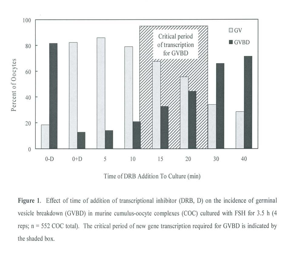
163 149
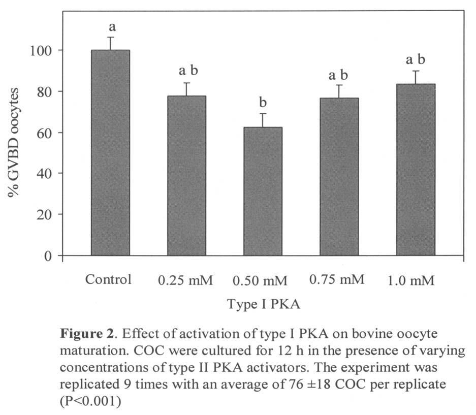
164 150
Animal Science 434! Tonic and Preovulatory Surge of GnRH! Tonic and Preovulatory Surge of GnRH! Lecture 11: The Follicular Phase of the Estrous Cycle!
 Tonic and Preovulatory Surge of GnRH! Animal Science 434! Lecture 11: The Follicular Phase of the Estrous Cycle!! (-)! Hypothalamus! GnRH! Estradiol! (-)! Tonic and Preovulatory Surge of GnRH! Anterior!
OVARY The surface of the ovary is covered with surface epithelium
 OVARY Cow The ovary, or female gonad, is: 1. an exocrine gland, producing oocytes 2. an endocrine gland, secreting hormones, i.e., estrogen and progesterone OVARY OVARY The surface of the ovary is covered
REPRODUCTIVE CYCLE OF FEMALE MAMMAL
 REPRODUCTIVE CYCLE OF FEMALE MAMMAL Fig. 8-12 Secondary follicles growing follicles increase in number of layers of granulosa cells Tertiary follicles maturing follicles antrum formation fluid filled space
The intra-follicular molecular biology mandating advancement of egg retrieval in some women
 The intra-follicular molecular biology mandating advancement of egg retrieval in some women David H. Barad, USA Director of Assisted Reproductive Technology, The Center for Human Reproduction New York
Regulators of Cell Cycle Progression
 Regulators of Cell Cycle Progression Studies of Cdk s and cyclins in genetically modified mice reveal a high level of plasticity, allowing different cyclins and Cdk s to compensate for the loss of one
基醫所. The Cell Cycle. Chi-Wu Chiang, Ph.D. IMM, NCKU
 基醫所 The Cell Cycle Chi-Wu Chiang, Ph.D. IMM, NCKU 1 1 Introduction to cell cycle and cell cycle checkpoints 2 2 Cell cycle A cell reproduces by performing an orderly sequence of events in which it duplicates
Gametogenesis. Dr Corinne de Vantéry Arrighi Dr Hervé Lucas
 WHO Collaborating Center for Research in Human Reproduction Clinic for Infertility and Gynecological Endocrinology University Hospital, Geneva, Switzerland Gametogenesis Dr Corinne de Vantéry Arrighi Dr
Chapter 27 The Reproductive System. MDufilho
 Chapter 27 The Reproductive System 1 Figure 27.19 Events of oogenesis. Before birth Meiotic events 2n Oogonium (stem cell) Mitosis Follicle development in ovary Follicle cells Oocyte 2n Primary oocyte
Chapter 2. Mitosis and Meiosis
 Chapter 2. Mitosis and Meiosis Chromosome Theory of Heredity What structures within cells correspond to genes? The development of genetics took a major step forward by accepting the notion that the genes
Cell cycle and apoptosis
 Cell cycle and apoptosis Cell cycle Definition Stages and steps Cell cycle Interphase (G1/G0, S, and G2) Mitosis (prophase, metaphase, anaphase, telophase, karyokinesis, cytokinesis) Control checkpoints
Animal Development. Lecture 3. Germ Cells and Sex
 Animal Development Lecture 3 Germ Cells and Sex 1 The ovary of sow. The ovary of mare. The ovary of cow. The ovary of ewe. 2 3 The ovary. A generalized vertebrate ovary. (Wilt and Hake, Ch 2, 2004) 4 The
Fertilization depends on mechanisms that help sperm meet eggs of the same species.
 Fertilization depends on mechanisms that help sperm meet eggs of the same species. www.uchsc.edu/ltc/fertilization.html Fertilization union of sperm and egg Is a chain of events. Interruption of any step
Cell Cycle and Cell Division
 122 Cell Cycle and Cell Division 1. Meiosis I is reductional division. Meiosis II is equational division due to [1988] (a) pairing of homologous chromosomes (b) crossing over (c) separation of chromatids
Oogenesis. Key Concepts. Female Reproductive Tract
 Oogenesis 1 Key Concepts Female Reproductive Tract Ovary Oogenesis Follicles Ovulation Corpus Luteum Molecular Activity Primordial Germ Cells (PGCs) 2 Female Reproductive Tract Ovary Oviduct Uterus Vagina
Model Answer. M.Sc. Zoology (First Semester) Examination Paper LZT 103 (Endocrinology)
 Examination Paper LZT 103 (Endocrinology)") Model Answer M.Sc. Zoology (First Semester) Examination-2013 Paper LZT 103 (Endocrinology) Section A 1. (i) d (ii) b (iii) b (iv) c (v) c (vi) a (vii) c (viii) a (ix) d (x) b Section B Q.2 Answer Hormonal
Model Answer M.Sc. Zoology (First Semester) Examination-2013 Paper LZT 103 (Endocrinology) Section A 1. (i) d (ii) b (iii) b (iv) c (v) c (vi) a (vii) c (viii) a (ix) d (x) b Section B Q.2 Answer Hormonal
Gametogenesis. Omne vivum ex ovo All living things come from eggs.
 Omne vivum ex ovo All living things come from eggs. William Harvery, 1651 Gametogenesis This lecture is the preface, so to speak, to embryology; that is, it introduces the development of the specialized
Spermatogenesis. What is it and what does it look like? How do hormones regulate spermatogenesis?
 Spermatogenesis What is it and what does it look like? How do hormones regulate spermatogenesis? FSH, androgens, growth factors Animal Physiology (Hill, Wise, Anderson): Ch. 15 435-438 1 Spermatogenesis:
To General Embryology Dr: Azza Zaki
 Introduction To General Embryology The Human Development is a continuous process that begins when an ovum from a female is fertilized by a sperm from a male. Cell division, growth and differentiation transform
Cell Divisions. The autosomes represent the whole body. * Male Sex Chromosomes: XY * Female Sex Chromosomes: XX
 Cell Divisions Each Cell (including gonads) has 46 chromosomes (23 pairs of chromosomes: 22 pairs of autosomes, 1 pair of sex chromosomes) which are located in the nucleus). The autosomes represent the
Biology 4361 Developmental Biology Exam 1 ID#: October 11, 2005
 Biology 4361 Developmental Biology Name: Key Exam 1 ID#: October 11, 2005 Multiple choice (one point each) 1. Primordial germ cells a. are immortal b. produce polar bodies c. are haploid d. are somatic
Chapter 8: Cellular Reproduction
 Chapter 8: Cellular Reproduction 1. The Cell Cycle 2. Mitosis 3. Meiosis 2 Types of Cell Division 2n 1n Mitosis: occurs in somatic cells (almost all cells of the body) generates cells identical to original
General Embryology. School of Medicine Department of Anatomy and Histology School of medicine The University of Jordan
 General Embryology 2019 School of Medicine Department of Anatomy and Histology School of medicine The University of Jordan https://www.facebook.com/dramjad-shatarat What is embryology? Is the science that
Biology 4361 Developmental Biology. October 11, Multiple choice (one point each)
") Biology 4361 Developmental Biology Exam 1 October 11, 2005 Name: ID#: Multiple choice (one point each) 1. Sertoli cells a. surround spermatocytes b. are the structural components of the seminiferous tubules
Biology 4361 Developmental Biology Exam 1 October 11, 2005 Name: ID#: Multiple choice (one point each) 1. Sertoli cells a. surround spermatocytes b. are the structural components of the seminiferous tubules
Embryology 3. Spermatogenesis:
 Embryology 3 Spermatogenesis: The 2 testis in males are each divided into lobes and lobules by connective tissue septa forming 250 lobule and in each lobule there are 1 to 4 seminefrous tubule ( so almost
Mitosis and the Cell Cycle
 Mitosis and the Cell Cycle Chapter 12 The Cell Cycle: Cell Growth & Cell Division Where it all began You started as a cell smaller than a period at the end of a sentence Getting from there to here Cell
2. Which of the following factors does not contribute to ion selectivity?
 General Biology Summer 2014 Exam II Sample Answers 1. Which of the following is TRUE about a neuron at rest? A. The cytosol is positive relative to the outside B. Na+ concentrations are higher inside C.
Genetics. Instructor: Dr. Jihad Abdallah Lecture 2 The cell cycle and Cell Division
 Genetics Instructor: Dr. Jihad Abdallah Lecture 2 The cell cycle and Cell Division 1 The cell cycle Living cells go through a series of stages known as the cell cycle. They undergo a continuous alternation
The Cell Life Cycle. S DNA replication, INTERPHASE. G 2 Protein. G 1 Normal THE CELL CYCLE. Indefinite period. synthesis. of histones.
 Mitosis & Meiosis The Cell Life Cycle INTERPHASE G 1 Normal cell functions plus cell growth, duplication of organelles, protein synthesis S DNA replication, synthesis of histones THE CELL CYCLE M G 2 Protein
General Biology. Overview: The Key Roles of Cell Division The continuity of life is based upon the reproduction of cells, or cell division
 General Biology Course No: BNG2003" Credits: 3.00 " " " 8. The Cell Cycle Prof. Dr. Klaus Heese Overview: The Key Roles of Cell Division The continuity of life is based upon the reproduction of cells,
Mitosis THE CELL CYCLE. In unicellular organisms, division of one cell reproduces the entire organism Multicellular organisms use cell division for..
 Mitosis THE CELL CYCLE In unicellular organisms, division of one cell reproduces the entire organism Multicellular organisms use cell division for.. Development from a fertilized cell Growth Repair Cell
meiosis asexual reproduction CHAPTER 9 & 10 The Cell Cycle, Meiosis & Sexual Life Cycles Sexual reproduction mitosis
 meiosis asexual reproduction CHAPTER 9 & 10 The Cell Cycle, Meiosis & Sexual Sexual reproduction Life Cycles mitosis Chromosomes Consists of a long DNA molecule (represents thousands of genes) Also consists
BIOLOGY - CLUTCH CH.12 - CELL DIVISION.
 !! www.clutchprep.com CONCEPT: CELL DIVISION Cell division is the process by which one cell splits into two or more daughter cells. Cell division generally requires that cells produce enough materials,
Chapter 13 Pre-Test Question 2
 Student View Summary View Diagnostics View Print View with Answers Settings per Student Questions part 1: Keimzellen und Befruchtung Due: 12:00pm on Wednesday, December 7, 2011 Note: You will receive no
Female Reproductive Physiology. Dr Raelia Lew CREI, FRANZCOG, PhD, MMed, MBBS Fertility Specialist, Melbourne IVF
 Female Reproductive Physiology Dr Raelia Lew CREI, FRANZCOG, PhD, MMed, MBBS Fertility Specialist, Melbourne IVF REFERENCE Lew, R, Natural History of ovarian function including assessment of ovarian reserve
The Cell Cycle 4/10/12. Chapter 12. Overview: The Key Roles of Cell Division
 LECTURE PRESENTATIONS For CAMPBELL BIOLOGY, NINTH EDITION Jane B. Reece, Lisa A. Urry, Michael L. Cain, Steven A. Wasserman, Peter V. Minorsky, Robert B. Jackson Chapter 12 The Cell Cycle Lectures by Erin
How Cells Divide. Chapter 10
 How Cells Divide Chapter 10 Bacterial Cell Division Bacteria divide by binary fission. -the single, circular bacterial chromosome is replicated -replication begins at the origin of replication and proceeds
Mohammad. Renad zakaria ---
 13 Mohammad Renad zakaria --- Before we start: - I didn t follow the record order, for organizing purposes. - I added extra information from our text box which is Guyton 12 th edition, pages 987-997, actually
General Biology. Overview: The Key Roles of Cell Division. Unicellular organisms
 General Biology Course No: BNG2003 Credits: 3.00 8. The Cell Cycle Prof. Dr. Klaus Heese Overview: The Key Roles of Cell Division The continuity of life is based upon the reproduction of cells, or cell
The Cell Cycle. Chapter 12. PowerPoint Lecture Presentations for Biology Eighth Edition Neil Campbell and Jane Reece
 Chapter 12 The Cell Cycle PowerPoint Lecture Presentations for Biology Eighth Edition Neil Campbell and Jane Reece Lectures by Chris Romero, updated by Erin Barley with contributions from Joan Sharp Overview:
The Cell Cycle. Chapter 12. Biology Eighth Edition Neil Campbell and Jane Reece. PowerPoint Lecture Presentations for
 Chapter 12 The Cell Cycle PowerPoint Lecture Presentations for Biology Eighth Edition Neil Campbell and Jane Reece Lectures by Chris Romero, updated by Erin Barley with contributions from Joan Sharp Copyright
Chapter 12. living /non-living? growth repair renew. Reproduction. Reproduction. living /non-living. fertilized egg (zygote) next chapter
 next chapter") Chapter 12 How cells divide Reproduction living /non-living? growth repair renew based on cell division first mitosis - distributes identical sets of chromosomes cell cycle (life) Cell Division in Bacteria
Chapter 12 How cells divide Reproduction living /non-living? growth repair renew based on cell division first mitosis - distributes identical sets of chromosomes cell cycle (life) Cell Division in Bacteria
Cell Cycle Notes chromatin, somatic cells gametes mitosis sister chromatids, centromere cytokinesis binary fission,
 Cell Cycle Notes 1. Importance of Cell Division a. For single celled organisms, cell division increases the number of individuals. b. In a multicellular organism, cell division functions to repair and
General Principles of Endocrine Physiology
 General Principles of Endocrine Physiology By Dr. Isabel S.S. Hwang Department of Physiology Faculty of Medicine University of Hong Kong The major human endocrine glands Endocrine glands and hormones
Cellular Reproduction, Part 1: Mitosis Lecture 10 Fall 2008
 Cell Theory 1 Cellular Reproduction, Part 1: Mitosis Lecture 10 Fall 2008 Cell theory: All organisms are made of cells All cells arise from preexisting cells How do new cells arise? Cell division the reproduction
Ovary. Ovary is the female gonad or sex gland. Gonad: an organ that produces gametes; a testis or ovary.
 Ovary The ovary is composed of two parts: Outer cortex : houses the follicels. Inner medulla: is the middle region composed of connective tissue, blood vessels and lymphatics. Ovary is the female gonad
-The cell s hereditary endowment of DNA -Usually packaged into chromosomes for manageability
 Binary Fission-Bacterial Cell Division -Asexual reproduction of prokaryotes -No mitosis -Circular DNA and organelles replicate, the copies migrate to opposite sides of the elongating cell, and the cell
HANDOUT # 1 GAMETOGENESIS
 Gametogenesis 1 HANDOUT # 1 GAMETOGENESIS Anatomy Department R.A. FADEL Gametogenesis 2 بسم هللا الرحمن الرحيم ت هأ م وى }46 } ا نل و و ا زل و ج و ن و و و و و ن ه و أ ل ه ث وى }45{ إ ول ن أ و ة م ن ان
Cellular Reproduction, Part 2: Meiosis Lecture 10 Fall 2008
 Mitosis & 1 Cellular Reproduction, Part 2: Lecture 10 Fall 2008 Mitosis Form of cell division that leads to identical daughter cells with the full complement of DNA Occurs in somatic cells Cells of body
BIOL 4374/BCHS 4313 Cell Biology Exam #1 February 13, 2001
 BIOL 4374/BCHS 4313 Cell Biology Exam #1 February 13, 2001 SS# Name This exam is worth a total of 100 points. The number of points each question is worth is shown in parentheses. Good luck! 1. (2) The
Ion currents and molecules involved in oocyte maturation, fertilization and embryo development
 Ion currents and molecules involved in oocyte maturation, fertilization and embryo development Dr. Elisabetta Tosti Animal Physiology and Evolution laboratory Stazione Zoologica, Naples, Italy Main steps
Unit 5: Cell Cycle, Mitosis, Meiosis & Drug Influence Influence on Nervous System
 Unit 5: Cell Cycle, Mitosis, Meiosis & Drug Influence Influence on Nervous System 1. Which of the following is NOT related to a cell s surface area to volume ratio? a. Cell size b. Number of nuclei c.
Campbell Biology in Focus (Urry) Chapter 9 The Cell Cycle. 9.1 Multiple-Choice Questions
 Chapter 9 The Cell Cycle. 9.1 Multiple-Choice Questions") Campbell Biology in Focus (Urry) Chapter 9 The Cell Cycle 9.1 Multiple-Choice Questions 1) Starting with a fertilized egg (zygote), a series of five cell divisions would produce an early embryo with how
Campbell Biology in Focus (Urry) Chapter 9 The Cell Cycle 9.1 Multiple-Choice Questions 1) Starting with a fertilized egg (zygote), a series of five cell divisions would produce an early embryo with how
Cell Signaling part 2
 15 Cell Signaling part 2 Functions of Cell Surface Receptors Other cell surface receptors are directly linked to intracellular enzymes. The largest family of these is the receptor protein tyrosine kinases,
Effect of Bovine Follicular Fluid Added to the Maturation Medium on Sperm Penetration in Pig Oocytes Matured In Vitro
 Article Effect of Bovine Follicular Fluid Added to the Maturation Medium on Sperm Penetration in Pig Oocytes Matured In Vitro Abstract Naoki ISOBE Research Associate Graduate School for International Development
Name. A.P. Biology Chapter 12 The Cell Cycle
 A.P. Biology Chapter 12 The Cell Cycle Name Living species MUST possess the ability to r if they are to flourish. The Cell Cycle follows the life of a cell from its o until its d. The Key Roles Of Cell
The reproductive lifespan
 The reproductive lifespan Reproductive potential Ovarian cycles Pregnancy Lactation Male Female Puberty Menopause Age Menstruation is an external indicator of ovarian events controlled by the hypothalamicpituitary
The Cell Cycle 4/10/12. Chapter 12. Overview: The Key Roles of Cell Division
 LECTURE PREENTATION For CAMPBELL BIOLOGY, NINTH EDITION Jane B. Reece, Lisa A. Urry, Michael L. Cain, teven A. Wasserman, Peter V. Minorsky, Robert B. Jackson Chapter 12 The Cell Cycle Overview: The Key
Biology is the only subject in which multiplication is the same thing as division
 Biology is the only subject in which multiplication is the same thing as division The Cell Cycle: Cell Growth, Cell Division 2007-2008 2007-2008 Getting from there to here Going from egg to baby. the original
Gametogenesis. To complete this worksheet, select: Module: Continuity Activity: Animations Title: Gametogenesis. Introduction
 Gametogenesis To complete this worksheet, select: Module: Continuity Activity: Animations Title: Gametogenesis Introduction 1. a. Define gametogenesis. b. What cells are gametes? c. What are the two cell
Ploidy and Human Cell Types. Cell Cycle and Mitosis. DNA and Chromosomes. Where It All Began 11/19/2014. Chapter 12 Pg
 Ploidy and Human Cell Types Cell Cycle and Mitosis Chapter 12 Pg. 228 245 Cell Types Somatic cells (body cells) have 46 chromosomes, which is the diploid chromosome number. A diploid cell is a cell with
Bacterial cell. Origin of replication. Septum
 Bacterial cell Bacterial chromosome: Double-stranded DNA Origin of replication Septum 1 2 3 Chromosome Rosettes of Chromatin Loops Scaffold protein Chromatin Loop Solenoid Scaffold protein Chromatin loop
Male Reproduction Organs. 1. Testes 2. Epididymis 3. Vas deferens 4. Urethra 5. Penis 6. Prostate 7. Seminal vesicles 8. Bulbourethral glands
 Outline Terminology Human Reproduction Biol 105 Lecture Packet 21 Chapter 17 I. Male Reproduction A. Reproductive organs B. Sperm development II. Female Reproduction A. Reproductive organs B. Egg development
Biology is the only subject in which multiplication is the same thing as division
 Biology is the only subject in which multiplication is the same thing as division 2007-2008 The Cell Cycle: Cell Growth, Cell Division 2007-2008 Where it all began You started as a cell smaller than a
Lecture 15. Signal Transduction Pathways - Introduction
 Lecture 15 Signal Transduction Pathways - Introduction So far.. Regulation of mrna synthesis Regulation of rrna synthesis Regulation of trna & 5S rrna synthesis Regulation of gene expression by signals
CELL CYCLE INTRODUCTION PART I ANIMAL CELL CYCLE INTERPHASE EVOLUTION/HEREDITY UNIT. Activity #3
 AP BIOLOGY EVOLUTION/HEREDITY UNIT Unit 1 Part 3 Chapter 12 Activity #3 INTRODUCTION CELL CYCLE NAME DATE PERIOD The nuclei in cells of eukaryotic organisms contain chromosomes with clusters of genes,
Developmental Biology Biology Fertilization. October 19, 2006
 Developmental Biology Biology 4361 Fertilization October 19, 2006 Fertilization Fertilization accomplishes two things: Sex (combining genes from two genomes) Reproduction (initiates reactions in the egg
LECTURE PRESENTATIONS
 LECTURE PRESENTATIONS For CAMPBELL BIOLOGY, NINTH EDITION Jane B. Reece, Lisa A. Urry, Michael L. Cain, Steven A. Wasserman, Peter V. Minorsky, Robert B. Jackson Chapter 12 The Cell Cycle Lectures by Erin
Breaking Up is Hard to Do (At Least in Eukaryotes) Mitosis
 Mitosis") Breaking Up is Hard to Do (At Least in Eukaryotes) Mitosis Chromosomes Chromosomes were first observed by the German embryologist Walther Fleming in 1882. Chromosome number varies among organisms most
Breaking Up is Hard to Do (At Least in Eukaryotes) Mitosis Chromosomes Chromosomes were first observed by the German embryologist Walther Fleming in 1882. Chromosome number varies among organisms most
BIOH122 Session 26 Gametogenesis. Introduction. 1. a. Define gametogenesis. b. What cells are gametes?
 BIOH122 Session 26 Gametogenesis Introduction 1. a. Define gametogenesis. b. What cells are gametes? c. What are the two cell division processes that occur during the cell cycle? d. Define the cell cycle.
Cell cycle and Apoptosis. Chalermchai Mitrpant
 Cell cycle and Apoptosis 2556 Chalermchai Mitrpant Overview of the cell cycle Outline Regulatory mechanisms controlling cell cycle Progression of the cell cycle Checkpoint of the cell cycle Phases of the
Chapter 8 The Cell Cycle
 What molecule stores your genetic information or determines everything about you? DNA a nucleic acid How are DNA molecules arranged in the nucleus? As you can see DNA is: Chapter 8 The Cell Cycle 1. Arranged
Fertilization: Beginning a New New Organism Or
 Fertilization: Beginning a New Organism 1. Contact and recognition between sperm and egg. In most cases, this ensures that the sperm and egg are of the same species. 2. Regulation of sperm entry into the
Principles of Anatomy and Physiology
 Principles of Anatomy and Physiology 14 th Edition CHAPTER 3 The Cellular Level of Organization Introduction The purpose of the chapter is to: 1. Introduce the parts of a cell 2. Discuss the importance
LECTURE PRESENTATIONS
 LECTURE PRESENTATIONS For CAMPBELL BIOLOGY, NINTH EDITION Jane B. Reece, Lisa A. Urry, Michael L. Cain, Steven A. Wasserman, Peter V. Minorsky, Robert B. Jackson Chapter 12 The Cell Cycle Lectures by Erin
Genome Integrity in Mammalian Oocytes
 Genome Integrity in Mammalian Oocytes ESHRE Workshop on mammalian folliculogenesis and oogenesis April 19 21 Stresa Italy 2003 Workshop Lisbon Genome Integrity Structure is chromatin in open or closed
Creating Identical Body Cells
 Creating Identical Body Cells 5.A Students will describe the stages of the cell cycle, including DNA replication and mitosis, and the importance of the cell cycle to the growth of organisms 5.D Students
The Cell Cycle CHAPTER 12
 The Cell Cycle CHAPTER 12 The Key Roles of Cell Division cell division = reproduction of cells All cells come from pre-exisiting cells Omnis cellula e cellula Unicellular organisms division of 1 cell reproduces
Chapter 12. The Cell Cycle
 Chapter 12 The Cell Cycle The Key Roles of Cell Division The ability of organisms to produce more of their own kind is the one characteristic that best distinguishes living things from nonliving things.
Breaking Up is Hard to Do (At Least in Eukaryotes) Mitosis
 Mitosis") Breaking Up is Hard to Do (At Least in Eukaryotes) Mitosis Prokaryotes Have a Simpler Cell Cycle Cell division in prokaryotes takes place in two stages, which together make up a simple cell cycle 1. Copy
Breaking Up is Hard to Do (At Least in Eukaryotes) Mitosis Prokaryotes Have a Simpler Cell Cycle Cell division in prokaryotes takes place in two stages, which together make up a simple cell cycle 1. Copy
Oocyte development in cattle: physiological and genetic aspects. Jack H. Britt
 Revista Brasileira de Zootecnia ISSN impresso: 1516-3598 R. Bras. Zootec., v.37, suplemento especial p.110-115, 2008 ISSN on-line: 1806-9290 www.sbz.org.br Oocyte development in cattle: physiological and
Chapter 12 The Cell Cycle: Cell Growth, Cell Division
 Chapter 12 The Cell Cycle: Cell Growth, Cell Division 2007-2008 Where it all began You started as a cell smaller than a period at the end of a sentence And now look at you How did you get from there to
Biology is the only subject in which multiplication is the same thing as division
 Biology is the only subject in which multiplication is the same thing as division 2007-2008 The Cell Cycle: Cell Growth, Cell Division 2007-2008 Where it all began You started as a cell smaller than a
LECTURE PRESENTATIONS
 LECTURE PRESENTATIONS For CAMPBELL BIOLOGY, NINTH EDITION Jane B. Reece, Lisa A. Urry, Michael L. Cain, Steven A. Wasserman, Peter V. Minorsky, Robert B. Jackson Chapter 12 The Cell Cycle Lectures by Erin
Derived copy of Fertilization *
 OpenStax-CNX module: m56433 1 Derived copy of Fertilization * Stephanie Fretham Based on Fertilization by OpenStax This work is produced by OpenStax-CNX and licensed under the Creative Commons Attribution
Female and Male Gametogenesis
 19 2 Female and Male Gametogenesis Nina Desai, Jennifer Ludgin, Rakesh Sharma, Raj Kumar Anirudh, and Ashok Agarwal 2.1 Introduction 21 2.2 Structure of the Ovary 21 2.3 Overview of Oogenesis 22 2.3.1
Origin of replication. Septum
 Bacterial cell Bacterial chromosome: Double-stranded DNA Origin of replication Septum 1 2 3 Chromosome Rosettes of Chromatin Loops Chromatin Loop Solenoid Scaffold protein Scaffold protein Chromatin loop
Cell Cycle. Trends in Cell Biology
 Cell Cycle Trends in Cell Biology Cell Cycle The orderly sequence of events by which a cell duplicates its contents and divides into two Daughter Cells Activities of a cell from one cell division to the
APGRU4L1 Chap 12 Extra Reading Cell Cycle and Mitosis
 APGRU4L1 Chap 12 Extra Reading Cell Cycle and Mitosis Dr. Ramesh Biology is the only subject in which multiplication is the same thing as division 2007-2008 The Cell Cycle: Cell Growth, Cell Division 2007-2008
Monday, October 6 Put these items into the appropriate category:
 Monday, October 6 Put these items into the appropriate category: Active Transport Facilitated Diffusion Osmosis Simple Diffusion The smell of rotten eggs spreading through the room Requires ATP expenditure
Ovarian follicular development in cattle
 Ovarian follicular development in cattle John P Kastelic Professor of Theriogenology Head, Department of Production Animal Health University of Calgary Calgary, Alberta, Canada Overview Prenatal development
Biology is the only subject in which multiplication is the same thing as division
 Biology is the only subject in which multiplication is the same thing as division 2007-2008 The Cell Cycle: Cell Growth, Cell Division 2007-2008 Getting from there to here Going from egg to baby. the original
BIOH111. o Cell Biology Module o Tissue Module o Integumentary system o Skeletal system o Muscle system o Nervous system o Endocrine system
 BIOH111 o Cell Biology Module o Tissue Module o Integumentary system o Skeletal system o Muscle system o Nervous system o Endocrine system Endeavour College of Natural Health endeavour.edu.au 1 Textbook
An Introduction to Genetics. 9.1 An Introduction to Genetics. An Introduction to Genetics. An Introduction to Genetics. DNA Deoxyribonucleic acid
 An Introduction to Genetics 9.1 An Introduction to Genetics DNA Deoxyribonucleic acid Information blueprint for life Reproduction, development, and everyday functioning of living things Only 2% coding
Biology is the only subject in which multiplication is the same thing as division. AP Biology
 Biology is the only subject in which multiplication is the same thing as division Chapter 12. The Cell Cycle: Cell Growth, Cell Division Where it all began You started as a cell smaller than a period at
Why do cells reproduce?
 Outline Cell Reproduction 1. Overview of Cell Reproduction 2. Cell Reproduction in Prokaryotes 3. Cell Reproduction in Eukaryotes 1. Chromosomes 2. Cell Cycle 3. Mitosis and Cytokinesis Examples of Cell
Cell Division and Mitosis
 Chromatin-Uncoiled DNA during interphase Cell Division and Mitosis Chromosomes-Tightly coiled DNA Chromatid-One half of a duplicated chromosome. Each is identical and called sister chromatids Centromere-The
Reproductive Endocrinology. Isabel Hwang Department of Physiology Faculty of Medicine University of Hong Kong Hong Kong May2007
 Reproductive Endocrinology Isabel Hwang Department of Physiology Faculty of Medicine University of Hong Kong Hong Kong May2007 isabelss@hkucc.hku.hk A 3-hormone chain of command controls reproduction with
Chapter 12 The Cell Cycle
 Chapter 12 The Cell Cycle Objectives Describe how cell reproduction contributes to repair and growth. Compare and contrast prokaryotic and eukaryotic cell division. Compare and contrast asexual and sexual
Topic 1 (Old Curriculum) Past Exam Questions Extended Response SOLUTIONS
 Past Exam Questions Extended Response SOLUTIONS") 1. skeletal muscle fibres are larger / have many nuclei / are not typical cells; fungal hyphae are (sometimes) not divided up into individual cells; unicellular organisms can be considered acellular; because
1. skeletal muscle fibres are larger / have many nuclei / are not typical cells; fungal hyphae are (sometimes) not divided up into individual cells; unicellular organisms can be considered acellular; because