This peer-reviewed article can be downloaded, printed and distributed freely for any purposes (see copyright notice below).
|
|
|
- Oswald Crawford
- 5 years ago
- Views:
Transcription

1 Diagnostic Pathology This Provisional PDF corresponds to the article as it appeared upon acceptance. Fully formatted PDF and full text (HTML) versions will be made available soon. Endometrioid adenocarcinoma with simultaneous endocervical and intestinal-type mucinous differentiation: report of a rare phenomenon and the immunohistochemical profile Diagnostic Pathology 2013, 8:128 doi: / Rebecca Buell-Gutbrod (rbuellgutbrod@wihri.org) C James Sung (Jsung@wihri.org) W Dwayne Lawrence (Dlawrence@wihri.org) M Ruhul Quddus (mquddus@wihri.org) ISSN Article type Case Report Submission date 28 February 2013 Acceptance date 21 June 2013 Publication date 2 August 2013 Article URL This peer-reviewed article can be downloaded, printed and distributed freely for any purposes (see copyright notice below). Articles in Diagnostic Pathology are listed in PubMed and archived at PubMed Central. For information about publishing your research in Diagnostic Pathology or any BioMed Central journal, go to For information about other BioMed Central publications go to Buell-Gutbrod et al. This is an open access article distributed under the terms of the Creative Commons Attribution License ( which permits unrestricted use, distribution, and reproduction in any medium, provided the original work is properly cited.
2 Endometrioid adenocarcinoma with simultaneous endocervical and intestinal-type mucinous differentiation: report of a rare phenomenon and the immunohistochemical profile Abstract Rebecca Buell-Gutbrod 1 rbuellgutbrod@wihri.org C James Sung 1 Jsung@wihri.org W Dwayne Lawrence 1 Dlawrence@wihri.org M Ruhul Quddus 1* * Corresponding author mquddus@wihri.org 1 Department of Pathology and Laboratory Medicine, Women & Infants Hospital and the Warren Alpert Medical School of Brown University, 101 Dudley Street, Providence, RI 02905, USA Intestinal differentiation in the endometrium is rare with only case reports in the international literature. We describe a case of simultaneous endocervical and intestinal-type mucinous differentiation with goblet cells arising in a FIGO grade 1 endometrioid adenocarcinoma. The patient had no involvement of the myometrium, cervix, or extra-uterine sites. There were no intestinal metaplastic changes of the endocervical canal. The etiology of this change is unknown, although recent reports suggest an association with hyperestrogenism. Virtual slides The virtual slides for this article can be found here: Keywords Endometrium, Endometrioid adenocarcinoma, Intestinal differentiation, Goblet cells Introduction Endocervical-type mucinous differentiation in endometrioid adenocarcinoma is not uncommon. However, intestinal differentiation in the endometrium is rare with only case reports in the international literature. These included two cases associated with endometrial
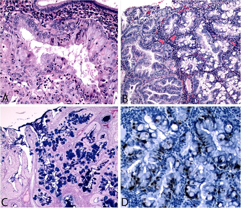
3 carcinoma where the intestinal differentiation was uncovered upon application of special stains [1] and two cases associated with an endometrial polyp and endometrial hyperplasia [2]. Reports of rare carcinoma of the endometrium, their immunohistocheamical staining patters and tissue reaction to endometrial carcinoma are available in the literature [3-5]. We present a case of simultaneous intestinal differentiation with goblet cells and endocervical type mucinous differentiation in a FIGO grade 1 endometrioid adenocarcinoma. Case presentation The patient is a 55 year old gravida 2, para 2 with a chief complaint of postmenopausal bleeding. Her past medical history is significant for morbid obesity (BMI 40), type II diabetes, hypertension, and asthma. CT scan showed heterogeneity of the uterus and bilateral hydrosalpinx, with no evidence of metastatic disease. The patient underwent an endometrial biopsy and a subsequent robotic assisted total laparoscopic hysterectomy and bilateral salpingo-oophorectomy. The post-surgical course was unremarkable and the patient was discharged on post-op day 3. Pathological findings Endometrial biopsy revealed a well differentiated endometrioid adenocarcinoma, FIGO grade 1, arising in a background of complex atypical hyperplasia. On hysterectomy, the uterus measured cm uterus with a smooth glistening serosal surface and an attached cervix. The specimen was opened to reveal a cm intrauterine mass occupying the posterior aspect of the uterine fundus. The remainder of the cm endometrium was pink-tan. The myometrium was grossly uninvolved by tumor. The endocervical canal was unremarkable. Bilateral adnexae consisted of white bosselated ovaries and previously ligated fallopian tube with a fimbriated ends. The endometrium was submitted in its entirety. Microscopically, the tumor consisted primarily of an endometrioid adenocarcinoma of usual type which focally showed endocervical-type mucinous differentiation (Figure 1A). Endocervical-type mucinous differentiation is characterized by tall columnar mucin filled cells with basally located nuclei. Two adjacent microscopic foci also showed readily visible intestinal-type mucinous differentiation with basally located hyperchromatic nuclei and goblet cells (Figure 1B). There was involvement of superficial adenomyosis by tumor, but no true myometrial invasion. As in the prior biopsy, a background of complex atypical endometrial hyperplasia was noted. The lower uterine segment and cervix were free of tumor and so was lymph-vascular invasion. No intestinal-type metaplastic change was noted in the endocervical canal. Bilateral fallopian tubes showed only benign changes and a small serous adenofibroma was discovered in the left ovary. Figure 1 Endometrioid adenocarcinoma with endocervical (A) and intestinal type (B) mucinous differentiation. Classic endometrioid adenocarcinoma is seen in the left side of panel B adjacent to the intestinal-type metaplasia. AB-PAS highlights goblet cells with deep blue staining (C). CDX-2 is positive in the intestinal type cells (D). Special stains and immunohistochemical (IHC) analysis was employed to further characterize the cells of interest. Alcian blue-perioidic acid-schiff stain (AB-PAS) stains acid mucins such as those in goblet cells blue while the neutral mucins of endocervical-type cells are pink as in the classic PAS stain. AB-PAS stain in this case confirms the morphologic impression of
4 goblet cells by staining the acid mucin a deep blue color while the endocervical-type mucinous cells show faint pink staining (Figure 1C). IHC of the areas with intestinal differentiation revealed Cytokeratin (CK) 7 positivity, while CK20 was negative. CDX2 (Figure 1D) and CEA were focally positive in the cells of interest, while synaptophysin and chromogranin were completely negative (Table 1). Table 1 Immunohistochemical profile of intestinal metaplasia in G1 EMCA Antibody Antibody information Results Cytokeratin 7 M; OV-TL 12/30; Dako Positive Cytokeratin 20 M; Ks20.8; Dako Negative CDX2 M; DAK-CDX-2; Dako Focally positive Carcinoembryonic Antigen (CEA) M; II-7; Dako Focally positive Synaptophysin M; SY38; Dako Negative Chromogranin P; Rabbit; Dako Negative M Monoclonal, P Polyclonal. Discussion and conclusions Mucinous differentiation in endometrioid adenocarcinomas is not uncommon and pure mucinous adenocarcinomas of the endometrium comprise 1-10% of all endometrioid adenocarcinomas. Most mucin producing tumors of the endometrium are well differentiated with endocervical-type mucin producing cells characterized by tall columnar cells with basally located nuclei. However, the presence of intestinal differentiation, and of goblet cells in particular, within the endometrium is rare. The largest series of endometrial goblet cells differentiation in the world literature reports only two cases, which were identified with the assistance of special stains [1]. Two cases of intestinal metaplasia, one in an endometrial polyp and one in association with complex hyperplasia without atypia, have recently been reported [2]. Neither was associated with endometrial carcinoma, in contrasts to ours. One was associated with concurrent intestinal metaplasia of the endocervical canal. Two cases of endometrial adenocarcinomas with signet ring cells have been reported in the literature [6] and an additional case of mucinous adenocarcinoma of the endometrium was found to have a gastric phenotype on immunohistochemistry mimicking a minimal deviation adenocarcinoma of the cervix [7]. As in this case, all were associated with more typical appearing endometrioid adenocarcinoma. Enteric-type mucin has been identified in endometrial adenocarcinomas, a finding which verifies the ability of the Mullerian epithelium to undergo metaplasia along an intestinal lineage [8]. No cases in that series, however, showed morphologic intestinal differentiation and there was no correlation with tumor grade. The presence of goblet cell within the endometrium raises the legitimate question for metastasis. Bland well-differentiated intestinal-type mucinous epithelium with goblet cell in the endometrium, endocervix, and fallopian tubes, has been shown to be metastatic lesions from gastrointestinal primaries [9]. Clinical evaluation for an occult primary gastrointestinal lesion as well as careful gross and histologic exam is necessary. Cytokeratins are useful in this setting as metaplastic Mullerian epithelium retains CK7 positivity, as in our case. In this current case, the patient had no clinical gastrointestinal lesion. The gross exam was that of a classic non-invasive carcinoma arising from endometrial. No involvement of the adnexa, serosa, or the myometrium by the tumor was noted and the cells with intestinal differentiation were intimately associated and readily visible with the endometrioid carcinoma.
5 Intestinal-type metaplasia is more common within the endocervix, with reports of cases in non-neoplastic and endocervical adenocarcinoma [1,10]. Intestinal metaplasia in the endocervix is associated with both adenocarcinoma in-situ and endocervical adenocarcinoma and the presence of intestinal-type cells on cervical biopsy warrants further workup. The etiology of intestinal-type differentiation within the endometrium is unclear. The two recently reported cases associated with non-neoplastic lesions were associated with probable hyperestrogenism [2], a finding replicated in this patient who was obese and had a background of endometrial hyperplasia. This is in contrast to the proposed mechanism of endocervical-type mucinous metaplasia which has been linked to use of tamoxifen and exogenous progestin [11,12]. With the addition of our case, the number of cases of intestinal type differentiation with goblet cells in the endometrium is increased to five, with two cases being benign and two cases associated with adenocarcinoma only detected with special stains [1,2]. This entity is extremely rare, this being the first case encountered in our sub-specialty practice and consult service. The clinical implication of finding intestinal-type differentiation in endometrial carcinoma, as in our case report, remains in the fact that the pathologists and clinicians needed to be aware that such differentiation may occur in primary endometrial carcinoma and that the finding of intestinal-type mucinous differentiation in endometrial carcinoma should also warrant careful clinico-pathologic work-up to exclude a metastatic carcinoma from the gastro-intestinal tract. Consent The patient has given written consent for the use of the images and case presentation for educational and scientific purposes provided the unique personal identification is not revealed. Competing interest The authors declare no competing financial interest. All the authors have actively participated in the diagnosis process or manuscript writing. Authors contributions RBG is the Stuart Lauchlan Fellow in Gynecologic and Breast Pathology and has written up the case report and MRQ is the attending Pathologist on the case. CJS and WDL were involved in interpreting the special stains. All authors read and approved the final manuscript. References 1. Nieuwenhuizen L, Khalil MK, Naik VR, Othman NH: Prevalence of goblet cell metaplasia in endocervical and endometrial adenocarcinoma: a histochemical study. Malays J Med Sci 2007, 14:56 61.
6 2. Nicolae A, Goyenaga P, McCluggage WG, Preda O, Nogales FF: Endometrial intestinal metaplasia: a report of two cases, including one associated with cervical intestinal and pyloric metaplasia. Int J Gynecol Pathol 2011, 30: Yasuda M, Katoh T, Hori S, et al: Endometrial intraepithelial carcinoma in association with polyp: review of eight cases. Diagn Pathol 2013, 8: Ribeiro-Silva A: Immunohistohemical features of a papillary squamous cell carcinoma of the endomertrium with transitional cell differentiation. Diagn Pathol 2007, 2: Uehara K, Yasuda M, Ichimura T, et al: Peritoneal granuloma associated with endometrial adenocarcinoma of the uterine corpus. Diagn Pathol 2011, 6: Boyd C, Cameron I, McCluggage WG: Endometrial adenocarcinoma with signet ring cells: report of two cases of an extremely rare phenomenon. Int J Gynecol Pathol 2010, 29: Abiko K, Baba T, Ogawa M, Mikami Y, Koyama T, Mandai M, Konishi I: Minimal deviation mucinous adenocarcinoma ( adenoma malignum ) of the uterine corpus. Pathol Int 2010, 60: McCluggage WG, Roberts N, Bharucha H: Enteric differentiation in endometrial adenocarcinomas: a mucin histochemical study. Int J Gynecol Pathol 1995, 14: Moore WF, Bentley RC, Kim KR, Olatidoye B, Gray SR, Robboy SJ: Goblet-cell mucinous epithelium lining the endometrium and endocervix: evidence of metastasis from an appendiceal primary tumor through the use of cytokeratin-7 and 20 immunostains. Int J Gynecol Pathol 1998, 17: Park KJ, Bramlage MP, Ellenson LH, Pirog EC: Immunoprofile of adenocarcinomas of the endometrium, endocervix, and ovary with mucinous differentiation. Appl Immunohistochem Mol Morphol 2009, 17: Clement PB, Young RH: Endometrioid carcinoma of the uterine corpus: a review of its pathology with emphasis on recent advances and problematic aspects. Adv Anat Pathol 2002, 9: Dallenbach-Hellweg G, Hahn U: Mucinous and clear cell adenocarcinomas of the endometrium in patients receiving antiestrogens (tamoxifen) and gestagens. Am J Pathol 1995, 14:7 15.
7 Figure 1
A Rare Case of Invasive Squamous Cell Carcinoma of Cervix Extending to Endometrium and Right Fallopian Tube
 A Rare Case of Invasive Squamous Cell Carcinoma of Cervix Extending to Endometrium and Right Fallopian Tube Kate Madhuri S 1, Gulhane Sushma R 2, Mane Sheetal V 3 1 Professor and Head, 2 Specialist cum
PREVALENCE OF GOBLET CELL METAPLASIA IN ENDOCERVICAL AND ENDOMETRIAL ADENOCARCINOMA : A HISTOCHEMICAL STUDY
 Malaysian Journal of Medical Sciences, Vol., No., January 007 (-) ORIGINAL ARTICLE PREVALENCE OF GOBLET CELL METAPLASIA IN ENDOCERVICAL AND ENDOMETRIAL ADENOCARCINOMA : A HISTOCHEMICAL STUDY Lauren Nieuwenhuizen,
MPH Quiz. 1. How many primaries are present based on this pathology report? 2. What rule is this based on?
 MPH Quiz Case 1 Surgical Pathology from hysterectomy performed July 11, 2007 Final Diagnosis: Uterus, resection: Endometrioid adenocarcinoma, Grade 1 involving most of endometrium, myometrial invasion
Histopathological Study of Spectrum of Lesions Seen in Surgically Resected Specimens of Fallopian Tube
 Original Article Print ISSN: 2321-6379 Online ISSN: 2321-595X DOI: 10.17354/ijss/2016/613 Histopathological Study of Spectrum of Lesions Seen in Surgically Resected Specimens of Fallopian Tube Pratima
What s (new) and Important in Reporting of Uterine Cancers Katherine Vroobel The Royal Marsden
 and Important in Reporting of Uterine Cancers Katherine Vroobel The Royal Marsden") What s (new) and Important in Reporting of Uterine Cancers Katherine Vroobel The Royal Marsden Maastricht Pathology 2018 Wednesday 20 th June Endometrioid adenocarcinoma High grade carcinomas (common)
What s (new) and Important in Reporting of Uterine Cancers Katherine Vroobel The Royal Marsden Maastricht Pathology 2018 Wednesday 20 th June Endometrioid adenocarcinoma High grade carcinomas (common)
Page # 1. Endometrium. Cellular Components. Anatomical Regions. Management of SIL Thomas C. Wright, Jr. Most common diseases:
 Endometrium Pathology of the Endometrium Thomas C. Wright Columbia University, New York, NY Most common diseases: Abnormal uterine bleeding Inflammatory conditions Benign neoplasms Endometrial cancer Anatomical
Adenocarcinoma of the Cervix
 Question 1. Each of the following statements about cervical adenocarcinoma is true except: Adenocarcinoma of the Cervix SAMS a) A majority of women with cervical adenocarcinoma have stage I tumors at diagnosis.
 DIAGNOSIS A. RIGHT OVARY: Krukenberg tumor (20 cm in maximum dimension, see comment). B. LEFT OVARY: Krukenberg tumor (8.5 cm in maximum dimension, see comment). C. UTERUS (130 Grams): Cervix: Metastatic
Int. J. Curr. Res. Med. Sci. (2017). 3(1): International Journal of Current Research in Medical Sciences
. 3(1): International Journal of Current Research in Medical Sciences") International Journal of Current Research in Medical Sciences ISSN: 2454-5716 www.ijcrims.com Volume 3, Issue 1-2017 Case Report DOI: http://dx.doi.org/10.22192/ijcrms.2017.03.01.006 A rare case report
International Journal of Current Research in Medical Sciences ISSN: 2454-5716 www.ijcrims.com Volume 3, Issue 1-2017 Case Report DOI: http://dx.doi.org/10.22192/ijcrms.2017.03.01.006 A rare case report
CASE 4 21/07/2017. Ectopic Prostatic Tissue in Cervix. Female 31. LLETZ for borderline nuclear abnormalities
 Female 31 CASE 4 LLETZ for borderline nuclear abnormalities PSA Ectopic Prostatic Tissue in Cervix AJSP 2006;30;209-215 usually incidental microscopic finding usually in ectocervical stroma? developmental
Please complete prior to the webinar. HOSPITAL REGISTRY WEBINAR FEMALE REPRODUCTIVE SYSTEM EXERCISES CASE 1: FEMALE REPRODUCTIVE
 Please complete prior to the webinar. HOSPITAL REGISTRY WEBINAR FEMALE REPRODUCTIVE SYSTEM EXERCISES PHYSICAL EXAMINATION CASE 1: FEMALE REPRODUCTIVE 3/5 Patient presents through the emergency room with
Female Genital Tract Lab. Dr. Nisreen Abu Shahin Assistant Professor of Pathology University of Jordan
 Female Genital Tract Lab Dr. Nisreen Abu Shahin Assistant Professor of Pathology University of Jordan Ovarian Pathology A 20-year-old female presented with vague left pelvic pain. Pelvic exam revealed
Mody. AIS vs. Invasive Adenocarcinoma of the Cervix
 Common Problems in Gynecologic Pathology Michael T. Deavers, M.D. Houston Methodist Hospital, Houston, Texas Common Problems in Gynecologic Pathology Adenocarcinoma in-situ (AIS) of the Cervix vs. Invasive
Objectives. Atypical Glandular Cells. Atypical Endocervical Cells. Reactive Endocervical Cells
 2013 California Society of Pathologists 66 th Annual Meeting San Francisco, CA Atypical Glandular Cells to Early Invasive Adenocarcinoma: Cervical Cytology and Histology Christina S. Kong, MD Associate
64 YO lady THBSO for prolapse At gross : A 3 cm endometrial polyp in the fundus
 Case 6 64 YO lady THBSO for prolapse At gross : A 3 cm endometrial polyp in the fundus Numerous irregular, large glands with leaf-like pattern Large glands with broad-based papillary infolding into the
Endometrial adenocarcinoma icd 10 code
 Endometrial adenocarcinoma icd 10 code Gogamz Menu Cancer of the endometrium, adenocarcinoma ;. (mucous membrane that lines the endometrial cavity). ICD - 10 -CM C54.1 is grouped within. ICD-10 -CM Diagnosis
Section 1. Biology of gynaecological cancers: our current understanding
 Section 1 Biology of gynaecological cancers: our current understanding Chapter 1 Morphological sub-types of ovarian carcinoma: new developments and pathogenesis W Glenn McCluggage 1 Introduction In most
Case 3 - GYN. History: 66 year old, routine Pap test. Dr. Stelow
 Case 3 - GYN History: 66 year old, routine Pap test Dr. Stelow Case 3 66 year year old woman Routine Pap Test Cytologic Features 3 dimensional clusters of cells with small to moderate amount of
Mu ath M.A. Rjoub Supervised by: Dr. Huda Zahawi, FRCPath. King Abdullah University Hospital )KAUH(
KAUH(") Mu ath M.A. Rjoub Supervised by: Dr. Huda Zahawi, FRCPath. King Abdullah University Hospital )KAUH( Clinical History A 56 year old single female, presented complaining of postmenopausal bleeding. She underwent
Mu ath M.A. Rjoub Supervised by: Dr. Huda Zahawi, FRCPath. King Abdullah University Hospital )KAUH( Clinical History A 56 year old single female, presented complaining of postmenopausal bleeding. She underwent
Atypical Hyperplasia/EIN
 EIN Atypical Hyperplasia/EIN Based on scientific and diagnostic advances, in 2014 the WHO moved that the precursor lesion for endometrioid carcinoma be atypical hyperplasia/ein, rather than what was previously
What is endometrial cancer?
 Uterine cancer What is endometrial cancer? Endometrial cancer is the growth of abnormal cells in the lining of the uterus. The lining is called the endometrium. Endometrial cancer usually occurs in women
New Cancer Cases By Site Breast 28% Lung 14% Colo-Rectal 10% Uterus 6% Thyroid 5% Lymphoma 4% Ovary 3%
 Uterine Malignancy New Cancer Cases By Site 2010 Breast 28% Lung 14% Colo-Rectal 10% Uterus 6% Thyroid 5% Lymphoma 4% Ovary 3% Cancer Deaths By Site 2010 Lung 26% Breast 15% Colo-Rectal 9% Pancreas 7%
UTERINE SARCOMA EXAMPLE OF A UTERINE SARCOMA USING PROPOSED TEMPLATE
 UTERINE SARCOMA EXAMPLE OF A UTERINE SARCOMA USING PROPOSED TEMPLATE Case: Adenosarcoma with heterologous elements and stromal overgrowth o TAH, BSO, omentectomy, staging biopsies of cul-de-sac, bladder
New Developments in Immunohistochemistry for Gynecologic Pathology
 New Developments in Immunohistochemistry for Gynecologic Pathology Michael T. Deavers, M.D. Professor, Departments of Pathology and Gynecologic Oncology Immunohistochemistry in Gynecologic Pathology Majority
Preface to the Second Edition
 Preface to the Second Edition This second edition of Diagnosis of Endometrial Biopsies and Curettings: A Practical Approach follows a number of favorable comments we received about the first edition. As
Endosalpingiosis. Case report
 Case report Endosalpingiosis Michael D. Holmes, M.D. Howard S. Levin M.D. Department of Pathology Lester A. Ballard, Jr., M.D. Department of Gynecology Endosalpingiosis, a term referring to tuballike epithelium
Minimal Deviation Adenocarcinoma, Mucinous Type, of the Uterine Cervix
 The Korean Journal of Pathology 2004; 38: 121-5 Minimal Deviation Adenocarcinoma, Mucinous Type, of the Uterine Cervix - Report of a Case with Extensive Metastasis to the Uterine Corpus and Bilateral Adnexae
JMSCR Vol 05 Issue 11 Page November 2017
 www.jmscr.igmpublication.org Impact Factor 5.84 Index Copernicus Value: 71.58 ISSN (e)-2347-176x ISSN (p) 2455-0450 DOI: https://dx.doi.org/10.18535/jmscr/v5i11.78 A Histomorphological Study of Carcinoma
Case 1. Pathology of gynecological cancer. What do we need to know (Case 1) Luca Mazzucchelli Istituto cantonale di patologia Locarno
 Luca Mazzucchelli Istituto cantonale di patologia Locarno") Case 1 Pathology of gynecological cancer. What do we need to know (Case 1) Luca Mazzucchelli Istituto cantonale di patologia Locarno SAMO Interdisciplinary Workshop on Gynecological Tumors Lucern, October
Case 1 Pathology of gynecological cancer. What do we need to know (Case 1) Luca Mazzucchelli Istituto cantonale di patologia Locarno SAMO Interdisciplinary Workshop on Gynecological Tumors Lucern, October
In situ and Invasive Endocervical Carcinoma: Problems and Pitfalls in Diagnosis
 In situ and Invasive Endocervical Carcinoma: Problems and Pitfalls in Diagnosis Rouba Ali-Fehmi,MD The Karmanos Cancer Institute, Wayne State University School of Medicine Global incidence of cervical
Endometrial Cancer. Incidence. Types 3/25/2019
 Endometrial Cancer J. Anthony Rakowski DO, FACOOG MSU SCS Board Review Coarse Incidence 53,630 new cases yearly 8,590 deaths yearly 4 th most common malignancy in women worldwide Most common GYN malignancy
Normal endometrium: A, proliferative. B, secretory.
 Normal endometrium: A, proliferative. B, secretory. Nội mạc tử cung Nội mạc tử cung Cyclic changes in endometrium.. Approximate relationship of useful microscopic changes. Arias-Stella reaction in endometrial
Disclosure. Case. Mixed Tumors of the Uterine Corpus and Cervix. I have nothing to disclose
 Mixed Tumors of the Uterine Corpus and Cervix Marisa R. Nucci, M.D. Division of Women s and Perinatal Pathology Department of Pathology Brigham and Women s Hospital Boston, MA UCSF Current Issues in Anatomic
The PAX8 gene is a member of the paired-box family of
 Assessment of the Utility of PAX8 Immunohistochemical Stain in Diagnosing Endocervical Glandular Lesions Li Liang, MD, PhD; Wenxin Zheng, MD; Jinsong Liu, MD, PhD; Sharon X. Liang, MD, PhD Context. PAX8,
Dysplastic intestinal-type metaplasia of appendiceal endometriosis: a mimic of low grade appendiceal mucinous neoplasm
 Mitchell et al. Diagnostic Pathology 2014, 9:39 CASE REPORT Open Access Dysplastic intestinal-type metaplasia of appendiceal endometriosis: a mimic of low grade appendiceal mucinous neoplasm Andrew Mitchell
3/28/2017. Disclosure of Relevant Financial Relationships. Clinical History. Pathology
 Disclosure of Relevant Financial Relationships Monalisa Sur MBBS, FCPath(S.A), MMED(WITS), FRCPath(U.K), FRCP( C) Professor, Department of Pathology and Molecular Medicine Division of Anatomical Pathology
Gynecologic Cytopathology: Glandular lesions
 Gynecologic Cytopathology: Glandular lesions Lin Wai Fung (MSc, MPH, CMIAC) 17/4/2014 Glandular lesions of the uterus Endocervix Endometrium Normal endocervical cells Sheets, strips well-preserved architecture:
ENODMETRIAL CARCINOMA: SPECIAL & NOT SO SPECIAL VARIANTS
 ENODMETRIAL CARCINOMA: SPECIAL & NOT SO SPECIAL VARIANTS Pacific Northwest Society of Pathologists Vancouver, B.C. September 26, 2015 Teri A. Longacre, M.D. longacre@stanford.edu Stanford University, Stanford,
NAACCR Webinar Series /7/17
 COLLECTING CANCER DATA: UTERUS 2017 2018 NAACCR WEBINAR SERIES Q&A Please submit all questions concerning webinar content through the Q&A panel. Reminder: If you have participants watching this webinar
Index. B Bilateral salpingo-oophorectomy (BSO), 69
, 69") A Advanced stage endometrial cancer diagnosis, 92 lymph node metastasis, 92 multivariate analysis, 92 myometrial invasion, 92 prognostic factors FIGO stage, 94 histological grade, 94, 95 histologic cell
A Advanced stage endometrial cancer diagnosis, 92 lymph node metastasis, 92 multivariate analysis, 92 myometrial invasion, 92 prognostic factors FIGO stage, 94 histological grade, 94, 95 histologic cell
Case Scenario 1. History
 History Case Scenario 1 A 53 year old white female presented to her primary care physician with post-menopausal vaginal bleeding. The patient is not a smoker and does not use alcohol. She has no family
The role of immunohistochemistry in surgical pathology of the uterine corpus and cervix
 The role of immunohistochemistry in surgical pathology of the uterine corpus and cervix Prof. Ben Davidson, MD PhD Department of Pathology, Norwegian Radium Hospital, Oslo University Hospital, Oslo, Norway
How to Recognize Gynecologic Cancer Cells from Pelvic Washing and Ascetic Specimens
 How to Recognize Gynecologic Cancer Cells from Pelvic Washing and Ascetic Specimens Wenxin Zheng, M.D. Professor of Pathology and Gynecology University of Arizona zhengw@email.arizona.edu http://www.zheng.gynpath.medicine.arizona.edu/index.html
CME/SAM. KRAS Mutations in Mucinous Lesions of the Uterus
 AJCP / Original Article KRAS Mutations in Mucinous Lesions of the Uterus Mai He, MD, PhD,,2 Cynthia L. Jackson, PhD, 2,3 Rebecca Buell Gubrod, MD,,2 Virginia Breese, 3 Margaret Steinhoff, MD,,2 W. D. Lawrence,
Wendy L Frankel. Chair and Distinguished Professor
 1 Wendy L Frankel Chair and Distinguished Professor Case 1 59 y/o woman Abdominal pain No personal or family history of cancer History of colon polyps Colonoscopy Polypoid rectosigmoid mass Biopsy 3 4
Uterine Cervix. Protocol applies to all invasive carcinomas of the cervix.
 Uterine Cervix Protocol applies to all invasive carcinomas of the cervix. Protocol revision date: January 2005 Based on AJCC/UICC TNM, 6 th edition and FIGO 2001 Annual Report Procedures Cytology (No Accompanying
Intravascular Endometrium Mimicking Vascular Invasion
 ISPUB.COM The Internet Journal of Pathology Volume 12 Number 1 A Papanicolau, G Lin Citation A Papanicolau, G Lin.. The Internet Journal of Pathology. 2010 Volume 12 Number 1. Abstract Intravascular endometrium
This protocol is intended to assist pathologists in providing
 Protocol for the Examination of Specimens From Patients With Carcinomas of the Endometrium A Basis for Checklists Steven G. Silverberg, MD, for the Members of the Cancer Committee, College of American
When Immunostains Can Get You in Trouble: Gynecologic Pathology p16: Panacea or Pandora s Box?
 When Immunostains Can Get You in Trouble: Gynecologic Pathology p16: Panacea or Pandora s Box? Teri A. Longacre, MD Stanford Medicine Stanford California pi6 in Gynecologic Pathology: Panacea or Pandora
6/5/2010. Outline of Talk. Endometrial Alterations That Mimic Cancer & Vice Versa: Metaplastic / reactive changes. Problems in Biopsies/Curettages
 Outline of Talk Endometrial Alterations That Mimic Cancer & Vice Versa: Problems in Biopsies/Curettages Metaplastic / reactive changes Mucinous change Microglandular hyperplasia-like change Squamous metaplasia
Case Report Ovarian Seromucinous Borderline Tumor and Clear Cell Carcinoma: An Unusual Combination
 Case Reports in Obstetrics and Gynecology Volume 2015, Article ID 690891, 5 pages http://dx.doi.org/10.1155/2015/690891 Case Report Ovarian Seromucinous Borderline Tumor and Clear Cell Carcinoma: An Unusual
Department of Pathology, Royal Group of Hospitals Trust, Belfast, Northern Ireland.
 UTERINE ADENOSARCOMA W Glenn McCluggage Department of Pathology, Royal Group of Hospitals Trust, Belfast, Northern Ireland. Definition of Adenosarcoma: A mixed tumor composed of benign neoplastic glandular
Definition of Synoptic Reporting
 Definition of Synoptic Reporting The CAP has developed this list of specific features that define synoptic reporting formatting: 1. All required cancer data from an applicable cancer protocol that are
Papillary Lesions of the Breast A Practical Approach to Diagnosis. (Arch Pathol Lab Med. 2016;140: ; doi: /arpa.
 Papillary Lesions of the Breast A Practical Approach to Diagnosis (Arch Pathol Lab Med. 2016;140:1052 1059; doi: 10.5858/arpa.2016-0219-RA) Papillary lesions of the breast Span the spectrum of benign,
Chapter 2: Initial treatment for endometrial cancer (including histologic variant type)
") Chapter 2: Initial treatment for endometrial cancer (including histologic variant type) CQ01 Which surgical techniques for hysterectomy are recommended for patients considered to be stage I preoperatively?
Chapter 2: Initial treatment for endometrial cancer (including histologic variant type) CQ01 Which surgical techniques for hysterectomy are recommended for patients considered to be stage I preoperatively?
Shina Oranratanaphan, Tarinee Manchana*, Nakarin Sirisabya
 Comparison of Synchronous Endometrial and Ovarian Cancers versus Primary with Metastasis RESEARCH COMMUNICATION Clinicopathologic Variables and Survival Comparison of Patients with Synchronous Endometrial
uterine cancer endometrial cancer
 2018 ICD-10-CM Diagnosis Code. Adenocarcinoma of endometrium ; Cancer of the. (mucous membrane that lines the endometrial cavity). ICD-10-CM C54.1 is grouped. Home ICD 9 Codes Endometrial Cancer ICD 9
Hyperchromatic Crowded Groups: What is Your Diagnosis? Session 3000
 Hyperchromatic Crowded Groups: What is Your Diagnosis? Session 3000 Thomas A. Bonfiglio, M.D. Professor Emeritus, Pathology and Laboratory Medicine University of Rochester Disclosures In the past 12 months,
of 20 to 80 and subsequently declines [2].
![of 20 to 80 and subsequently declines [2].](/thumbs/80/81450506.jpg "of 20 to 80 and subsequently declines [2].") - - According to the 2014 World Health Organization (WHO) classification and tumor morphology, primary ovarian tumors are subdivided into three categories: epithelial (60%), germ cell (30%), and sex-cord
- - According to the 2014 World Health Organization (WHO) classification and tumor morphology, primary ovarian tumors are subdivided into three categories: epithelial (60%), germ cell (30%), and sex-cord
Mucin-producing urothelial-type adenocarcinoma of prostate: report of two cases of a rare and diagnostically challenging entity
 & 2005 USCAP, Inc All rights reserved 0893-3952/05 $30.00 www.modernpathology.org Mucin-producing urothelial-type adenocarcinoma of prostate: report of two cases of a rare and diagnostically challenging
Article begins on next page
 Association of cervical microglandular hyperplasia with exogenous progestin exposure Rutgers University has made this article freely available. Please share how this access benefits you. Your story matters.
Cytology and Surgical Pathology of Gynecologic Neoplasms
 Cytology and Surgical Pathology of Gynecologic Neoplasms Current Clinical Pathology ANTONIO GIORDANO, MD, PHD SERIES EDITOR For further titles published in this series, go to http://www.springer.com/springer/series/7632
BOSNIAN-TURKISH CYTOPATHOLOGY SCHOOL June 18-19, 2016 Sarajevo. Case Discussions. 60 year old woman Routine gynecologic control LBC
 BOSNIAN-TURKISH CYTOPATHOLOGY SCHOOL June 18-19, 2016 Sarajevo Case Discussions Prof Dr Sıtkı Tuzlalı Tuzlalı Pathology Laboratory 60 year old woman Routine gynecologic control LBC 1 2 Endometrial thickening
Staging and Treatment Update for Gynecologic Malignancies
 Staging and Treatment Update for Gynecologic Malignancies Bunja Rungruang, MD Medical College of Georgia No disclosures 4 th most common new cases of cancer in women 5 th and 6 th leading cancer deaths
University Journal of Pre and Para Clinical Sciences
 ISSN 2455 2879 Volume 2 Issue 1 2016 Metaplastic carcinoma breast a rare case report Abstract : Metaplastic carcinoma of the breast is a rare malignancy with two distinct cell lines described as a breast
Case Scenario 1. Pathology report Specimen from mediastinoscopy Final Diagnosis : Metastatic small cell carcinoma with residual lymphatic tissue
 Case Scenario 1 Oncology Consult: Patient is a 51-year-old male with history of T4N3 squamous cell carcinoma of tonsil status post concurrent chemoradiation finished in October two years ago. He was hospitalized
Epithelial Ovarian Cancer 8/2/2013. Tu-be or Not Tu-be: Is the Fallopian Tube the Source of Ovarian Cancer?
 Tu-be or Not Tu-be: Is the Fallopian Tube the Source of Ovarian Cancer? Ann E. Smith Sehdev, MD Director, Center for Gynecologic Pathology Cascade Pathology, Portland, Oregon Ann E. Smith Sehdev has no
PAPILLARY PROLIFERATION OF THE ENDOMETRIUM: A BENIGN LESION SIMULATING ADENOCARCINOMA.
 PAPILLARY PROLIFERATION OF THE ENDOMETRIUM: A BENIGN LESION SIMULATING ADENOCARCINOMA. Teresa Pusiol, Maria Grazia Zorzi, Doriana Morichetti U.O. Anatomia Patologica Ospedale S. Maria del Carmine Rovereto
Endometrial Metaplasia, Hyperplasia & Other Cancer Mimics: a Consultant s Experience
 Endometrial Metaplasia, Hyperplasia & Other Cancer Mimics: a Consultant s Experience Pacific Northwest Society of Pathologists Vancouver, B.C. September 26, 2015 Teri A. Longacre, M.D. longacre@stanford.edu
Endometrial adenocarcinoma icd 10 code
 P ford residence southampton, ny Endometrial adenocarcinoma icd 10 code Jun 24, 2014. Billable Medical Code for Malignant Neoplasm of Corpus Uteri, Except Isthmus Diagnosis Code for Reimbursement Claim:
05/07/2018. Types of challenges. Challenging cases in uterine pathology. Case 1 ` 65 year old female Post menopausal bleeding Uterine Polyp
 Types of challenges Challenging cases in uterine pathology Nafisa Wilkinson Gynaecological Pathologist UCLH London Lack of complete history often, NO clinical history at all! Cases from other centres often
Histopathological Spectrum of Lesions in Fallopian Tube
 IOSR Journal of Dental and Medical Sciences (IOSR-JDMS) e-issn: 2279-0853, p-issn: 2279-0861.Volume 16, Issue 1 Ver. III (January. 2017), PP 75-80 www.iosrjournals.org Histopathological Spectrum of Lesions
Cancers of unknown primary : Knowing the unknown. Prof. Ahmed Hossain Professor of Medicine SSMC
 Cancers of unknown primary : Knowing the unknown Prof. Ahmed Hossain Professor of Medicine SSMC Definition Cancers of unknown primary site (CUPs) Represent a heterogeneous group of metastatic tumours,
MULLERIAN PAPILLOMA ENTITY RECOGNITION FAILURE 04/04/2016 OUT OF SIGHT, OUT OF MIND: LESSER KNOWN LESIONS OF THE VULVOVAGINAL TRACT
 OUT OF SIGHT, OUT OF MIND: LESSER KNOWN LESIONS OF THE VULVOVAGINAL TRACT 23 rd ANNUAL SEMINAR IN PATHOLOGY 30 APRIL 2016 W. Dwayne Lawrence MD MSc (Path.) Chief of Pathology and Laboratory Medicine Women
The Diagnostic Challenges of Low Grade and High Grade Tubo-Ovarian Serous Carcinomas. W Glenn McCluggage Belfast, Northern Ireland
 The Diagnostic Challenges of Low Grade and High Grade Tubo-Ovarian Serous Carcinomas W Glenn McCluggage Belfast, Northern Ireland Enterprise Interest None OVARIAN SEROUS CARCINOMA (OSC) RECENT DEVELOPMENTS
One of the commonest gynecological cancers,especially in white Americans.
 Gynaecology Dr. Rozhan Lecture 6 CARCINOMA OF THE ENDOMETRIUM One of the commonest gynecological cancers,especially in white Americans. It is a disease of postmenopausal women with a peak incidence in
Bladder Case 1 SURGICAL PATHOLOGY REPORT. Procedure: Cystoscopy, transurethral resection of bladder tumor (TURBT)
") Bladder Case 1 February 17, 2007 Specimen (s) received: Bladder Tumor Pre-operative Diagnosis: Bladder Cancer Post operative Diagnosis: Bladder Cancer Procedure: Cystoscopy, transurethral resection of
Bladder Case 1 February 17, 2007 Specimen (s) received: Bladder Tumor Pre-operative Diagnosis: Bladder Cancer Post operative Diagnosis: Bladder Cancer Procedure: Cystoscopy, transurethral resection of
GOBLET CELL CARCINOID. Hanlin L. Wang, MD, PhD University of California Los Angeles
 GOBLET CELL CARCINOID Hanlin L. Wang, MD, PhD University of California Los Angeles Disclosure of Relevant Financial Relationships USCAP requires that all planners (Education Committee) in a position to
GOBLET CELL CARCINOID
 GOBLET CELL CARCINOID Hanlin L. Wang, MD, PhD University of California Los Angeles Disclosure of Relevant Financial Relationships USCAP requires that all planners (Education Committee) in a position to
Vulvar Villoglandular Adenocarcinoma of Colonic Type: A Rare Finding
 Case Report Middle East Journal of Cancer; January 2018; 9(1): 71-75 Vulvar Villoglandular Adenocarcinoma of Colonic Type: A Rare Finding Heidarali Esmaeili*, Mahzad Azimpouran*, Elaheh Olad-Saheb-Madarek**
Pathology of the female genital tract
 Pathology of the female genital tract Common illnesses of the female genital tract Before menarche Developmental anomalies Tumors (ovarial teratoma) Amenorrhea Fertile years PCOS, ovarian cysts Endometriosis
Endometrial line thickness in different conditions.
 Endometrial line thickness in different conditions 1 Endometrial thickens in response to Rising estrogen levels during the menstrual cycle and then shedding endometrial at the times of menses 2 The thickens
Uterine Tumors Resembling Ovarian Sex Cord Tumor in Postmenopausal Woman
 DOI 10.1007/s13224-014-0545-0 CASE REPORT in Postmenopausal Woman Byun Jung Mi Kim Ki Tae Yoon Hye Kyoung Jeong Dae Hoon Kim Young Nam Lee Kyung Bok Sung Moon Su Received: 14 January 2014 / Accepted: 22
ARRO Case: Early-stage Endometrial Cancer
 ARRO Case: Early-stage Endometrial Cancer Ankit Modh, MD (PGY-4) Faculty Advisor: Mohamed A Elshaikh, MD Department of Radiation Oncology Henry Ford Cancer Institute Case Presentation 70 y/o African American
Low-grade serous neoplasia. Robert A. Soslow, MD
 Low-grade serous neoplasia Robert A. Soslow, MD soslowr@mskcc.org Outline Orientation Ovarian tumor overview Non serous borderline tumors Serous borderline tumors Clinical summary Morphologic description
Minfen Zhang 1, Elena Lucas 3, Hanzhen Xiong 1, Shaoyan Liu 1, Kyle Molberg 3, Qingping Jiang 1,2,3* and Wenxin Zheng 3,4*
 Zhang et al. Journal of Ovarian Research (2018) 11:44 https://doi.org/10.1186/s13048-018-0417-9 CASE REPORT Superficially invasive cervical squamous cell carcinoma metastatic to ovarian endometriotic cyst
International Society of Gynecological Pathologists Symposium 2007
 International Society of Gynecological Pathologists Symposium 2007 Anais Malpica, M.D. Department of Pathology The University of Texas M.D. Anderson Cancer Center Grading of Ovarian Cancer Histologic grade
PRE TEST CERVICAL SCREENING MANAGEMENT COLPOSCOPY PATHOLOGIC DIAGNOSIS AND TREATMENT
 PRE TEST CERVICAL SCREENING MANAGEMENT COLPOSCOPY PATHOLOGIC DIAGNOSIS AND TREATMENT QUESTION #1 WHICH OF THE FOLLOWING IS NOT A RISK FACTOR FOR CERVICAL CANCER? A. HIGH RISK HPV B. CIGARETTE SMOKING C.
Standards and datasets for reporting cancers. Dataset for histological reporting of endometrial cancer. December 2017
 Standards and datasets for reporting cancers Dataset for histological reporting of endometrial cancer December 2017 Authors: Dr Raji Ganesan, Birmingham Women s NHS Foundation Trust Dr Naveena Singh, Barts
Urinary Bladder: WHO Classification and AJCC Staging Update 2017
 Urinary Bladder: WHO Classification and AJCC Staging Update 2017 Houston Society of Clinical Pathologists 58 th Annual Spring Symposium Houston, TX April 8, 2017 Jesse K. McKenney, MD Classification
Immunohistochemical Expression of Cytokeratin 5/6 in Gynaecological Tumors.
 ISPUB.COM The Internet Journal of Pathology Volume 13 Number 2 Immunohistochemical Expression of Cytokeratin 5/6 in Gynaecological Tumors. A Baghla, S Choudhry, A Kataria Citation A Baghla, S Choudhry,
Endometrial hyperplasia vs. Intraepithelial neoplasia. Martin Chang, MD PhD FRCPC Pathology Update Friday November 9, 2012
 Endometrial hyperplasia vs. Intraepithelial neoplasia Martin Chang, MD PhD FRCPC Pathology Update Friday November 9, 2012 Disclosure No relevant financial conflicts to declare. Case 1 Gland crowding Gland
Greater Manchester & Cheshire Guidelines for Pathology Reporting for Oesophageal and Gastric Malignancy
 Greater Manchester & Cheshire Guidelines for Pathology Reporting for Oesophageal and Gastric Malignancy Authors: Dr Gordon Armstrong, Dr Sue Pritchard 1. General Comments 1.1 Cancer reporting: Biopsies
Article begins on next page
 Primary Carcinosarcoma of the Uterine Cervix-Report of a case and review of the literature Rutgers University has made this article freely available. Please share how this access benefits you. Your story
Presentation material is for education purposes only. All rights reserved URMC Radiology Page 1 of 98
 Presentation material is for education purposes only. All rights reserved. 2011 URMC Radiology Page 1 of 98 Radiology / Pathology Conference February 2011 Brooke Koltz, Cytopathology Resident Presentation
Case Report Osteoclastic Giant Cell Rich Squamous Cell Carcinoma of the Uterine Cervix: A Case Report and Review of the Literature
 Case Reports in Pathology, Article ID 415328, 4 pages http://dx.doi.org/10.1155/2014/415328 Case Report Osteoclastic Giant Cell Rich Squamous Cell Carcinoma of the Uterine Cervix: A Case Report and Review
NEUROENDOCRINE CARCINOMA OF THE UTERUS Kanika Jain 1, Debasis Dutta 2, Rashmi Nigam 3 & Kanika Chopra *4
 NEUROENDOCRINE CARCINOMA OF THE UTERUS Kanika Jain 1, Debasis Dutta 2, Rashmi Nigam 3 & Kanika Chopra *4 1,2,3&*4 Department Of Minimal Invasive Gynaecology, Institute Of Obstetrics And Gynaecology, Sir
Endometrium. Protocol applies to all carcinomas of the endometrium. Procedures Cytology (No Accompanying Checklist) Biopsy Curettage Hysterectomy
 Biopsy Curettage Hysterectomy") Endometrium Protocol applies to all carcinomas of the endometrium. Protocol revision date: January 2005 Based on AJCC/UICC TNM, 6 th edition and FIGO 2001 Annual Report Procedures Cytology (No Accompanying
Endometrium Protocol applies to all carcinomas of the endometrium. Protocol revision date: January 2005 Based on AJCC/UICC TNM, 6 th edition and FIGO 2001 Annual Report Procedures Cytology (No Accompanying
Papillary Lesions of the breast
 Papillary Lesions of the breast Emad Rakha Professor of Breast Pathology The University of Nottingham Papillary lesions of the breast are a heterogeneous group of disease, which are characterised by neoplastic
Kidney Case 1 SURGICAL PATHOLOGY REPORT
 Kidney Case 1 Surgical Pathology Report February 9, 2007 Clinical History: This 45 year old woman was found to have a left renal mass. CT urography with reconstruction revealed a 2 cm medial mass which