Accepted Manuscript. Anthony Paproki, Craig Engstrom, Shekhar S. Chandra, Ales Neubert, Jurgen Fripp, Stuart Crozier
|
|
|
- Darrell Short
- 5 years ago
- Views:
Transcription

1 Accepted Manuscript Automated Segmentation and Analysis of Normal and Osteoarthritic Knee Menisci from Magnetic Resonance Images - Data from the Osteoarthritis Initiative Anthony Paproki, Craig Engstrom, Shekhar S. Chandra, Ales Neubert, Jurgen Fripp, Stuart Crozier PII: S (14) DOI: /j.joca Reference: YJOCA 3199 To appear in: Osteoarthritis and Cartilage Received Date: 7 March 2014 Revised Date: 9 June 2014 Accepted Date: 28 June 2014 Please cite this article as: Paproki A, Engstrom C, Chandra SS, Neubert A, Fripp J, Crozier S, Automated Segmentation and Analysis of Normal and Osteoarthritic Knee Menisci from Magnetic Resonance Images - Data from the Osteoarthritis Initiative, Osteoarthritis and Cartilage (2014), doi: /j.joca This is a PDF file of an unedited manuscript that has been accepted for publication. As a service to our customers we are providing this early version of the manuscript. The manuscript will undergo copyediting, typesetting, and review of the resulting proof before it is published in its final form. Please note that during the production process errors may be discovered which could affect the content, and all legal disclaimers that apply to the journal pertain.
2 1 2 Automated Segmentation and Analysis of Normal and Osteoarthritic Knee Menisci from Magnetic Resonance Images - Data from the Osteoarthritis Initiative Anthony Paproki 1,2,*, A.P, anthony.paproki@uqconnect.edu.au School of Information Technology and Electrical Engineering, The University of Queensland, St Lucia, QLD 4027, Australia Craig Engstrom 3, C.E, c.engstrom@uq.edu.au Shekhar S. Chandra 1, S.S.C, shekhar.chandra@uq.edu.au Ales Neubert 1,2, A.N, ales.neubert@uqconnect.edu.au Jurgen Fripp 1, J.F, jurgen.fripp@csiro.au Stuart Crozier 2, S.C, stuart@itee.uq.edu.au 1. The Australian e-health Research Centre, CSIRO Computational Informatics, Royal Brisbane and Women s Hospital, Herston QLD 4029, Australia 2. School of Information Technology and Electrical Engineering, The University of Queensland, St Lucia, QLD 4027, Australia 3. School of Human Movement Studies, The University of Queensland, St Lucia, QLD 4072, Australia 1
3 31 Abstract Objective: To validate an automatic scheme for the segmentation and quantitative analysis of the medial (MM) and lateral meniscus (LM) in magnetic resonance (MR) images of the knee joint Method: We analysed sagittal water-excited dual-echo steady-state MR images of the knee joint from a subset of the Osteoarthritis Initiative cohort. The MM and LM were automatically segmented in the MR images based on a 3D deformable model approach. Quantitative parameters including volume, subluxation and tibial- coverage were automatically calculated from the segmentations for comparison (Wilcoxon tests) between knees with variable radiographic osteoarthritis (roa), medial and lateral joint space narrowing (mjsn, ljsn) and pain characteristics. Automatic segmentations and estimated parameters were evaluated for accuracy using manual delineations of the menisci in 88 pathological knee MR examinations at baseline and 12 months time-points. Results: The median (95% confidence-interval) Dice similarity index ( ) between the manual and automated segmentations for the MM and LM were 78.3%( ), 83.9%( ) at baseline and 75.3%( ), 83.0%( ) at 12 months. Pearson coefficients between automatic and manual segmentation parameters ranged from r=0.70 to r=0.92. MM in roa and mjsn knees had significantly greater subluxation and smaller tibial- coverage than no-roa and no-mjsn knees. LM in roa knees had significantly greater volumes and tibial- coverage than no-roa knees. Conclusion: Our automated method successfully segmented the menisci in normal and osteoarthritic knee MR images and detected meaningful morphological differences in the MM and LM with respect to roa and JSN. Our approach will facilitate analyses of the menisci in prospective MR cohorts such as the OAI for investigations into pathophysiological changes occurring in early OA development Keywords: medial meniscus, lateral meniscus, automated segmentation, morphometric analysis, osteoarthritis, MRI 60 2
4 1 2 Automated Segmentation and Analysis of Normal and Osteoarthritic Knee Menisci from Magnetic Resonance Images - Data from the Osteoarthritis Initiative Anthony Paproki 1,2,*, A.P, anthony.paproki@uqconnect.edu.au School of Information Technology and Electrical Engineering, The University of Queensland, St Lucia, QLD 4027, Australia Craig Engstrom 3, C.E, c.engstrom@uq.edu.au Shekhar S. Chandra 1, S.S.C, shekhar.chandra@uq.edu.au Ales Neubert 1,2, A.N, ales.neubert@uqconnect.edu.au Jurgen Fripp 1, J.F, jurgen.fripp@csiro.au Stuart Crozier 2, S.C, stuart@itee.uq.edu.au 1. The Australian e-health Research Centre, CSIRO Computational Informatics, Royal Brisbane and Women s Hospital, Herston QLD 4029, Australia 2. School of Information Technology and Electrical Engineering, The University of Queensland, St Lucia, QLD 4027, Australia 3. School of Human Movement Studies, The University of Queensland, St Lucia, QLD 4072, Australia 1
5 31 Abstract Objective: To validate an automatic scheme for the segmentation and quantitative analysis of the medial (MM) and lateral meniscus (LM) in magnetic resonance (MR) images of the knee Method: We analysed sagittal water-excited dual-echo steady-state MR images of the knee from a subset of the Osteoarthritis Initiative cohort. The MM and LM were automatically segmented in the MR images based on a deformable model approach. Quantitative parameters including volume, subluxation and tibial-coverage were automatically calculated for comparison (Wilcoxon tests) between knees with variable radiographic osteoarthritis (roa), medial and lateral joint space narrowing (mjsn, ljsn) and pain. Automatic segmentations and estimated parameters were evaluated for accuracy using manual delineations of the menisci in 88 pathological knee MR examinations at baseline and 12 months time-points. Results: The median (95% confidence-interval) Dice similarity index ( ) between manual and automated segmentations for the MM and LM were 78.3%( ), 83.9%( ) at baseline and 75.3%( ), 83.0%( ) at 12 months. Pearson coefficients between automatic and manual segmentation parameters ranged from r=0.70 to r=0.92. MM in roa/mjsn knees had significantly greater subluxation and smaller tibial-coverage than no-roa/no-mjsn knees. LM in roa knees had significantly greater volumes and tibial-coverage than no- roa knees. Conclusion: Our automated method successfully segmented the menisci in normal and osteoarthritic knee MR images and detected meaningful morphological differences with respect to roa and JSN. Our approach will facilitate analyses of the menisci in prospective MR cohorts such as the OAI for investigations into pathophysiological changes occurring in early OA development. 2
6 1 Introduction Quantitative analyses of the medial meniscus (MM) and lateral meniscus (LM) from three-dimensional (3D) magnetic resonance (MR) imaging offer opportunities to better understand the pathophysiological processes involved in the structural and functional degeneration of the menisci associated with osteoarthritis (OA) 1-3. Recent semi 4-11 and fully-quantitative 8-15 MR studies have reported significant differences in the volume, tibial-coverage and subluxation of the menisci between knees with distinctive radiographic OA (roa), medial and lateral joint space narrowing (mjsn, ljsn) or pain scores. While MR scoring methods provide good reproducibility and reliability for clinical evaluation of the menisci 4-6, acquisition of detailed quantitative data on these structures through MR segmentation offers increased measurement precision for investigating the in-vivo 3D morphological and biochemical characteristics of these fibro-cartilaginous discs (e.g. T2, T1ρ imaging 16-19, analysis of volume changes with OA or post surgery ). Manual segmentation of the menisci from 3D MR images is a time- and expertise intensive process (35 minutes reported for segmentation of a single coronal water-excited double-echo steady-state (wedess) MR 9 ). Specifically, it requires numerous subjective interpretations for separating adjacent structures with comparable signal contrasts which predispose to low intra-rater reproducibility and high inter-rater variability 12. A desirable direction is the automation the MR segmentation and analysis. Several semi-automatic methods for the 3D segmentation of the menisci have been developed to reduce both analysis time and rater biases 19,20. However these still require expert training and varying levels of manual intervention. In terms of fully automated segmentation approaches 22-25, good accuracy, as measured with the Dice similarity index (DSI) 26, has been achieved for the MM (75±10%) and LM volumes (77±10%) 24 and a total meniscal volume (81±3%) 25 although these methods were only validated on healthy menisci. To the best of our knowledge, results and validation of fully automatic segmentations of the menisci from MR images of individuals with knee roa have not been published. There are substantial technical challenges for automated segmentation of the menisci with pathological damage or degeneration which give rise to a spectrum of structural and biochemical tissue changes which, as illustrated in Fig. 1, are variably associated with increases in signal heterogeneity and shape variability 1, Consequently, segmentation approaches that assume homogeneous signal intensity in the menisci are not well suited for morphometric analyses of the menisci in knees with roa 19,20, and although methods that combined shape- and image-priors provided 3
7 promising leads 24,25, only preliminary results on the automatic segmentation of healthy meniscus have been reported in relatively small populations (N<14). The objectives of this study were to 1) develop a fully-automatic method for the segmentation and quantitative analysis of the individual MM and LM from MR images of the knee, 2) quantitatively evaluate the accuracy of the automatic segmentation and estimations of derived parameters such as volume, subluxation and tibial coverage and 3) to explore the sensitivity of the method in the detection of meaningful changes in meniscus parameters across individuals with various roa grades, mjsn, ljsn and pain. [Figure 1] / [Table 1] Material and Method Patient and MR Image Datasets The MR images used in this study were obtained from the Osteoarthritis Initiative (OAI) database, which is available for public access at Three Datasets (A), (B) and (C) of sagittal 3D wedess MR images of the knee featuring a high spatial-resolution (0.37x0.37mm matrix, 0.7mm slice thickness) and signal-to-noise ratio well-suited for accurate morphological analyses of the meniscus, were selected from the OAI image release 0.E.1, 1.E.1, 3.E.1 and 5.E.1. Imaging protocol and knee positioning were standardized across all subjects 27. Dataset (A) consisted of MR examinations of knee pathology from 88 patients selected from the OAI baseline and 12-month image releases. The MM and LM were manually segmented in all the MR examinations of Dataset (A) and kindly provided by Imorphics (Manchester, UK). The manual segmentations were performed by a single operator trained by a musculoskeletal radiologist (Charles Hutchinson) and an expert segmenter (Mike Bowes) and had passed the Imorphics cartilage segmentation training protocol, requiring an intra-observer coefficient of variation lower than 3% on paired test images. The segmentations were reviewed by the expert segmenter. These manual segmentations, which were performed blind to the present study, were used to train and validate of our automated segmentation algorithm. Datasets (B) and (C) consisted of 22 and 129 subjects (left and right knees) selected from the OAI Progression (definite roa) and Incidence (asymptomatic with increased risks of developing OA) cohorts at baseline, 12, 24 and 36 months. Automated segmentations of the baseline MR images from Datasets (B) and (C) were undertaken for visual assessments of the performance of the segmentation method (results provided) and exploratory data analyses on meniscal volume, subluxation and tibial coverage in a larger cohort with a wider spectrum of healthy and 4
8 pathological meniscal morphologies. Additional results of automated analyses of the menisci in the longitudinal Datasets (B) and (C) are reported as supplementary material. Relevant demographics and clinical data are provided in Table 1. [Figure 2] Automatic Menisci Segmentation The proposed automatic MR image segmentation method is based on a 3D active shape model (ASM) scheme 29 which involves deforming statistical shape models (SSMs) of the MM and LM in the MR image based on a template matching procedure 30,31. A 3D SSM mathematically describes the direction and the magnitude of shape variability of a training-set of triangulated surfaces 29. These models are characterised by a mean-shape which changes in a plausible manner (anatomically credible) based on a set of shape-parameters (illustrated in Supplementary Fig. S1.a). In ASM-fitting schemes, SSMs are frequently used to restrain the deformation from converging towards unlikely shapes deviating excessively from typical shapes of the training-set. In this work, three separate SSMs (1. combined menisci, 2. individual MM and 3. individual LM) were trained based on the manual segmentations of Dataset (A) at baseline. These SSMs were deformed in the MR images using image-feature models 30,31, which comprised 1D template intensity profiles typically surrounding the menisci in the training-set (illustrated in Supplementary Fig. S1.b). The ASM utilised these image-feature models in order to find the intensity profiles most similar to that of the template profiles in the new image to segment. Technical background regarding the generation of the models is provided in Supplementary Data A. The segmentation pipeline, detailed bellow and in Fig. 2.a, involved four steps: (1) image preprocessing, (2) ASM initialisation, (3) ASM-fitting and (4) post-processing. The method was implemented in C++ based on the Insight 32 and Visualisation Toolkits 33 (implementation details are provided in Supplementary Table S1 ). I. MR Image Preprocessing In this first stage, the MR image to segment denoted was normalised to a fixed intensity range (0±200) using linear rescaling and preprocessed using a median smoothing algorithm (radius 1x1x1) in order to reduce the image noise and increase signal homogeneity within structures. 5
9 II. Affine initialisation 92 In the initialisation stage, an average menisci surface denoted was aligned to a likely meniscus region of based on the registration of an average knee image to. Underlying methods utilised to generate the average knee image and menisci surface is described in 'Supplementary Data A'. The average knee image was first registered to using an affine registration algorithm 34, and the obtained transformation was propagated to the average surface, resulting in a surface approximately aligned with the meniscus region in. To refine the initialisation, the meniscus region was extracted from both the average knee image and (2mm around ), and the registration process was repeated with the cropped images. For an individual with multiple time-point scans, the MR images were first co-registered and averaged into a subject-specific mean image using groupwise registration 35, and the mean-image obtained was used for the initialisation of all the time-points. The initial pose and shape parameters of the ASM were then estimated from this obtained surface. An example of an initial segmentation obtained after this stage (by voxelising into segmentation masks) is provided in Fig. 2.a(II). III. Active Shape Model Fitting. The SSMs were then deformed towards the most likely shape and position in based on the template profile matching process illustrated in Fig. 2.a (shaded area) and described in detail elsewhere 30,31. Summarising, for a given point k of, a grey level profile, longer than that of the image-feature model is extracted along the surface normal (positive and negative direction) and compared to the template profiles, of the image feature model. As shown in Fig. 2.b, the template profiles, are translated along the case profile, and the normalised-cross-correlation 0,1 30,31 is computed at each position. The translation offset of the profile, which maximises is then used to translate the point k of along its normal, thus deforming the surface. Once all the points of have been translated, the deformed surface is restrained to a bounded space representative of typical menisci shapes by either constraining the shape-parameters of the SSM or smoothing the surface. The process is then iterated until the maximum number of iteration is reached 6
10 (supplementary Table S1). Optimizing the ASM-fitting process for the segmentation of the menisci involved three parts. A combined SSM encoding the pose variability was deformed in to refine the initial pose of the menisci. This step was performed using a 2 level Gaussian image-pyramid scheme to avoid converging towards local minima. In a second pass, individual SSMs of the MM and LM describing the local shape variability were separately deformed in to obtain likely morphologies. SSMs were used to constrain the deformation during the 2 first stages of the fitting process 29. To account for the shape variability not described by the SSMs and allow the ASMs to deform towards shapes slightly different than that of the training-set, a third pass deformed separate MM and LM ASMs in without SSM constraints. Finally, smoothing was applied to remove noise from the deformed surface and the surface was voxelised to create the initial segmentation masks. IV. Segmentation Post-Processing To correct any small over-segmentation, a post processing classification method was applied to the menisci masks. As shown in Fig. 2.c, the tissue intensity properties of the MM and LM were estimated by a Gaussian distribution (mean µ, variance σ 2 ), and each voxel was assigned a probability of being meniscal tissue based upon its distance to µ. Since the intensity of meniscal tissues is expected to be lower than that of the surrounding articular cartilages in the wedess MR images, voxels featuring intensities lower than µ+σ were classified as meniscal tissue and other voxels were discarded. To account for inherent signal intensity heterogeneity and tears within the menisci, potentially excluded from the meniscal tissue due to high signal intensity, defects in the internal portions of the image mask were marked as unclassified outliers and treated as meniscal tissue for quantitative analyses. [Figure 3] Quantitative Analysis Based upon the segmentations and 3D reconstructions, the menisci were automatically analysed for volume, tibial coverage and subluxation parameters, which are often altered in individuals with knee roa 7-11, The volumes were computed by numerical integration of image-voxels belonging to the segmented menisci. The menisci subluxation and tibial-coverage parameters, illustrated in Fig. 3.a,b, required the identification of the 7
11 tibial bone and plateau (bone-cartilage interface), which were automatically obtained in each MR image following the method described by Fripp et al. 36. The MM and LM coverage areas were calculated as the percentage of the medial and lateral tibial plateau surfaces (Fig. 3, yellow) covered by the individual menisci (Fig. 3, orange) 8,14. The subluxation parameter was computed as the maximum distance between the external margin of the meniscus and that of the tibial plateau (Fig. 3.b green and red curves) when the meniscus position was external relative to the tibial plateau, otherwise the minimum distance (signed negatively) was used 8,14. Validation Strategy The automated segmentation algorithm was applied to all 88 MR images of Dataset (A) with manual segmentations at baseline (V00) and 12 month (V01) time-points and quantitatively validated using a leaveone-out strategy (each case currently segmented was omitted from the training stage). The automatic and manual menisci segmentations were compared using the sensitivity, specificity, DSI 26 and mean absolute surface distance 37 (MASD) values as per Eq. 1: = ( + ) 100 = ( + ) 100 (1) = 2 ( + ) 100 = ( (, ) + (, ))/2) in which TP, TN, FP, FN are the number of true positives, true negatives, false positives and false negatives, and A and M are the automatic and manual segmentation masks respectively. The sensitivity, specificity, DSI and MASD quantified the percentage of true positives, true negatives, the spatial overlap and the average forward and backward Euclidean distances (D(x,y)) between automatic and manual segmentations 37. For both the MM and LM, differences in DSI values were examined using Wilcoxon rank-sum tests across roa grades and Wilcoxon signed-rank tests between time-points (significance-level 0.05). Non-parametric tests were used due to a negative skew in the DSI distributions. Associations between meniscal parameters estimated from the automatic and manual segmentation data were investigated using the Pearson product-moment correlation coefficient 38, the intraclass correlation 8
12 coefficient (ICC - two-way random single measure) 39 and Bland-Altman analyses 40. Coefficients above 0.75 were interpreted as good, while coefficients between 0.5 and <0.75 were interpreted as moderate. To account for outliers and the negative skew of the DSI distributions, the correlation analyses were performed on Dataset (A) trimmed by 5% of the DSI extrema Using the baseline imaging data pooled over all datasets, meniscal volume, subluxation and tibial coverage were compared for differences 1) between roa groups (such that no(confirmed)-roa = grade 0 or I, mildroa=grade II and advanced-roa=grade III-IV), 2) between medial and lateral JSN groups (grades 0, I and II) and 3) between pain-score groups (WOMAC=0, 0<WOMAC<=10 and 10<WOMAC<=20) using Wilcoxon ranksum tests adjusted for false discovery rate 41 (significance-level: 0.05). All statistical analyses were performed using R 3.0. [Table 2] Results Segmentation Validation There was good spatial overlap between the manual and automated segmentations of the MM (median(95%ci) DSI V00 =78.3%( ), DSI V01 =75.3%( )) and LM volumes (DSI V00 =83.9%( ), DSI V01 =83.0%( )) at both time points (Table 2). For each meniscus, there were no significant differences in DSI values across the roa grades (p>0.05 for all Wilcoxon rank-sum tests) and between V00 and V01 (Wilcoxon signed-rank tests p>0.05). Segmentations for the MM and LM in representative cases corresponding to the interquartile mean, maximum and minimum DSI are provided in Fig. 4.a,b to visualise the typically good spatial overlap between the automatic and manual approaches. Severe damage to either or both of the menisci, as shown for the MM in Fig. 4.c, resulted in segmentation difficulties and low DSI values (<=60%) in a small number of cases (15/ % for MM and 3/ % for LM) There were strong or moderate correlations (Fig. 5) between the manual and automated meniscal parameters at both V00 and V01 for the MM (r V00 =0.80, ICC V00 =0.80; r V01 =0.78, ICC V01 =0.78) and LM (r V00 =0.91, ICC V00 =0.90; r V01 =0.89, ICC V01 =0.88) volume, the MM (r V00 =0.83, ICC V00 =0.83; r V01 =0.70, ICC V01 =0.69) and LM (r V00 =0.92, ICC V00 =0.91; r V01 =0.89, ICC V01 =0.89) subluxation and the MM (r V00 =0.82, ICC V00 =0.81; r V01 =0.81, ICC V01 =0.79) and LM (r V00 =0.83, ICC V00 =0.82; r V01 =0.71, ICC V01 =0.70) tibial coverage. 9
13 Comparisons between the manual and automated volume data using Bland-Altman plots showed for both the MM and LM an even distribution of the differences between methods across the range of meniscal measures (no apparent funnelling effects) with a bias of (-4.45%, 6.46%), (-0.525mm, mm) and (-1.98%, -1.64%) for the (MM, LM) volume, subluxation and tibial coverage, respectively. Automatic segmentation and quantitative analysis results obtained for each patient of Datasets (A), (B) and (C) can be publicly accessed online at Observations across automated segmentations of both the MM and LM of Datasets (B) and (C) were visually comparable to those obtained and evaluated for (A), indicating an overall robustness of the method. [Figure 4/5] Quantitative Analysis As reported in Table 3, in roa and mjsn knees, the MM had significantly more subluxation and less tibial coverage than no-roa/no-mjsn knees. So did the MM of advanced-roa knees compared to mild-roa knees. mjsn and advanced-roa knees also had significantly greater MM volume than no-mjsn and no-roa knees respectively. The subluxation of the MM was significantly greater in knees with advanced-roa compared to mild-roa knees. For the LM, knees with roa had significantly greater meniscal volume and tibial-coverage than no-roa/noljsn knees. The volume of the LM was also greater in knees with ljsn. No significant differences were noted between different groups of pain score in any of the meniscal parameters. Automated segmentations of the MM and LM were also performed to obtain volume, subluxation and tibial coverage measures at baseline, 12, 24 and 36 months follow-up for the OAI Progression and Incidence datasets. At this stage descriptive data, as reported in Supplementary Table S2, have been generated with additional data input such as roa grade and compartmental JSN progression required for downstream analyses and validation. [Table 3] 229 Computational Time All the experiments were performed on a dual 6-core Intel Xeon Westmere X5670 (2.93GHz) workstation. Using our fully-automatic method, the mean±sd CPU time required to segment the MM and LM from an 10
14 individual MR examination was 27.2±1.8 minutes (min=24.3, max=32.3), and the time required to perform the quantitative analysis was 2.4±0.2 minutes (min=2.1, max=3.4) minutes. [Figure 6] Discussion This study is the first to successfully provide automatic segmentation and quantitative analysis of both the MM and LM from MR images of individuals with knee roa. Leave-one-out experiments showed good spatial agreement between the manual and automated segmentations of the individual MM and LM, with overall median DSI values of 77.1% and 83.5% in analyses of knee MR examinations for individuals presenting a large spectrum of JSN, sclerotic bone and osteophytes (roa grades II-IV). For both the MM and LM in individuals with knee roa, our scheme may provide a good alternative to the semi-automatic method of Swanson et al. 20 which performed direct segmentations of T2-Maps (more challenging to segment) and achieved a DSI of 69%. In terms of DSIs, our approach compared favourably to previous automated segmentation approaches of these structures in healthy states 24 and although the mean DSI (81.9%) obtained by Zhang et al. 25 slightly outperforms our current results, a direct comparison is difficult since the segmentation of individual menisci was not reported. The automatic estimation of quantitative parameters was sufficiently accurate (0.70<r<0.92) to discern meaningful cross-sectional differences in the volume, tibial-coverage and subluxation of the meniscus between groups with variable roa characteristics. In particular, the MM showed overall greater subluxation and smaller tibial-coverage area in individuals with roa and mjsn, concurrent with recent findings 8, The LM showed a greater median volume in knees with roa and mjsn. The tibial coverage of the LM was found significantly greater in knees with roa (4.4% relative difference, which corroborate results from a recent study although this difference was not reported significant 15 ), but was not significantly different between knees with and without ljsn. The good DSI values and successful identification of significant differences between groups, particularly in relation with the meniscal subluxation and tibial-coverage (eg. 0.43mm and 2.0% absolute difference detected in subluxation and tibial-coverage of the MM between roa and no-roa knees) which have been associated with cartilage loss 3,8, suggest that the present method would be suitable to efficiently analyse and monitor the evolution meniscus morphological characteristics with OA development in large populations. The automatic method also provides opportunities for investigations into the biochemical changes of the meniscus with OA, 11
15 requiring accurate co-registration schemes (eg. Xue et al. 43 ) to align the high-resolution MR image with the biochemical MR sequence (eg. T1ρ, T2, dgemeric MR). However, as with all automated methods, quality control procedures are required to detect the small number of segmentation failures. Our initial experience found web applications 42 (e.g. to be efficient to perform this task, although further investigations are required to ensure their effectiveness. The primary advantages of our method are: (1) it does not require any manual MR image processing, (2) it provides good segmentation of the MM and LM as separate labels, (3) it performs well on knees with roa and visual inspections showed equivalent performance in healthy knees, (4) it readily segments torn menisci and finally (5) it does not require prior identification of the bones or articular cartilages within the knee. There are some limitations with the present research. First, the method was only evaluated on wedess MR images acquired as part of the OAI. Further validation is required for to assess the applicability of the method on clinically focused sequences such as intermediately-weighted 2D fast-spin-echo (FSE) and 3D-FSE. Regarding the performance of the method on the OAI wedess MR images, another possible limitation of the method was the decrease DSI values with roa severity (for MM) and between time-points (Table 2). The primary reason for these differences relates to the increase in meniscus shape complexity and MR signal heterogeneity associated with disease progression, which blurred the boundaries with articular-cartilages and weakened the features driving the ASM. These differences were not significant (Table 2: p>0.05 for all comparisons), suggesting that the method maintained reliable segmentations in the majority of the cases. Training the models of the segmentation algorithm using V01 yielded equivalent results, with a non-significant decrease in DSI values between time-points (Wilcoxon signed-rank test MM: p=0.11, r=0.12; LM: 0.53, r=0.05), which reduced the likelihood that this difference was induced by a strong time-point or training bias. In comparison with the segmentation of the LM, analyses of the anatomically more mobile MM presented greater challenges and a lower median DSI value was obtained. The primary cause of this was the greater variability of shapes and MR tissue-contrasts encountered in the MR images for this structure as a result of a more substantial expression of structural and biochemical alterations with OA. A median DSI of 77% (overall) still compares favourably with existing analyses of this structure in healthy states 24, highlighting the potential of the current segmentation approach for automated analyses of pathological menisci. The tip of the horns and the peripheral margins mid-way along the MM and LM were the areas that segmented least accurately (Fig. 6). These results stem from the unclear demarcations between the meniscal horns and ligaments and between the peripheral edges of the meniscus and fat (Fig. 1). From the high specificity and comparatively low 12
16 sensitivity reported in Table 2, we concluded that under-segmentation was the most common segmentation error obtained. Several cases such as the MM shown in Fig. 4.c exhibited severe tissue destruction and our automated method failed in this specific instance of very advanced tissue loss. Our experience showed that these failed segmentations could be easily detected from the web applications previously mentioned, and with a failure rate (DSI<=60%) of 8.5% for MM and 1.7% for LM, we consider the method suitable for analyses of the menisci in a framework of early OA assessment. In conclusion, our automated scheme is well suited to efficiently process and analyse large prospective MR cohorts, thereby presenting opportunities to facilitate epidemiological and interventional studies into morphological changes of the meniscus. The proposed method provides good accuracy for segmentation of the MM and LM meniscus from wedess MR images from individuals with variably severe knee roa (overall median DSI of 77.1% for MM and 83.5% for LM). Subsequent quantitative analyses obtained Pearson correlations ranging from 0.70 to 0.92 between manual and automatic volume, subluxation and tibial-coverage of the meniscus. Cross-sectional comparisons of the MM and LM parameters from various roa and compartmental JSN groups provided results that corroborated previous manual findings. Author s Contribution All listed authors provided substantial contributions in (1) the conception and design of the study, or acquisition of data, or analysis and interpretation of data. All authors (2) participated in the redaction and revision of the paper. Finally, all authors (3) approved the final version of the article submitted. In particular, AP, JF, and SC participated in the conception and design of the study. AP, SSC, AN, and JF implemented substantial parts of the method presented. JF, CE, and SC obtained the funding resources for the project. And finally, AP, CE, JF, and SC provided helpful background, statistical expertise and clinical insights crucial for the research study. 319 Conflict of interest The authors declare that they have no conflict of interest. 13
17 Acknowledgments The authors would like to thank Mike Bowes (Imorphics) and Dr Charles Hutchinson (University of Manchester) for kindly providing the manual segmentations for public access. Role of the funding sources This research was supported under Australian Research Council s Linkage Projects funding scheme LP The OAI is a public-private partnership comprised of five contracts (N01-AR ; N01-AR ; N01- AR ; N01-AR ; N01-AR ) funded by the National Institutes of Health, a branch of the Department of Health and Human Services, and conducted by the OAI Study Investigators. Private funding partners include Merck Research Laboratories; Novartis Pharmaceuticals Corporation, GlaxoSmithKline; and Pfizer, Inc. Private sector funding for the OAI is managed by the Foundation for the National Institutes of Health. This manuscript was prepared using an OAI public use data set and does not necessarily reflect the opinions or views of the OAI investigators, the NIH, or the private funding partners. References 1. Englund M, Guermazi A, Lohmander LS. The Meniscus in Knee Osteoarthritis. Rheumatic Disease Clinics of North America 2009;35(3): Chan W, Lang P, Stevens M, Sack K, Majumdar S, Stoller D, et al. Osteoarthritis of the knee: comparison of radiography, CT, and MR imaging to assess extent and severity. American Journal of Roentgenology 1991;157(4): Hunter DJ, Zhang YQ, Niu JB, Tu X, Amin S, Clancy M, et al. The association of meniscal pathologic changes with cartilage loss in symptomatic knee osteoarthritis. Arthritis and rheumatism 2006;54(3):
18 Peterfy C.G, Guermazi A, Zaim S, Tirman P.F., Miaux Y, White D et al., Whole-Organ Magnetic Resonance Imaging Score (WORMS) of the knee in osteoarthritis, Osteoarthritis and Cartilage 2004;12(3): Hunter DJ, Lo GH, Gale D, Grainger AJ, Guermazi A, Conaghan PG. The reliability of a new scoring system for knee osteoarthritis MRI and the validity of bone marrow lesion assessment: BLOKS (Boston Leeds Osteoarthritis Knee Score). Ann Rheum Dis 2008;67(2): Hunter D.J, Guermazi A, Lo G.H, Grainger A.J, Conaghan P.G, Boudreau R.M, et al. Evolution of semiquantitative whole joint assessment of knee OA: MOAKS (MRI Osteoarthritis Knee Score), Osteoarthritis and Cartilage 2011;19(8): Berthiaume MJ, Raynauld JP, Martel-Pelletier J, Labonté F, Beaudoin G, Bloch Da, et al. Meniscal tear and extrusion are strongly associated with progression of symptomatic knee osteoarthritis as assessed by quantitative magnetic resonance imaging. Annals of the Rheumatic Diseases 2005;64(4): Bloecker K, Guermazi A, Wirth W, Benichou O, Kwoh CK, Hunter DJ, et al. Tibial coverage, meniscus position, size and damage in knees discordant for joint space narrowing - data from the Osteoarthritis Initiative. Osteoarthritis and Cartilage 2013;21(3): Bloecker K, Guermazi A, Wirth W, Kwoh CK, Resch H, Hunter D.J, et al. Correlation of semiquantitative vs quantitative MRI meniscus measures in osteoarthritic knees: results from the Osteoarthritis Initiative. Skeletal radiology 2014;43(2): Am Jung K, Lee SC, Hwang SH, Yang KH, Kim DH, Sohn JH, et al. High frequency of meniscal hypertrophy in persons with advanced varus knee osteoarthritis. Rheumatology international 2010;30(10): Hwang S, Jung K, Lee W, Yang K, Lee D, Carter A, et al. Morphological changes of the lateral meniscus in end-stage lateral compartment osteoarthritis of the knee. Osteoarthritis and Cartilage 2012;20(2): Siorpaes K, Wenger A, Bloecker K, Wirth W, Hudelmaier M, Eckstein F. Interobserver reproducibility of quantitative meniscus analysis using coronal multiplanar DESS and IWTSE MR imaging. Magnetic Resonance in Medicine 2012;67(5): Bloecker K, Wirth W, Hunter DJ, Duryea J, Guermazi A, Kwoh, CK, et al. Contribution of regional 3D meniscus and cartilage morphometry by MRI to joint space width in fixed flexion knee radiography--a between-knee comparison in subjects with unilateral joint space narrowing. European journal of radiology 2013;82(12):
19 Wirth W, Frobell RB, Souza RB, Li X, Wyman BT, Le Graverand MPH, et al. A three-dimensional quantitative method to measure meniscus shape, position, and signal intensity using MR images: A pilot study and preliminary results in knee osteoarthritis. Magnetic Resonance in Medicine 2010;63(5): Wenger A, Wirth W, Hudelmaier M, Noebauer-Huhmann I, Trattnig S, Bloecker K, et al. Meniscus Body Position, Size, and Shape in Persons With and Persons Without Radiographic Knee Osteoarthritis: Quantitative Analyses of Knee Magnetic Resonance Images From the Osteoarthritis Initiative. Arthritis & Rheumatism 2013;65(7): Li X, Pai A, Blumenkrantz G, Carballido-Gamio J, Link T, Ma B, et al. Spatial distribution and relationship of T1ρ and T2 relaxation times in knee cartilage with osteoarthritis. Magnetic Resonance in Medicine 2009;61(6): Mayerhoefer ME, Welsch GH, Riegler G, Mamisch TC, Materka A, Weber M, et al. Feasibility of texture analysis for the assessment of biochemical changes in meniscal tissue on T1 maps calculated from delayed gadolinium-enhanced magnetic resonance imaging of cartilage data: comparison with conventional relaxation time measurements. Investigative Radiology 2010;45(9): Wang L, Chang G, Xu J, Vieira RLR, Krasnokutsky S, Abramson S, et al. T1ρ MRI of menisci and cartilage in patients with osteoarthritis at 3T. European Journal of Radiology 2012;81(9): Rauscher I, Stahl R, Cheng J, Li X, Huber MB, Luke A, et al. Meniscal Measurements of T1ρ and T2 at MR Imaging in Healthy Subjects and Patients with Osteoarthritis. Radiology 2008;249(2): Swanson M, Prescott J, Best T, Powell K, Jackson R, Haq F, et al. Semi-automated segmentation to assess the lateral meniscus in normal and osteoarthritic knees. Osteoarthritis and Cartilage 2010;18(3): Bowers ME, Tung GA, Fleming BC, Crisco JJ, Rey J. Quantification of meniscal volume by segmentation of 3 T magnetic resonance images. Journal of Biomechanics 2007;40(12): Sasaki T, Hata Y, Ando Y, Ishikawa M, Ishikawa H. Fuzzy rule-based approach to segment the menisci regions from MR images. In: Medical Imaging 99. International Society for Optics and Photonics 1999; Tamez-Pena JG, Totterman S, Parker KJ. Unsupervised statistical segmentation of multispectral volumetric MRI images. In: Medical Imaging 99. International Society for Optics and Photonics 1999;
20 Fripp J, Bourgeat P, Engstrom C, Ourselin S, Crozier S, Salvado O. Automated segmentation of the menisci from MR images. In: IEEE International Symposium on Biomedical Imaging 2009; Zhang K, Lu W, Marziliano P. The unified extreme learning machines and discriminative random fields for automatic knee cartilage and meniscus segmentation from multi-contrast MR images. Machine Vision and Applications 2013;24(7): Dice LR. Measures of the amount of ecologic association between species. Ecology 1945;26(3): Peterfy CG, Schneider E, Nevitt M. The osteoarthritis initiative: report on the design rationale for the magnetic resonance imaging protocol for the knee. Osteoarthritis and Cartilage 2008;16(12): Kellgren J, Lawrence J. Radiological assessment of osteo-arthrosis. Annals of the Rheumatic Diseases 1957;16(4): Cootes TF, Taylor CJ, Cooper DH, Graham J. Active Shape Models-Their Training and Application. Computer Vision and Image Understanding 1995;61(1): Chandra S, Dowling J, Shen K, Raniga P, Pluim JF, Greer PB, et al. Patient specific prostate segmentation in 3-D magnetic resonance images. IEEE Transactions on Medical Imaging 2012;31: Neubert A, Fripp J, Engstrom C, Schwarz R, Lauer L, Salvado O, et al. Automated detection, 3D segmentation and analysis of high resolution spine MR images using statistical shape models. Physics in Medicine and Biology 2012;57(24): Ibanez L, Schroeder W, Ng L, Cates J. The ITK software guide: the insight segmentation and registration toolkit. Kitware Inc 2003; Schroeder W, Martin K, Lorensen B. An Object-Oriented Approach To 3D Graphics. Prentice Hall Rivest-Hénault D, Dowson N, Greer P, Dowling J. Symmetric affine registration of CT and MR pelvis scans for MR-based planning and adaptive prostate radiation therapy (Accepted). IOP Journal of Physics: Conference Series Reuter M, Schmansky JN, Rosas HD, Fischl B. Within-subject template estimation for unbiased longitudinal image analysis. NeuroImage 2012;61(4): Fripp J, Crozier S, Warfield SK, Ourselin S. Automatic segmentation of the bone and extraction of the bone-cartilage interface from magnetic resonance images of the knee. Physics in Medicine and Biology 2007;52(6):
21 Gerig G, Jomier M, Chakos M. Valmet: A new validation tool for assessing and improving 3D object segmentation. In: Medical Image Computing and Computer-Assisted Intervention MICCAI vol of Lecture Notes in Computer Science. Springer. Springer Berlin Heidelberg 2001; Rodgers JL, Nicewander WA. Thirteen ways to look at the correlation coefficient. American Statistician ;4: Koch GG. Intraclass correlation coefficient. Encyclopedia of Statistical Sciences 1983;4: Martin Bland J, Altman D. Statistical methods for assessing agreement between two methods of clinical measurement. The Lancet 1986;327(8476): Benjamini Y, Hochberg Y. Controlling the false discovery rate: a practical and powerful approach to multiple testing. Journal of the Royal Statistical Society. Series B (Methodological) 1995;57(1): Bourgeat P, Dore V, Villemagne VL, Rowe CC, Salvado O, Fripp J. MilxXplore: a web-based system to explore large imaging datasets. Journal of the American Medical Informatics Association 2013;20(6): Xue N, Doellinger M, Fripp J, Ho CP, Surowiec RK, Schwarz R. Automatic model-based semantic registration of multimodal MRI knee data. Journal of Magnetic Resonance Imaging Vapnik V. The nature of statistical learning theory. Springer Breiman L. Random forests. Machine learning 2001;45(1): Fripp J, Crozier S, Warfield SK, Ourselin S. Automatic Segmentation and Quantitative Analysis of the Articular Cartilages From Magnetic Resonance Images of the Knee. IEEE Transactions on Medical Imaging 2010;29(1): Lorensen WE, Cline HE. Marching cubes: A high resolution 3D surface construction algorithm. In: Proceedings of the 14th annual conference on Computer graphics and interactive techniques. SIGGRAPH 87. New York, NY, USA 1987; Combes B, Prima S. An Efficient EM-ICP Algorithm for Symmetric Consistent Non-linear Registration of Point Sets. In: Medical Image Computing and Computer-Assisted Intervention MICCAI vol of Lecture Notes in Computer Science. Springer Berlin / Heidelberg Gower JC. Generalized procrustes analysis. Psychometrika 1975;40(1): Jolliffe IT. Principal Component Analysis. 2nd ed. Springer-Verlag, New York
22 1 Figure legends Figure 1: (a) Manual segmentation of the menisci in a 3D wedess MR image acquired in the sagittal plane (patient , female, age 57, height 168.5cm, BMI 31.8kg/m 2, roa grade III). (left) Coronal view, MM=medial meniscus, LM = lateral meniscus, FM=femur, T=tibia, C=cartilage, F=fat. (Right) Axial view, AH=anterior horn, PH = posterior horn. (b) A 3D sagittal wedess MR image of healthy menisci demonstrating high tissue intensity homogeneity and clear demarcation between the surrounding cartilage and fat tissues in (left) coronal and (right) axial views (patient , male, age 51, height 161.8cm, BMI 27.4, roa grade 0). (c) The menisci in a patient with moderate/severe roa of the knee joint demonstrating lesions in the menisci in (left) coronal and (right) axial views (patient , male, age 65, 184.7cm, BMI 31.1kg/m 2, roa grade III). Figure 2: (a) Segmentation method flow diagram (axial view illustration; case , roa grade III) demonstrating processing of the MM (orange) and LM (green) after affine initialisation, combined menisci ASM pose estimation, constrained MM and LM ASM fitting, MM and LM relaxation and tissue classification. Although shown in 2D, segmentation occurs in 3D. (b) Grey level profile matching. For a surface point k and associated profile, of length 2rL+1 (with r =1.5 a padding ratio allowing the extraction of profiles larger than that of the image-feature-model), the template profiles, of length 2L+1 are translated along, and 30,31 is computed for each position. The profile and displacement maximising describe the displacement of the point k along its normal. The green line at the centre of each profile represents the menisci surface. (c) Postprocessing stage for a MM with over-segmentation (green arrow) and a tear (blue arrow) visualised in the axial view. (1) Segmentation of the MM following ASM-fitting stage. (2) Tissue probability estimation within the MM (darker shades of blue denote a lower probability of meniscal tissue). (3) Tissue classification based on probability estimation. (4) Final segmentation of the MM following dilation and erosion, which allowed closure of the defect associated with the high signal intensity of the tear Figure 3: Schematic representation of the computation of the tibial-coverage and subluxation parameters. (a) A 3D rendering of the MM and LM (displayed as semi-transparent surfaces) tibial coverage areas (orange MM.Cov, LM.Cov) on the medial and lateral tibial plateau (yellow MM.TA and LM.TA). (b) Computation of the subluxation for the MM. The red (t ext ) and green (m ext ) points are the outermost points of the tibial plateau
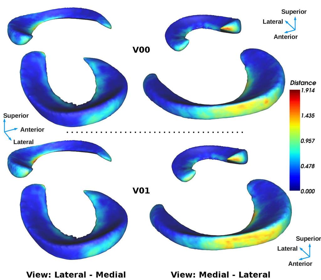
23 31 32 (MM.TA) and the MM which, in this case, maximise the subluxation. The distance between these two points defines the subluxation parameter (blue arrow). A: anterior, P: posterior Figure 4: Qualitative assessment between manual (green overlay) and automatic (blue overlay) meniscal segmentations viewed as per right knee. (left) 3 axial slices focused on the MM, (middle) manual segmentation, (right) automatic segmentation. In (a), from top to bottom, MM segmentation in cases situated at the interquartile mean (Case , DSI=77.6%, roa grade III), interquartile minimum (Case , DSI=71.6%, roa grade III), and interquartile maximum (Case , DSI=81.9%, roa grade III). Similarly, in (b), from top to bottom, LM segmentation in cases situated at the interquartile mean (Case , DSI=83.4%, roa grade II), interquartile minimum (Case , DSI=80.6%, roa grade IV), and interquartile maximum (Case , DSI=85.8%, roa grade III). (c) is an illustration of segmentation failure caused by severe truncation of the MM (Case , DSI=37.0%, roa grade III). Figure 5: (a), (b) and (c) present the correlation and Bland-Atlman analyses performed for the MM (left, green) and LM (right, blue) volume, subluxation and tibial coverage parameters. The scatter-plots present the automatic segmentation parameters against the manual segmentation parameters, and the Bland-Altman analyses present the relative (for volume and tibial coverage) or absolute (for the subluxation) difference between automatic segmentation parameters and the manual segmentation parameters. The absolute error (expressed in mm) is used for the subluxation due to the presence of zero valued parameters. Figure 6: Mapping of the median Hausdorff Distance (maximum forward/backward distance between automatic and manual surface) 37 between deformed ASM surfaces and the manual segmentations onto the menisci mean shape at V00 (top) and V01 (bottom) (all subjects from Dataset (A)). The blue and red areas characterised the smallest and largest distances to the manual segmentations. The tip of the horns and the external surface of the mid-compartment of both menisci were the areas most problematic to segment Supplementary Figure S1: (a) The major mode of variation (strongest λ of C) for the combined menisci SSM, MM SSM, and LM SSM, explained between -3 and +3 standard deviations from the mean-shape. (b) An example of N grey level profiles extracted along the surface normal vectors for a given surface. In this
24 60 61 example, the profiles extracted along the surface normals had a length L of 12. (c) The average knee image and surface used as atlas in the affine registration (initialisation).
25 Table 1: Demographic data of subjects analysed from the 3 datasets used in the present study. Readings for the roa grades and JSN were based on site readings performed by a certified radiologist. roa grades (near Kellgren/Lawrence grade 28 ) at baseline are defined such that 0 = normal knee, 1 = not confirmed roa, 2 = definite-mild roa, 3 = moderate roa, 4 = severe roa. mjsn and ljsn are defined such that a grade of 0 = no- 1 JSN (equivalent to Osteoarthritis Research Society International (OARSI) JSN grades 0), 1 = mild-jsn (equivalent to OARSI grades I-II) and 2 = severe-jsn (equivalent to OARSI grades III). The pain score is defined as the Western Ontario and McMaster Universities Arthritis Index (WOMAC). A B C Male Female Male Female Male Female N Age (yrs) 62.02± ± ± ± ± ±8.6 Height (cm) 176.7± ± ± ± ± ±6.46 Mass (kg) 96.31± ± ± ± ± ±14.46 BMI (kg/m 2 ) 30.51± ± ± ± ± ±5.27 Pain score ([0,20]) 5.07± ± ± ± ± ±4.37 Time-points 2 4* 4* Left and Right No Yes Yes** # Knees baseline # Total Knees roa Grade (0;1;2;3;4) (0, 0, 15, 56, 17) (0, 9, 17, 14, 2) (203, 33, 10, 7, 1) m.jsn score (0;1;2) (16, 55, 17) (30, 10, 2) (240, 12, 2) l.jsn score (0;1;2) (74, 14, 0) (26, 16, 0) (251, 3, 0) *30 and 104 patient knees were available at 4 Time-Points in (B) and (C), other patient were missing time-points ** Except for 3 cases
26 1 Table 2: Evaluation of the accuracy (median(md), 95% confidence-interval(ci)) of the automated segmentation algorithm for the MM and LM volumes after the affine initialisation, ASM-fitting and classification (final) stages for the MM and LM volumes at V00 and V01. Final segmentation results are reported for the overall population and per OA grade. 2 3 V00 Sensitivity (%) Specificity (%) DSI (%) MASD (MM) MD 95% CI MD 95% CI MD 95% CI MD 95% CI (p, r)* Medial Meniscus (V00) Affine ASM-fitting Final (overall) OA Grade II OA Grade III p=0.72, r= OA Grade IV p=0.25, r=0.21 Lateral Meniscus (V00) Affine ASM-fitting Final (overall) OA Grade II OA Grade III p=0.49,r= OA Grade IV p=0.88, r=0.03 V01 Sensitivity (%) Specificity (%) DSI (%) MASD (MM) MD 95% CI MD 95% CI MD 95% CI MD 95% CI (p, r)* Medial Meniscus (V01) Affine ASM-fitting Final (overall) p=0.07, r= OA Grade II OA Grade III p=0.07,r= OA Grade IV p=0.13,r=0.27 Lateral Meniscus (V01) Affine ASM-fitting Final (overall) p=0.18, r= OA Grade II OA Grade III p=0.79,r= OA Grade IV p=0.94,r=0.02 *p-values and effect-size given in italics are the results of the Wilcoxon sum-rank tests between the DSI values of OA grade II verse the DSI values of OA grade III and IV. Underlined values given at time-point V01 correspond to the Wilcoxon signed-rank test results between the DSI values obtained at V00 and V01.
27 Table 3: Median values (MD), interquartile range (IQR), significance values and effect-sizes for MM and LM volume, subluxation, and tibial-coverage comparisons between knees with no-roa, mild-roa, and advancedroa, between knees with no-jsn, mild-jsn and severe-jsn and between 3 groups of patients with increasing WOMAC scores ([0], ]0;10] and ]10;20]). For the LM, only knees with no-jsn or mild-jsn were available. MM.Vol and LM.Vol are expressed in mm 3, MM.Sub and LM.Sub are expressed in mm and MM.Cov and LM.Cov are pressed in %. Radiographic OA no-roa mild-roa advanced-roa p-value; effect-size MD IQR MD IQR MD IQR no-roa vs mild-roa no-roa vs advanced-roa mild-roa vs advanced-roa MM.Vol ;0.09 <0.001; ,0.16 LM.Vol <0.001;0.28 <0.001; ,0.02 MM.Sub ;0.15 <0.001;0.54 <0.001;0.47 LM.Sub ; , ,0.01 MM.Cov ;0.14 <0.001; ,0.29 LM.Cov ;0.15 <0.001; ,0.015 Medial and Lateral JSN no-jsn mild-jsn severe-jsn p-value; r-value MD IQR MD IQR MD IQR no-jsn vs mild-jsn no-jsn vs severe-jsn mild-jsn vs severe-jsn MM.Vol <0.001; , ,0.11 LM.Vol <0.001; MM.Sub <0.001;0.44 <0.001; ,0.17 LM.Sub ; MM.Cov <0.001;0.31 <0.001; ,0.25 LM.Cov ; WOMAC Score 0 (n=77) ]0;10] (n=273) ]10;20] (n=34) p-value; r-value MD IQR MD IQR MD IQR 0 vs ]0;10] 0 vs ]10;20] ]0;10] vs ]10;20] MM.Vol ; , ,0.01 LM.Vol ; , ,0.03 MM.Sub ; , ,0.03 LM.Sub ; , ,0.09 MM.Cov ; , ,0.03 LM.Cov ; , ,0.04 no-roa = roa grade 0 or 1, mild-roa = grade II, advanced-roa = grade III-IV; MM and LM parameters were tested against medial and lateral JSN respectively, with grade 0 = no-jsn, 1 = mild-jsn, 2 = severe-jsn. Differences between groups were tested using Wilcoxon rank-sum tests, with a significance level p=0.05. P-values were adjusted for multiple comparisons using false discovery rate 41. 1

28

29 T CCEP TED

30

31 Medial Lateral Posterior Anterior

32
33
34 1 Supplementary Data C: Longitudinal results Table S2. Median and interquartile range (IQR) of the automatic parameters computed for the MM and LM at baseline, 12, 24 and 36 months follow-up for the OAI Progression and Incidence datasets. 2 3 Medial Meniscus Baseline 12 Months 24 Months 36 Months Median IQR Median IQR Median IQR Median IQR Volume (mm 3 ) OAI Progression (B) OAI Incidence (C) Subluxation (mm) OAI Progression (B) OAI Incidence (C) Tibial Coverage (%) OAI Progression (B) OAI Incidence (C) Lateral Meniscus Baseline 12 Months 24 Months 36 Months Median IQR Median IQR Median IQR Median IQR Volume (mm 3 ) OAI Progression (B) OAI Incidence (C) Subluxation (mm) OAI Progression (B) OAI Incidence (C) Tibial Coverage (%) OAI Progression (B) OAI Incidence (C)
35 1 Supplementary Data B: Algorithm Parameters 2 Table S1. ASM-fitting implementation parameters. The segmentation of all subjects utilised the same parameters, which have been tuned based on training and observations Fitting stage Description (1) Combined menisci ASM-fitting* (2) Individual MM- LM ASM-fitting (3) Individual MM- LM ASM-relaxation Pyramid level 1* Pyramid level 2* SSM # of modes SSM deviation from Profile length (2L+1) Profile spacing Fitting Iterations Constraint type 4 ±2.0SD 2x30+1 = mm 10 SSM 4 ±2.0SD 2x12+1 = mm 20 SSM MM 35 ±1.0SD 2x8+1= mm 25 SSM LM 35 ±3.0SD 2x8+1= mm 120 SSM Smoothing MM NA** NA** 2x20+1= mm 10 (15 it) Smoothing LM NA** NA** 2x12+1= mm 10 (15 it) * Performed using a 2 level Gaussian image pyramid (Level 1:0.729x0.729x1.397mm; Level 2: 0.365x0.365x0.698mm) **No SSM utilised for the relaxation 1

36 T CCEP TE
Osteoarthritis and Cartilage 18 (2010) 1402e1407
 1402e1407") Osteoarthritis and Cartilage 18 (2010) 1402e1407 Comparison of BLOKS and WORMS scoring systems part II. Longitudinal assessment of knee MRIs for osteoarthritis and suggested approach based on their performance:
Osteoarthritis and Cartilage 18 (2010) 1402e1407 Comparison of BLOKS and WORMS scoring systems part II. Longitudinal assessment of knee MRIs for osteoarthritis and suggested approach based on their performance:
Fast cine-magnetic resonance imaging point tracking for prostate cancer radiation therapy planning
 Journal of Physics: Conference Series OPEN ACCESS Fast cine-magnetic resonance imaging point tracking for prostate cancer radiation therapy planning Recent citations - Motion prediction in MRI-guided radiotherapy
Thanks. As something to help remember. Thanks 07/02/14. Arthroscopy TKR V Uni. Arthroscopy TKR V Uni. Role of arthroscopy to choose operation
 07/02/14 Thanks Role of arthroscopy to choose operation TKR Versus Uni Francois and Philippe Faculty Audience Audiovisual Pierre and Vincent Myles Coolican Val d Isere 2014 Thanks Francois and Philippe
The Association of Meniscal Pathologic Changes With Cartilage Loss in Symptomatic Knee Osteoarthritis
 ARTHRITIS & RHEUMATISM Vol. 54, No. 3, March 2006, pp 795 801 DOI 10.1002/art.21724 2006, American College of Rheumatology The Association of Meniscal Pathologic Changes With Cartilage Loss in Symptomatic
Computer based delineation and follow-up multisite abdominal tumors in longitudinal CT studies
 Research plan submitted for approval as a PhD thesis Submitted by: Refael Vivanti Supervisor: Professor Leo Joskowicz School of Engineering and Computer Science, The Hebrew University of Jerusalem Computer
Knee Menisci Segmentation using Convolutional Neural Networks: Data from the Osteoarthritis Initiative 1
 Zuse Institute Berlin Takustr. 7 14195 Berlin Germany ALEXANDER TACK, ANIRBAN MUKHOPADHYAY, STEFAN ZACHOW Knee Menisci Segmentation using Convolutional Neural Networks: Data from the Osteoarthritis Initiative
Tibial coverage, meniscus position, size and damage in knees discordant for joint space. narrowing data from the Osteoarthritis Initiative
 Tibial coverage, meniscus position, size and damage in knees discordant for joint space narrowing data from the Osteoarthritis Initiative K. Bloecker 1,2, A. Guermazi 3,4, W. Wirth 1,5, O. Benichou 6,
MRI of Cartilage. D. BENDAHAN (PhD)
") MRI of Cartilage D. BENDAHAN (PhD) Centre de Résonance Magnétique Biologique et Médicale UMR CNRS 7339 Faculté de Médecine de la Timone 27, Bd J. Moulin 13005 Marseille France david.bendahan@univ-amu.fr
MRI of Cartilage D. BENDAHAN (PhD) Centre de Résonance Magnétique Biologique et Médicale UMR CNRS 7339 Faculté de Médecine de la Timone 27, Bd J. Moulin 13005 Marseille France david.bendahan@univ-amu.fr
Accurate Automated Volumetry of Cartilage of the Knee using Convolutional Neural Networks: Data from the Osteoarthritis Initiative 1
 Zuse Institute Berlin Takustr. 7 14195 Berlin Germany ALEXANDER TACK, STEFAN ZACHOW Accurate Automated Volumetry of Cartilage of the Knee using Convolutional Neural Networks: Data from the Osteoarthritis
D. Doré 1, C. Ding 1,2, J.P. Pelletier 3, J. Martel-Pelletier 3, F. Cicuttini 2, G. Jones 1.
 Responsiveness of qualitative and quantitative MRI measures over 2.7 years D. Doré 1, C. Ding 1,2, J.P. Pelletier 3, J. Martel-Pelletier 3, F. Cicuttini 2, G. Jones 1. 1 Menzies Research Institute Tasmania,
Osteoarthritis and Cartilage 18 (2010) 1393e1401
 1393e1401") Osteoarthritis and Cartilage 18 (2010) 1393e1401 Comparison of BLOKS and scoring systems part I. Cross sectional comparison of methods to assess cartilage morphology, meniscal damage and bone marrow lesions
Osteoarthritis and Cartilage 18 (2010) 1393e1401 Comparison of BLOKS and scoring systems part I. Cross sectional comparison of methods to assess cartilage morphology, meniscal damage and bone marrow lesions
A Method to Monitor Local Changes in MR Signal Intensity in Articular Cartilage: A Potential Marker for Cartilage Degeneration in Osteoarthritis
 A Method to Monitor Local Changes in MR Signal Intensity in Articular Cartilage: A Potential Marker for Cartilage Degeneration in Osteoarthritis Josephine H. Naish 1, Graham Vincent 2, Mike Bowes 2, Manish
Meniscus body position and its change over four years in asymptomatic adults: a cohort study using data from the Osteoarthritis Initiative (OAI)
") Bruns et al. BMC Musculoskeletal Disorders 2014, 15:32 RESEARCH ARTICLE Open Access Meniscus body position and its change over four years in asymptomatic adults: a cohort study using data from the Osteoarthritis
Bruns et al. BMC Musculoskeletal Disorders 2014, 15:32 RESEARCH ARTICLE Open Access Meniscus body position and its change over four years in asymptomatic adults: a cohort study using data from the Osteoarthritis
Comparative Study of K-means, Gaussian Mixture Model, Fuzzy C-means algorithms for Brain Tumor Segmentation
 Comparative Study of K-means, Gaussian Mixture Model, Fuzzy C-means algorithms for Brain Tumor Segmentation U. Baid 1, S. Talbar 2 and S. Talbar 1 1 Department of E&TC Engineering, Shri Guru Gobind Singhji
Meniscal Tears with Fragments Displaced: What you need to know.
 Meniscal Tears with Fragments Displaced: What you need to know. Poster No.: C-1339 Congress: ECR 2015 Type: Authors: Keywords: DOI: Educational Exhibit M. V. Ferrufino, A. Stroe, E. Cordoba, A. Dehesa,
International Cartilage Repair Society
 OsteoArthritis and Cartilage (2007) 15, 487e492 ª 2006 Osteoarthritis Research Society International. Published by Elsevier Ltd. All rights reserved. doi:10.1016/j.joca.2006.11.002 Novel fast semi-automated
Valgus Malalignment Is a Risk Factor for Lateral Knee Osteoarthritis Incidence and Progression
 ARTHRITIS & RHEUMATISM Vol. 65, No. 2, February 2013, pp 355 362 DOI 10.1002/art.37726 2013, American College of Rheumatology Valgus Malalignment Is a Risk Factor for Lateral Knee Osteoarthritis Incidence
Group-Wise FMRI Activation Detection on Corresponding Cortical Landmarks
 Group-Wise FMRI Activation Detection on Corresponding Cortical Landmarks Jinglei Lv 1,2, Dajiang Zhu 2, Xintao Hu 1, Xin Zhang 1,2, Tuo Zhang 1,2, Junwei Han 1, Lei Guo 1,2, and Tianming Liu 2 1 School
Knee Articular Cartilage in an Asymptomatic Population : Comparison of T1rho and T2 Mapping
 TR_002 Technical Reports Knee Articular Cartilage in an Asymptomatic Population : Comparison of T1rho and T2 Mapping Min A Yoon 1,*, Suk-Joo Hong 1, Chang Ho Kang 2, Baek Hyun Kim 3 1 Korea University
Central Reading of Knee X-rays for Kellgren & Lawrence Grade and Individual Radiographic Features of Tibiofemoral Knee OA
 Central Reading of Knee X-rays for Kellgren & Lawrence Grade and Individual Radiographic Features of Tibiofemoral Knee OA 1. Overview... 1 1.1 SAS dataset... 1 1.2 Contents of dataset... 1 1.3 Merging
Automatic Atlas-based Three-label Cartilage Segmentation from MR Knee Images
 Automatic Atlas-based Three-label Cartilage Segmentation from MR Knee Images Liang Shan Department of Computer Science UNC Chapel Hill shan@cs.unc.edu Marc Niethammer Department of Computer Science UNC
RECENT ADVANCES IN CLINICAL MR OF ARTICULAR CARTILAGE
 In Practice RECENT ADVANCES IN CLINICAL MR OF ARTICULAR CARTILAGE By Atsuya Watanabe, MD, PhD, Director, Advanced Diagnostic Imaging Center and Associate Professor, Department of Orthopedic Surgery, Teikyo
Classification and Statistical Analysis of Auditory FMRI Data Using Linear Discriminative Analysis and Quadratic Discriminative Analysis
 International Journal of Innovative Research in Computer Science & Technology (IJIRCST) ISSN: 2347-5552, Volume-2, Issue-6, November-2014 Classification and Statistical Analysis of Auditory FMRI Data Using
Automatic segmentation of lumbar vertebrae on digitised radiographs using linked active appearance models
 Automatic segmentation of lumbar vertebrae on digitised radiographs using linked active appearance models M.G. Roberts a, T.F. Cootes a, J.E. Adams a a Department of Imaging Science and Biomedical Engineering
ARTICLE IN PRESS. International Cartilage Repair Society
 Osteoarthritis and Cartilage (2009) jj, jjjejjj ª 2006 Osteoarthritis Research Society International. Published by Elsevier Ltd. All rights reserved. doi:10.1016/j.joca.2008.11.014 Strong association of
Conservative surgical treatments for osteoarthritis: A Finite Element Study
 Conservative surgical treatments for osteoarthritis: A Finite Element Study Diagarajen Carpanen, BEng (Hons), Franziska Reisse, BEng(Hons), Howard Hillstrom, PhD, Kevin Cheah, FRCS, Rob Walker, PhD, Rajshree
A Pattern Classification Approach to Aorta Calcium Scoring in Radiographs
 A Pattern Classification Approach to Aorta Calcium Scoring in Radiographs Marleen de Bruijne IT University of Copenhagen, Denmark marleen@itu.dk Abstract. A method for automated detection of calcifications
Automated Volumetric Cardiac Ultrasound Analysis
 Whitepaper Automated Volumetric Cardiac Ultrasound Analysis ACUSON SC2000 Volume Imaging Ultrasound System Bogdan Georgescu, Ph.D. Siemens Corporate Research Princeton, New Jersey USA Answers for life.
Planning knee OA prevention studies in obese populations Lessons from the Multicenter Osteoarthritis Study (MOST) and Osteoathritis Initiative (OAI)
 and Osteoathritis Initiative (OAI)") Planning knee OA prevention studies in obese populations Lessons from the (MOST) and Osteoathritis Initiative (OAI) Michael Nevitt, PhD UCSF Epidemiology and Biostatistics OAI and MOST Coordinating Centers
Planning knee OA prevention studies in obese populations Lessons from the (MOST) and Osteoathritis Initiative (OAI) Michael Nevitt, PhD UCSF Epidemiology and Biostatistics OAI and MOST Coordinating Centers
Quantification of Biomechanical Imaging Biomarkers. Sudhakar Tummala
 Quantification of Biomechanical Imaging Biomarkers Sudhakar Tummala February 9, 2012 Contents 1 Introduction 1 1.1 Imaging Osteoarthritis and Biomarkers............ 2 1.1.1 MRI principle.......................
PET in Radiation Therapy. Outline. Tumor Segmentation in PET and in Multimodality Images for Radiation Therapy. 1. Tumor segmentation in PET
 Tumor Segmentation in PET and in Multimodality Images for Radiation Therapy Wei Lu, Ph.D. Department of Radiation Oncology Mallinckrodt Institute of Radiology Washington University in St. Louis Outline
Automatic Segmentation of Jaw Tissues in CT Using Active Appearance Models and Semi-automatic Landmarking
 Automatic Segmentation of Jaw Tissues in CT Using Active Appearance Models and Semi-automatic Landmarking Sylvia Rueda 1,JoséAntonioGil 1, Raphaël Pichery 2,andMarianoAlcañiz 1 1 Medical Image Computing
Quadriceps Muscle and Intermuscular Fat Volumes in the Thighs of Men in the OAI are Associated with Physical Function and Knee Pain
 Quadriceps Muscle and Intermuscular Fat Volumes in the Thighs of Men in the OAI are Associated with Physical Function and Knee Pain Karen A. Beattie, Monica R. Maly, Sami Shaker, Norma J. MacIntyre McMaster
Bone marrow lesions in knee osteoarthritis: MR-assessment by manual segmentation and computer-assisted tresholding
 Bone marrow lesions in knee osteoarthritis: MR-assessment by manual segmentation and computer-assisted tresholding Poster No.: P-0073 Congress: ESSR 2012 Type: Scientific Exhibit Authors: F. K. Nielsen,
The Relationship Between Cartilage Loss on Magnetic Resonance Imaging and Radiographic Progression in Men and Women With Knee Osteoarthritis
 ARTHRITIS & RHEUMATISM Vol. 52, No. 10, October 2005, pp 3152 3159 DOI 10.1002/art.21296 2005, American College of Rheumatology The Relationship Between Cartilage Loss on Magnetic Resonance Imaging and
Automatic Classification System for Lumbar Spine X-ray Images
 Automatic Classification System for Lumbar Spine X-ray Images Soontharee Koompairojn Kien A. Hua School of Electrical Engineering and Computer Science University of Central Florida Orlando, FL 32816 {soonthar,
MRI KNEE WHAT TO SEE. Dr. SHEKHAR SRIVASTAV. Sr.Consultant KNEE & SHOULDER ARTHROSCOPY
 MRI KNEE WHAT TO SEE Dr. SHEKHAR SRIVASTAV Sr.Consultant KNEE & SHOULDER ARTHROSCOPY MRI KNEE - WHAT TO SEE MRI is the most accurate and frequently used diagnostic tool for evaluation of internal derangement
Investigating the loading behaviour of intact and meniscectomy knee joints and the impact on surgical decisions
 Investigating the loading behaviour of intact and meniscectomy knee joints and the impact on surgical decisions M. S. Yeoman 1 1. Continuum Blue Limited, One Caspian Point, Caspian Way, CF10 4DQ, United
Four Tissue Segmentation in ADNI II
 Four Tissue Segmentation in ADNI II Charles DeCarli, MD, Pauline Maillard, PhD, Evan Fletcher, PhD Department of Neurology and Center for Neuroscience, University of California at Davis Summary Table of
Cancer Cells Detection using OTSU Threshold Algorithm
 Cancer Cells Detection using OTSU Threshold Algorithm Nalluri Sunny 1 Velagapudi Ramakrishna Siddhartha Engineering College Mithinti Srikanth 2 Velagapudi Ramakrishna Siddhartha Engineering College Kodali
Medial Knee Osteoarthritis Precedes Medial Meniscal Posterior Root Tear with an Event of Painful Popping
 Medial Knee Osteoarthritis Precedes Medial Meniscal Posterior Root Tear with an Event of Painful Popping Dhong Won Lee, M.D, Ji Nam Kim, M.D., Jin Goo Kim, M.D., Ph.D. KonKuk University Medical Center
A rapid, novel method of volumetric assessment of MRI-detected subchondral bone marrow lesions in knee osteoarthritis
 Osteoarthritis and Cartilage 21 (2013) 806e814 A rapid, novel method of volumetric assessment of MRI-detected subchondral bone marrow lesions in knee osteoarthritis C. Ratzlaff y *, A. Guermazi z, J. Collins
An Articulated Statistical Shape Model of the Human Knee
 An Articulated Statistical Shape Model of the Human Knee Matthias Bindernagel, Dagmar Kainmueller, Heiko Seim, Hans Lamecker, Stefan Zachow, Hans-Christian Hege Zuse Institute Berlin bindernagel@zib.de
Knee Cartilage Extraction and Bone-Cartilage Interface Analysis from 3D MRI Data Sets
 Knee Cartilage Extraction and Bone-Cartilage Interface Analysis from 3D MRI Data Sets José G. Tamez-Peña, Monica Barbu-McInnis and Saara Totterman VirtualScopics, 350 Linden Oaks, Rochester NY, 14625,
dgemric Effectively Predicts Cartilage Damage Associated with Femoroacetabular Impingement
 Riccardo Lattanzi 1,2 Catherine Petchprapa 2 Daniele Ascani 1 Roy I. Davidovitch 3 Thomas Youm 3 Robert J. Meislin 3 Michael. Recht 2 1 The Bernard and Irene Schwartz Center for Biomedical Imaging, New
1 Introduction. Abstract: Accurate optic disc (OD) segmentation and fovea. Keywords: optic disc segmentation, fovea detection.
 segmentation and fovea. Keywords: optic disc segmentation, fovea detection.") Current Directions in Biomedical Engineering 2017; 3(2): 533 537 Caterina Rust*, Stephanie Häger, Nadine Traulsen and Jan Modersitzki A robust algorithm for optic disc segmentation and fovea detection
Current Directions in Biomedical Engineering 2017; 3(2): 533 537 Caterina Rust*, Stephanie Häger, Nadine Traulsen and Jan Modersitzki A robust algorithm for optic disc segmentation and fovea detection
A fully automated system for quantification of knee bone marrow lesions using MRI and the osteoarthritis initiative cohort
 ORIGINAL RESEARCH A fully automated system for quantification of knee bone marrow lesions using MRI and the osteoarthritis initiative cohort Pierre Dodin 1, François Abram 1, Jean-Pierre Pelletier 2, Johanne
Meniscus T2 Relaxation Time at Various Stages of Knee Joint Degeneration
 Meniscus T2 Relaxation Time at Various Stages of Knee Joint Degeneration Richard Kijowski, Michael Fazio, Benjamin Beduhn, and Fang Liu Department of Radiology University of Wisconsin School of Medicine
Accuracy and Precision of Quantitative Assessment of Cartilage Morphology by Magnetic Resonance Imaging at 3.0T
 ARTHRITIS & RHEUMATISM Vol. 52, No. 10, October 2005, pp 3132 3136 DOI 10.1002/art.21348 2005, American College of Rheumatology Accuracy and Precision of Quantitative Assessment of Cartilage Morphology
Imorphics OA Knee MRI Measurements
 Precision. Insight. Innovation. White Paper: Imorphics OA Knee MRI Measurements At left: Automated segmentation of an osteoarthritic knee This knee shows advanced osteophytes. As well as the major bones,
Change in Femur Shape during Postnatal Development and Growth of C57BL/6 Mice
 Change in Femur Shape during Postnatal Development and Growth of C57BL/6 Mice Introduction: Disorders of skeletal development, including hip dysplasia, slipped capital femoral epiphysis, and Legg-Calve-Perthes
A Reliable Method for Brain Tumor Detection Using Cnn Technique
 IOSR Journal of Electrical and Electronics Engineering (IOSR-JEEE) e-issn: 2278-1676,p-ISSN: 2320-3331, PP 64-68 www.iosrjournals.org A Reliable Method for Brain Tumor Detection Using Cnn Technique Neethu
EARLY STAGE DIAGNOSIS OF LUNG CANCER USING CT-SCAN IMAGES BASED ON CELLULAR LEARNING AUTOMATE
 EARLY STAGE DIAGNOSIS OF LUNG CANCER USING CT-SCAN IMAGES BASED ON CELLULAR LEARNING AUTOMATE SAKTHI NEELA.P.K Department of M.E (Medical electronics) Sengunthar College of engineering Namakkal, Tamilnadu,
Conclusions and future directions
 Chapter 9 Conclusions and future directions This chapter summarises the findings within this thesis, examines its main contributions, and provides suggestions for further directions. 9.1 Thesis summary
A new Method on Brain MRI Image Preprocessing for Tumor Detection
 2015 IJSRSET Volume 1 Issue 1 Print ISSN : 2395-1990 Online ISSN : 2394-4099 Themed Section: Engineering and Technology A new Method on Brain MRI Preprocessing for Tumor Detection ABSTRACT D. Arun Kumar
This presentation is the intellectual property of the author. Contact them for permission to reprint and/or distribute.
 MRI of the Knee Jennifer Swart, M.D. Musculoskeletal Radiology South Texas Radiology Group Outline Coils, Patient Positioning Acquisition Parameters, Planes and Pulse Sequences Knee Arthrography Normal
Automatic cardiac contour propagation in short axis cardiac MR images
 International Congress Series 1281 (2005) 351 356 www.ics-elsevier.com Automatic cardiac contour propagation in short axis cardiac MR images G.L.T.F. Hautvast a,b, T, M. Breeuwer a, S. Lobregt a, A. Vilanova
White Rose Research Online URL for this paper: Version: Accepted Version
 This is a repository copy of Osteoarthritic bone marrow lesions almost exclusively colocate with denuded cartilage: a 3D study using data from the Osteoarthritis Initiative. White Rose Research Online
International Journal of Engineering Trends and Applications (IJETA) Volume 4 Issue 2, Mar-Apr 2017
 Volume 4 Issue 2, Mar-Apr 2017") RESEARCH ARTICLE OPEN ACCESS Knowledge Based Brain Tumor Segmentation using Local Maxima and Local Minima T. Kalaiselvi [1], P. Sriramakrishnan [2] Department of Computer Science and Applications The Gandhigram
RESEARCH ARTICLE OPEN ACCESS Knowledge Based Brain Tumor Segmentation using Local Maxima and Local Minima T. Kalaiselvi [1], P. Sriramakrishnan [2] Department of Computer Science and Applications The Gandhigram
This presentation is the intellectual property of the author. Contact them at for permission to reprint and/or distribute.
 MRI of the Knee Jennifer Swart, M.D. Musculoskeletal Radiology South Texas Radiology Group Financial Disclosure Dr. Jennifer Swart has no relevant financial relationships with commercial interests to disclose.
Osteoarthritis and Cartilage 19 (2011) 689e699
 689e699") Osteoarthritis and Cartilage 19 (2011) 689e699 MRI-based extended ordered values more efficiently differentiate cartilage loss in knees with and without joint space narrowing than region-specific approaches
Osteoarthritis and Cartilage 19 (2011) 689e699 MRI-based extended ordered values more efficiently differentiate cartilage loss in knees with and without joint space narrowing than region-specific approaches
MRI Assessments of Cartilage
 SNR IMPACTS THE ACCURACY AND PRECISION OF KNEE ARTICULAR CARTILAGE T2 RELAXATION TIME MEASUREMENTS B.J. Dardzinski 1, E. Schneider 2 1 Merck Sharp & Dohme Corp., West Point, PA USA 2 Imaging Institute,
Coronal Tibiofemoral Subluxation in Knee Osteoarthritis
 Coronal Tibiofemoral Subluxation in Knee Osteoarthritis Saker Khamaisy, MD 1,2 * ; Hendrik A. Zuiderbaan, MD 1 ; Meir Liebergall, MD 2; Andrew D. Pearle, MD 1 1Hospital for Special Surgery, Weill Medical
Automated detection of abnormal changes in cortical thickness: A tool to help diagnosis in neocortical focal epilepsy
 Automated detection of abnormal changes in cortical thickness: A tool to help diagnosis in neocortical focal epilepsy 1. Introduction Epilepsy is a common neurological disorder, which affects about 1 %
FieldStrength. Achieva 3.0T enables cutting-edge applications, best-in-class MSK images
 FieldStrength Publication for the Philips MRI Community Issue 33 December 2007 Achieva 3.0T enables cutting-edge applications, best-in-class MSK images Palo Alto Medical Clinic Sports Medicine Center employs
Tumor cut segmentation for Blemish Cells Detection in Human Brain Based on Cellular Automata
 Tumor cut segmentation for Blemish Cells Detection in Human Brain Based on Cellular Automata D.Mohanapriya 1 Department of Electronics and Communication Engineering, EBET Group of Institutions, Kangayam,
Statistically Optimized Biopsy Strategy for the Diagnosis of Prostate Cancer
 Statistically Optimized Biopsy Strategy for the Diagnosis of Prostate Cancer Dinggang Shen 1, Zhiqiang Lao 1, Jianchao Zeng 2, Edward H. Herskovits 1, Gabor Fichtinger 3, Christos Davatzikos 1,3 1 Center
Automatic Definition of Planning Target Volume in Computer-Assisted Radiotherapy
 Automatic Definition of Planning Target Volume in Computer-Assisted Radiotherapy Angelo Zizzari Department of Cybernetics, School of Systems Engineering The University of Reading, Whiteknights, PO Box
Brain Tumor Detection and Segmentation in MR images Using GLCM and. AdaBoost Classifier
 2015 IJSRSET Volume 1 Issue 3 Print ISSN : 2395-1990 Online ISSN : 2394-4099 Themed Section: Engineering and Technology Brain Tumor Detection and Segmentation in MR images Using GLCM and ABSTRACT AdaBoost
8/10/2016. PET/CT Radiomics for Tumor. Anatomic Tumor Response Assessment in CT or MRI. Metabolic Tumor Response Assessment in FDG-PET
 PET/CT Radiomics for Tumor Response Evaluation August 1, 2016 Wei Lu, PhD Department of Medical Physics www.mskcc.org Department of Radiation Oncology www.umaryland.edu Anatomic Tumor Response Assessment
International Journal of Research (IJR) Vol-1, Issue-6, July 2014 ISSN
 Vol-1, Issue-6, July 2014 ISSN") Developing an Approach to Brain MRI Image Preprocessing for Tumor Detection Mr. B.Venkateswara Reddy 1, Dr. P. Bhaskara Reddy 2, Dr P. Satish Kumar 3, Dr. S. Siva Reddy 4 1. Associate Professor, ECE Dept,
Developing an Approach to Brain MRI Image Preprocessing for Tumor Detection Mr. B.Venkateswara Reddy 1, Dr. P. Bhaskara Reddy 2, Dr P. Satish Kumar 3, Dr. S. Siva Reddy 4 1. Associate Professor, ECE Dept,
MRI Image Processing Operations for Brain Tumor Detection
 MRI Image Processing Operations for Brain Tumor Detection Prof. M.M. Bulhe 1, Shubhashini Pathak 2, Karan Parekh 3, Abhishek Jha 4 1Assistant Professor, Dept. of Electronics and Telecommunications Engineering,
EXTRACT THE BREAST CANCER IN MAMMOGRAM IMAGES
 International Journal of Civil Engineering and Technology (IJCIET) Volume 10, Issue 02, February 2019, pp. 96-105, Article ID: IJCIET_10_02_012 Available online at http://www.iaeme.com/ijciet/issues.asp?jtype=ijciet&vtype=10&itype=02
An Overview of RAMRIQ: An Automated MRI Rheumatoid Arthritis Quantitative Assessment System
 Precision. Insight. Innovation. White Paper: An Overview of RAMRIQ: An Automated MRI Rheumatoid Arthritis Quantitative Assessment System At left: Volume of synovitis in the hand and wrist. Areas of enhancement
Meniscal Root Tears: Evaluation, Imaging, and Repair Techniques
 Meniscal Root Tears: Evaluation, Imaging, and Repair Techniques R O B E R T N A S C I M E N TO, M D, M S C H I E F OF S P O RT S M E D I C I N E & SH O U L D E R S U R G E RY N E W TO N- W E L L E S L
COMPUTERIZED SYSTEM DESIGN FOR THE DETECTION AND DIAGNOSIS OF LUNG NODULES IN CT IMAGES 1
 ISSN 258-8739 3 st August 28, Volume 3, Issue 2, JSEIS, CAOMEI Copyright 26-28 COMPUTERIZED SYSTEM DESIGN FOR THE DETECTION AND DIAGNOSIS OF LUNG NODULES IN CT IMAGES ALI ABDRHMAN UKASHA, 2 EMHMED SAAID
Physical activity, alignment and knee osteoarthritis: data from MOST and the OAI
 Osteoarthritis and Cartilage 21 (2013) 789e795 Physical activity, alignment and knee osteoarthritis: data from MOST and the OAI D.T. Felson yzz *, J. Niu y, T. Yang y, J. Torner #, C.E. Lewis k, P. Aliabadi
NMF-Density: NMF-Based Breast Density Classifier
 NMF-Density: NMF-Based Breast Density Classifier Lahouari Ghouti and Abdullah H. Owaidh King Fahd University of Petroleum and Minerals - Department of Information and Computer Science. KFUPM Box 1128.
International Cartilage Repair Society
 Osteoarthritis and Cartilage (2008) 16, 1539e1544 ª 2008 Osteoarthritis Research Society International. Published by Elsevier Ltd. All rights reserved. doi:10.1016/j.joca.2008.04.012 A pilot study of the
Post-injury painful and locked knee
 H R J Post-injury painful and locked knee, p. 54-59 Clinical Case - Test Yourself Musculoskeletal Imaging Post-injury painful and locked knee Ioannis I. Daskalakis 1, 2, Apostolos H. Karantanas 1, 2 1
Correspondence should be addressed to Thomas Kurien;
 Case Reports in Orthopedics Volume 2016, Article ID 6043497, 5 pages http://dx.doi.org/10.1155/2016/6043497 Case Report Resection and Resolution of Bone Marrow Lesions Associated with an Improvement of
In vivo diffusion tensor imaging (DTI) of articular cartilage as a biomarker for osteoarthritis
 of articular cartilage as a biomarker for osteoarthritis") In vivo diffusion tensor imaging (DTI) of articular cartilage as a biomarker for osteoarthritis Jose G. Raya 1, Annie Horng 2, Olaf Dietrich 2, Svetlana Krasnokutsky 3, Luis S. Beltran 1, Maximilian F.
In vivo diffusion tensor imaging (DTI) of articular cartilage as a biomarker for osteoarthritis Jose G. Raya 1, Annie Horng 2, Olaf Dietrich 2, Svetlana Krasnokutsky 3, Luis S. Beltran 1, Maximilian F.
Proceedings of the UGC Sponsored National Conference on Advanced Networking and Applications, 27 th March 2015
 Brain Tumor Detection and Identification Using K-Means Clustering Technique Malathi R Department of Computer Science, SAAS College, Ramanathapuram, Email: malapraba@gmail.com Dr. Nadirabanu Kamal A R Department
Biometric identification using knee X-rays
 Biometric identification using knee X-rays Lior Shamir 1,, Shari Ling 2, Salim Rahimi 3, Luigi Ferrucci 2, Ilya G. Goldberg 1 1. Laboratory of Genetics, National Institute on Aging, National Institutes
Primary Level Classification of Brain Tumor using PCA and PNN
 Primary Level Classification of Brain Tumor using PCA and PNN Dr. Mrs. K.V.Kulhalli Department of Information Technology, D.Y.Patil Coll. of Engg. And Tech. Kolhapur,Maharashtra,India kvkulhalli@gmail.com
Meniscal Tears: Role of Axial MRI Alone and in Combination with Other Imaging Planes
 Nefise Cagla Tarhan 1,2 Christine. Chung 1 urea Valeria Rosa Mohana-orges 1 Tudor Hughes 1 Donald Resnick 1 Received September 30, 2003; accepted after revision February 2, 2004. 1 Department of Radiology,
Ischemic Stroke Lesion Segmentation Proceedings 5th October 2015 Munich, Germany
 0111010001110001101000100101010111100111011100100011011101110101101012 Ischemic Stroke Lesion Segmentation www.isles-challenge.org Proceedings 5th October 2015 Munich, Germany Preface Stroke is the second
Copyright 2007 IEEE. Reprinted from 4th IEEE International Symposium on Biomedical Imaging: From Nano to Macro, April 2007.
 Copyright 27 IEEE. Reprinted from 4th IEEE International Symposium on Biomedical Imaging: From Nano to Macro, April 27. This material is posted here with permission of the IEEE. Such permission of the
MR imaging of the knee in marathon runners before and after competition
 Skeletal Radiol (2001) 30:72 76 International Skeletal Society 2001 ARTICLE W. Krampla R. Mayrhofer J. Malcher K.H. Kristen M. Urban W. Hruby MR imaging of the knee in marathon runners before and after
Computer-based 3d Puzzle Solving For Pre-operative Planning Of Articular Fracture Reductions In The Ankle, Knee, And Hip
 Computer-based 3d Puzzle Solving For Pre-operative Planning Of Articular Fracture Reductions In The Ankle, Knee, And Hip Andrew M. Kern, MS, Donald Anderson. University of Iowa, Iowa City, IA, USA. Disclosures:
Copyright 2008 Society of Photo Optical Instrumentation Engineers. This paper was published in Proceedings of SPIE, vol. 6915, Medical Imaging 2008:
 Copyright 2008 Society of Photo Optical Instrumentation Engineers. This paper was published in Proceedings of SPIE, vol. 6915, Medical Imaging 2008: Computer Aided Diagnosis and is made available as an
Brain Tumor segmentation and classification using Fcm and support vector machine
 Brain Tumor segmentation and classification using Fcm and support vector machine Gaurav Gupta 1, Vinay singh 2 1 PG student,m.tech Electronics and Communication,Department of Electronics, Galgotia College
Segmentation of White Matter Lesions from Volumetric MR Images
 Segmentation of White Matter Lesions from Volumetric MR Images S. A. Hojjatoleslami, F. Kruggel, and D. Y. von Cramon Max-Planck-Institute of Cognitive Neuroscience Stephanstraße 1A, D-04103 Leipzig, Germany
Meniscal Extrusion Progresses Shortly after the Medial Meniscus Posterior Root Tear
 Original Article Knee Surg Relat Res 217;29(4):295-31 https://doi.org/1.5792/ksrr.17.27 pissn 2234-726 eissn 2234-2451 Knee Surgery & Related Research Meniscal Extrusion Progresses Shortly after the Medial
arxiv: v1 [cs.cv] 13 Jul 2018
![arxiv: v1 [cs.cv] 13 Jul 2018](/thumbs/84/89699669.jpg "arxiv: v1 [cs.cv] 13 Jul 2018") Multi-Scale Convolutional-Stack Aggregation for Robust White Matter Hyperintensities Segmentation Hongwei Li 1, Jianguo Zhang 3, Mark Muehlau 2, Jan Kirschke 2, and Bjoern Menze 1 arxiv:1807.05153v1 [cs.cv]
Multi-Scale Convolutional-Stack Aggregation for Robust White Matter Hyperintensities Segmentation Hongwei Li 1, Jianguo Zhang 3, Mark Muehlau 2, Jan Kirschke 2, and Bjoern Menze 1 arxiv:1807.05153v1 [cs.cv]
Classification of Alzheimer s disease subjects from MRI using the principle of consensus segmentation
 Classification of Alzheimer s disease subjects from MRI using the principle of consensus segmentation Aymen Khlif and Max Mignotte 1 st September, Maynooth University, Ireland Plan Introduction Contributions
Cover Page. The handle holds various files of this Leiden University dissertation.
 Cover Page The handle http://hdl.handle.net/1887/35124 holds various files of this Leiden University dissertation. Author: Wokke, Beatrijs Henriette Aleid Title: Muscle MRI in Duchenne and Becker muscular
kmri_qcart_eckstein_descrip 8/7/2017 1
 Central Assessments of MRI Images for Quantitative Measurements of Cartilage Morphology from knee MRI Scans (Felix Eckstein: Chondrometrics and Paracelsus) 1. Overview... 2 1.1 SAS dataset... 2 1.2 Contents
Discriminative Analysis for Image-Based Studies
 Discriminative Analysis for Image-Based Studies Polina Golland 1, Bruce Fischl 2, Mona Spiridon 3, Nancy Kanwisher 3, Randy L. Buckner 4, Martha E. Shenton 5, Ron Kikinis 6, Anders Dale 2, and W. Eric
Joint Space Width Criteria Can Reduce Knee Osteoarthritis Trial Heterogeneity: Phase 2 Post-Hoc Data from Wnt Pathway Inhibitor, SM04690
 Joint Space Width Criteria Can Reduce Knee Osteoarthritis Trial Heterogeneity: Phase 2 Post-Hoc Data from Wnt Pathway Inhibitor, SM04690 Jeyanesh R.S. Tambiah 1, Christian Lattermann 2, Christopher J.