Meniscus T2 Relaxation Time at Various Stages of Knee Joint Degeneration
|
|
|
- Eric Hall
- 6 years ago
- Views:
Transcription

1 Meniscus T2 Relaxation Time at Various Stages of Knee Joint Degeneration Richard Kijowski, Michael Fazio, Benjamin Beduhn, and Fang Liu Department of Radiology University of Wisconsin School of Medicine and Public Health Madison, Wisconsin

2 Meniscus T2 Relaxation Time Meniscus T2 relaxation time shown to correlate strongly with water content and moderately with dynamic compressive and shear modulus Meniscus T2 relaxation time may provide information regarding disease-related and treatment-related changes in composition, microstructure, and biomechanical properties Son, et al. Osteoarthritis Cartilage. 21:796, 2013
3 Meniscus T2 Relaxation Time T2 of entire medial and lateral menisci significantly higher in subjects with knee osteoarthritis Rauscher, et al. Radiology. 249:591, 2008
4 Meniscus T2 Relaxation Time Subjects with knee osteoarthritis had much higher frequency of meniscus tears than subjects without knee osteoarthritis Rauscher, et al. Radiology. 249:591, 2008

5 Meniscus T2 Relaxation Time Increased meniscus T2 in subjects with knee osteoarthritis due to Meniscus degeneration? OR High T2 fluid between fibers of torn meniscus? Rauscher, et al. Radiology. 249:591, 2008
6 Purpose To investigate changes in T2 relaxation time in torn and untorn meniscus in patients with varying degrees of knee joint degeneration

7 Methods
 and 8-channel phasedarray extremity coil on knees of 121")

8 Study Design T2 mapping sequence acquired using 3T scanner (MR750, GE Healthcare) and 8-channel phasedarray extremity coil on knees of 121 patients with isolated tears of posterior horn of medial or lateral meniscus identified on knee arthroscopy performed within 2 months following MRI examination


9 Imaging Parameters of T2 Mapping Sequence TR: 1500 ms TEs: 7, 16, 25, 34, 52 ms Flip Angle: 90 o Bandwidth: 31.3 khz FOV: 16 cm Matrix: 256 x 192 Slice Thickness: 3 mm Signal Averages: 1 Scan Time: 6 min


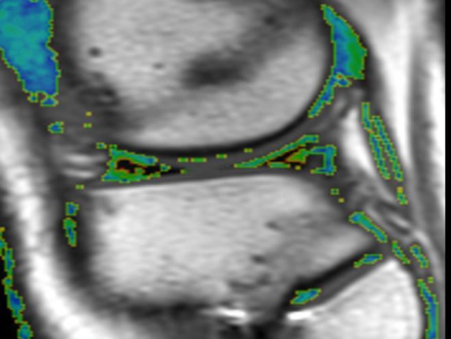
10 Analysis of Knee T2 Maps
11 Analysis of Knee T2 Maps

12 Analysis of Knee T2 Maps T2 of torn posterior horn of meniscus with tear T2 of untorn anterior horn of meniscus with tear T2 of anterior and posterior horns of contralateral untorn meniscus

13 Review of Knee Radiographs Knee radiographs reviewed to determine severity of knee joint degeneration using Kellgren-Lawrence (KL) grading scale KL0=No osteoarthritis KL1= Tiny osteophytes of doubtful clinical significance KL2=Definitive osteophytes with minimal joint space loss

14 Review of Knee Arthroscopy Reports Arthroscopy reports reviewed to determine. Numerical score representing severity of cartilage loss within medial and lateral compartments Type of posterior horn meniscus tear Vertical Horizontal Complex Root

15 Statistical Analysis Kruskal-Wallis tests and Chi-square tests used to compare measured variables between KL grades and meniscus tear types Spearman correlation coefficient used to assess relationship between meniscus T2 and severity of cartilage loss in same compartment

16 Results
17 Meniscus T2 Versus KL Grade
18 Meniscus T2 Versus KL Grade


19 Meniscus T2 Versus KL Grade KL0 KL1 KL2

20 Meniscus T2 Versus Cartilage Loss Significant direct moderate correlation (rho=0.535, p<0.001) between meniscus T2 and severity of cartilage loss in same compartment
21 Variables for Different Tear Types
22 Variables for Different Tear Types
23 Variables for Different Tear Types
24 Variables for Different Tear Types

25 Conclusions Meniscus T2 is significantly higher in both torn and untorn portions of medial and lateral meniscus in patients with higher KL grades T2 can detect early degeneration-related changes in composition and microstructure of intact meniscus prior to development of meniscus tear Direct correlation between meniscus T2 and severity of adjacent cartilage loss reflects important inter-relationship between changes in meniscus and cartilage during knee joint degeneration

26 Conclusions Vertical meniscus tears tend to occur in younger individuals who have less meniscus degeneration and less overall knee joint degeneration when compared to other tear types as vertical tears are due to trauma as opposed to joint degeneration Only distinguishing feature between complex, horizontal, and root meniscus tears is that complex tears have higher T2 in torn portion of meniscus
27 THANK YOU!
In vivo diffusion tensor imaging (DTI) of articular cartilage as a biomarker for osteoarthritis
 of articular cartilage as a biomarker for osteoarthritis") In vivo diffusion tensor imaging (DTI) of articular cartilage as a biomarker for osteoarthritis Jose G. Raya 1, Annie Horng 2, Olaf Dietrich 2, Svetlana Krasnokutsky 3, Luis S. Beltran 1, Maximilian F.
In vivo diffusion tensor imaging (DTI) of articular cartilage as a biomarker for osteoarthritis Jose G. Raya 1, Annie Horng 2, Olaf Dietrich 2, Svetlana Krasnokutsky 3, Luis S. Beltran 1, Maximilian F.
Medial Knee Osteoarthritis Precedes Medial Meniscal Posterior Root Tear with an Event of Painful Popping
 Medial Knee Osteoarthritis Precedes Medial Meniscal Posterior Root Tear with an Event of Painful Popping Dhong Won Lee, M.D, Ji Nam Kim, M.D., Jin Goo Kim, M.D., Ph.D. KonKuk University Medical Center
Proximal tibial bony and meniscal slopes are higher in ACL injured subjects than controls: a comparative MRI study
 Proximal tibial bony and meniscal slopes are higher in ACL injured subjects than controls: a comparative MRI study Ashraf Elmansori, Timothy Lording, Raphaël Dumas, Khalifa Elmajri, Philippe Neyret, Sebastien
This presentation is the intellectual property of the author. Contact them for permission to reprint and/or distribute.
 MRI of the Knee Jennifer Swart, M.D. Musculoskeletal Radiology South Texas Radiology Group Outline Coils, Patient Positioning Acquisition Parameters, Planes and Pulse Sequences Knee Arthrography Normal
The Meniscus. History. Anatomy. Anatomy. Blood Supply. Attachments
 History The Meniscus W. Randall Schultz, MD, MS Austin, TX January 23, 2016 Meniscus originally thought to represent vestigial tissue 1883 first reported meniscal repair (Annandale) Total menisectomy treatment
This presentation is the intellectual property of the author. Contact them at for permission to reprint and/or distribute.
 MRI of the Knee Jennifer Swart, M.D. Musculoskeletal Radiology South Texas Radiology Group Financial Disclosure Dr. Jennifer Swart has no relevant financial relationships with commercial interests to disclose.
MRI of Bucket-Handle Te a rs of the Meniscus of the Knee 1
 MRI of ucket-handle Te a rs of the Meniscus of the Knee 1 Joon Yong Park, M.D., Young-uk Lee M.D., Eun-Chul Chung M.D., Hae-Won Park M.D., E u n - Kyung Youn M.D., Shin Ho Kook, M.D., Young Rae Lee, M.D.
Prevalence of Meniscal Radial Tears of the Knee Revealed by MRI After Surgery
 Downloaded from www.ajronline.org by 46.3.207.114 on 12/22/17 from IP address 46.3.207.114. Copyright RRS. For personal use only; all rights reserved Thomas Magee 1 Marc Shapiro David Williams Received
Conservative surgical treatments for osteoarthritis: A Finite Element Study
 Conservative surgical treatments for osteoarthritis: A Finite Element Study Diagarajen Carpanen, BEng (Hons), Franziska Reisse, BEng(Hons), Howard Hillstrom, PhD, Kevin Cheah, FRCS, Rob Walker, PhD, Rajshree
RECENT ADVANCES IN CLINICAL MR OF ARTICULAR CARTILAGE
 In Practice RECENT ADVANCES IN CLINICAL MR OF ARTICULAR CARTILAGE By Atsuya Watanabe, MD, PhD, Director, Advanced Diagnostic Imaging Center and Associate Professor, Department of Orthopedic Surgery, Teikyo
Meniscal Tears with Fragments Displaced: What you need to know.
 Meniscal Tears with Fragments Displaced: What you need to know. Poster No.: C-1339 Congress: ECR 2015 Type: Authors: Keywords: DOI: Educational Exhibit M. V. Ferrufino, A. Stroe, E. Cordoba, A. Dehesa,
MR imaging of the knee in marathon runners before and after competition
 Skeletal Radiol (2001) 30:72 76 International Skeletal Society 2001 ARTICLE W. Krampla R. Mayrhofer J. Malcher K.H. Kristen M. Urban W. Hruby MR imaging of the knee in marathon runners before and after
FieldStrength. Achieva 3.0T enables cutting-edge applications, best-in-class MSK images
 FieldStrength Publication for the Philips MRI Community Issue 33 December 2007 Achieva 3.0T enables cutting-edge applications, best-in-class MSK images Palo Alto Medical Clinic Sports Medicine Center employs
Arthrographic study of the rheumatoid knee.
 Annals of the Rheumatic Diseases, 1981, 40, 344-349 Arthrographic study of the rheumatoid knee. Part 2. Articular cartilage and menisci KYOSUKE FUJIKAWA, YOSHINORI TANAKA, TSUNEYO MATSUBAYASHI, AND FUJIO
What is the most effective MRI specific findings for lateral meniscus posterior root tear in ACL injuries
 What is the most effective MRI specific findings for lateral meniscus posterior root tear in ACL injuries Kazuki Asai 1), Junsuke Nakase 1), Kengo Shimozaki 1), Kazu Toyooka 1), Hiroyuki Tsuchiya 1) 1)
The Characteristic Findings to Assess Meniscal Healing Status After Meniscal Repair on MRI-T2 Mapping
 The Characteristic Findings to Assess Meniscal Healing Status After Meniscal Repair on MRI-T2 Mapping Shinya Yamasaki, MD, PhD 1,2, Yusuke Hashimoto, MD, PhD 2, Takuya Kinoshita, MD 2, Kazuya Nishino,
Post-injury painful and locked knee
 H R J Post-injury painful and locked knee, p. 54-59 Clinical Case - Test Yourself Musculoskeletal Imaging Post-injury painful and locked knee Ioannis I. Daskalakis 1, 2, Apostolos H. Karantanas 1, 2 1
Role of magnetic resonance imaging in the evaluation of traumatic knee joint injuries
 Original Research Article Role of magnetic resonance imaging in the evaluation of traumatic knee joint injuries Dudhe Mahesh 1*, Rathi Varsha 2 1 Resident, 2 Professor, Department of Radio-Diagnosis, Grant
(Alternate title) Evaluation of Meniscal Extrusion with Posterior Root Disruption and Repair using Ultrasound
 Evaluation of Meniscal Extrusion with Posterior Root Disruption and Repair using Ultrasound") Evaluation of lateral meniscal position with weight bearing: Ultrasonography to measure extrusion with intact, torn and repaired posterior root attachments (Alternate title) Evaluation of Meniscal Extrusion
Evaluation of lateral meniscal position with weight bearing: Ultrasonography to measure extrusion with intact, torn and repaired posterior root attachments (Alternate title) Evaluation of Meniscal Extrusion
Knee: Meniscus Back to Basics
 Knee: Meniscus Back to Basics Kyung Jin Suh kyungjin.suh@gmail.com Doctor Radiology, Daegu, KOREA Medial Lateral 7.7 10.2 11.6 9.6 10.6 mm Posterior > Anterior horn 10.6 mm Posterior = Anterior horn Medial
CLINICAL PRESENTATION AND RADIOLOGY QUIZ QUESTION
 Donald L. Renfrew, MD Radiology Associates of the Fox Valley, 333 N. Commercial Street, Suite 100, Neenah, WI 54956 12/01/2012 Radiology Quiz of the Week # 101 Page 1 CLINICAL PRESENTATION AND RADIOLOGY
Knee Articular Cartilage in an Asymptomatic Population : Comparison of T1rho and T2 Mapping
 TR_002 Technical Reports Knee Articular Cartilage in an Asymptomatic Population : Comparison of T1rho and T2 Mapping Min A Yoon 1,*, Suk-Joo Hong 1, Chang Ho Kang 2, Baek Hyun Kim 3 1 Korea University
Case Report: Knee MR Imaging of Haemarthrosis in a Case of Haemophilia A
 Clinical > Pediatric Imaging Case Report: Knee MR Imaging of Haemarthrosis in a Case of Haemophilia A M. A. Weber, J. K. Kloth University Hospital Heidelberg, Department of Diagnostic and Interventional
MENISCAL INJURY. Meniscus. Anterior Roots. Medial Meniscus. Lateral Meniscus. Posterior Roots. MRI and Arthroscopic Findings
 Meniscus Anterior Roots MENISCAL INJURY MRI and Arthroscopic Findings Medial Meniscus AH PH PH AH Lateral Meniscus Rawiwan Pattaweerakul Naresuan University Hospital Posterior Roots Meniscus Normal Meniscus
KNEE ALIGNMENT SYSTEM (KAS) MRI Protocol
 MRI Protocol") KNEE ALIGNMENT SYSTEM (KAS) MRI Protocol Sample referral sticker Referral Sticker Insert here Corin 17 Bridge Street Pymble NSW Australia 2073 P: +61 (0)2 9497 7400 F: +61 (0)2 9497 7498 E: KAS.customerservice@coringroup.com
KNEE ALIGNMENT SYSTEM (KAS) MRI Protocol Sample referral sticker Referral Sticker Insert here Corin 17 Bridge Street Pymble NSW Australia 2073 P: +61 (0)2 9497 7400 F: +61 (0)2 9497 7498 E: KAS.customerservice@coringroup.com
Sensitivity and Specificity in Detection of Labral Tears with 3.0-T MRI of the Shoulder
 Magee and Williams MRI for Detection of Labral Tears Musculoskeletal Imaging Clinical Observations C M E D E N T U R I C L I M G I N G JR 2006; 187:1448 1452 0361 803X/06/1876 1448 merican Roentgen Ray
Modified Oblique Sagittal Magnetic Resonance Imaging of Rotator Cuff Tears: Comparison with Standard Oblique Sagittal Images
 Journal of Magnetics 22(3), 519-524 (2017) ISSN (Print) 1226-1750 ISSN (Online) 2233-6656 https://doi.org/10.4283/jmag.2017.22.3.519 Modified Oblique Sagittal Magnetic Resonance Imaging of Rotator Cuff
T1rho and T2 relaxation times of the normal adult knee meniscus at 3T: analysis of zonal differences
 Takao et al. BMC Musculoskeletal Disorders (2017) 18:202 DOI 10.1186/s12891-017-1560-y RESEARCH ARTICLE Open Access T1rho and T2 relaxation times of the normal adult knee meniscus at 3T: analysis of zonal
The Effects of Music intervention on Functional connectivity. Supplemental Information
 Yang et al. 0 The Effects of Music intervention on Functional connectivity strength of Brain in Schizophrenia Supplemental Information Mi Yang,#, Hui He #, Mingjun Duan,, Xi Chen, Xin Chang, Yongxiu Lai,
Research Article Relationship between Pain and Medial Meniscal Extrusion in Knee Osteoarthritis
 Advances in Orthopedics Volume 2015, Article ID 210972, 4 pages http://dx.doi.org/10.1155/2015/210972 Research Article Relationship between Pain and Medial Meniscal Extrusion in Knee Osteoarthritis Hiroaki
ADVANCED IMAGING OF THE KNEE
 MENISCAL ANATOMY ADVANCED IMAGING OF THE KNEE MENISCAL ABNORMALITIES MENISCAL FUNCTION MENISCAL FUNCTION load transmission shock absorption stability The menisci DO NOT function as primary stabilizers
MRI of the Knee: Part 2 - menisci. Mark Anderson, M.D. University of Virginia Health System
 MRI of the Knee: Part 2 - menisci Mark Anderson, M.D. University of Virginia Health System Learning Objectives At the end of the presentation, each participant should be able to: describe the normal anatomy
New Evidence Suggests that Work Related Knee Pain with Degenerative Complications May Not Require Surgery
 4 th Quarter 2017 New Evidence Suggests that Work Related Knee Pain with Degenerative Complications May Not Require Surgery By: Michael Erdil MD, FACOEM Introduction It is estimated there are approximately
Epidemiology. Meniscal Injury & Repair. Meniscus Anatomy. Meniscus Anatomy
 Epidemiology 60-70/100,000 per year Meniscal Injury & Repair Arthroscopic Mensiscectomy One of the most common orthopaedic procedures 20% of all surgeries at some centers Male:Female ratio - 2-4:1 Younger
Quantitative Analysis of Vascular Canals in Vertebral Endplate
 Quantitative Analysis of Vascular Canals in Vertebral Endplate Kristine Tan 1, Won C. Bae, PhD 1, Tomonori Yamaguchi, MS 1,2, Kelli Xu, BS 1, Iris Shieh, BS 1, Jade He, BS 1, Robert L. Sah, MD, ScD 1,
Meniscal Root Tears: Evaluation, Imaging, and Repair Techniques
 Meniscal Root Tears: Evaluation, Imaging, and Repair Techniques R O B E R T N A S C I M E N TO, M D, M S C H I E F OF S P O RT S M E D I C I N E & SH O U L D E R S U R G E RY N E W TO N- W E L L E S L
Disclosures. Background. Background
 Kinematic and Quantitative MR Imaging Evaluation of ACL Reconstructions Using the Mini-Two Incision Method Compared to the Anteromedial Portal Technique Drew A. Lansdown, MD Christina Allen, MD Samuel
Western Michigan University Homer Stryker School of Medicine 2. Steadman Philippon Research Institute 3. The Steadman Clinic
 Biomechanical Consequences of Lateral Meniscal Posterior Root Avulsions Influence of the Meniscofemoral Ligaments and ACL on Tibiofemoral Contact Mechanics Andrew G. Geeslin, MD 1 ; David Civitarese, BA
Osteoarthritis and Cartilage 18 (2010) 1393e1401
 1393e1401") Osteoarthritis and Cartilage 18 (2010) 1393e1401 Comparison of BLOKS and scoring systems part I. Cross sectional comparison of methods to assess cartilage morphology, meniscal damage and bone marrow lesions
Osteoarthritis and Cartilage 18 (2010) 1393e1401 Comparison of BLOKS and scoring systems part I. Cross sectional comparison of methods to assess cartilage morphology, meniscal damage and bone marrow lesions
Christian David Hunt. Chapel Hill Approved by: Brian Pietrosimone. Troy Blackburn. Matthew Harkey
 ASSOCIATION BETWEEN QUADRICEPS STRENGTH AND TIBIOFEMORAL CARTILAGE PROTEOGLYCAN DENSITY AT 6 MONTHS FOLLOWING ANTERIOR CRUCIATE LIGAMENT RECONSTRUCTION Christian David Hunt A thesis submitted to the Faculty
Orthopedic Hardware Imaging Part II: MRI v. Metal
 Orthopedic Hardware Imaging Trent Roth, MD And Lauren Ladd, MD Indiana University School of Medicine IU Health Physicians-Radiology Recap: Imaging Techniques Radiography Standard for initial and surveillance
The Impact of Age on Knee Injury Treatment
 The Impact of Age on Knee Injury Treatment Focus on the Meniscus Dr. Alvin J. Detterline, MD Sports Medicine and Orthopaedic Surgery Towson Orthopaedic Associates University of Maryland St. Joseph Medical
The Meniscal Roots: Gross Anatomic Correlation with 3-T MRI Findings
 rody et al. Meniscal Root MRI Musculoskeletal Imaging Pictorial Essay Jeffrey M. rody 1 Michael J. Hulstyn 2 raden. Fleming 3 Glenn. Tung 1 rody JM, Hulstyn MJ, Fleming, Tung G Keywords: anatomy, knee,
Meniscal tears on 3T MR: Patterns, pearls and pitfalls
 Meniscal tears on 3T MR: Patterns, pearls and pitfalls Poster No.: C-2221 Congress: ECR 2010 Type: Educational Exhibit Topic: Musculoskeletal Authors: J. C. Kandathil; Singapore/SG Keywords: Knee injuries,
MRI KNEE WHAT TO SEE. Dr. SHEKHAR SRIVASTAV. Sr.Consultant KNEE & SHOULDER ARTHROSCOPY
 MRI KNEE WHAT TO SEE Dr. SHEKHAR SRIVASTAV Sr.Consultant KNEE & SHOULDER ARTHROSCOPY MRI KNEE - WHAT TO SEE MRI is the most accurate and frequently used diagnostic tool for evaluation of internal derangement
Meniscal Tears: Role of Axial MRI Alone and in Combination with Other Imaging Planes
 Nefise Cagla Tarhan 1,2 Christine. Chung 1 urea Valeria Rosa Mohana-orges 1 Tudor Hughes 1 Donald Resnick 1 Received September 30, 2003; accepted after revision February 2, 2004. 1 Department of Radiology,
T2 Values of Femoral Cartilage of the Knee Joint: Comparison between Pre-Contrast and Post-Contrast Images
 Original Article Musculoskeletal Imaging http://dx.doi.org/.3348/kjr.14.15.1.123 pissn 1229-6929 eissn 05-83 Korean J Radiol 14;15(1):123-129 T2 Values of Femoral Cartilage of the Knee Joint: Comparison
D. Doré 1, C. Ding 1,2, J.P. Pelletier 3, J. Martel-Pelletier 3, F. Cicuttini 2, G. Jones 1.
 Responsiveness of qualitative and quantitative MRI measures over 2.7 years D. Doré 1, C. Ding 1,2, J.P. Pelletier 3, J. Martel-Pelletier 3, F. Cicuttini 2, G. Jones 1. 1 Menzies Research Institute Tasmania,
Discoid Medial Meniscus Tear, with a Literature Review of Treatments
 Case Report Knee Surg Relat Res 2017;29(3):237-242 https://doi.org/10.5792/ksrr.15.054 pissn 2234-0726 eissn 2234-2451 Knee Surgery & Related Research Discoid Medial Meniscus Tear, with a Literature Review
BEFORE THE ARKANSAS WORKERS' COMPENSATION COMMISSION CLAIM NOS. G & G607740
 BEFORE THE ARKANSAS WORKERS' COMPENSATION COMMISSION CLAIM NOS. G204575 & G607740 KERRY D. LEE, EMPLOYEE ARKANSAS PATHOLOGY ASSOCIATES, EMPLOYER C L A I M ANT RESPONDENT BRIDGEFIELD CASUALTY INS. CO.,
Rehabilitation Guidelines for Knee Arthroscopy
 Rehabilitation Guidelines for Knee Arthroscopy The knee is the body's largest joint, and the place where the femur, tibia, and patella meet to form a hinge-like joint. These bones are supported by a large
Causation Review - Meniscal Tears.
 Causation Review - Meniscal Tears. Classification Of Meniscal Tears Of The Knee And The Evidence That Certain Tears Are Degenerative In Origin; Including The Relationship Of Meniscal Tears To Knee Osteoarthritis.
Disclosures: C.B. Raub: None. B.C. Hansen: None. T. Yamaguchi: None. M.M. Temple-Wong: None. K. Masuda: None. R.L. Sah: None.
 En Face Microscopy of Rabbit Knee Articular Cartilage Following Anterior Cruciate Ligament Transection Reveals Early Matrix Damage, Chondrocyte Loss and Cloning Christopher B. Raub, PhD, Bradley C. Hansen,
LATERAL MENISCUS SLOPE AND ITS CLINICAL RELEVANCE IN PATIENTS WITH A COMBINED ACL TEAR AND POSTERIOR TIBIA COMPRESSION
 LATERAL MENISCUS SLOPE AND ITS CLINICAL RELEVANCE IN PATIENTS WITH A COMBINED ACL TEAR AND POSTERIOR TIBIA COMPRESSION R. ŚMIGIELSKI, B. DOMINIK, U, ZDANOWICZ, Z. GAJEWSKI, K. SKIERBISZEWSKA, K. SIEWRUK,
A comparison of arthroscopic diagnosis of ramp lesion and pre-operative MRI evaluation
 A comparison of arthroscopic diagnosis of ramp lesion and pre-operative MRI evaluation Yasuma S, Nozaki M, Kobayashi M, Kawanishi Y Yoshida M, Mitsui H, Nagaya Y, Iguchi H, Murakami H Department of Orthopaedic
Repeatability of 2D FISP MR Fingerprinting in the Brain at 1.5T and 3.0T
 Repeatability of 2D FISP MR Fingerprinting in the Brain at 1.5T and 3.0T Guido Buonincontri 1,2, Laura Biagi 1,3, Alessandra Retico 2, Michela Tosetti 1,3, Paolo Cecchi 4, Mirco Cosottini 1,4,5, Pedro
Role of Magnetic Resonance Imaging in Patients with Knee Trauma
 Original Research Article Role of Magnetic Resonance Imaging in Patients with Knee Trauma Bhautik Kapadia 1, Bhumika Suthar 2* 1 Associate Professor, 2 Assistant Professor, Department of Radiodiagnosis,
SUPPLEMENT: DYNAMIC FUNCTIONAL CONNECTIVITY IN DEPRESSION. Supplemental Information. Dynamic Resting-State Functional Connectivity in Major Depression
 Supplemental Information Dynamic Resting-State Functional Connectivity in Major Depression Roselinde H. Kaiser, Ph.D., Susan Whitfield-Gabrieli, Ph.D., Daniel G. Dillon, Ph.D., Franziska Goer, B.S., Miranda
Publication for the Philips MRI Community
 FieldStrength Publication for the Philips MRI Community Issue 38 Summer 2009 Pediatric MSK imaging benefits from tailored scan protocols Vanderbilt University Children s Hospital builds dedicated scans
A Patient s Guide to Osteoarthritis of the Knee. William T. Grant, MD
 A Patient s Guide to Osteoarthritis of the Knee Dr. Grant is a talented orthopedic surgeon with more than 30 years of experience helping people return to their quality of life. He and GM Pugh, PA-C pride
UCLA UCLA Previously Published Works
 UCLA UCLA Previously Published Works Title MR-IMAGING OF TIBIAL COLLATERAL LIGAMENT INJURY - COMPARISON WITH CLINICAL EXAMINATION Permalink https://escholarship.org/uc/item/2bs9g934 Journal SKELETAL RADIOLOGY,
MENISCAL INJURIES. (copyright s h palmer 2009) MENISCAL FUNCTION
 MENISCAL FUNCTION") (copyright s h palmer 2009) MENISCAL FUNCTION MENISCAL INJURIES Menisci are important for weight bearing, load distribution, joint stability and proprioception. Figure 1: A normal medial meniscus Any load
(copyright s h palmer 2009) MENISCAL FUNCTION MENISCAL INJURIES Menisci are important for weight bearing, load distribution, joint stability and proprioception. Figure 1: A normal medial meniscus Any load
Management of the Early Degenerate Knee. Kieran Barnard Hip and Knee Pathway Lead
 Management of the Early Degenerate Knee Kieran Barnard Hip and Knee Pathway Lead This history Moseley et al (2002) Randomised placebo controlled trial. A total of 180 patients with osteoarthritis of the
Stability of Post Traumatic Osteochondritis Dissecans of the Knee: MR Imaging Findings
 Chin J Radiol 2005; 30: 199-204 199 Stability of Post Traumatic Osteochondritis Dissecans of the Knee: MR Imaging Findings YU-CHUNG HUNG 1 JON-KWAY HUANG 1,2 Department of Radiology 1, Mackay Memorial
Shoulder Instability. Fig 1: Intact labrum and biceps tendon
 Shoulder Instability What is it? The shoulder joint is a ball and socket joint, with the humeral head (upper arm bone) as the ball and the glenoid as the socket. The glenoid (socket) is a shallow bone
Injury Mechanisms of the Cervical Intervertebral Disc During Simulated Whiplash. Spine: Volume 29(11) June 1, 2004 pp
 June 1, 2004 pp") 1 Injury Mechanisms of the Cervical Intervertebral Disc During Simulated Whiplash Spine: Volume 29(11) June 1, 2004 pp 1217-1225 Panjabi, Manohar M. PhD; Ito, Shigeki MD; Pearson, Adam M. BA; Ivancic,
1 Injury Mechanisms of the Cervical Intervertebral Disc During Simulated Whiplash Spine: Volume 29(11) June 1, 2004 pp 1217-1225 Panjabi, Manohar M. PhD; Ito, Shigeki MD; Pearson, Adam M. BA; Ivancic,
Eisuke Nomura, Hisatada Hiraoka, and Hiroya Sakai. 1. Introduction. 2. Case Report
 Case Reports in Orthopedics Volume 2016, Article ID 1026861, 5 pages http://dx.doi.org/10.1155/2016/1026861 Case Report Spontaneous Recurrent Hemarthrosis of the Knee: A Report of Two Cases with a Source
Arthroscopy / MRI Correlation Conference. Department of Radiology, Section of MSK Imaging Department of Orthopedic Surgery 7/19/16
 Arthroscopy / MRI Correlation Conference Department of Radiology, Section of MSK Imaging Department of Orthopedic Surgery 7/19/16 Case 1: 29 YOM with recurrent shoulder dislocations Glenoid Axial T1FS
Summary. Introduction
 Osteoarthritis and Cartilage (1999) 7, 526 532 1999 OsteoArthritis Research Society International 1063 4584/99/060526+07 $12.00/0 Article No. joca.1999.0256, available online at http://www.idealibrary.com
Comparative study of imaging at 3.0 T versus 1.5 T of the knee
 Skeletal Radiol (2009) 38:761 769 DOI 10.1007/s00256-009-0683-0 SCIENTIFIC ARTICLE Comparative study of imaging at 3.0 T versus 1.5 T of the knee Scott Wong & Lynne Steinbach & Jian Zhao & Christoph Stehling
Case Report Double-Layered Lateral Meniscus Accompanied by Meniscocapsular Separation
 Case Reports in Orthopedics Volume 2015, Article ID 357463, 5 pages http://dx.doi.org/10.1155/2015/357463 Case Report Double-Layered Lateral Meniscus Accompanied by Meniscocapsular Separation Aki Fukuda,
Inside-Out Meniscal Repair Still the Gold Standard?
 Inside-Out Meniscal Repair Still the Gold Standard? ERIC BERKSON, MD MGH SPORTS MEDICINE TEAM PHYSICIAN, BOSTON RED SOX ASSISTANT PROFESSOR, HARVARD MEDICAL SCHOOL JULY 26, 2018 Disclosures Neither I nor
Ultrasound assessment of internal derangement of the knee
 Acta Orthop. Belg., 2006, 72, 72-76 ORIGINAL STUDY sound assessment of internal derangement of the knee Zaka KHAN, Zia FARUQUI, Olajide OGYUNBIYI, Guy ROSSET, Javaid IQBAL From Armed Forces Hospital, Southern
Icd 10 for lateral meniscus tear
 Free, official coding info for 2018 ICD-10 -CM S83.282A - includes detailed rules, index and annotation crosswalks,. Short description: Other meniscus derangements, unsp medial meniscus, unsp knee; The
How Much Tesla Is Too Much?
 How Much Tesla Is Too Much? Johnny U. V. Monu, MB, BS; MSc Professor of Radiology and Orthopedics University of Rochester School of Medicine Rochester, New York Historical Timeline Clinical Imaging 1970
A comparative study of MRI versus arthroscopic findings in ACL and meniscal injuries of the knee
 International Journal of Research in Orthopaedics Kulkarni OP et al. Int J Res Orthop. 2018 Mar;4(2):198-202 http://www.ijoro.org Original Research Article DOI: http://dx.doi.org/10.18203/issn.2455-4510.intjresorthop20180123
SMRT Student Scope Submission
 SMRT Student Scope Submission Title & Author Title: MRI in the Detection and Diagnosis of Acoustic Neuroma Author: Manya Lovesky Andersen, student Massachusetts College of Pharmacy and Health Sciences,
Modeling of human knee joint and finite element analysis of landing impact motion
 ISSN 1746-7659, England, UK Journal of Information and Computing Science Vol. 13, No. 1, 2018, pp.044-048 Modeling of human knee joint and finite element analysis of landing impact motion Bao Chunyu 1,3,Meng
JMSCR Vol 05 Issue 01 Page January
 www.jmscr.igmpublication.org Impact Factor 5.244 Index Copernicus Value: 83.27 ISSN (e)-2347-176x ISSN (p) 2455-0450 DOI: https://dx.doi.org/10.18535/jmscr/v5i1.28 Diagnostic Accuracy of Magnetic Resonance
MRI of Cartilage. D. BENDAHAN (PhD)
") MRI of Cartilage D. BENDAHAN (PhD) Centre de Résonance Magnétique Biologique et Médicale UMR CNRS 7339 Faculté de Médecine de la Timone 27, Bd J. Moulin 13005 Marseille France david.bendahan@univ-amu.fr
MRI of Cartilage D. BENDAHAN (PhD) Centre de Résonance Magnétique Biologique et Médicale UMR CNRS 7339 Faculté de Médecine de la Timone 27, Bd J. Moulin 13005 Marseille France david.bendahan@univ-amu.fr
KNEE ARTHROSCOPY PATIENT INFORMATION SHEET
 KNEE ARTHROSCOPY PATIENT INFORMATION SHEET Introduction It has been recommended that you undergo an arthroscopy of your knee. This information sheet is designed to explain what is involved in an arthroscopy,
Personalized Solutions. MRI Protocol for PSI and Signature Guides
 Personalized Solutions MRI Protocol for PSI and Signature Guides 2 Personalized Solutions MRI Protocol for PSI and Signature Guides Purpose and Summary This protocol is applicable for the Zimmer Biomet
HOW DO WE DIAGNOSE LAMENESS IN YOUR HORSE?
 HOW DO WE DIAGNOSE LAMENESS IN YOUR HORSE? To help horse owners better understand the tools we routinely use at VetweRx to evaluate their horse s soundness, the following section of this website reviews
FAI syndrome with or without labral tear.
 Case This 16-year-old female, soccer athlete was treated for pain in the right groin previously. Now has acute onset of pain in the left hip. The pain was in the groin that was worse with activities. Diagnosis
Save the meniscus Mais pourquoi?
 Save the meniscus Mais pourquoi? #$%&' ()"*+!," Philippe Neyret E Servien S Lustig P Verdonk One or more of the authors of the next presentation have identified no potential conflicts of interest 2 Consequences
Concentrations of serum cartilage oligomeric matrix protein after anterior cruciate ligament injury.
 Concentrations of serum cartilage oligomeric matrix protein after anterior cruciate ligament injury. -Comparing with MRI T2 mapping technique- Yohei Nishida, M.D. 1) Yusuke Hashimoto, M.D. Ph.D. 1), Shinya
Evaluation of normal ankle cartilage by MRI T1ρ mapping
 Evaluation of normal ankle cartilage by MRI T1ρ mapping Ryuhei Katsui 1 MD Akira Taniguchi 2 MD, Ph. D Munehiro Ogawa 2 MD, Ph. D Hiroaki Kurokawa 2 MD Norihiro Samoto 3 MD, Ph. D Yasuhito Tanaka 2 MD,
Incidental Meniscal Findings on Knee MRI
 Transcript Details This is a transcript of an educational program accessible on the ReachMD network. Details about the program and additional media formats for the program are accessible by visiting: https://reachmd.com/programs/clinicians-roundtable/incidental-meniscal-findings-on-knee-mri/3772/
The Association of Meniscal Pathologic Changes With Cartilage Loss in Symptomatic Knee Osteoarthritis
 ARTHRITIS & RHEUMATISM Vol. 54, No. 3, March 2006, pp 795 801 DOI 10.1002/art.21724 2006, American College of Rheumatology The Association of Meniscal Pathologic Changes With Cartilage Loss in Symptomatic
Rehabilitation Guidelines for Meniscal Repair
 Rehabilitation Guidelines for Meniscal Repair The knee is the body's largest joint, and the place where the femur, tibia, and patella meet to form a hinge-like joint. These bones are supported by a large
Medical Diagnosis for Michael s Knee
 Medical Diagnosis for Michael s Knee Introduction The following report mainly concerns the diagnosis and treatment of the patient, Michael. Given that Michael s clinical problem surrounds an injury about
MR Imaging of a Posterior Root Tear of the Medial Meniscus: Diagnostic Accuracy of Various Tear Configurations and
 MR Imaging of a Posterior Root Tear of the Medial Meniscus: Diagnostic Accuracy of Various Tear Configurations and Associated Knee Abnormalities 1 Hyang Mi Lee, M.D., Jae Chan Shim, M.D., Jin Goo Kim,
Articular cartilage of the knee 3 years after ACL reconstruction: A quantitative T2 relaxometry analysis of 10 knees
 Articular cartilage of the knee 3 years after ACL reconstruction: A quantitative T2 relaxometry analysis of 10 knees The Harvard community has made this article openly available. Please share how this
Degeneratiivne menisk
 Degeneratiivne menisk Conflict of interest: none Microcirculation of the meniscus Age Dependent Inferior/Superior Geniculates Red zone peripheral 1/3 (3mm) Red/White zone Mid 1/3 (3-5mm) White zone Inner
Clinical Application of Scaffolds for Partial Meniscus Replacement
 1 Clinical Application of Scaffolds for Partial Meniscus Replacement 2 3 4 5 6 7 8 9 10 11 12 13 14 15 16 17 18 19 20 21 22 23 24 25 26 Cathal&J.&Moran,&MD,&FRCS(Orth)& 1,&Daniel&P&Withers&MB,&FRCS&(Orth)&
Why Talk About Technique? MRI of the Knee:
 Why Talk About Technique? MRI of the Knee: Part 1 - Imaging Techniques Mark Anderson, M.D. University of Virginia Health Sciences Center Charlottesville, Virginia Always had an interest teach our fellows
MRI Characteristics of Healed and Unhealed Peripheral Vertical Meniscal Tears
 Musculoskeletal Imaging Original Research Kijowski et al. MRI of Peripheral Meniscal Tears Musculoskeletal Imaging Original Research Richard Kijowski 1 Humberto G. Rosas 1 Kenneth S. Lee 1 rnold Cheung
ACL AND PCL INJURIES OF THE KNEE JOINT
 ACL AND PCL INJURIES OF THE KNEE JOINT Dr.KN Subramanian M.Ch Orth., FRCS (Tr & Orth), CCT Orth(UK) Consultant Orthopaedic Surgeon, Special interest: Orthopaedic Sports Injury, Shoulder and Knee Surgery,
Original Article Effect of anterior cruciate ligament rupture of knee joint on meniscus and cartilage: a finite element analysis of knee joint
 Int J Clin Exp Med 2017;10(12):16468-16475 www.ijcem.com /ISSN:1940-5901/IJCEM0034238 Original Article Effect of anterior cruciate rupture of knee joint on meniscus and : a finite element analysis of knee
the new accurate analysis and classification system of knee joint osteoarthritis
 the new accurate analysis and classification system of knee joint osteoarthritis i3a Soft- and Hardware Components created and assembled in Austria Due to the rapidly increasing fraction of aging people