Title. CitationInternal Medicine, 46(8): Issue Date Doc URL. Type. File Information
|
|
|
- Albert Lee
- 5 years ago
- Views:
Transcription
1 Title Scapular Winging as a Symptom of Cervical Flexion My Author(s)Yaguchi, Hiroaki; Takahashi, Ikuko; Tashiro, Jun; Ts CitationInternal Medicine, 46(8): Issue Date Doc URL Type article (author version) File Information Flexion_myelopathy.pdf Instructions for use Hokkaido University Collection of Scholarly and Aca
2 CASE REPORT Scapular winging as a symptom of cervical flexion myelopathy Hiroaki Yaguchi, Ikuko Takahashi, Jun Tashiro, Sachiko Tsuji, Ichiro Yabe and Hidenao Sasaki Department of Neurology, Hokkaido University Graduate School of Medicine, Kita-15 Nishi-7, Kita-ku, Sapporo , Japan Corresponding; Ichiro Yabe, Department of Neurology, Hokkaido University Graduate School of Medicine, Kita-15 Nishi-7, Kita-ku, Sapporo , Japan Tel; Fax; ; yabe@med.hokudai.ac.jp Key words; flexion myelopathy, Hirayama disease, scapular winging, juvenile muscular atrophy of distal upper extremity, JMADUE
3 Abstract A 23-year-old man complained of weakness of the right arm that he first noted six years prior to his visit. Neurological examination revealed atrophy and weakness of the triceps and serratus anterior muscle on the right side, which resulted in scapular winging on that side. MRI with neck flexion revealed compression of the cervical cord enabling a diagnosis of flexion myelopathy. Proximal muscle weakness and atrophy in flexion myelopathies including Hirayama disease are extremely rare. Here, we report a case of unilateral, proximal upper limb atrophy with scapular winging, attributed to middle cervical flexion myelopathy.
4 Introduction Flexion Myelopathy is caused by a compression of the spinal cord during flexor movements and has been attributed to a combination of many factors. The forward movement of the dural canal in flexion and no dural folding in extension are considered to be important factors (1). Among the flexion myelopathies, juvenile muscular atrophy of distal upper extremity (JMADUE, Hirayama disease), which was first reported in 12 patients by Hirayama et al. in 1959 (2), is now a well-known entity in Japan, however, similar cases have also been described in various countries (3). The pathomechanism of Hirayama disease has been attributed to cervical flexion myelopathy involving the lower cervical cord, presenting a predominantly unilateral hand and forearm involvement (1,3). Recent neuroradiological evaluation using MRI revealed atrophy, flattening and forward displacement of the lower cervical cord, and forward displacement of the posterior dura with expansion of the posterior epidural spaces in a large number of cases (3). However, proximal muscle weakness and atrophy in flexion myelopathies including Hirayama disease are extremely rare (3). Here, we report a case of unilateral, proximal upper limb atrophy with scapular winging,
5 attributed to middle cervical flexion myelopathy. Case Report The patient was a 23-year-old man who complained of weakness of the right arm that he first noted six years prior to his visit. His past history included temporal lobe epilepsy, for which he was taking sodium valproate (600 mg / day). He had not let the neck and shoulder muscles exercise intensely. Neurological examination revealed atrophy and weakness of the triceps, serratus anterior muscle on the right side which resulted in scapular winging on that side (Fig. 1A), without any sensory disturbances. In addition, mild weakness of the right common digital extensor muscle without atrophy was also observed. The oblique amyotrophy of the forearm, characteristic of Hirayama disease, was not present. The deep tendon reflexes, except for mild hyporeflex of the right triceps, were normal in both upper and lower limbs without Babinski sign. He was not aware of cold paresis but trembling of the fingers was noted. The results of laboratory tests were within normal limits, including creatine kinase, aldolase, myoglobin, sodium, potassium, calcium, c-reactive protein, endocrine and
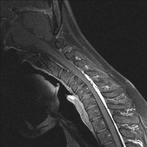
6 metabolic parameters. In neck and thoracic CT, abnormality that caused the long thoracic nerve palsy was not detected. Cerebrospinal fluid examination results were also normal, including the IgG index. He showed no signs of conduction block with a conventional motor nerve conduction study of the median and ulnar nerves. Median somatosensory evoked potentials were normal. Electromyogram in the right triceps muscle showed polyphase and high amplitude motor unit potentials and reduced recruitment, indicative of neurogenic change. Because he was undergoing medical treatment for his temporal lobe epilepsy, a magnetic evoked potential could not be recorded. MRI with neck flexion revealed compression of the cervical cord at the level of the C4/5 vertebrae, with predominant flattening of the right side in the axial view (Fig. 1C,D), compatible with the neurological findings. This image suggested a diagnosis of flexion myelopathy. In this case, weakness of the right upper extremity was not progressive. We decided to conduct follow ups without an operation.
7 Discussion Tashiro et al. described the clinical requirements for the diagnosis of Hirayama disease (3). One of the diagnostic criteria is distal dominant muscle weakness and atrophy in the forearm and hand, which means the peak in the flexion position is generally at the C6 level. Maximal tension is distributed from the C7 to Th1 vertebral level, as it takes the shortest route through the posteriorly convex spinal canal, and muscle weakness is distributed from the C7 to Th1 myelomere. Therefore, our case was not initially diagnosed as Hirayama disease. In Hirayama disease, spinal MRI in cervical flexion shows forward displacement of the dural sac and compressive flattening of the lower cervical cord with widely opened epidural spaces, suggestive of the venous plexus with a flow void (1). In this case, the peak in the flexion position was at the C4/5 level on cervical MRI and muscle weakness from the C5 to C7 myelomere was apparent. Thus, by cervical MRI this case seemed to be a flexion myelopathy, with the same pathomechanism as Hirayama disease. Similar cases have been reported in various countries (3).
8 In the Japanese literature, two cases with the same mechanism of flexion myelopathy, presenting muscular atrophy of proximal upper limbs, unilaterally dominant or unilateral, have been reported (4, 5) and in both cases the peak in the flexion position was at the C4 level on the neuroradiological image, but no scapular winging has been documented. Hence our case could be classified as a specific type of flexion myelopathy, almost identical to Hirayama disease, but the presentation of scapular winging and this particular level of cord involvement have not been previously reported for this disorder. This suggests that patients presenting with scapular winging must be carefully distinguished from those with flexion myelopathy.
9 References 1 Kikuchi S, Tashiro K. Juvenile Muscular Atrophy of Distal Upper Extremity (Hirayama s Disease). In: Jones HR Jr, De Vivo DC, eds. Neuromuscular Disorder of Infancy, Childhood, and Adolescence. A Clinician s Approach. Philadelphia: Butterworth Heinemann, 2003 : Hirayama K, Toyokura Y, Tsubaki T. Juvenile muscular atrophy of unilateral upper extremity; a new clinical entity. Psychiatr Neurol Jpn 1959; 61: (In Japanese, abstract in English.) 3. Tashiro K, Kikuchi S, Itoyama Y, et al. Nationwide survey of juvenile muscular atrophy of distal upper extremity ( Hirayama disease ) in Japan. Amyotrophic Lateral Sclerosis 2006; 7: Masaki T, Hashida H, Sakuta M, et al. A case of flexion myelopathy presenting juvenile segmental muscular atrophy of upper extremities - a successful treatment by cervical spine immobilization. Rinsho Shinkeigaku 1990; 30: (in Japanese, abstract in English. ) 5. Ando T, Fukatsu H, Kameyama T, et al. A case of flexion myelopathy presenting with
10 reversible muscular weakness and atrophy of unilateral proximal upper limb. Rinsho Shinkeigaku 1993; 33: ( in Japanese, abstract in English. )
11 Figure Legend (A) Scapular winging. (B) Cervical fat-suppressed and Gd-enhanced T1-weighted MRI scan in a neutral neck position shows the posterior dural wall near the spinal canal. (C, D) With neck flexion, a forward displacement of the posterior wall of the dura with an expanded venous plexus is visualized in the epidural spaces, resulting in an asymmetrical compression of the cervical cord.


12 A D R C4/5 L B C C4 C4
FOUR CASES OF CLASSICAL HIRAYAMA DISEASE WITH DIFFERENT STAGES OF EVOLUTION Venkatesan Nagarajan 1, Rajesh Venkat I 2, Mahesh I 3, Muthuraj k 4
 FOUR CASES OF CLASSICAL HIRAYAMA DISEASE WITH DIFFERENT STAGES OF EVOLUTION Venkatesan Nagarajan 1, Rajesh Venkat I 2, Mahesh I 3, Muthuraj k 4 HOW TO CITE THIS ARTICLE: Venkatesan Nagarajan, Rajesh Venkat
Title. CitationNeurology and Clinical Neuroscience, 5(1): Issue Date Doc URL. Rights. Type. Additional There Information
: Issue Date Doc URL. Rights. Type. Additional There Information") Title Pseudodystonia in sarcoid myopathy Uwatoko, Hisashi; Yabe, Ichiro; Shirai, Shinichi; Ta Author(s) Hidenao CitationNeurology and Clinical Neuroscience, 5(1): 34-35 Issue Date 2017-01 Doc URL http://hdl.handle.net/2115/68037
Title Pseudodystonia in sarcoid myopathy Uwatoko, Hisashi; Yabe, Ichiro; Shirai, Shinichi; Ta Author(s) Hidenao CitationNeurology and Clinical Neuroscience, 5(1): 34-35 Issue Date 2017-01 Doc URL http://hdl.handle.net/2115/68037
Distal chronic spinal muscular atrophy involving the hands
 Journal ofneurology, Neurosurgery, and Psychiatry, 1978, 41, 653-658 Distal chronic spinal muscular atrophy involving the hands D. J. O'SULLIVAN AND J. G. McLEOD From St Vincent's Hospital, and Department
Juvenile amyotrophy of distal upper extremity, also
 J Neurosurg Spine 20:191 195, 2014 AANS, 2014 A severe case of Hirayama disease successfully treated by anterior cervical fusion Case report Igor Paredes, M.D., 1 Jesus Esteban, Ph.D., 2 Ana Ramos, Ph.D.,
12 Interesting MSK Cases
 12 Interesting MSK Cases James F Griffith Department of Imaging and Interventional Radiology Prince of Wales Hospital Case 1: 12-year-old boy Slipped and fell. Anterior knee pain and swelling Knee pain
Electrophysiological differences between Hirayama disease, amyotrophic lateral sclerosis and cervical spondylotic amyotrophy
 Jin et al. BMC Musculoskeletal Disorders 2014, 15:349 RESEARCH ARTICLE Open Access Electrophysiological differences between Hirayama disease, amyotrophic lateral sclerosis and cervical spondylotic amyotrophy
JMSCR Vol 06 Issue 04 Page April 2018
 www.jmscr.igmpublication.org Impact Factor (SJIF): 6.379 Index Copernicus Value: 71.58 ISSN (e)-2347-176x ISSN (p) 2455-0450 DOI: https://dx.doi.org/10.18535/jmscr/v6i4.78 Electrophysiological Characteristics
Hirayama Disease. Yen-Lin Huang, MD, Chi-Jen Chen, MD* neuroimaging.theclinics.com KEYWORDS DEMOGRAPHICS
 Hirayama Disease Yen-Lin Huang, MD, Chi-Jen Chen, MD* KEYWORDS Hirayama disease Cervical myelopathy Neck flexion Adolescent Hirayama disease is a benign, self-limiting cervical myelopathy first brought
The Upper Limb III. The Brachial Plexus. Anatomy RHS 241 Lecture 12 Dr. Einas Al-Eisa
 The Upper Limb III The Brachial Plexus Anatomy RHS 241 Lecture 12 Dr. Einas Al-Eisa Brachial plexus Network of nerves supplying the upper limb Compression of the plexus results in motor & sensory changes
Human Anatomy and Physiology I Laboratory Spinal and Peripheral Nerves and Reflexes
 Human Anatomy and Physiology I Laboratory Spinal and Peripheral Nerves and Reflexes 1 This lab involves the second section of the exercise Spinal Cord, Spinal Nerves, and the Autonomic Nervous System,
STRUCTURAL BASIS OF MEDICAL PRACTICE EXAMINATION 5. September 30, 2011
 STRUCTURAL BASIS OF MEDICAL PRACTICE EXAMINATION 5 September 30, 2011 PART l. Answer in the space provided. (12 pts) 1. Identify the structures. (2 pts) EXAM NUMBER A. Suprascapular nerve B. Axillary nerve
Central motor conduction in brachial monomelic amyotrophy
 Original Article Central motor conduction in brachial monomelic amyotrophy Pramod K. Pal, Nalini Atchayaram, Gaurav Goel 1, Ebenezer Beulah Departments of Neurology and 1 Neuroimaging and Interventional
How to Think like a Neurologist Review of Exam Process and Assessment Findings
 Lehigh Valley Health Network LVHN Scholarly Works Neurology Update for the Non-Neurologist 2013 Neurology Update for the Non-Neurologist Feb 20th, 5:10 PM - 5:40 PM How to Think like a Neurologist Review
Differential Diagnosis of Neuropathies and Compression. Dr Ashwin Pinto Consultant Neurologist Wessex Neurological Centre
 Differential Diagnosis of Neuropathies and Compression Dr Ashwin Pinto Consultant Neurologist Wessex Neurological Centre Outline of talk Mononeuropathies median and anterior interosseous nerve ulnar nerve
Chapter 13: The Spinal Cord and Spinal Nerves
 Chapter 13: The Spinal Cord and Spinal Nerves Spinal Cord Anatomy Protective structures: Vertebral column and the meninges protect the spinal cord and provide physical stability. a. Dura mater, b. Arachnoid,
region of the upper limb between the shoulder and the elbow Superiorly communicates with the axilla.
 1 region of the upper limb between the shoulder and the elbow Superiorly communicates with the axilla. Inferiorly, a number of important structures pass between arm & forearm through cubital fossa. 2 medial
Pain Assessment Patient Interview (location/nature of symptoms), Body Diagram. Observation and Examination: Tests and Measures
, Body Diagram. Observation and Examination: Tests and Measures") Examination of Upper Quarter Neurogenic Pain Jane Fedorczyk, PT, PhD, CHT Thomas Jefferson University, Philadelphia, PA Center of Excellence for Hand and Upper Limb Rehabilitation I. History Mechanism
Examination of Upper Quarter Neurogenic Pain Jane Fedorczyk, PT, PhD, CHT Thomas Jefferson University, Philadelphia, PA Center of Excellence for Hand and Upper Limb Rehabilitation I. History Mechanism
Year 2004 Paper one: Questions supplied by Megan
 QUESTION 47 A 58yo man is noted to have a right foot drop three days following a right total hip replacement. On examination there is weakness of right ankle dorsiflexion and toe extension (grade 4/5).
Two Cases of Benign Monomelic Amyotrophy of the Lower Extremities
 Journal of the K. S. C. N. Vol. 2, No. 2 Two Cases of Benign Monomelic Amyotrophy of the Lower Extremities Dong Kuck Lee, M.D. Department of Neurology, School of Medicine, Catholic University of Daegu
Thoracic Spine Applied Anatomy. Jason Zafereo, PT, OCS, FAAOMPT
 Thoracic Spine Applied Anatomy Jason Zafereo, PT, OCS, FAAOMPT Clinical i l Orthopedic Rehabilitation ti Education Objectives Discuss concepts relevant to thoracic pain of red flag origin Discuss concepts
Acute Cervical Motor Radiculopathy Induced by Neck and Limb Immobilization in a Patient with Parkinson Disease
 CASE REPORT Acute Cervical Motor Radiculopathy Induced by Neck and Limb Immobilization in a Patient with Parkinson Disease Toshio Shimizu, Tetsuo Komori and Hideaki Hayashi Abstract A 68-year-old woman
Cervical Spine Exercise and Manual Therapy for the Autonomous Practitioner
 Cervical Spine Exercise and Manual Therapy for the Autonomous Practitioner Eric Chaconas PT, PhD, DPT, FAAOMPT Assistant Professor and Assistant Program Director Doctor of Physical Therapy Program Eric
Un d e r normal circumstances, the anteroposterior. Hirayama disease. Clinical article
 J Neurosurg Spine 12:629 634, 2010 Hirayama disease Clinical article Mu h-sh i Lin, M.D., 1 3 Wo o n-ma n Ku n g, M.D., 1,2 We n-ta Ch i u, M.D., Ph.D., 4,5 Ro n g -Ku o Ly u, M.D., 6 Ch i-je n Ch e n,
Fig Cervical spinal nerves. Cervical enlargement C7. Dural sheath. Subarachnoid space. Thoracic. Spinal cord Vertebra (cut) spinal nerves
 spinal nerves") Fig. 13.1 C1 Cervical enlargement C7 Cervical spinal nerves Dural sheath Subarachnoid space Thoracic spinal nerves Spinal cord Vertebra (cut) Lumbar enlargement Medullary cone T12 Spinal nerve Spinal nerve
Fig. 13.1 C1 Cervical enlargement C7 Cervical spinal nerves Dural sheath Subarachnoid space Thoracic spinal nerves Spinal cord Vertebra (cut) Lumbar enlargement Medullary cone T12 Spinal nerve Spinal nerve
Spine Pain Management Program
 Spine Pain Management Program Please complete the following information: Patient Name: Patient ID Number: Patient DOB: The procedure being requested: Epidural Injection Please check the indication (reason)
MRI of chronic spinal cord injury
 The British Journal of Radiology, 76 (2003), 347 352 DOI: 10.1259/bjr/11881183 E 2003 The British Institute of Radiology Pictorial review MRI of chronic spinal cord injury 1 K POTTER, FRCR and 1 A SAIFUDDIN,
Comprehension of the common spine disorder.
 Objectives Comprehension of the common spine disorder. Disc degeneration/hernia. Spinal stenosis. Common spinal deformity (Spondylolisthesis, Scoliosis). Osteoporotic fracture. Anatomy Anatomy Anatomy
Clinical examination of the shoulder girdle
 Clinical of the shoulder girdle CHAPTER CONTENTS Symptoms referred to the shoulder girdle........ e72 Symptoms referred from the shoulder girdle...... e72 History........................... e72 Inspection.........................
CHAPTER 13 LECTURE OUTLINE
 CHAPTER 13 LECTURE OUTLINE I. INTRODUCTION A. The spinal cord and spinal nerves mediate reactions to environmental changes. B. The spinal cord has several functions. 1. It processes reflexes. 2. It is
Can angled sagittal MRI of neural foramen combined with neurological findings determine the affected nerve root in cervical radiculopathy?
 Can angled sagittal MRI of neural foramen combined with neurological findings determine the affected nerve root in cervical radiculopathy? Masatoshi Morimoto, MD., Akihiro Nagamachi, MD. PhD., Kosuke Sugiura,
Note: Please refer to handout Spinal Plexuses and Representative Spinal Nerves for
 Chapter 13 Outline Note: Please refer to handout Spinal Plexuses and Representative Spinal Nerves for what you need to know from Exhibits 13.1 13.4 I. INTRODUCTION A. The spinal cord and spinal nerves
Thoracic Spine Applied Anatomy. Jason Zafereo, PT, OCS, FAAOMPT
 Thoracic Spine Applied Anatomy Jason Zafereo, PT, OCS, FAAOMPT Clinical i l Orthopedic Rehabilitation ti Education 1 Objectives Discuss red flag signs for the thoracic region Apply key concepts from the
Title. CitationInternational Cancer Conference Journal, 4(1): Issue Date Doc URL. Rights. Type. File Information
: Issue Date Doc URL. Rights. Type. File Information") Title Lymph node metastasis in the suprasternal space from Homma, Akihiro; Hatakeyama, Hiromitsu; Mizumachi, Ta Author(s) Tomohiro; Fukuda, Satoshi CitationInternational Cancer Conference Journal, 4(1):
Title Lymph node metastasis in the suprasternal space from Homma, Akihiro; Hatakeyama, Hiromitsu; Mizumachi, Ta Author(s) Tomohiro; Fukuda, Satoshi CitationInternational Cancer Conference Journal, 4(1):
Nerves of the upper limb Prof. Abdulameer Al-Nuaimi. E. mail:
 Nerves of the upper limb Prof. Abdulameer Al-Nuaimi E-mail: a.al-nuaimi@sheffield.ac.uk E. mail: abdulameerh@yahoo.com Brachial plexus Median nerve After originating from the brachial plexus in the axilla,
Disclosures: T. Yoshii: None. T. Yamada: None. T. Taniyama: None. S. Sotome: None. T. Kato: None. S. Kawabata: None. A. Okawa: None.
 Dynamic Changes in Spinal Cord Compression by Cervical Ossification of the Posterior Longitudinal Ligament Evaluated by Kinematic Computed Tomography Myelogram Toshitaka Yoshii, Tsuyoshi Yamada, Takashi
Multichannel somato sensory evoked potential study demonstrated abnormalities in cervical cord function in brachial monomelic amyotrophy
 Original Article Multichannel somato sensory evoked potential study demonstrated abnormalities in cervical cord function in brachial monomelic amyotrophy A. Nalini, S. Praveen-Kumar, Beulah Ebenezer, S.
The Elbow Scanning Protocol
 The Elbow Scanning Protocol Diagnostic Imaging of the Elbow: Introduction The elbow maybe considered as consisting of four quadrants, anterior, medial, lateral and posterior. Ultrasound would normally
Spine Pain Management Program
 Spine Pain Management Program Please complete the following information: Patient Name: Patient ID Number: Patient DOB: The procedure being requested: Epidural Adhesiolysis Please check the indication (reason)
Upper Limb Muscles Muscles of Axilla & Arm
 Done By : Saleh Salahat Upper Limb Muscles Muscles of Axilla & Arm 1) Muscles around the axilla A- Muscles connecting the upper to thoracic wall (4) 1- pectoralis major Origin:- from the medial half of
*Our main subject is the brachial plexus but it's important to understand the spinal cord first in order to understand the brachial plexus.
 *Our main subject is the brachial plexus but it's important to understand the spinal cord first in order to understand the brachial plexus. *Vertebral column is formed by the union of 33 sequential vertebrae
Regional Review of Musculoskeletal System: Head, Neck, and Cervical Spine Presented by Michael L. Fink, PT, DSc, SCS, OCS Pre- Chapter Case Study
 Regional Review of Musculoskeletal System: Presented by Michael L. Fink, PT, DSc, SCS, OCS (20 minutes CEU Time) Subjective A 43-year-old male, reported a sudden onset of left-sided neck and upper extremity
MULTIPLE CHOICE. Choose the one alternative that best completes the statement or answers the question.
 EPC Ch 24 Quiz w-key Name MULTIPLE CHOICE. Choose the one alternative that best completes the statement or answers the question. 1) Which of the following best explains the presentation and prognosis of
Richard Dobrusin DO FACOFP
 Richard Dobrusin DO FACOFP Define Thoracic Outlet Syndrome (TOS) Describe the Mechanisms of Dysfunction List Diagnostic tests for (TOS) Understand (TOS) referral patterns Discuss Treatment Options Definition:
Pathophysiology and treatment for cervical flexion myelopathy
 Eur Spine J (2002) 11 :276 285 DOI 10.1007/s005860100344 ORIGINAL ARTICLE Yoshinori Fujimoto Shinichi Oka Nobuhiro Tanaka Kohichiro Nishikawa Hiroyuki Kawagoe Itsushi Baba Pathophysiology and treatment
Anatomy of the Musculoskeletal System
 Anatomy of the Musculoskeletal System Kyle E. Rarey, Ph.D. Department of Anatomy & Cell Biology and Otolaryngology University of Florida College of Medicine Outline of Presentation Vertebral Column Upper
Case Report: CASE REPORT OF FACET ARTHROPATHY INDUCED NERVE ROOT COMPRESSION RESULTING IN MOTOR WEAKNESS AND PAIN
 Cox Technic Case Report #100 published at www.coxtechnic.com (sent October 2011 on 10/11/11 ) 1 Case Report: CASE REPORT OF FACET ARTHROPATHY INDUCED NERVE ROOT COMPRESSION RESULTING IN MOTOR WEAKNESS
Nerve Injury. 1) Upper Lesions of the Brachial Plexus called Erb- Duchene Palsy or syndrome.
 Upper Lesions of the Brachial Plexus called Erb- Duchene Palsy or syndrome.") Nerve Injury - Every nerve goes to muscle or skin so if the nerve is injured this will cause paralysis in the muscle supplied from that nerve (paralysis means loss of function) then other muscles and other
Nerve Injury - Every nerve goes to muscle or skin so if the nerve is injured this will cause paralysis in the muscle supplied from that nerve (paralysis means loss of function) then other muscles and other
Spinal Column. Anatomy Of The Spine
 Anatomy Of The Spine The spine is a flexible column, composed of a stack of individual bones. Each bone is called a vertebra. There are seven vertebrae in the neck (cervical vertebrae) twelve in the thoracic
The arm: *For images refer back to the slides
 The arm: *For images refer back to the slides Muscles of the arm: deltoid, triceps (which is located at the back of the arm), biceps and brachialis (it lies under the biceps), brachioradialis (it lies
J Korean Soc Spine Surg 2011 Sep;18(3): Originally published online September 30, 2011;
: Originally published online September 30, 2011;") Journal of Korean Society of Spine Surgery Acute Spontaneous Cervical Spinal Epidural Hematoma with Spontaneous Resolution -A Case Report- Young-Do Koh, M.D.,Seung Hwan Kook, M.D. J Korean Soc Spine Surg
Journal of Korean Society of Spine Surgery Acute Spontaneous Cervical Spinal Epidural Hematoma with Spontaneous Resolution -A Case Report- Young-Do Koh, M.D.,Seung Hwan Kook, M.D. J Korean Soc Spine Surg
Case 3. Your Diagnosis?
 Case 3 45 year-old presenting with a history of injury to the right shoulder whilst working in the freezing work. He was loading a sheep over an incline with his arm around the sheep. He felt pain in the
Multifocal motor neuropathy: diagnostic criteria that predict the response to immunoglobulin treatment
 Multifocal motor neuropathy: diagnostic criteria that predict the response to immunoglobulin treatment 7 MMN RM Van den Berg-Vos, H Franssen, JHJ Wokke, HW Van Es, LH Van den Berg Annals of Neurology 2000;
Selective laminoplasty for cervical spondylotic myelopathy: a comparative study with a minimum 5-year follow-up
 Selective laminoplasty for cervical spondylotic myelopathy: a comparative study with a minimum 5-year follow-up Minori Kato*, Hiroaki Nakamura**, Koji Tamai**, Kazunori Hayashi**, Akira Matsumura**, Sadahiko
Benign monomelic amyotrophy in a 7-year-old girl with proximal upper limb involvement: case report
 The Turkish Journal of Pediatrics 2011; 53: 471-476 Case Report Benign monomelic amyotrophy in a 7-year-old girl with proximal upper limb involvement: case report Öznur Yılmaz 1, İpek Alemdaroğlu 1, Ayşe
Gateway to the upper limb. An area of transition between the neck and the arm.
 Gateway to the upper limb An area of transition between the neck and the arm. Pyramidal space inferior to shoulder @ junction of arm & thorax Distribution center for the neurovascular structures that serve
A rare case of cervical epidural extramedullary plasmacytoma presenting with monoparesis
 Romanian Neurosurgery Volume XXXI Number 1 2017 January - March Article A rare case of cervical epidural extramedullary plasmacytoma presenting with monoparesis Okan Turk, Ibrahim Burak Atci, Hakan Yilmaz,
Chiropractic ICD-10 Common Codes List
 Chiropractic ICD-10 Common Codes List This is a preliminary list of Common ICD-10 Codes for chiropractic diagnoses. This is a common code list to be used as a guide for coding and is not intended to represent
Spine Pain Management Program
 Spine Pain Management Program Please complete the following information: Patient Name: Patient ID Number: Patient DOB: The procedure being requested: Facet Injection Please check the indication (reason)
Thoracic Outlet Syndrome
 Thoracic Outlet Syndrome Part 1: The Scalene Triangle TOS: Vascular Symptom Presentation Venous persistent/intermittent edema heaviness and fatigue deep pain in neck/shoulder increased pain at night warm
Cervical Spine: Pearls and Pitfalls
 Cervical Spine: Pearls and Pitfalls Presenters Dr. Rob Donkin Functional Anatomy Current research Cervical Radiculopathy Dr. Gert Ferreira Red flags Case Study Kinesio Taping Chris Neethling Gonstead adjusting
10/5/2017. Cervical Manual Evaluation and Mobilizations. Upper Cervical Stability Testing Alar Ligament
 Cervical Manual Evaluation and Mobilizations Upper Cervical Stability Testing Alar Ligament Upper Cervical Stability Testing Transverse Ligament 1 Upper Cervical Stability Testing Transverse Plane Positive
Clinical Examination. of the. Cervicothoracic Region. Neck Disability Index. Serious Pathological Conditions. Medical Screening Questionnaire
 Clinical Examination Clinical Examination of the Cervicothoracic Region Screening for associated serious pathological conditions Neck disability index Physical Exam Serious Pathological Conditions Cervical
Lumbosacral plexus lesion Lumbosacral plexus disorders G54.1 Neuralgic amyotrophy Neuralgic amyotrophy G
 ICD-9-CM and ICD-10-CM NEUROMUSCULAR DIAGNOSIS CODES Focal Neuropathy ICD-9-CM ICD-10-CM Mononeuropathy G56.00 Carpal tunnel syndrome 354.00 Other median nerve lesion 354.10 Lesion of ulnar nerve 354.20
Nerves of Upper limb. Dr. Brijendra Singh Professor & Head Department of Anatomy AIIMS Rishikesh
 Nerves of Upper limb Dr. Brijendra Singh Professor & Head Department of Anatomy AIIMS Rishikesh 1 Objectives Origin, course & relation of median & ulnar nerves. Motor & sensory distribution Carpal tunnel
Posture. Posture Evaluation. Good Posture. Correct Posture. Postural Analysis. Endomorphs
 Posture Posture Evaluation Martha Macht Sliwinski PT PhD The alignment and positioning of the body in relation to gravity, center of mass and base of support The physical therapist uses posture tests and
Region of upper limb attachment to the trunk Proximal segment of limb overlaps parts of the trunk (thorax and back) and lower lateral neck.
 and lower lateral neck.") Region of upper limb attachment to the trunk Proximal segment of limb overlaps parts of the trunk (thorax and back) and lower lateral neck. includes Pectoral Scapular Deltoid regions of the upper limb
Region of upper limb attachment to the trunk Proximal segment of limb overlaps parts of the trunk (thorax and back) and lower lateral neck. includes Pectoral Scapular Deltoid regions of the upper limb
Planning the Objective Exam. Objective Examination of the Cervical Spine. Clearing Tests. Observation. Functional Demonstration.
 Objective Examination of the Cervical Spine Taking the complaint and identifying the damaged structure Planning the Objective Exam With a clear picture from the subjective exam, the objective exam should
BRACHIAL PLEXUS. DORSAL SCAPULAR NERVE (C5) supraclavicular branch innervates rhomboids (major and minor) and levator scapulae
 supraclavicular branch innervates rhomboids (major and minor) and levator scapulae") THE BRACHIAL PLEXUS DORSAL SCAPULAR NERVE (C5) supraclavicular branch innervates rhomboids (major and minor) and levator scapulae SCHEMA OF THE BRACHIAL PLEXUS THE BRACHIAL PLEXUS PHRENIC NERVE supraclavicular
THE BRACHIAL PLEXUS DORSAL SCAPULAR NERVE (C5) supraclavicular branch innervates rhomboids (major and minor) and levator scapulae SCHEMA OF THE BRACHIAL PLEXUS THE BRACHIAL PLEXUS PHRENIC NERVE supraclavicular
S pinal muscle atrophy of the distal upper extremities, with
 627 PAPER Peripheral and segmental spinal abnormalities of median and ulnar somatosensory evoked potentials in Hirayama s disease A Polo, M Curro Dossi, A Fiaschi, G P Zanette, N Rizzuto... See end of
Fractures of the Thoracic and Lumbar Spine
 A spinal fracture is a serious injury. Nader M. Hebela, MD Fellow of the American Academy of Orthopaedic Surgeons http://orthodoc.aaos.org/hebela Cleveland Clinic Abu Dhabi Cleveland Clinic Abu Dhabi Neurological
Lab Workbook. ANATOMY Manual Muscle Testing Lower Trapezius Patient: prone
 ANATOMY Manual Muscle Testing Lower Trapezius Patient: prone Lab Workbook Fixation: place on hand below the scapula on the opposite side Test: adduction and depression of the scapula with lateral rotation
BRACHIAL PLEXUS 11/12/2014 كيف تتكون الضفيرة FORMATION ENLARGEMENT (INTUMESCENCE) OF THE SPINAL CORD. Grey matter. Cervical intumescence - C 6 - T 2
 OF THE SPINAL CORD. Grey matter. Cervical intumescence - C 6 - T 2") BRACHIAL PLEXUS Prof. Fawzy Elnady ENLARGEMENT (INTUMESCENCE) OF THE SPINAL CORD Grey matter Cervical intumescence - C 6 - T 2 Lumbar intumescence - L 4 S 2 كيف تتكون الضفيرة FORMATION The ventral rami
BRACHIAL PLEXUS Prof. Fawzy Elnady ENLARGEMENT (INTUMESCENCE) OF THE SPINAL CORD Grey matter Cervical intumescence - C 6 - T 2 Lumbar intumescence - L 4 S 2 كيف تتكون الضفيرة FORMATION The ventral rami
Inhibition Associated with somatic dysfunctions, no matter which components are impaired Implies consideration of all components in treatment planning
 Somatic Dysfunction Impaired or altered function of related components of the somatic system including the skeletal, arthrodial, myofascial structures and their related vascular, lymphatic and neural elements.
Index. Note: Page numbers of article titles are in boldface type.
 Neurol Clin N Am 20 (2002) 605 617 Index Note: Page numbers of article titles are in boldface type. A ALS. See Amyotrophic lateral sclerosis (ALS) Amyotrophic lateral sclerosis (ALS) active denervation
Restraints to Movement... 4 Restraints to flexion... 4 Primary restraint into Extension... 4
 CERVICAL SPINE... 4 Neck Pain Categories... 4 Kinematics... Error! Bookmark not defined. Ranges of Motion C2-7... 4 Coupled Movements... 4 Ranges of Motion C0-2... 4 Coupled Movements... 4 Restraints to
STRUCTURAL BASIS OF MEDICAL PRACTICE EXAMINATION 5 October 6, 2006
 STRUCTURAL BASIS OF MEDICAL PRACTICE EXAMINATION 5 October 6, 2006 PART l. Answer in the space provided. (8 pts) 1. Identify the structures. (2 pts) B C A. _pisiform B. _ulnar artery A C. _flexor carpi
Regional Human Anatomy (HBA 461/561/540): Course Objectives
: Course Objectives") Regional Human Anatomy (HBA 461/561/540): Course Objectives This is a 5-credit course that consists of 1-hour lectures followed by 3-hour labs. It is organized into three modules (see syllabus): Module
Regional Human Anatomy (HBA 461/561/540): Course Objectives This is a 5-credit course that consists of 1-hour lectures followed by 3-hour labs. It is organized into three modules (see syllabus): Module
Overview. Spinal Anatomy Spaces & Meninges Spinal Cord. Anatomy of the dura. Anatomy of the arachnoid. Anatomy of the spinal meninges
 European Course in Neuroradiology Module 1 - Anatomy and Embryology Dubrovnik, October 2018 Spinal Anatomy Spaces & Meninges Spinal Cord Johan Van Goethem Overview spinal meninges & spaces spinal cord
Spine Pain Management Program
 Spine Pain Management Program Please complete the following information: Patient Name: Patient ID Number: Patient DOB: The procedure being requested is: Please check the indication (reason) for this procedure
Human Anatomy. Spinal Cord and Spinal Nerves
 Human Anatomy Spinal Cord and Spinal Nerves 1 The Spinal Cord Link between the brain and the body. Exhibits some functional independence from the brain. The spinal cord and spinal nerves serve two functions:
The Spinal Cord & Spinal Nerves
 The Spinal Cord & Spinal Nerves Together with brain forms the CNS Functions spinal cord reflexes integration (summation of inhibitory and excitatory) nerve impulses highway for upward and downward travel
Cervical laminectomy for spinal cord compression. Information for patients Neurosurgery
 Cervical laminectomy for spinal cord compression Information for patients Neurosurgery What is a compression of the spinal cord and how has it been caused? The bones in our back are called vertebras and
3/10/17 Spinal a Injury 1
 Spinal Injury 1 'Paralysed' Watmough vows he'll have the backbone for Game Two after treatment for neck injury Watmough will have cortisone injected into his spine this morning to speed up the recovery
Electrodiagnostics for Back & Neck Pain. Steven Andersen, MD Providence Physiatry Clinic
 Electrodiagnostics for Back & Neck Pain Steven Andersen, MD Providence Physiatry Clinic Electrodiagnostics Electromyography (EMG) Needle EMG exam (NEE) Nerve conduction studies (NCS) Motor Sensory Late
HIGH LEVEL - Science
 Learning Outcomes HIGH LEVEL - Science Describe the structure and function of the back and spine (8a) Outline the functional anatomy and physiology of the spinal cord and peripheral nerves (8a) Describe
Chapter 13. The Spinal Cord & Spinal Nerves. Spinal Cord. Spinal Cord Protection. Meninges. Together with brain forms the CNS Functions
 Spinal Cord Chapter 13 The Spinal Cord & Spinal Nerves Together with brain forms the CNS Functions spinal cord reflexes integration (summation of inhibitory and excitatory) nerve impulses highway for upward
Daniel J. Blizzard, MD, MS
 Daniel J. Blizzard, MD, MS None Common degenerative (usually) condition caused by compression on the spinal cord that is characterized by clumsiness and difficulty with fine motor tasks in the hands and
PNS and ANS Flashcards
 1. Name several SOMATIC SENSES Light touch (being touched by a feather), heat, cold, vibration, pressure, pain are SOMATIC SENSES. 2. What are proprioceptors; and how is proprioception tested? PROPRIOCEPTORS
CERVICAL SPONDYLOSIS AND CERVICAL SPONDYLOTIC MYELOPATHY
 CERVICAL SPONDYLOSIS AND CERVICAL SPONDYLOTIC MYELOPATHY A NEUROSURGEON S VIEW A Preventable Journey to a wheelchair bound-life Dr H. BOODHOO F.C.S (Neurosurgery) Cervical Spondylosis Spinal Osteoarthritis
Objectives. Comprehension of the common spine disorder
 Objectives Comprehension of the common spine disorder Disc degeneration/hernia Spinal stenosis Common spinal deformity (Spondylolisthesis, Scoliosis) Osteoporotic fracture Destructive spinal lesions Anatomy
Assessment of the Brachial Plexus EMG Course CNSF Halifax Fraser Moore, Canadian Society of Clinical Neurophysiology McGill University
 Assessment of the Brachial Plexus EMG Course CNSF Halifax 2018 Fraser Moore, Canadian Society of Clinical Neurophysiology McGill University Angela Scott, Association of Electromyography Technologists of
Intraoperative spinal cord monitoring with Tce-MEP for cervical laminoplasty
 Intraoperative spinal cord monitoring with Tce-MEP for cervical laminoplasty Nobuhiro Tanaka 1, 2), Kazuyoshi Nakanishi 2), Naosuke Kamei 2), Toshio Nakamae 2), Shinji Kotaka 2), Yoshinori Fujimoto 1),
Gregory M Yoshida, MD. Lateral curvature of the spine in the coronal plane > 10 degrees on an upright film
 Gregory M Yoshida, MD Lateral curvature of the spine in the coronal plane > 10 degrees on an upright film Measurement Angle made by the endplates of the two most tilted vertebra from horizontal Cobb angle
Title. CitationJournal of the Neurological Sciences, 313(1-2): 147- Issue Date Doc URL. Type. File Information
: 147- Issue Date Doc URL. Type. File Information") Title Nationwide survey on the epidemiology of syringomyel Sakushima, Ken; Tsuboi, Satoshi; Yabe, Ichiro; Hida, Author(s) Sasaki, Hidenao CitationJournal of the Neurological Sciences, 313(1-2): 147- Issue
Title Nationwide survey on the epidemiology of syringomyel Sakushima, Ken; Tsuboi, Satoshi; Yabe, Ichiro; Hida, Author(s) Sasaki, Hidenao CitationJournal of the Neurological Sciences, 313(1-2): 147- Issue
Physical Examination of the Shoulder
 General setup Patient will be examined in both the seated and supine position so exam table needed 360 degree access to patient Expose neck and both shoulders (for comparison); female in gown or sports
102 Results RESULTS. Age Mean=S.D Range 42= years -84 years Number % <30 years years >50 years
 102 Results RESULTS A total of 50 cases were studied 39 males and 11females.Their age ranged between 16 years and 84 years (mean 42years). T1 and T2WI were acquired for all cases in sagittal and axial
SpineFAQs. Neck Pain Diagnosis and Treatment
 SpineFAQs Neck Pain Diagnosis and Treatment Neck pain is a common reason people visit their doctor. Neck pain typically doesn't start from a single injury. Instead, the problem usually develops over time
Orthopadic cors. Topic : -Cervical spondylitis. -Development disorders(spondylolysis and Spodylolsithesis)
") Orthopadic cors Topic : -Cervical spondylitis. -Development disorders(spondylolysis and Spodylolsithesis) Cervical spondylitis. Definition : - a painful condition of the cervical spine resulting from the
Orthopadic cors Topic : -Cervical spondylitis. -Development disorders(spondylolysis and Spodylolsithesis) Cervical spondylitis. Definition : - a painful condition of the cervical spine resulting from the
Guide to the use of nerve conduction studies (NCS) & electromyography (EMG) for non-neurologists
 & electromyography (EMG) for non-neurologists") Guide to the use of nerve conduction studies (NCS) & electromyography (EMG) for non-neurologists What is NCS/EMG? NCS examines the conduction properties of sensory and motor peripheral nerves. For both
Guide to the use of nerve conduction studies (NCS) & electromyography (EMG) for non-neurologists What is NCS/EMG? NCS examines the conduction properties of sensory and motor peripheral nerves. For both
THORACIC OUTLET SYNDROME (T.O.S.) Prof J van Marle
 Prof J van Marle") THORACIC OUTLET SYNDROME (T.O.S.) Prof J van Marle 1. Definition Clinical syndrome caused by compression of the neurovascular bundle as it passes through the thoracic outlet, a narrow space bordered by
THORACIC OUTLET SYNDROME (T.O.S.) Prof J van Marle 1. Definition Clinical syndrome caused by compression of the neurovascular bundle as it passes through the thoracic outlet, a narrow space bordered by