Diagnostic imaging of. Musculoskeletal diseases
|
|
|
- Valerie Barker
- 5 years ago
- Views:
Transcription
1 Diagnostic imaging of Musculoskeletal diseases
2 Investigating methods 1. X ray tha basic technique 2. CT more accurate anatomy, reconstructions 3. MRI bones no, but bone marrow 4. Scintigraphy : osteoblast activity, radiopharmacon uptake (Tc) 5. Ultrasound joints, muscles, tendons, 6. Densitometry : the calcium content 7. Angiography eg. preop,(limbsaving) selective chemotherapy
3 Plain radiograph 40% of all investigations in a general radiological department : trauma, degenerative
4 Bones are fit for x-ray investigation x-ray attenuation of the calcium is much higher, than those of the surrounding soft tissues The linear resolution is very good for evaluating the fine bony structure


5 Normal variants of no clinical importance mistaken with fractures pl. persisting apophysis, accessory bones Persist. apophysis. os Vesalianum access. sesamy-bone
6 A kórfolyamatok röntgenológiai alapjelenségei - calcipenia diffuse Calcium decreasing - atrophy (circumscribed, e.g. in the region of one joint) Sudeck-atrophy - osteolysis circumscribed bone disappearing - usuration conturdefect because of external pressure - erosio following an inflammation - osteosclerosis - periosteal reaction - osteonecrosis nutritive artery blocked


7 Diffuse calcipenia not only in osteoporosis! normal normal

8 Differencies of the calcium content in the lumbal spine
9 Only 30-40% decreasing of the mineral substance is to be seen in the plain film

10 Osteosclerosis Marble bone disease (osteopetrosis)

11 circumscribed sclerosis ROD Paget s disease Ruggers jersey spine

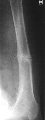
12 periosteal diseases Codman-triangle inflammtion = thickens calcification any damage/hurt, chr. heart and pulmonary disaeases, toxic impacts: thickens and calcify new-bone production surround - adjacent haematoma - malignant tumors: lamellar, spiculated
13 CT - transversal imaging also in deep soft tissue details - thin slices isotropic volume data = good multiplanar reconstructions 3D: ortopedy, traumatology - better resolution, bones and surrounding soft tissues - contrast media enhancement indicates


14 Conventional x ray (plain radiograph) summation effect CT no summ.


15
16 Bone scintigraphy - entire skeleton together - high sensitivity - low specificity systemic /spreaded disorders high activity : inflammation, damage or tumor - 3 phases blood perfusion bloodpool metabolic phase: radiofarmakon uptake = osteoblast activity
17 Osteodensitometry mineral bone-mass photon-absorptive techique single photon absorptiometry - isotope (SPA) - x-ray (SXA) dual-photon-absorptiometry - izotope (DPA) - x-ray (DEXA) - quantitative CT (QCT és pqct) quantitative ultrasound bone-densitometry (QUS)
18 ODM: bone mass g/cm 2 DEXA: low energy x-ray -vertebra, femur neck Comparing with the bone density of healthy y o persons: T score according to the age: Z score WHO criteria Normal ± 1 SD Osteopenia (- 1 SD) (- 2,5 SD) Osteoporosis - 2,5 SD lower than Serious porosis < - 2,5 and 1 vertebral compression
19 - bony structure: no - soft tissues : yes periosteum: haematoma, subperiosteal abscess joints: fluis, tendons, ligaments biopsy
20 Ultrasound Newborn screening for displasia
21 Musculoskeletal diseases developmental metabolic degenerative inflammation tumors blood supply
22 Developmental - generalised bone alterations eg. osteogenesis imperfecta - circumscribed developmental disorders in segmentation and vertabral corpus
23 developmental disorders generalised - prenatal form: lethal - postnatal form: blue sclera deafness frquent fractures autosomal dominant hereditary Disturbed collagen sinthesis osteogenesis imperfecta
24 developmental disorders heraditary autosomal dominant Short limbs, Big head dwarf chondrodystrophia foetalis
 hereditary (dominant /")

25 Osteopetrosis (marmor bone disease) hereditary (dominant / recessive forms ) osteoclast disorder, bone-resorption se ALP level normal!
26 Circumscribed dvelopmental disturbances spondylolysis Scotty dog sign spina bifida cervical rib hemivertebra
27 Inflammatory diseases Osteomyelitis arthritis tuberculosa
28 Metabolic bone-diseases the bone resorption and new formation: remodeling homones and local factors (eg. PTH, sexual and steroid hormons, D3vit.)
29 In the matabolic bone diseases:balance lost increased resorption osteoporosis calcipenic osteopathia increased bone formation sclerosis
30 Metabolic osteopathies osteoporosis osteomalacia hyperparathyreoidism myeloma multiplex
31 the age of the patient, anamnesis, laboratory data) markers of bone-metabolism!


32 wedge-like biconcave vertebral compression crush
 2-4.")
33 Metacarpal index (radiogrammetria) Barnett és Nordin 1961 digital x-ray radiogrammetry revitalised by the digital technique (DXR) 2-4. metacarpus, ulna, radius

34 Osteomalacia osteoid mineralisation disturbed


35 Hyperparathyreoidism (pth adenoma) subperiosteal resorption 2., 3. midle phalanx radial side Perpendicular to the cortical more frequent in the secondary HPT


36 Bonecysts pathologic fracture of the fibula generalised form M. Recklinghausen


37 Myeloma multiplex osteoclast inducating factor diffuse calcipenia lythic spots
38 Renal osteodystrophy (ROD) uraemic patients compound disease: osteomalacy and HPT symptoms paraarticular calcification Ruggers jersey spine


39 Trauma MR CT
40 Pathologic fracture Stress fracture fatigue fracture normal bone, repetitive or cyclical stresses insufficiency fracture weakened bones (osteoporosis), normal forces or microtrauma pathologic fractures tumor /inflammation /cysts locally weakened bones
41 Femoral neck fracture lateral medial
42 Nuclear medicine isotope high osteoblast activity - izotóp deposition early sign of the fracture femoral neck os scophoideum fracture

43 scaphoid fracture
44 Tumors benign, malignant osteoma osteochondroma ostesarcoma


45 Metastasis (haematogenous) shigillocellular cc osteolytic mamma hypernephroma osteoplastic prostata mixed form mamma prostata
46 Degenerative joint diseases arthrosis spondylosis rheumatoid arthritis (RA) gout atlanto-axial instability myelon compression
47 in the joints or around metabolic material depositions
48 - purinmetabolism disturbance - urat level high in the blood - deposits in the joints Prevalency: 1,5-2,6 / 1000 Males of middle ages or older (90%) 28/1000 Females in the premenopausa rare Tophus: chronic gout Na-urat deposits
49 This document was created with Win2PDF available at The unregistered version of Win2PDF is for evaluation or non-commercial use only.
Department of Radiology, University of Szeged. Imaging of the skeleton
 Imaging of the skeleton Methods of examination: plain x-ray (radiography, densitometry) x-ray with contrast material (fistulography, angiography) ultrasound (b-mode, Doppler, color, duplex) computed tomography
Pediatric metabolic bone diseases
 Pediatric metabolic bone diseases Classification and overview of clinical and radiological findings M. Mearadji International Foundation for Pediatric Imaging Aid www.ifpia.com Introduction Metabolic bone
CHAPTER 13 SKELETAL SYSTEM
 CHAPTER 13 SKELETAL SYSTEM Structure and Function Functions of the skeletal system Provides shape and support Protects internal organs Stores minerals and fat Produces blood cells and platelets Assists
CHAPTER 6 MUSCULOSKELETAL SYSTEM DISEASES, DISORDERS, AND DIAGNOSTIC TERMS. Ms. Doshi
 CHAPTER 6 MUSCULOSKELETAL SYSTEM DISEASES, DISORDERS, AND DIAGNOSTIC TERMS Ms. Doshi Worksheet 1 pp 154-156 Review Exercises Infections Disease cellulitis myocellulitis osteitis osteomyelitis osteochondritis
ESSENTIALS OF PLAIN FILM INTERPRETATION: SPINE DR ASIF SAIFUDDIN
 ESSENTIALS OF PLAIN FILM INTERPRETATION: SPINE DR ASIF SAIFUDDIN Consultant Musculoskeletal Radiologist Royal National Orthopaedic Hospital Stanmore,UK. INTRODUCTION 2 INTRODUCTION 3 INTRODUCTION Spinal
Accuracy of DEXA scanning & other methods for determining BMD.
 BMD- Measurement Site Accuracy of DEXA scanning & other methods for determining BMD. Ann Larkin In general, densitometry techniques can be performed in either the axial or the appendicular skeleton, depending
Metabolic and Endocrine Disorders
 Metabolic and Endocrine Disorders Christine B. Chung, M.D. Professor of Radiology Musculoskeletal Division UCSD and VAHCS Diffuse Regional Rickets and Osteomalacia Scurvy Parathyroid Disorders Hyperparathyroidism
Primary bone tumors > metastases from other sites Primary bone tumors widely range -from benign to malignant. Classified according to the normal cell
 Primary bone tumors > metastases from other sites Primary bone tumors widely range -from benign to malignant. Classified according to the normal cell counterpart and line of differentiation. Among the
Hths 2231 Laboratory 13 Alterations in Musculoskeletal
 Watch Movie: Osteoporosis Answer the movie questions on the worksheet. Complete activities 1-4. Activity #1: Click on the website link in activity 1 to review the structure and function of bone. Activity
The role of CT and MRI in evaluation of Osteoid Oteoma
 The role of CT and MRI in evaluation of Osteoid Oteoma Elene Iordanishvili Tbilisi Sate Medical University Instructor: Prof. Dr. Ketevan Kotetishvili Department of Physics Georgian Technical University
The Skeletal System Vertebral column Sacrum. Osseous tissue For the body and soft organs. Magnesium, sodium, fluoride Levers for muscle action
 10/1/2016 Cranium Facial s Skull Clavicle Scapula Sternum Rib Humerus Vertebra Radius Ulna Carpals Thoracic cage (ribs and sternum) The Skeletal System Vertebral column Sacrum Phalanges Metacarpals Femur
Bone Mineral Densitometry with Dual Energy X-Ray Absorptiometry
 Bone Mineral Densitometry with Dual Energy X-Ray Absorptiometry R Gilles, Laurentius Ziekenhuis Roermond 1. Introduction Osteoporosis is characterised by low bone mass, disruption of the micro-architecture
36 1 The Skeletal System Slide 1 of 40
 1 of 40 The Skeleton All organisms need structural support. Unicellular organisms have a cytoskeleton. Multicellular animals have either an exoskeleton (arthropods) or an endoskeleton (vertebrates). 2
Musculoskeletal System
 Musculoskeletal System The musculoskeletal system gives the body strength, structure, and capability of movement. Bones are the framework. Ligaments and tendons are the nails Muscles are the way we move
Understanding Osteoporosis
 Understanding Osteoporosis Professor Juliet E. Compston Published by Family Doctor Publications Limited in association with the British Medical Association IMPORTANT NOTICE This book is intended not as
Brain Atrophy. Brain Atrophy
 Aging Central Nervous System Processes Age related brain atrophy Non-age related brain atrophy Cerebrovascular disease Cerebral infarction Hypertensive hemorrhage Carotid artery stenosis and occlusion
hypercalcemia of malignancy hyperparathyroidism PHPT the most common cause of hypercalcemia in the outpatient setting the second most common cause
 hyperparathyroidism A 68-year-old woman with documented osteoporosis has blood tests showing elevated serum calcium and parathyroid hormone (PTH) levels: 11.2 mg/dl (8.8 10.1 mg/dl) and 88 pg/ml (10-60),
What is bone? Specialized form of connective tissue: mineralized collagen matrix, therefore very rigid and strong while still retaining some degree of
 Bone What is bone? Specialized form of connective tissue: mineralized collagen matrix, therefore very rigid and strong while still retaining some degree of flexibility Other types of connective tissue:
Case study Group 2 presentation
 Case study Group 2 presentation Patient profile HN 3095-57 Female 60 years old Hometown : Sa Kaeo province Occupation : farmer No drug and food allergy Chief complain Left neck mass 10 years PTA that gradually
Functions of the Skeletal System. Chapter 6: Osseous Tissue and Bone Structure. Classification of Bones. Bone Shapes
 Chapter 6: Osseous Tissue and Bone Structure Functions of the Skeletal System 1. Support 2. Storage of minerals (calcium) 3. Storage of lipids (yellow marrow) 4. Blood cell production (red marrow) 5. Protection
Overview. Bone Biology Osteoporosis Osteomalacia Paget s Disease Cases. People Centred Positive Compassion Excellence
 Overview Osteoporosis and Metabolic Bone Disease Dr Chandini Rao Consultant Rheumatologist Bone Biology Osteoporosis Osteomalacia Paget s Disease Cases Bone Biology Osteoporosis Increased bone remodelling
MRI XR, CT, NM. Principal Modality (2): Case Report # 2. Date accepted: 15 March 2013
: Case Report # 2. Date accepted: 15 March 2013") Radiological Category: Musculoskeletal Principal Modality (1): Principal Modality (2): MRI XR, CT, NM Case Report # 2 Submitted by: Hannah Safia Elamir, D.O. Faculty reviewer: Naga R. Chinapuvvula, M.D.
Radiological Category: Musculoskeletal Principal Modality (1): Principal Modality (2): MRI XR, CT, NM Case Report # 2 Submitted by: Hannah Safia Elamir, D.O. Faculty reviewer: Naga R. Chinapuvvula, M.D.
BONE REMODELLING. Tim Arnett. University College London. Department of Anatomy and Developmental Biology
 BONE REMODELLING Tim Arnett Department of Anatomy and Developmental Biology University College London The skeleton, out of sight and often out of mind, is a formidable mass of tissue occupying about 9%
Interpreting DEXA Scan and. the New Fracture Risk. Assessment. Algorithm
 Interpreting DEXA Scan and the New Fracture Risk Assessment Algorithm Prof. Samir Elbadawy *Osteoporosis affect 30%-40% of women in western countries and almost 15% of men after the age of 50 years. Osteoporosis
Skeletal metastases are the most common variety of bone tumors and should always be considered in the differential diagnosis, particularly in older
 Dr Brajesh Nandan Skeletal metastases are the most common variety of bone tumors and should always be considered in the differential diagnosis, particularly in older patients. Cancers of the breast, prostate,
Pediatric Orthopedic Pathology Pathology 2 Dr. Gary Mumaugh
 Pediatric Orthopedic Pathology Pathology 2 Dr. Gary Mumaugh Congenital Defects - Clubfoot (congenital equinovarus) Forefoot is adducted and supinated o Positional equinovarus o Idiopathic congenital equinovarus
Osteomyelitis in infancy and childhood: A clinical and diagnostic overview M. Mearadji
 Osteomyelitis in infancy and childhood: A clinical and diagnostic overview M. Mearadji International Foundation for Pediatric Imaging Aid Introduction Osteomyelitis is a relative common disease in infancy
DISEASES WITH ABNORMAL MATRIX
 DISEASES WITH ABNORMAL MATRIX MSK-1 FOR 2 ND YEAR MEDICAL STUDENTS Dr. Nisreen Abu Shahin CONGENITAL DISEASES WITH ABNORMAL MATRIX OSTEOGENESIS IMPERFECTA (OI): also known as "brittle bone disease" a group
DXA When to order? How to interpret? Dr Nikhil Tandon Department of Endocrinology and Metabolism All India Institute of Medical Sciences New Delhi
 DXA When to order? How to interpret? Dr Nikhil Tandon Department of Endocrinology and Metabolism All India Institute of Medical Sciences New Delhi Clinical Utility of Bone Densitometry Diagnosis (DXA)
Osteoporosis. Dr. C. C. Visser. MBChB MMed (Med Phys) Diploma Musculoskeletal Medicine (UK) Member: Society of Orthopaedic Medicine (UK)
 Diploma Musculoskeletal Medicine (UK) Member: Society of Orthopaedic Medicine (UK)") Osteoporosis Dr. C. C. Visser MBChB MMed (Med Phys) Diploma Musculoskeletal Medicine (UK) Member: Society of Orthopaedic Medicine (UK) Effect of age on trabecular bone. Fatfree dry bone cylinders obtained
Osteoporosis Dr. C. C. Visser MBChB MMed (Med Phys) Diploma Musculoskeletal Medicine (UK) Member: Society of Orthopaedic Medicine (UK) Effect of age on trabecular bone. Fatfree dry bone cylinders obtained
The Radiology Assistant : Bone tumor - ill defined osteolytic tumors and tumor-like lesions
 Bone tumor - ill defined osteolytic tumors and tumor-like lesions Henk Jan van der Woude and Robin Smithuis Radiology department of the Onze Lieve Vrouwe Gasthuis, Amsterdam and the Rijnland hospital,
An Introduction to the Skeletal System Skeletal system includes Bones of the skeleton Cartilages, ligaments, and connective tissues
 An Introduction to the Skeletal System Skeletal system includes Bones of the skeleton Cartilages, ligaments, and connective tissues Functions of the Skeletal System Support Storage of minerals (calcium)
Renal osteodystrophy revisited: A didactic review of imaging, pathophysiology, and differential diagnosis
 Renal osteodystrophy revisited: A didactic review of imaging, pathophysiology, and differential diagnosis Poster No.: C-2174 Congress: ECR 2010 Type: Educational Exhibit Topic: Musculoskeletal Authors:
Chapter 6 & 7 The Skeleton
 Chapter 6 & 7 The Skeleton Try this Make clockwise circles with your RIGHT foot, while doing this, draw the number 6 in the air with you RIGHT hand what happens to your foot???? Bony Background Adult body
Bone Development. Two Types of OssificaDon 10/18/14. Osteogenesis ( ) bone Dssue formadon Stages. Bones and Skeletal Tissues: Part B
 bone Dssue formadon Stages. Bones and Skeletal Tissues: Part B") Bone Development 6 Bones and Skeletal Tissues: Part B Osteogenesis ( ) bone Dssue formadon Stages Bone formadon begins in the 2nd month of development Postnatal bone growth undl early adulthood Bone remodeling
Bone Development 6 Bones and Skeletal Tissues: Part B Osteogenesis ( ) bone Dssue formadon Stages Bone formadon begins in the 2nd month of development Postnatal bone growth undl early adulthood Bone remodeling
Annotations Part III Vertebral Fracture Initiative. International Osteoporosis Foundation March 2011
 Annotations Part III Vertebral Fracture Initiative International Osteoporosis Foundation March 2011 Slide 1-3 Topics to be covered: What is vertebral fracture assessment? How does VFA compare to standard
Functional Orthopedic Imaging Capturing Motion, Flow and Perfusion. Case Study Brochure Centre University Hospital Nancy.
 Capturing Motion, Flow and Perfusion dynamic volume CT Case Study Brochure Centre University Hospital Nancy http://www.toshibamedicalsystems.com Toshiba Medical Systems Corporation 2013. All rights reserved.
Metabolic and Endocrine Bone Disease Imaging. Tumoral Calcinosis
 Metabolic and Endocrine Bone Disease Imaging Tumoral Calcinosis Osteoporosis Osteoporosis is the most common metabolic bone disorder. It has been defined by the National Institutes of Health as an agerelated
Skeletal System worksheet
 Skeletal System worksheet Name Section A: Intro to Skeletal System The skeletal system performs vital functions that enable us to move through our daily lives. Support - The skeleton provides support and
FIRST COAST SERVICE OPTIONS FLORIDA MEDICARE PART B LOCAL COVERAGE DETERMINATION
 FIRST COAST SERVICE OPTIONS FLORIDA MEDICARE PART B LOCAL COVERAGE DETERMINATION CPT/HCPCS Codes 78300 Bone and/or joint imaging; limited area 78305 multiple areas 78306 whole body 78315 three phase study
Bone Health in the Cancer Patient. Stavroula Otis, M.D. Primary Care and Oncology: Practical Lessons Conference Brea Community Center May 10, 2018
 Bone Health in the Cancer Patient Stavroula Otis, M.D. Primary Care and Oncology: Practical Lessons Conference Brea Community Center May 10, 2018 Overview Healthy bone is in a constant state of remodelling
MUSCULOSKELETAL STUDY GUIDE
 MUSCULOSKELETAL STUDY GUIDE The following pages summarize potential content for the Certifying and Maintenance Examinations in the Musculoskeletal section. The subject matter that may be used for a test
A 64 y.o. man presents to the hospital with persistent cough and hemoptysis. Fernando Mut Montevideo - Uruguay
 A 64 y.o. man presents to the hospital with persistent cough and hemoptysis Fernando Mut Montevideo - Uruguay Teaching case Bone # 1 A 64 y.o. man presents to the hospital with persistent cough and hemoptysis.
Objectives. Comprehension of the common spine disorder
 Objectives Comprehension of the common spine disorder Disc degeneration/hernia Spinal stenosis Common spinal deformity (Spondylolisthesis, Scoliosis) Osteoporotic fracture Destructive spinal lesions Anatomy
Musculoskeletal Development and Sports Injuries in Pediatric Patients
 Dynamic Chiropractic October 21, 2010, Vol. 28, Issue 22 Musculoskeletal Development and Sports Injuries in Pediatric Patients By Deborah Pate, DC, DACBR Physical activity is extremely important for everyone,
Bone Densitometry. Dr. Tudor H. Hughes M.D., FRCR Department of Radiology University of California School of Medicine San Diego, California
 Bone Densitometry Dr. Tudor H. Hughes M.D., FRCR Department of Radiology University of California School of Medicine San Diego, California Interpretation of DEXA Osteoporosis Osteoporosis is the most common
Chapter 6: Osseous Tissue and Bone Structure
 Chapter 6: Osseous Tissue and Bone Structure I. An Introduction to the Skeletal System, p. 180 Objective: Describe the functions of the skeletal system The skeletal system includes: - bones of the skeleton
Basic Radiographic Principles Part II
 Basic Radiographic Principles Part II Kristopher Avant, D.O. October 19 th, 2016 I have no disclosures relevant to the material presented in this discussion. Good Stuff!!! 1 Really? Really! Musculoskeletal
Osseous Tissue and Bone Structure
 C h a p t e r 6 Osseous Tissue and Bone Structure PowerPoint Lecture Slides prepared by Jason LaPres Lone Star College - North Harris Copyright 2009 Pearson Education, Inc., publishing as Pearson Benjamin
Gross Anatomy. Landmarks on a typical long bone. Membranes. Diaphysis Epiphysis Membranes. Periosteum Endosteum
 BONE STRUCTURE Gross Anatomy Landmarks on a typical long bone Diaphysis Epiphysis Membranes Membranes Periosteum Endosteum Diaphysis Long tubular diaphysis is the shaft of the bone Collar of compact bone
The scapula is located on the back side of the ribcage and helps provide part of the shoulder joint and movement for the arms.
 The scapula is located on the back side of the ribcage and helps provide part of the shoulder joint and movement for the arms. Scapula Humerus (Upper Arm Bone) Radius and Ulna Radius on Top Ulna on Bottom
Chapter 09: Musculoskeletal Disorders Test Bank MULTIPLE CHOICE
 Instant download and all chapters Test Bank Gould's Pathophysiology for the Health Professions 5th Edition VanMeter https://testbanklab.com/download/test-bank-goulds-pathophysiology-health-professions-
4/28/2010. Fractures. Normal Bone and Normal Ossification Bone Terms. Epiphysis Epiphyseal Plate (physis) Metaphysis
 Metaphysis") Fractures Normal Bone and Normal Ossification Bone Terms Epiphysis Epiphyseal Plate (physis) Metaphysis Diaphysis 1 Fracture Classifications A. Longitudinal B. Transverse C. Oblique D. Spiral E. Incomplete
Fractures Normal Bone and Normal Ossification Bone Terms Epiphysis Epiphyseal Plate (physis) Metaphysis Diaphysis 1 Fracture Classifications A. Longitudinal B. Transverse C. Oblique D. Spiral E. Incomplete
Trebeculae. Step 4. compact bone. Diploë Pearson Education, Inc.
 Trebeculae compact bone Step 4 Diploë Abnormalities in bone growth Fibrodysplasia ossificans progressiva (FOP) autosomal dominant, Codon 206: Arg à Hist 1 : 2, 000, 000 endothelial cells à mesenchymal
Chealon Miller, HMS IV Gillian Lieberman, MD. November Stress Fractures. Chealon Miller, Harvard Medical School Year IV Gillian Lieberman, MD
 November 2005 Stress Fractures Chealon Miller, Harvard Medical School Year IV Our Patient G.F. 29 year old female runner c/o left shin pain and swelling Evaluated at OSH with MRI showing a mass Referred
Skeletal System worksheet
 Skeletal System worksheet Name Section A: Intro to Skeletal System The skeletal system performs vital functions that enable us to move through our daily lives. Support - The skeleton provides support and
General osteology. General anatomy of the human skeleton. Development and classification of bones. The bone as a multifunctional organ.
 General osteology. General anatomy of the human skeleton. Development and classification of bones. The bone as a multifunctional organ. Composed by Natalia Leonidovna Svintsitskaya, Associate professor
Skeletal Tissue Study Slides. Chapter 6
 Skeletal Tissue Study Slides Chapter 6 Functions of the skeletal system include all of the following, except A. support. B. storage. C. protection. D. blood cell production. E. movement. ANSWER Functions
Prevalence of Osteoporosis p. 262 Consequences of Osteoporosis p. 263 Risk Factors for Osteoporosis p. 264 Attainment of Peak Bone Density p.
 Dedication Preface Acknowledgments Continuing Education An Introduction to Conventions in Densitometry p. 1 Densitometry as a Quantitative Measurement Technique p. 2 Accuracy and Precision p. 2 The Skeleton
Skeletal Manifestations
 Skeletal Manifestations of Metabolic Bone Disease Mishaela R. Rubin, MD February 21, 2008 The Three Ages of Women Gustav Klimt 1905 1 Lecture Outline Osteoporosis epidemiology diagnosis secondary causes
ISPUB.COM. Spectrum Of MRI Findings In Musculoskeletal Tuberculosis: Pictoral Essay. P Chudgar INTRODUCTION SPINE
 ISPUB.COM The Internet Journal of Radiology Volume 8 Number 2 Spectrum Of MRI Findings In Musculoskeletal Tuberculosis: Pictoral Essay P Chudgar Citation P Chudgar.. The Internet Journal of Radiology.
What Lung Cancer Patients Need to Know About Bone Health. A Publication of The Bone and Cancer Foundation
 What Lung Cancer Patients Need to Know About Bone Health A Publication of The Bone and Cancer Foundation Contents THIS PUBLICATION PROVIDES IMPORTANT INFORMATION ABOUT THE RELATIONSHIP BETWEEN LUNG CANCER
Bone Metastases. Sukanda Denjanta, M.Sc., BCOP Pharmacy Department, Chiangrai Prachanukroh Hospital
 Bone Metastases Sukanda Denjanta, M.Sc., BCOP Pharmacy Department, Chiangrai Prachanukroh Hospital 1 Outline Pathophysiology Signs & Symptoms Diagnosis Treatment Spinal Cord Compression 2 General Information
= Developmental disorders of chondro-osseous tissue
 = Developmental disorders of chondro-osseous tissue Common Orthopedic Problems Dwarfism Short Stature Pathologic Short Stature Normal Variant Short Stature Proportionate Short Stature Midget Endocrine/nutritional
Malignant bone tumors. Incidence Myeloma 45% Osteosarcoma 24% Chondrosarcoma 12% Lyphoma 8% Ewing s Sarcoma 7%
 Malignant bone tumors Incidence Myeloma 45% Osteosarcoma 24% Chondrosarcoma 12% Lyphoma 8% Ewing s Sarcoma 7% Commonest primary bone sarcoma is osteosarcoma X ray Questions to ask 1. Solitary or Multiple
Bone Tumors Clues and Cues
 William Herring, M.D. 2002 Bone Tumors Clues and Cues In Slide Show mode, advance the slides by pressing the spacebar All Photos Retain the Copyright of their Authors Clues by Appearance of Lesion Patterns
The Skeletal System. Chapter 7a. Skeletal System Introduction Functions of the skeleton Framework of bones The skeleton through life
 The Skeletal System Skeletal System Introduction Functions of the skeleton Framework of bones The skeleton through life Chapter 7a Support Protection Movement Storage areas Minerals Lipids Hemopoiesis
Bio 103 Skeletal System 45
 45 Lecture Outline: SKELETAL SYSTEM [Chapters 7, 8] Introduction A. Components B. Functions 1. 2. 3. 4. Classification and Parts A. Bone Shapes 1. Long: 2. Short: 3. Flat: 4. Irregular: 5. Sesamoid: B.
DISEASE OF THE MUSCULOSKELETAL SYSTEM FUNCTIONS BONES. Determines body size and shape. Mechanical support for movement. Protect vital organs
 https://upload.wikimedia.org/wikipedia/commons/thumb/c/ca/human_skeleton_front_en.svg/220px-human_skeleton_front_en.svg.png DISEASE OF THE MUSCULOSKELETAL SYSTEM Thanisa Sanmanee, M.D. FUNCTIONS Determines
MARK D. MURPHEY MD, FACR. Physician-in-Chief, AIRP. Chief, Musculoskeletal Imaging
 ALPHABET SOUP AND CYSTIC LESIONS OF THE BONE MARK D. MURPHEY MD, FACR Physician-in-Chief, AIRP Chief, Musculoskeletal Imaging ALPHABET SOUP AND CYSTIC LESIONS OF THE BONE Giant cell tumor (GCT) Unicameral
MRI is able to depict abnormalities weeks before a radiographic lesion. It has comparable sensitivity and superior specificity with bone scintigraphy.
 Early identification of stress fractures RAD Magazine, 36, 426, 15-17 By Dr Shilpa Patel Specialist Registrar Radiology Dr Simon Spencer Specialist Registrar Radiology Dr Rosy Jalan Consultant Radiologist
Topics. Musculoskeletal Infection Extremities. Detection of Infection. Role of Imaging in Extremity Infection. Detection of Infection
 Topics Musculoskeletal Infection Extremities Nuttaya Pattamapaspong M.D. Department of Radiology, Faculty of Medicine, Chiang Mai University, Chiang Mai, Thailand Role of imaging in extremity infection
Exercise Science Section 2: The Skeletal System
 Exercise Science Section 2: The Skeletal System An Introduction to Health and Physical Education Ted Temertzoglou Paul Challen ISBN 1-55077-132-9 Role of the Skeleton Protection Framework Attachments for
Anabolic Therapy With Teriparatide Indications Beyond Osteoporosis
 Anabolic Therapy With Teriparatide Indications Beyond Osteoporosis Andreas Panagopoulos MD, PhD Upper Limb & Sports Medicine Orthopaedic Surgeon Assistant Professor, University of Patras Outline Teriparatide
Bone Mass Measurement BONE MASS MEASUREMENT HS-042. Policy Number: HS-042. Original Effective Date: 8/25/2008
 Easy Choice Health Plan, Inc. Harmony Health Plan of Illinois, Inc. Missouri Care, Inc. Ohana Health Plan, a plan offered by WellCare Health Insurance of Arizona, Inc. WellCare Health Insurance of Illinois,
Clinical Study Comparison of QCT and DXA: Osteoporosis Detection Rates in Postmenopausal Women
 International Endocrinology Volume 3, Article ID 895474, 5 pages http://dx.doi.org/.55/3/895474 Clinical Study Comparison of QCT and DXA: Osteoporosis Detection Rates in Postmenopausal Women Na Li, Xin-min
Osteodensitometry in primary and secondary osteoporosis
 Osteodensitometry in primary and secondary osteoporosis Institut für Qualität und Wirtschaftlichkeit im Gesundheitswesen (IQWiG) Research question The main goal of the present research was the assessment
Warm-Up Activity. Fill in the names of the bones in the skeleton diagram.
 Warm-Up Activity Fill in the names of the bones in the skeleton diagram. Warm-Up 1. What are the 4 types of bones? Give an example of each. 2. Give 3 ways you can tell a female skeleton from a male skeleton.
Plain Film CT. Principal Modality (2): Case Report # [] Date accepted: 15 March 2014
![Plain Film CT. Principal Modality (2): Case Report # [] Date accepted: 15 March 2014](/thumbs/89/100531147.jpg "Plain Film CT. Principal Modality (2): Case Report # [] Date accepted: 15 March 2014") Radiological Category: Musculoskeletal Principal Modality (1): Principal Modality (2): Plain Film CT Case Report # [] Submitted by: Dr. Jason E. Lally, M.D. Faculty reviewer: Dr. Naga Ramesh Chinapuvvula,
Radiological Category: Musculoskeletal Principal Modality (1): Principal Modality (2): Plain Film CT Case Report # [] Submitted by: Dr. Jason E. Lally, M.D. Faculty reviewer: Dr. Naga Ramesh Chinapuvvula,
Special Imaging MUSCULOSKELETAL INFECTION. Special Imaging. Special Imaging. 18yr old male pt What is it? Additional Imaging
 MUSCULOSKELETAL INFECTION Additional Imaging May assist in diagnosis and, possibly, treatment Help create the picture May help differentiate from neoplasia 18yr old male pt What is it? Lymphoma Ewings
Albert Leung, HMS III Gillian Lieberman, M.D. BIDMC Radiology Clerkship February 22, 2010
 Albert Leung, HMS III Gillian Lieberman, M.D. BIDMC Radiology Clerkship February 22, 2010 Overview Index Patient Periosteal Reactions Differential Diagnosis Principles of Osteoid Osteomas Bone Anatomy
LUMBAR IS IT IMPORTANT? S. Tantawy,, M.D.
 بسم االله الرحمن الرحيم DEXA LATERAL LUMBAR IS IT IMPORTANT? By S. Tantawy,, M.D. Osteopenia,, bone mineral deficiency in the absence of fracture, is an indicator of the bone structural integrity and compared
Building Bone Density-Research Issues
 Building Bone Density-Research Issues Helping to Regain Bone Density QUESTION 1 What are the symptoms of Osteoporosis? Who is at risk? Symptoms Bone Fractures Osteoporosis 1,500,000 fractures a year Kyphosis
GLOSSARY. This glossary includes definitions for patients who have cancer with bone involvement. New definitions will be added periodically.
 GLOSSARY This glossary includes definitions for patients who have cancer with bone involvement. New definitions will be added periodically. For more in-depth information, please refer to the Bone and Cancer
Bone pathology. László Kereskai MD
 Bone pathology László Kereskai MD Anatomy and function Organic osteoid matrix and mineral calcium hydroxyapatite Dynamic tissue: resorption, renewal, remodeling Osteoprogenitor cells: pluripotent mesenchymal
Lecture 2: Skeletogenesis
 Jilin University School of Stomatology Skeletogenesis Lecture 2: Skeletogenesis Aug. 18, 2015 Yuji Mishina, Ph.D. mishina@umich.edu Student will describe Development of Bone - the general anatomy of bone
Chapter 5 The Skeletal System
 Chapter 5 The Skeletal System The Skeletal System Parts of the skeletal system Bones (skeleton) Joints Cartilages Ligaments (bone to bone)(tendon=bone to muscle) Divided into two divisions Axial skeleton:
Review Course «Musculoskeletal Oncology» October 6, 2011 UNIKLINIK BALGRIST. Imaging of Bone and Soft Tissue. Tumors
 Imaging of Bone and Soft Tissue Tumors Approach from a radiologist s point of view Florian Buck Radiology Radio- Radio- Oncologist Oncologist Orthopedist Orthopedist Patient Management Oncologist Oncologist
Figure ) The area that causes the lengthwise growth of a long bone is indicated by letter. Diff: 2 Page Ref:
 The area that causes the lengthwise growth of a long bone is indicated by letter. Diff: 2 Page Ref:") Essentials of Anatomy and Physiology, 9e (Marieb) Chapter 5 The Skeletal System Short Answer Figure 5.1 Using Figure 5.1, identify the following: 1) Spongy bone is indicated by letter. Diff: 1 Page Ref:
Essentials of Anatomy and Physiology, 9e (Marieb) Chapter 5 The Skeletal System Short Answer Figure 5.1 Using Figure 5.1, identify the following: 1) Spongy bone is indicated by letter. Diff: 1 Page Ref:
Abdullah Alaraj. Mohammad Alfarra. Mousa al abbadi. Modified by : Saif Yamin. 1 P a g e
 #2 Abdullah Alaraj Mohammad Alfarra Mousa al abbadi Modified by : Saif Yamin 1 P a g e introduction to what we will cover: -dysostosis is caused by genetic abnormalities in homeobox genes, cytokines and
Musculoskeletal System
 Musculoskeletal System CPT CPT copyright 2011 American Medical Association. All rights reserved. Fee schedules, relative value units, conversion factors and/or related components are not assigned by the
THE SKELETAL SYSTEM. Chapter 6
 THE SKELETAL SYSTEM Chapter 6 Bone is made of several different tissues working together Bone or osseous tissue Cartilage Dense connective tissue epithelium Various blood forming tissues Adipose tissue
Bone Mineral Density Studies in Adult Populations
 Bone Mineral Density Studies in Adult Populations Last Review Date: July 14, 2017 Number: MG.MM.RA10aC6 Medical Guideline Disclaimer Property of EmblemHealth. All rights reserved. The treating physician
Epidemiology of Low back pain
 Low Back Pain Definition Pain felt in your lower back may come from the spine, muscles, nerves, or other structures in that region. It may also radiate from other areas like the mid or upper back, a inguinal
Chapter 6 Skeletal System
 Chapter 6 Skeletal System Functions of the skeletal system/bone 1. Support skeletal system is the internal framework of the body 2. Protection protects internal organs 3. Movement muscles & bones work
Awaisheh. Mousa Al-Abbadi. Abdullah Alaraj. 1 Page
 f #3 Awaisheh Abdullah Alaraj Mousa Al-Abbadi 1 Page *This sheet was written from Section 1 s lecture, in the first 10 mins the Dr. repeated all the previous material relating to osteoporosis from the
Densitometry Techniques
 2 Densitometry Techniques CONTENTS PLAIN RADIOGRAPHY IN THE ASSESSMENT OF BONE DENSITY QUALITATIVE MORPHOMETRY QUALITATIVE SPINAL MORPHOMETRY THE SINGH INDEX QUANTITATIVE MORPHOMETRIC TECHNIQUES CALCAR
Skeletal System Practice Quiz and Exercises ANSWERS
 Skeletal System Practice Quiz and Exercises ANSWERS 1) Give the meaning of the following terms (4 marks) a) Prone b) Medial c) Posterior d) Ipsilateral a) Lying face down b) Nearer the midline c) Nearer
FISH VERTEBRAE RADIOLOGIC VIGNETTE DONALD L. RESNICK
 ~ 1073 RADIOLOGIC VIGNETTE FISH VERTEBRAE DONALD L. RESNICK The term fish verfebru is applied to a vertebral body that has an abnormal shape characterized by biconcavity due to depression of its superior