Basic Radiographic Principles Part II
|
|
|
- Ambrose Cameron
- 6 years ago
- Views:
Transcription


1 Basic Radiographic Principles Part II Kristopher Avant, D.O. October 19 th, 2016 I have no disclosures relevant to the material presented in this discussion. Good Stuff!!! 1

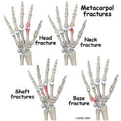
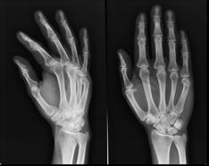
2 Really? Really! Musculoskeletal Objectives Ø Components in Interpretation Ø Normal Findings Ø Abnormal Findings Ø Common Pathology Ø Zebras Musculoskeletal Ø Classification and Names = Overwhelming!!! Ø Basics Ø Open vs. Closed Ø Location Ø Proximal Shaft Distal Ø Which bone Ø Fracture Type Ø Avulsion - Spiral Oblique - Transverse - Comminuted Ø Alignment Open Distal Radius Fx that is Comminuted and Dorsally angulated 2


3 Upper Extremity Ø Hand Ø Metacarpal Ø Phalanx Fractures Ø Dislocations Ø Wrist Ø Volar Ø Dorsal Ø Distal Radius Ø Carpal Fracture / Dislocation Ø Special Names Ø Elbow Ø Supracondylar Ø Condyles Ø Olecranon Ø Radial Head Ø Fracture / Dislocation Ø Shoulder Ø Clavicle Ø Proximal Humerus Ø Fracture / Dislocations Upper Extremity Imaging of the Hand 3

 Ø Wife")

4 History Ø First x-ray in 1895 Ø Wilhelm Roentgen ( ) Ø Wife s hand Know the Bones Shadows? Sesamoid Bones 4

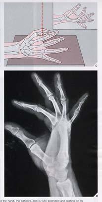
5 Basic Views Basic Views Posteroanterior (PA) view Basic Views Lateral View 5


6 Basic Views One view is no view! Basic Views Where s the fracture? Ø Cortical breaks Ø Radiolucent lines Fracture Concepts 6


7 Special Views Standard Pronated oblique view Special Views Supinated oblique view Special Views Supinated oblique view Arthritis & Fracture 7


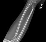
8 Joints Arthritis presents with: Ø Joint space narrowing Ø Sclerosis Ø Cysts Ø Osteophytes Joints Bone Density 8

9 Tuft Fracture 9

10 PIP Fx/Dislocations Metacarpal Fractures Boxer s Fracture 10

11 Carpal Fracture / Dislocations Ø Scaphoid Fractures Ø Perilunate dislocations Scaphoid Fracture Ø Can be occult! Ø Proximal Ø Middle Ø Distal Metacarpals capitate Lunate Radius Galulas Lines 11


12 Perilunate Dislocation Ø ~40% missed in ER Ø Can be devastating when missed Perilunate Dislocation Metacarpals Capitate Lunate Radius Galulas Lines 12


13 Most Common Missed Fracture? Diagnosis Distal Radius Fracture Ø Colles Ø Smith s Ø Barton s Ø Volar Ø Dorsal Ø Chauffeur Ø Galleazi 13


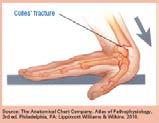
14 Distal Radius Bad vs. Not So Bad Colles Fracture Ø Distal Radius Fracture Ø Dorsal angulation 14


15 Smith s Fracture Ø Distal Radius Fracture Ø Volar angulation Barton s Fracture Ø Distal Radius fracture Ø Volar or Dorsal lip Chauffeur s Fracture Ø Radial styloid fracture 15


16 Galleazi Fracture Ø Radius Fracture Ø DRUJ dislocation Forearm Fractures Various Flavors! 16


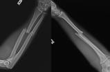
17 Elbow Fx/Dislocations Distal Humerus Fractures Ø Supracondylar Ø Medial & Lateral Condyle Ø Capitellum Supracondylar 17

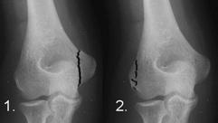
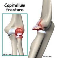
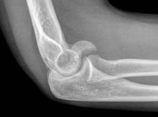
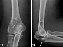
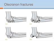
18 Medial & Lateral Condyle Capitellum Fracture Olecranon Fractures 18


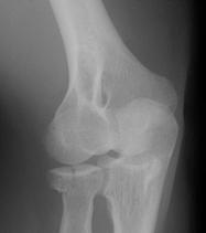
19 Radial Head Fractures Radial Head Fractures Redneck Power!! 19


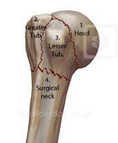
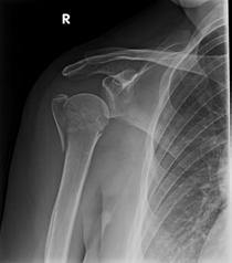
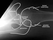
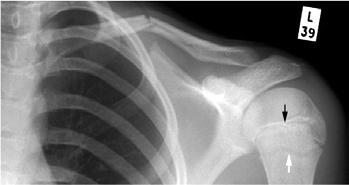
20 Shoulder! Proximal Humerus Axillary View 20
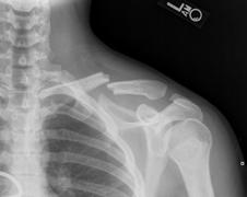
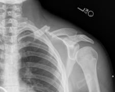
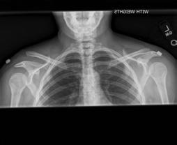
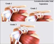

21 Clavicles Acromioclavicular Separation Upper Extremity Review 21

22 Lower Extremity Ø Pelvis & Hip Ø Hip Fractures Ø Rami Fractures Ø Knee Ø Supracondylar Ø Patella Ø Tibial Plateau Ø Tibial Spine Lower Extremity Ø Ankle Ø Malleolar Fractures Ø Maisonneuve Ø Foot Ø Lisfranc Injury 22


23 Hip Fractures Types of Fractures Femoral Neck Fx 23
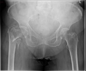
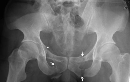
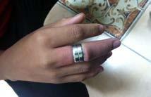
24 Intertrochanteric Fx Pubic Rami Fractures Distal Femur 24


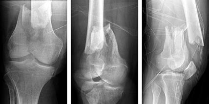
25 Distal Femur Patella Fractures Bipartite Patella 25

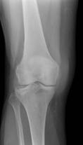
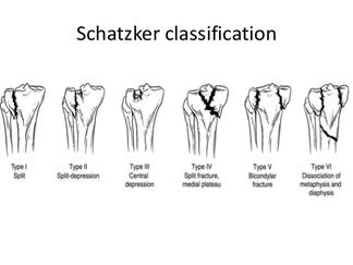
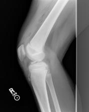
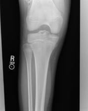
26 Tibial Plateau Schatzker Classification Tibial Spine 26

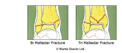
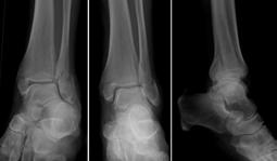
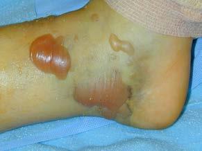

27 Malleolar Fractures Bimalleolar / Trimalleolar Fracture Blisters 27


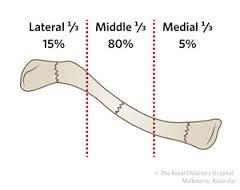
28 Maisonneuve Fracture Ø Spiral fracture of the proximal fibula with a tear of the syndesmosis Ø Associated fracture of the medial malleolus or rupture of the deltoid ligament Lisfranc Injury Ø Ligament that runs between medial cuneiform and 2nd metatarsal Pediatric MSK Imaging Ø Salter-Harris Classification Ø Growth plates / Ossification centers look weird!! Ø Immobilize if painful Ø Compare with contralateral side if necessary 28
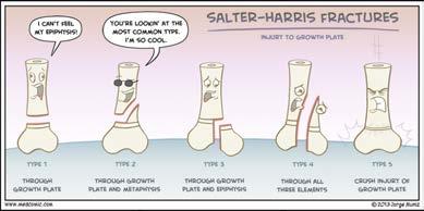
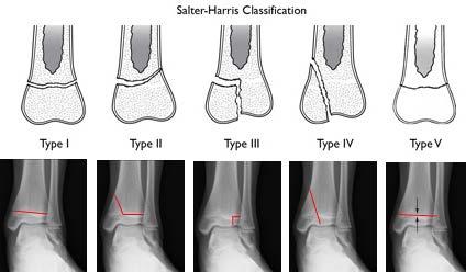
29 Salter-Harris Salter-Harris Pediatric Proximal Humerus 29


30 Pediatric Elbow Alignment Ø Fat Pads Ø Anterior Humeral Line Ø Radiocapitellar Line Advanced Imaging Ø Ultrasound Ø Bone Scan Ø CT Scan Ø MRI 30
 and")


31 Bone Scan Ø Technetium-labeled bone scintigraphy Ø High sensitivity, low specificity Ø Increased uptake indicates increased blood flow (immediate) and bone turnover (delayed phase) Ø Useful for detecting: Ø Osteomyelitis Ø CRPS CT Scan Ø Quick 3D view possible Ø Useful for detecting: Ø Suspected fractures not easily seen on X-ray Ø Assess union Ø Evaluate cortical integrity with bony tumors Ø Study fracture pattern and plan surgical approach Ø Expensive, Timely MRI Scan Ø Useful for detecting: Ø Soft Tissue Anatomy (mass/swelling) Ø Ligament injuries May use arthrography Ø Vascularity (Kienböck s, Scaphoid Non-union) 31

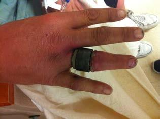
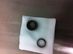
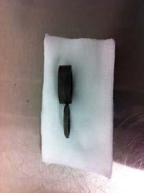
32 Other Modalities Removing a Ring 32

33 33

34 34
PEM GUIDE CHILDHOOD FRACTURES
 PEM GUIDE CHILDHOOD FRACTURES INTRODUCTION Skeletal injuries account for 10-15% of all injuries in children; 20% of those are fractures, 3 out of 4 fractures affect the physis or growth plate. Always consider
4/28/2010. Fractures. Normal Bone and Normal Ossification Bone Terms. Epiphysis Epiphyseal Plate (physis) Metaphysis
 Metaphysis") Fractures Normal Bone and Normal Ossification Bone Terms Epiphysis Epiphyseal Plate (physis) Metaphysis Diaphysis 1 Fracture Classifications A. Longitudinal B. Transverse C. Oblique D. Spiral E. Incomplete
Fractures Normal Bone and Normal Ossification Bone Terms Epiphysis Epiphyseal Plate (physis) Metaphysis Diaphysis 1 Fracture Classifications A. Longitudinal B. Transverse C. Oblique D. Spiral E. Incomplete
Index. Note: Page numbers of article titles are in boldface type.
 Note: Page numbers of article titles are in boldface type. A Abscess, epidural, 822 824 Achilles tendon rupture, 894 895, 981 982 Acromioclavicular separations, shoulder pain in, 751 753 Adhesive capsulitis,
Commonly Missed Injuries of the Extremities
 Commonly Missed Injuries of the Extremities Dr. Tudor H. Hughes M.D., FRCR Department of Radiology University of California School of Medicine San Diego, California 1. Base of skull 2. Odontoid process
Pediatric Fractures. Objectives. Epiphyseal Complex. Anatomy and Physiology. Ligaments. Bony matrix
 1 Pediatric Fractures Nicholas White, MD Assistant Professor of Pediatrics Eastern Virginia Medical School Attending, Pediatric Emergency Department Children s Hospital of The King s Daughters Objectives
Trauma Films for Upper Body. LCDR. Naruebade Rungrattanawilai RTN M.D., LL.B. FRCOST, DMOC
 Trauma Films for Upper Body LCDR. Naruebade Rungrattanawilai RTN M.D., LL.B. FRCOST, DMOC Objective A 42 year-old housekeeper with history of motorcycle accident. There was no external wound but she have
Appendicular skeleton: ABCs Image Interpretation Search strategy
 NOVEMBER 2013 volume 51 number 2 THE SOUTH AFRICAN RADIOGRAPHER peer reviewed ARTICLE OF INTEREST Appendicular skeleton: ABCs Image Interpretation Search strategy IJ Williams MSc in Medical Imaging; B
Orthopedic X-Rays most commonly missed
 Orthopedic X-Rays most commonly missed Vukiet Tran, MD, MHSc, MBA University Health Network Toronto, Canada 1 COI Disclosure I am the current Medical Director for Best Doctors Canada. Presenter: Dr. Vu
Appendicular Skeletal Trauma
 Appendicular Skeletal Trauma Dr. Tudor H. Hughes M.D., FRCR Department of Radiology University of California School of Medicine San Diego, California Types of cognitive error Satisfaction of search; Once
Surgical Care at the District Hospital. EMERGENCY & ESSENTIAL SURGICAL CARE
 Surgical Care at the District Hospital 1 18 Orthopedic Trauma Key Points 2 18.1 Upper Extremity Injuries Clavicle Fractures Diagnose fractures from the history and by physical examination Treat with a
Radiographic Positioning Summary (Basic Projections RAD 222)
") Lower Extremity Radiographic Positioning Summary (Basic Projections RAD 222) AP Pelvis AP Hip (Unilateral) (L or R) AP Femur Mid and distal AP Knee Lateral Knee Pt lies supine on table Align MSP to Center
Lower Extremity Radiographic Positioning Summary (Basic Projections RAD 222) AP Pelvis AP Hip (Unilateral) (L or R) AP Femur Mid and distal AP Knee Lateral Knee Pt lies supine on table Align MSP to Center
Index. Note: Page numbers of article titles are in boldface type.
 Note: Page numbers of article titles are in boldface type. A Acetabular fractures, 462 464 Achilles tendon rupture, 389 Acromioclavicular dislocations, 302 Acromion fractures, 301 Ankle, anatomy of, 376
The Appendicular Skeleton
 8 The Appendicular Skeleton PowerPoint Lecture Presentations prepared by Jason LaPres Lone Star College North Harris 8-1 The Pectoral Girdle The Pectoral Girdle Also called shoulder girdle Connects the
Trauma-related Pediatric Orthopedic Emergencies. Javier Gonzalez del Rey, M.D. Professor Pediatrics Cincinnati Children s Hospital Medical Center
 Trauma-related Pediatric Orthopedic Emergencies Javier Gonzalez del Rey, M.D. Professor Pediatrics Cincinnati Children s Hospital Medical Center Room # 10 7 month old sick since birth Room # 11 5 y/o Fell
10/12/2010. Upper Extremity. Pectoral (Shoulder) Girdle. Clavicle (collarbone) Skeletal System: Appendicular Skeleton
 Girdle. Clavicle (collarbone) Skeletal System: Appendicular Skeleton") Skeletal System: Appendicular Skeleton Pectoral girdle Pelvic girdle Upper limbs Lower limbs 8-1 Pectoral (Shoulder) Girdle Consists of scapula and clavicle Clavicle articulates with sternum (Sternoclavicular
Skeletal System: Appendicular Skeleton Pectoral girdle Pelvic girdle Upper limbs Lower limbs 8-1 Pectoral (Shoulder) Girdle Consists of scapula and clavicle Clavicle articulates with sternum (Sternoclavicular
Biology 218 Human Anatomy. Adapted from Martini Human Anatomy 7th ed. Chapter 7 The Skeletal System Appendicular Division
 Adapted from Martini Human Anatomy 7th ed. Chapter 7 The Skeletal System Appendicular Division Introduction The appendicular skeleton includes: Pectoral girdle Shoulder bones Upper limbs Pelvic girdle
Pectoral (Shoulder) Girdle
 Girdle") Chapter 8 Skeletal System: Appendicular Skeleton Pectoral girdle Pelvic girdle Upper limbs Lower limbs 8-1 Pectoral (Shoulder) Girdle Consists of scapula and clavicle Clavicle articulates with sternum
Chapter 8 Skeletal System: Appendicular Skeleton Pectoral girdle Pelvic girdle Upper limbs Lower limbs 8-1 Pectoral (Shoulder) Girdle Consists of scapula and clavicle Clavicle articulates with sternum
Basic Principles of Fractures & Easily Missed Fractures. Mr Irfan Merchant Trauma & Orthopaedic Registrar Bedford Hospital, East of England
 Basic Principles of Fractures & Easily Missed Fractures Mr Irfan Merchant Trauma & Orthopaedic Registrar Bedford Hospital, East of England Objectives Types Fracture Patterns Fracture Healing Assessing
PEDIATRIC CASTING AND SPLINTING HEATHER KONG, M.D. SHRINERS HOSPITAL FOR CHILDREN PORTLAND OCTOBER 7, 2017
 PEDIATRIC CASTING AND SPLINTING HEATHER KONG, M.D. SHRINERS HOSPITAL FOR CHILDREN PORTLAND OCTOBER 7, 2017 DISCLOSURES I have no financial relationship with any company or product discussed in this presentation.
Imaging the musculoskeletal system. An Introduction
 Imaging the musculoskeletal system An Introduction Objectives Discuss: commonly used imaging modalities in the musculoskeletal system normal imaging anatomy in the extremities fracture description Imaging
Montreal Children s Hospital McGill University Health Center Emergency Department Fracture Guideline
 Montreal Children s Hospital McGill University Health Center Emergency Department Guideline Disclaimers This document is designed to assist physicians working in our emergency department in caring for
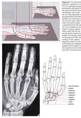
SKELETAL SYSTEM 206. AXIAL SKELETON 80 APPENDICULAR SKELETON 126 (see Figure 6.1) Clavicle. Clavicle. Pectoral girdles. Scapula. Scapula.
 Clavicle. Clavicle. Pectoral girdles. Scapula. Scapula.") SKELETAL SYSTEM 206 AXIAL SKELETON 80 APPENDICULAR SKELETON 126 (see Figure 6.1) Pectoral girdles 4 Clavicle Scapula 2 2 Clavicle Scapula Humerus 2 Humerus Upper limbs 60 Radius 2 Ulna Carpal bones Metacarpal
SKELETAL SYSTEM 206 AXIAL SKELETON 80 APPENDICULAR SKELETON 126 (see Figure 6.1) Pectoral girdles 4 Clavicle Scapula 2 2 Clavicle Scapula Humerus 2 Humerus Upper limbs 60 Radius 2 Ulna Carpal bones Metacarpal
Chapter 8B. The Skeletal System: Appendicular Skeleton. The Appendicular Skeleton. Clavicle. Pectoral (Shoulder) Girdle
 Girdle") The Appendicular Skeleton Chapter 8B The Skeletal System: Appendicular Skeleton 126 bones Pectoral (shoulder) girdle Pelvic (hip) girdle Upper limbs Lower limbs Functions primarily to facilitate movement
The Appendicular Skeleton Chapter 8B The Skeletal System: Appendicular Skeleton 126 bones Pectoral (shoulder) girdle Pelvic (hip) girdle Upper limbs Lower limbs Functions primarily to facilitate movement
Case. Case 8/29/ yo man with fever, cough. Vitals: Temp 102, HR 130, RR 20, bp 120/80. Ill appearing, crackles R side chest. Now what?
 Kate Aberger, MD August 28, 2016 Help from : Raphael Brancato DO, and Jordan Jeong DO Case 56 yo man with fever, cough Vitals: Temp 102, HR 130, RR 20, bp 120/80 Ill appearing, crackles R side chest Now
Chapter 8. The Appendicular Skeleton. Lecture Presentation by Lee Ann Frederick University of Texas at Arlington Pearson Education, Inc.
 Chapter 8 The Appendicular Skeleton Lecture Presentation by Lee Ann Frederick University of Texas at Arlington An Introduction to the Appendicular Skeleton The Appendicular Skeleton 126 bones Allows us
Chapter 8 The Skeletal System: The Appendicular Skeleton. Copyright 2009 John Wiley & Sons, Inc.
 Chapter 8 The Skeletal System: The Appendicular Skeleton Appendicular Skeleton It includes bones of the upper and lower limbs Girdles attach the limbs to the axial skeleton The pectoral girdle consists
ORTHOSCAN MOBILE DI POSITIONING GUIDE
 ORTHOSCAN MOBILE DI POSITIONING GUIDE Table of Contents SHOULDER A/P of Shoulder... 4 Tangential (Y-View) of Shoulder... 5 Lateral of Proximal Humerus... 6 ELBOW A/P of Elbow... 7 Extended Elbow... 8 Lateral
Biology 218 Human Anatomy
 Chapter 8 Adapted from Tortora 10 th ed. LECTURE OUTLINE A. Introduction (p. 203) 1. The appendicular skeleton contains 126 bones that form: i. two pectoral (shoulder) girdles two upper limbs i one pelvic
Hand and wrist emergencies
 Chapter1 Hand and wrist emergencies Carl A. Germann Distal radius and ulnar injuries PEARL: Fractures of the distal radius and ulna are the most common type of fractures in patients younger than 75 years.
EMERGENCY PITFALLS IN ORTHOPAEDIC TRAUMA. Thierry E. Benaroch, MD, FRCS MCH Trauma Rounds February 9, 2009
 EMERGENCY PITFALLS IN ORTHOPAEDIC TRAUMA Thierry E. Benaroch, MD, FRCS MCH Trauma Rounds February 9, 2009 MORAL OF THE STORY Fracture distal radius and intact ulna W/O radius fracture will most likely
Chapter 8. The Pectoral Girdle & Upper Limb
 Chapter 8 The Pectoral Girdle & Upper Limb Pectoral Girdle pectoral girdle (shoulder girdle) supports the arm consists of two on each side of the body // clavicle (collarbone) and scapula (shoulder blade)
Activity: Synopsis of Fractures and Dislocations. Approval Date: 3/1/2018. Termination Date: 2/29/2021
 Activity: Synopsis of Fractures and Dislocations Approval Date: 3/1/2018 Termination Date: 2/29/2021 Target Audience: All local physicians working in the fields of primary care, physical medicine and rehabilitation,
Lab Activity 9. Appendicular Skeleton Martini Chapter 8. Portland Community College BI 231
 Lab Activity 9 Appendicular Skeleton Martini Chapter 8 Portland Community College BI 231 Appendicular Skeleton Upper & Lower extremities Shoulder Girdle Pelvic Girdle 2 Humerus 3 Humerus: Proximal End
PRESENTED BY: JOHN STIMLER, DO, CPC, CHC, FACEP BSA HEALTHCARE AND BSA HEALTHCARE ADVISORY GROUP
 PRESENTED BY: JOHN STIMLER, DO, CPC, CHC, FACEP BSA HEALTHCARE AND BSA HEALTHCARE ADVISORY GROUP TOPICS (1) Fracture types ICD-10-CM diagnostic coding CPT procedure coding Fracture care treatments: Manipulated
Amy Warenda Czura, Ph.D. 1 SCCC BIO130 Lab 7 Appendicular Skeleton & Articulations
 The Skeletal System II: Appendicular Skeleton and Articulations Exercises 11, 13 (begins: page 145 in 9 th and 10 th editions) Exercises 10, 11 (begins: page 147 in 11 th edition, page 149 in 12 th edition)
The Appendicular Skeleton
 8 The Appendicular Skeleton PowerPoint Lecture Presentations prepared by Jason LaPres Lone Star College North Harris An Introduction to the Appendicular Skeleton Learning Outcomes 8-1 Identify the bones
radiologymasterclass.co.uk
 http://radiologymasterclass.co.uk Hip X-ray anatomy - Normal AP (anterior-posterior) Shenton's line is formed by the medial edge of the femoral neck and the inferior edge of the superior pubic ramus Loss
Upper Extremity Injury Management. Jonathan Pirie MD, Med, FRCPC, FAAP
 Upper Extremity Injury Management Jonathan Pirie MD, Med, FRCPC, FAAP Learning Objectives At the end of this session, you will be able to manage common fractures of the: 1. Humerus 2. Elbow 3. Forearm
Chapter XIX.1. Fractures May 2002
 Case Based Pediatrics For Medical Students and Residents Department of Pediatrics, University of Hawaii John A. Burns School of Medicine Chapter XIX.1. Fractures May 2002 Annemarie Uliasz The skeletal
Exercise Science Section 2: The Skeletal System
 Exercise Science Section 2: The Skeletal System An Introduction to Health and Physical Education Ted Temertzoglou Paul Challen ISBN 1-55077-132-9 Role of the Skeleton Protection Framework Attachments for
A. Incorrect! The appendicular skeleton includes bones of the shoulder, arm, hand, pelvis, leg and foot.
 Anatomy and Physiology - Problem Drill 08: The Skeletal System III No. 1 of 10 1. Which of the following statements about the appendicular skeleton is correct? A. The appendicular skeleton includes bones
Scaphoid Fractures. Mohammed Alasmari. Orthopaedic Surgery Demonstrator Majmaah University
 Scaphoid Fractures Mohammed Alasmari Orthopaedic Surgery Demonstrator Majmaah University 1 2 Scaphoid Fractures Introduction Anatomy History Clinical examination Radiographic evaluation Classification
Exercise 11. The Appendicular Skeleton
 Exercise 11 The Appendicular Skeleton The Appendicular Skeleton The appendicular skeleton contains 126 bones. Consists of the upper and lower limbs, the pectoral girdles, and the pelvic girdles. The pectoral
Appendicular Skeleton. Dr. Carmen E. Rexach Anatomy 35 Mt. San Antonio College
 Appendicular Skeleton Dr. Carmen E. Rexach Anatomy 35 Mt. San Antonio College Pectoral girdle clavicle scapula Upper limb brachium antebrachium carpus manus Pelvic girdle oscoxae Lower limb femoral region
Country Health SA Medical Imaging
 Country Health SA Medical Imaging REMOTE OPERATORS POSITIONING GUIDE Contents Image Evaluation Page 4 Positioning Guides Section 1 - THORAX 1.1 Chest Page 5 1.2 Bedside Chest Page 7 1.3 Ribs Page 8 Section
RADIOGRAPHY OF THE ANKLE and LOWER LEG
 RADIOGRAPHY OF THE ANKLE and LOWER LEG Patient Position: ANKLE AP Projection Part Position: True Slight to place foot s long axis Center to Central Ray: to IR Midway Note: Ankle joint is to tips of malleoli
Basic Care of Common Fractures Utku Kandemir, MD
 Basic Care of Common Fractures Utku Kandemir, MD Assistant Clinical Professor Trauma & Sports Medicine Dept. of Orthopaedic Surgery UCSF / SFGH History Physical Exam Radiology Treatment History Acute trauma
Top 10 Ortho Urgent Care Injuries. J.C. Clark, M.D. ORA Orthopedics
 Top 10 Ortho Urgent Care Injuries J.C. Clark, M.D. ORA Orthopedics 10. Proximal Humerus Fractures Treatment Simple sling ICE, pain meds Button-down shirts Recliner to sleep in It will be up to the surgeon
Upper Extremity Fractures
 Upper Extremity Fractures Ranie Whatley, RN,FNP-C David W. Gray, MD Skeletal Trauma 10 to 15 % of all Childhood Injuries Physeal (Growth Plate) Injuries are ~ 15% of all Skeletal Injuries Orthopaedic Assessment
Radiologic Pitfalls. Pelvis/ Hip Hip DL Femoral neck Another ring fracture Sacrum Acetabulum
 Radiologic Pitfalls Michelle Lin, MD UCSF Associate Professor of Clinical Emergency Medicine San Francisco General Hospital (Michelle.Lin@emergency.ucsf.edu) ERRORS IN RADIOGRAPH INTERPRETATION Commonly
Common. Common Hand Problems in Elite Athletes
 Common Hand Problems in Elite Athletes Fred Corley M.D. Dept. of Orthopaedic Surgery UTHSCSA I have no disclosures concerning this talk. The University of Texas Health Science Center @ San Antonio - Orthopaedics
Appendicular Skeleton. Prof. Abdulameer Al-Nuaimi
 Appendicular Skeleton Prof. Abdulameer Al-Nuaimi a.alnuaimi@sheffield.ac.uk abdulameerh@yahoo.com Hi Prof, It is great to hear from you, I really enjoyed your teaching last year. You taught me the hardest
NE Nebraska Trauma Conference Tristan Hartzell, MD November 8, 2017
 NE Nebraska Trauma Conference 2017 Tristan Hartzell, MD November 8, 2017 Traumatic arm injuries in the elderly Fractures Hand Wrist Elbow Shoulder Soft tissue injuries Definitions Elderly? old or aging
SYLLABUS. Required Text: Yochum TR, Essentials of Skeletal Radiology, 3 rd ed (Chapters 9 and 10). See Reading Assignments on page 8
. See Reading Assignments on page 8") SYLLABUS Name of Course: Length of Course: Course Description: RADIOLOGY II ACS 335/835 (lec/lab) 3 units, 53 hours This course is a continuation in the radiology diagnostic series; it is designed to reinforce
SYLLABUS Name of Course: Length of Course: Course Description: RADIOLOGY II ACS 335/835 (lec/lab) 3 units, 53 hours This course is a continuation in the radiology diagnostic series; it is designed to reinforce
Biology 152 Appendicular Skeleton Anatomy Objectives
 Biology 152 Appendicular Skeleton Anatomy Objectives We will learn proper bone names, left/right/medial, and the parts of bones in this exercise. Start by learning the names of the bones. As you gain comfort
PRE-LAB EXERCISES. Before we get started, look up the definitions of these common bone marking terms: Canal: Condyle: Facet: Fissure:
 1 PRE-LAB EXERCISES When studying the skeletal system, the bones are often sorted into two broad categories: the axial skeleton and the appendicular skeleton. This lab focuses on the appendicular skeleton,
Anatomy of the Musculoskeletal System
 Anatomy of the Musculoskeletal System Kyle E. Rarey, Ph.D. Department of Anatomy & Cell Biology and Otolaryngology University of Florida College of Medicine Outline of Presentation Vertebral Column Upper
Ouch, That s Gotta Hurt! Pediatric Fractures & Injuries
 Ouch, That s Gotta Hurt! Pediatric Fractures & Injuries Greg Canty, MD Medical Director, Sports Medicine Center Attending Physician, Emergency Medicine Children s Mercy Kansas City 2011 Children s Mercy
EPIPHYSEAL PLATE IN FEMUR
 Reviewing: Epiphyseal Plates (younger skeletons) eventually will disappear. Bones grow lengthwise up and down from each plate, and in a circular collar like fashion around the diaphysis. These plates will
11/4/2018 SUBTLETIES OF LOWER EXTREMITY TRAUMA IMAGING SPEAKER DISCLOSURES
 SUBTLETIES OF LOWER EXTREMITY TRAUMA IMAGING Charles S. Resnik, M.D. Professor of Radiology University of Maryland School of Medicine Upon completion of this presentation, participants will be better able
Chapter 8 The Skeletal System: The Appendicular Skeleton. Copyright 2009 John Wiley & Sons, Inc.
 Chapter 8 The Skeletal System: The Appendicular Skeleton Appendicular Skeleton The primary function is movement It includes bones of the upper and lower limbs Girdles attach the limbs to the axial skeleton
TRIQUETRUM FRACTURE. The triquetrum bone is one of the small bones that make up the carpus.
 TRIQUETRUM FRACTURE Introduction The triquetrum bone is one of the small bones that make up the carpus. It is also known as the triquetral bone, (and in the past the pyramidal or triangular bone) Triquetrum
Codes for internal or external fixation are to be used only when internal or external fixation is not already listed as part of the basic procedure.
 code it ALLOMATRIX Injectable Putty DBM Putty HPS Device odes HPS codes are developed and maintained by MS and are used to report items such as medical devices, implants, drugs and supplies. -codes are
Principles of Anatomy and Physiology
 Principles of Anatomy and Physiology 14 th Edition CHAPTER 8 The Skeletal System: The Appendicular Skeleton The Appendicular Skeleton The 126 bones of the appendicular skeleton are primarily concerned
MY PATIENT HAS KNEE PAIN. David Levi, MD Chief, Division of Musculoskeletal l limaging Atlantic Medical Imaging
 MY PATIENT HAS KNEE PAIN David Levi, MD Chief, Division of Musculoskeletal l limaging Atlantic Medical Imaging Causes of knee pain Non traumatic Trauma Osteoarthritis Patellofemoral pain Menisci or ligaments
Hands PA; Obl. Lat.; Norgaard s Thumb AP; Lat. PA. PA; Lat.: Obls.; Elongated PA with ulnar deviation
 Projections Region Basic projections Additional / Modified projections Upper Limbs Hands PA; Obl. Lat.; Norgaard s Thumb ; Lat. PA Fingers PA; Lat. Wrist PA; Lat. Obls. Scaphoid Lunate Trapezium Triquetral
Upper Extremity Page Lower Extremity Special Cases
 MSK MRI PROTOCOLS Contents Upper Extremity Shoulder Elbow Wrist Finger Thumb Lower Extremity Hip Pelvis Thigh Knee Lower Extremity/Shin Ankle Foot Special Cases Soft Tissue Mass Metal Protocol Page MSK
1:00 pm Welcome and Introduction Paul Tornetta III, MD and J. Tracy Watson, MD
 AAOS/OTA Daily Dilemmas in Trauma: Your Topics, Expert Solutions #3248 May 2-4, 2019 Orlando, FL Paul Tornetta III, MD and J. Tracy Watson, MD; Course Directors SCHEDULE THURSDAY, MAY 2 12:00 1:00 pm Registration/Exhibits
Common Limb Fractures. Mr Sheraz Malik MB BS MRCS Instructor Mr Paul Ofori-Atta Mb ChB FRCS President Motc Life UK April 2009
 Common Limb Fractures Mr Sheraz Malik MB BS MRCS Instructor Mr Paul Ofori-Atta Mb ChB FRCS President Motc Life UK April 2009 Objectives To be able to describe all characteristics of a fracture Describe
Radiology Positioning Practical Test #2 Table (By Jung Park):
:") Radiology Positioning Practical Test #2 Table (By Jung Park): (Lower Extremity): patient is fully gowned / no artifacts / properly shielded (exposure for femur and below : hold still, don t move ) (exposure
Radiology Positioning Practical Test #2 Table (By Jung Park): (Lower Extremity): patient is fully gowned / no artifacts / properly shielded (exposure for femur and below : hold still, don t move ) (exposure
Pediatric Elbow Radiology. Seema Awatramani, MD Friday, April 5, 2018 ACOEP Spring Seminar
 Pediatric Elbow Radiology Seema Awatramani, MD Friday, April 5, 2018 ACOEP Spring Seminar Disclosure I have no relevant financial relationships with the manufacturer(s) of any commercial product(s) and/or
St Mary Orthopaedic Conference. Steven A. Caruso, MD Trenton Orthopaedic Group Trauma and Complex Fracture Surgeon October 25, 2014
 St Mary Orthopaedic Conference Steven A. Caruso, MD Trenton Orthopaedic Group Trauma and Complex Fracture Surgeon October 25, 2014 Nothing to disclose Goals To discuss common orthopaedic pathologies and
Important Parts of Bones
 Important Parts of Bones For 2015 Know: Humerus (posterior) Clavical Femur (Anterior) Foot Hand Mandible Os Coxa Scapula Skull (Anterior, Inferior, Lateral) Sternum Humerus (posterior) A. olecranon fossa
RADIOGRAPHY OF THE WRIST
 RADIOGRAPHY OF THE WRIST Patient Position: WRIST PA Projection, elbow in same plane Part Position: Hand ; fingers centered to IR Central Ray: Structures Shown: NOTE: Optional AP projection best demonstrates
Tibia metaphysis fracture icd 9
 Tibia metaphysis fracture icd 9 The Borg System is 100 % Tibia metaphysis fracture icd 9 S82.244F is a billable/specific ICD-10-CM code that can be used to indicate a diagnosis for reimbursement purposes.
Lecture (10) Bone Fractures. Resources: - Lecture by dr.alboukai - Diagnostic imaging book
 Bone Fractures. Resources: - Lecture by dr.alboukai - Diagnostic imaging book") Lecture (10) Bone Fractures Hanan Alsalman Hanan Alrabiah Reem Aljurayyad Ayshah Almahboob Ghadeer Alwuhyad Khawlah AlOthman Dalal Alqadi Suliman Alshammari Maha AlKubaidan Rawabi Alghamdi Resources: -
Lecture (10) Bone Fractures Hanan Alsalman Hanan Alrabiah Reem Aljurayyad Ayshah Almahboob Ghadeer Alwuhyad Khawlah AlOthman Dalal Alqadi Suliman Alshammari Maha AlKubaidan Rawabi Alghamdi Resources: -
Upper Extremity Page Lower Extremity Special Cases
 MSK MRI PROTOCOLS Contents Upper Extremity Page Shoulder Elbow Wrist Finger Thumb Lower Extremity Hip Pelvis Thigh Knee Lower Extremity/Shin Ankle Foot Special Cases Soft Tissue Mass Metal Protocol MSK
Anatomy and Physiology 2016
 Anatomy and Physiology 2016 O = Temporal line I = coronoid process (Mandible) A = elevates mandible (chewing) O = galea aponeurotica (layer of dense fibrous tissue which covers the upper part of the cranium)
RADIOGRAPHY OF THE ELBOW & HUMERUS
 RADIOGRAPHY OF THE ELBOW & HUMERUS Patient Position: ELBOW AP Projection in same plane Part Position: Hand in ; patient Centered to Humeral epicondyles Central Ray: Structures Shown: AP Elbow Criteria
SNAP, CRACKLE, POP. Randy L Aldret, EdD, ATC, LAT Stephanie Aldret, DO, CAQSM OOA Winter CME Seminar January 26, 2018
 SNAP, CRACKLE, POP Randy L Aldret, EdD, ATC, LAT Stephanie Aldret, DO, CAQSM OOA Winter CME Seminar January 26, 2018 1 OBJECTIVES Review terms to describe fractures Identify fractures that can be treated
Episode 52 Commonly Missed Uncommon Orthopedic Injuries. Lisfranc Injuries. Drs. Ivy Cheng & Hossein Medhian. Prepared by Dr. Keerat Grewal, Oct 2014
 Prepared by Dr. Keerat Grewal, Oct 2014 Episode 52 Commonly Missed Uncommon Orthopedic Injuries Drs. Ivy Cheng & Hossein Medhian Lisfranc Injuries Q: What is a Lisfranc injury? Lisfranc injuries are a
SCAHPO-LUNATE DISSOCIATION
 SCAHPO-LUNATE DISSOCIATION Introduction Scapho-lunate dissociation is the most common significant ligamentous injury of the wrist. The condition is also sometimes referred to as rotary subluxation of the
Index. orthopedic.theclinics.com. Note: Page numbers of article titles are in boldface type.
 Index Note: Page numbers of article titles are in boldface type. A Acetabular fractures thromboembolic disease after, 341 Achilles tendon rupture ACL. See Anterior cruciate ligament (ACL) Adolescent idiopathic
Dr.Israa H. Mohsen. Lecture 5. The vertebral column
 Anatomy Lecture 5 Dr.Israa H. Mohsen The vertebral column The vertebral column a flexible structure consisting of 33 vertebrae holds the head and torso upright, serves as an attachment point for the legs,
Disclosures. Syndesmosis Injury. Syndesmosis Ligaments. Objectives. Mark M. Casillas, M.D.
 Disclosures Syndesmosis Injury No relevant disclosures Mark M. Casillas, M.D. 1 Objectives Syndesmosis Ligaments Understand the syndesmosis anatomy and function Classify syndesmosis injuries Describe treatment
bio4165 lab quiz 1 Posterior View Anterior View Lateral View Anterior View bio fall.quarter lab.quiz.1...page.1 of 6
 B A Posterior View D C E Lateral View bio.4165...fall.quarter.2005...lab.quiz.1...page.1 of 6 F I G 35 Posterior View H bio.4165...fall.quarter.2005...lab.quiz.1...page.2 of 6 J Posterior View L K Inferior
Fractures and dislocations around elbow in adult
 Lec: 3 Fractures and dislocations around elbow in adult These include fractures of distal humerus, fracture of the capitulum, fracture of the radial head, fracture of the olecranon & dislocation of the
AO SEC Course on Nonoperative Fracture Treatment. June 17 19, 2010 University Teaching Hospital, Lusaka/Zambia
 AO SEC Course on Nonoperative Fracture Treatment June 17 19, 2010 University Teaching Hospital, Lusaka/Zambia 2 AO Foundation Socio Economic Committee AO Foundation Socio Economic Committee 3 A word of
CLINICAL PRESENTATION AND RADIOLOGY QUIZ QUESTION
 Donald L. Renfrew, MD Radiology Associates of the Fox Valley, 333 N. Commercial Street, Suite 100, Neenah, WI 54956 10/13/2012 Radiology Quiz of the Week # 94 Page 1 CLINICAL PRESENTATION AND RADIOLOGY
Orthopaedische valkuilen op de spoedgevallendienst. Journal Club 28 januari 2016 Spoedgevallendienst UZ - Gent
 Orthopaedische valkuilen op de spoedgevallendienst Journal Club 28 januari 2016 Spoedgevallendienst UZ - Gent 2 - Orthopedic Pearls and Pitfalls, Carl Menckhoff, MD Department of Emergency Medicine, Georgia
The skeleton consists of: Bones: special connective tissue, hard. Cartilage: special connective tissue, less hard than bones. Joints: joint is the
 The skeleton consists of: Bones: special connective tissue, hard. Cartilage: special connective tissue, less hard than bones. Joints: joint is the location at witch two bones make contact, whereas ligaments
Upper Extremity Trauma.
 Upper Extremity Trauma www.fisiokinesiterapia.biz Topics Clavicle Shoulder Dislocation Humerus Elbow Forearm Distal Radius Clavicle Fractures Clavicle Fractures Mechanism Fall onto shoulder (87%) Direct
What you don t want to miss
 March 25, 2009 Vishal Michael Shah, M.D. What you don t want to miss Spectrum of Injuries Contusions Sprains Dislocations Fractures Lacerations Tendon Avulsions Ligament Tears Overuse Injuries FINGER
Anatomy and Physiology II. Review Shoulder Girdle New Material Upper Extremities - Bones
 Anatomy and Physiology II Review Shoulder Girdle New Material Upper Extremities - Bones Anatomy and Physiology II Shoulder Girdle Review Questions From Last Lecture Can you identify the following muscles?
Mayo Clinic Disorders of the Wrist
 Mayo Clinic Disorders of the Wrist Thursday, May 19, 2016 Pre-Conference Laboratory Workshop Anatomy of the Wrist & Wrist Arthroscopy 6:30 a.m. Registration and Breakfast 7:30 a.m. Welcome and Introduction
Sick Call Screener Course
 Sick Call Screener Course Musculoskeletal System Upper Extremities (2.7) 2.7-2-1 Enabling Objectives 1.46 Utilize the knowledge of musculoskeletal system anatomy while assessing a patient with a musculoskeletal
Ligaments of Elbow hinge: sagittal plane so need lateral and medial ligaments
 Ligaments of Elbow hinge: sagittal plane so need lateral and medial ligaments Ulnar Collateral ligament on medial side; arising from medial epicondyle and stops excess valgus movement (lateral movement)
Multiple Choice Identify the letter of the choice that best completes the statement or answers the question.
 RA202 positioning class three- EXM Multiple Choice Identify the letter of the choice that best completes the statement or answers the question. 1. Which of the following hand projections would be used
Contents SECTION 1: GENERAL TRAUMA AND RECONSTRUCTIVE HIP SURGERY
 SECTION 1: GENERAL TRAUMA AND RECONSTRUCTIVE HIP SURGERY 1. Acetabular and Pelvic Fractures...3 2. Acetabular Orientation (Total Hips)...6 3. Acetabular Osteotomy...7 4. Achilles Tendon Ruptures...9 5.