Abdominal Ultrasound : Aorta, Kidneys, Bladder
|
|
|
- Jayson Nicholson
- 5 years ago
- Views:
Transcription

1 Abdominal Ultrasound : Aorta, Kidneys, Bladder Nilam J. Soni, MD, MSc Associate Professor of Medicine Divisions of Hospital Medicine and Pulmonary/Critical Care Medicine Department of Medicine University of Texas Health Science Center San Antonio
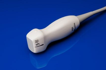
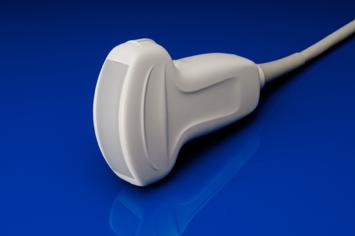
2 Low-frequency Transducers Phased-array Curvilinear
3 Anatomy of Aorta From Point-of-Care Ultrasound, 1 st edition, 2014
4 Image Acquisition Transverse Longitudinal
5 Transverse Longitudinal
6 Complete Exam of Aorta From Point-of-Care Ultrasound, 1 st edition, 2014

7 Celiac Trunk SMA From Point-of-Care Ultrasound, 1 st edition, 2014

8 Celiac Trunk Seagull Sign Common Hepatic Artery Splenic Artery Celiac Trunk
9 Celiac Trunk Seagull Sign Common Hepatic Artery Splenic Artery Celiac Trunk
10 Proximal Aorta Splenic Vein Celiac Trunk SMA Aorta From Point-of-Care Ultrasound, 1 st edition, 2014
11 Proximal Aorta From Point-of-Care Ultrasound, 1 st edition, 2014

12 Mid-Abdominal Aorta Splenic Vein SMA Au IVC Renal Arteries Vertebral Body From Point-of-Care Ultrasound, 1 st edition, 2014

13 Proximal Aorta
14 Distal Aorta Right and Left Common Iliac Arteries From Point-of-Care Ultrasound, 1 st edition, 2014
15 Distal Aorta From Point-of-Care Ultrasound, 1 st edition, 2014
16 Abdominal Aortic Aneurysm?

17 Abdominal Aortic Aneurysm?
18 Measurement of Aortic Diameter Measure outer wall to outer wall in both transverse and longitudinal views Normal < 3cm proximally (<2cm distally) Diameter >4.5cm referral to vascular surgery
19 AAA? From Point-of-Care Ultrasound, 1 st edition, 2014

20 AAA? From Point-of-Care Ultrasound, 1 st edition, 2014
 Measure in transverse and longitudinal planes")
21 Aorta Pearls Bowel gas limits visualization NPO Firm pressure or sweep transducer Types of AAA Fusiform Saccular (Pseudoaneurysm) Measure in transverse and longitudinal planes


22 Kidney Anatomy 2 Anatomical Sections Sonographic Double Density Renal Parenchyma = Cortex + Medulla Renal Sinus = Fatty tissue + Calyces + Vessels

23 Image Acquisition From Point-of-Care Ultrasound, 1 st edition, 2014
 and vessels 5.")
24 Normal Kidneys 1. Perinephric fat + Gerona's fascia = hyperechoic 2. Renal cortex = hypoechoic 3. Medullary pyramids = hypo-/anechoic 4. Renal sinus = hyperechoic with small pockets of urine (not contiguous) and vessels 5. Ureter normally obscured by bowel gas
25 Normal
26 Atrophy Hydronephrosis Stone Cysts Mass Renal Pathologies

27 Atrophic Kidney

28 Hydronephrosis Mild = central dilation with preservation of renal pyramids Moderate = blunting of renal pyramids, rounding of calices, bear-claw appearance, preservation of cortex Severe = cortical thinning, calyceal ballooning, distortion of architecture From Point-of-Care Ultrasound, 1 st edition, 2014
29 Mild Hydronephrosis From Point-of-Care Ultrasound, 1 st edition, 2014

30 Severe Hydronephrosis From Point-of-Care Ultrasound, 1 st edition, 2014
31 Bladder Indications Estimate bladder volume Confirm catheter placement Ureteral obstruction (ureteral jets) Detect stones Work-up for renal failure

32 Posterior and inferior to symphysis pubis (tilt US beam into pelvis) Ureters enter trigone on postero-inferior wall Prostate normally <5cm transversely Bladder Anatomy From Point-of-Care Ultrasound, 1 st edition, 2014
33 Image Acquisition Transverse View Longitudinal View From Point-of-Care Ultrasound, 1 st edition, 2014

34 Normal Male Bladder Transverse View
35 Foley + Decompressed Bladder

36 Distended Bladder + Foley + Enlarged Prostate

37 Bladder Volume Estimation Volume (ml) = 0.75 x width x length x height Transverse View Longitudinal View


38 Ultrasound-guided Lumbar Puncture Nilam J. Soni, MD, MSc Associate Professor of Medicine Divisions of General & Hospital Medicine / Pulmonary & Critical Care Medicine University of Texas Health San Antonio

39 Anatomy Lumbar Spine -Spinal cord ends at L 1 -L 2 in adults -Lumbar puncture can be safely performed below L 2 L1 L1 L2 L3 L4 L4 L5 L5
40 Lumbar Spine Transverse Plane Longitudinal Plane Spinous Process Spinous Process Interspinous Space Spinous Process

41 Lumbar Puncture Mapping Transverse Plane
42 Midline Shadow = Spinous Process L 3 L 4 Transverse Plane

43 Lumbar Puncture Site Mapping Longitudinal Plane

44 Lumbar Puncture Mapping

45 L5 = Deep and Small

46 Lumbar Puncture Mapping Mark 2 widest interspinous spaces Patient must remain in position Higher success in sitting position

47 Lumbar Spine

48 Lumbar Spine PITFALL: Lamina / articular processes can be mistaken for spinous processes No muscle should be seen superficial to spinous processes Erector spinae muscles Lamina / articular processes Paramedian View
49 Lumbar Spine

50 Lumbar Spine Paramedian View
51 Questions?
My Patient Has Abdominal Pain PoCUS of the Biliary Tract and the Urinary Tract
 My Patient Has Abdominal Pain PoCUS of the Biliary Tract and the Urinary Tract Objectives PoCUS for Biliary Disease PoCUS for Renal Colic PoCUS for Urinary Retention Biliary Disease A patient presents
Abdomen and Retroperitoneum Ultrasound Protocols
 Abdomen and Retroperitoneum Ultrasound Protocols Reviewed By: Anna Ellermeier, MD Last Reviewed: March 2018 Contact: (866) 761-4200, Option 1 **NOTE for all examinations: 1. If documenting possible flow
Abdominal Ultrasonography
 Abdominal Ultrasonography David A. Masneri, DO, FACEP, FAAEM Assistant Professor of Emergency Medicine Assistant Director, Emergency Medicine Residency Medical Director, Operational Medicine Division Center
Basic of Ultrasound Physics E FAST & Renal Examination. Dr Muhammad Umer Ihsan MBBS,MD, DCH CCPU,DDU1,FACEM
 Basic of Ultrasound Physics E FAST & Renal Examination Dr Muhammad Umer Ihsan MBBS,MD, DCH CCPU,DDU1,FACEM What is Sound? Sound is Mechanical pressure waves What is Ultrasound? Ultrasounds are sound waves
Abdominal Ultrasound. Diane Hallinen, MD. Bloodroot
 Abdominal Ultrasound Diane Hallinen, MD Bloodroot Abdominal Ultrasound Vasculature Hepatobiliary Spleen Kidney Bladder Bowel Where to put the probe? Vasculature We are going to talk about Celiac Trunk
ANATOMY OF PELVICAYCEAL SYSTEM -DR. RAHUL BEVARA
 1 ANATOMY OF PELVICAYCEAL SYSTEM -DR. RAHUL BEVARA 2 KIDNEY:ANATOMY OVERVIEW Kidneys are retroperitoneal, in posterior abdominal region, extending from T12 L3 Bean-shaped Right kidney is lower than left
US in non-traumatic acute abdomen. Lalita, M.D. Radiologist Department of radiology Faculty of Medicine ChiangMai university
 US in non-traumatic acute abdomen Lalita, M.D. Radiologist Department of radiology Faculty of Medicine ChiangMai university Sagittal Orientation Transverse (Axial) Orientation Coronal Orientation Intercostal
Abdominal Ultrasound
 Abdominal Ultrasound Imaging Control Buttons Depth The organ imaged should take up 3/4 of the screen Frequency = Penetration Use high frequencies (harmonics) for fluid filled and superficial structures
Guidelines, Policies and Statements D5 Statement on Abdominal Scanning
 Guidelines, Policies and Statements D5 Statement on Abdominal Scanning Disclaimer and Copyright The ASUM Standards of Practice Board have made every effort to ensure that this Guideline/Policy/Statement
Excretory urography (EU) or IVP US CT & radionuclide imaging
 or IVP US CT & radionuclide imaging") Excretory urography (EU) or IVP US CT & radionuclide imaging MRI arteriography studies requiring catherization or direct puncture of collecting system EU & to a lesser extent CT provide both functional
Excretory urography (EU) or IVP US CT & radionuclide imaging MRI arteriography studies requiring catherization or direct puncture of collecting system EU & to a lesser extent CT provide both functional
Point-of-Care Ultrasound Guide for Landmarks, Recording, and Report Content. TJUH/MHD EM Ultrasound Division 2012
 Point-of-Care Ultrasound Guide for Landmarks, Recording, and Report Content TJUH/MHD EM Ultrasound Division 2012 Table of Contents 1 - Objectives 2 - Procedural 3 - AAA 4 - Abdominal OB 5 - Transvaginal
Certificate in Clinician Performed Ultrasound (CCPU) Syllabus. Renal Hydronephrosis & Calculi
 Syllabus. Renal Hydronephrosis & Calculi") Certificate in Clinician Performed Ultrasound (CCPU) Syllabus Renal Hydronephrosis & Calculi Page 1 of 6 01/17 Renal Hydronephrosis and Calculi Syllabus Purpose: This unit is designed to cover the theoretical
Certificate in Clinician Performed Ultrasound (CCPU) Syllabus Renal Hydronephrosis & Calculi Page 1 of 6 01/17 Renal Hydronephrosis and Calculi Syllabus Purpose: This unit is designed to cover the theoretical
L o o k L i s t e n F e e l S c a n. Your Pocus Cards For Your Every Day Scanning.
 L o o k L i s t e n F e e l S c a n Your Pocus Cards For Your Every Day Scanning E-FAST Extended Focused Assessment by Sonography in Trauma Subcostal Heart View Pleural Sliding on M-mode (Sea-shore sign)
Chapter 6: Genitourinary and Gastrointestinal Systems 93
 Chapter 6: Genitourinary and Gastrointestinal Systems 93 Chapter 6 Genitourinary and Gastrointestinal Systems Embryology Three sets of excretory organs or kidneys develop in human embryos: Pronephros:
URINARY SYSTEM I. Kidneys II. Nephron Unit and Urine Formation
 URINARY SYSTEM I. Kidneys A. Location and Structure 1. Retroperitoneal 2. Between T12 and L3 3. Rt. kidney slightly lower 4. Two bean shaped organs 5. Adrenal gland 6. Internal construction a. Renal cortex
Abdominal ultrasound:
 Abdominal ultrasound: Non-traumatic acute abdomen Wittanee Na-ChiangMai, MD Department of Radiology ChiangMai University 26/04/2017 Contents Technique of examination Normal anatomy Emergency conditions
Anatomy of the renal system. Professor Nawfal K. Al-Hadithi
 Anatomy of the renal system Professor Nawfal K. Al-Hadithi Objectives To describe the posterior abdominal wall To identify the main anatomical landmarks of the kidneys & ureters To describe the suprarenal
The Kidneys. (L., ren; Gk, nephros; hence the adjectives renal and nephric) & Suprarenal (Adrenal) Glands. Dr Maan Al-Abbasi PhD, MBChB
 & Suprarenal (Adrenal) Glands. Dr Maan Al-Abbasi PhD, MBChB") The Kidneys (L., ren; Gk, nephros; hence the adjectives renal and nephric) & Suprarenal (Adrenal) Glands Dr Maan Al-Abbasi PhD, MBChB Functions of Urinary System Regulate electrolytes (K+, Na+, etc) Regulate
The Kidneys (L., ren; Gk, nephros; hence the adjectives renal and nephric) & Suprarenal (Adrenal) Glands Dr Maan Al-Abbasi PhD, MBChB Functions of Urinary System Regulate electrolytes (K+, Na+, etc) Regulate
Q129. Which of the following is NOT true about lymph node?
 Q129. Which of the following is NOT true about lymph node? (1). Normal lymph node is not seen in the ultrasound image (2). It is general that high frequency probe is used due to normal lymph node is located
ISUOG Basic Training. Distinguishing between Normal & Abnormal Appearances of the Urinary Tract. Seshadri Suresh, India
 ISUOG Basic Training Distinguishing between Normal & Abnormal Appearances of the Urinary Tract Seshadri Suresh, India Learning objectives 13 & 14 At the end of the lecture you will be able to: describe
Basic Abdominal Sonography
 24S Basic Abdominal Sonography Procedural Overview JOHN FATCHETT II, RDMS is provided. Patient preparation (i.e., fasting) scanning techniques, spleen, transducer. evaluation of abdominal anatomy in the
The Urinary System Pearson Education, Inc.
 26 The Urinary System Introduction The urinary system does more than just get rid of liquid waste. It also: Regulates plasma ion concentrations Regulates blood volume and blood pressure Stabilizes blood
Fetal Renal Malformations: The Role of Ultrasound in Diagnosis & Management
 Fetal Renal Malformations: The Role of Ultrasound in Diagnosis & Management 12 weeks Alfred Abuhamad, M.D. Eastern Virginia Medical School 13 weeks 2nd trimester Medullary pyramids Renal Sinus Cortex 2nd
Abdominal Aortic Aneurysm (AAA)
") Abdominal Aortic Aneurysm (AAA) Vascular Workshop: Objectives Anatomy Keith VanHaltren Indications Technique Cases Abdominal Aorta: Normal Size Abdominal aortic aneurysm: Definition Normal diameter of
Abdominal Aortic Aneurysm (AAA) Vascular Workshop: Objectives Anatomy Keith VanHaltren Indications Technique Cases Abdominal Aorta: Normal Size Abdominal aortic aneurysm: Definition Normal diameter of
Policies, Standards, and Guidelines. Guidelines for Abdominal Ultrasound Examination
 Policies, Standards, and Guidelines Guidelines for Abdominal Ultrasound Examination Approved by Council Feb 2018 Disclaimer and Copyright The ASUM Standards of Practice Board have made every effort to
Gross Anatomy of the Urinary System
 Gross Anatomy of the Urinary System Lecture Objectives Overview of the urinary system. Describe the external and internal anatomical structure of the kidney. Describe the anatomical structure of the ureter
Background & Indications Probe Selection
 Teresa S. Wu, MD, FACEP Director, EM Ultrasound Program & Fellowship Co-Director, Simulation Based Training Program & Fellowship Associate Program Director, EM Residency Program Maricopa Medical Center
Urinary System VASTACCESS, INC.
 Urinary System www.vastaccess.com 2 Urinary Tract Kidney Ureter Urinary Bladder Urethra Prostate (male) Membranous (male) Spongy (male) 3 Kidney Relations Suprarenal (Adrenal) Glands Liver Duodenum Transverse
Urinary system Ultrasound (Renal & Urinary bladder)
") Urinary system Ultrasound (Renal & Urinary bladder) Edited & Presented by ; Hussien A.B ALI DINAR. Msc.Phd ISRRT Associate Member Lecturer (National university) Reporting Sonographer (PHC) Objective By
Urinary system Ultrasound (Renal & Urinary bladder) Edited & Presented by ; Hussien A.B ALI DINAR. Msc.Phd ISRRT Associate Member Lecturer (National university) Reporting Sonographer (PHC) Objective By
CHAPTER 10: ULTRASOUND NEURAXIS & POST ABDOMINAL WALL
 CHAPTER 10: ULTRASOUND NEURAXIS & POST ABDOMINAL WALL Introduction 197 Transverse scans 198 Spinous process and laminas... 198 Spinal canal and facet joints 198 Transverse process... 199 The acoustic window
Focused Assessment Sonography of Trauma (FAST) Scanning Protocol
 Scanning Protocol") Focused Assessment Sonography of Trauma (FAST) Scanning Protocol Romolo Gaspari CHAPTER 3 GOAL OF THE FAST EXAM Demonstrate free fluid in abdomen, pleural space, or pericardial space. EMERGENCY ULTRASOUND
Focused Assessment Sonography of Trauma (FAST) Scanning Protocol Romolo Gaspari CHAPTER 3 GOAL OF THE FAST EXAM Demonstrate free fluid in abdomen, pleural space, or pericardial space. EMERGENCY ULTRASOUND
Figure 26.1 An Introduction to the Urinary System
 Chapter 26 Figure 26.1 An Introduction to the Urinary System Components of the Urinary System Kidney Produces urine Ureter Transports urine toward the urinary bladder Urinary Bladder Temporarily stores
Abdominal Aortic Aneurysm. Ultrasound Logbook. Name
 Abdominal Aortic Aneurysm Ultrasound Logbook ame Contents AAA Accreditation Requirements 15 Abdominal Aorta Report Records 3 Formative Assessments 1 Summative Assessment AAA Accreditation Abdominal Aortic
Obstetrics Content Outline Obstetrics - Fetal Abnormalities
 Obstetrics Content Outline Obstetrics - Fetal Abnormalities Effective February 2007 10 16% renal agenesis complete absence of the kidneys occurs when ureteric buds fail to develop Or degenerate before
URINARY SYSTEM ANATOMY
 URINARY SYSTEM ANATOMY Adapted from Human Anatomy & Physiology Marieb and Hoehn (9 th ed.) OVERVIEW Metabolism of nutrients by the body produces wastes that must be removed from the body. Although excretory
Role of imaging in evaluation of genitourinary i trauma Spectrum of GU injuries Relevance of imaging findings in determining management Focus on MDCT
 Genitourinary Tract Injuries 6 th Nordic Course Scott D. Steenburg, MD Assistant Professor University of Maryland Department of Radiology Division of Trauma and Emergency Radiology R Adams Cowley Shock
The functional anatomy of the urinary system. Human Anatomy Department Dr. Anastasia Bendelic
 The functional anatomy of the urinary system Human Anatomy Department Dr. Anastasia Bendelic Plan Development of the kidneys and their abnormalities Development of the urinary ways and their abnormalities
1 Normal Anatomy and Variants
 1 Normal Anatomy and Variants 1.1 Normal Anatomy MR Technique. e standard MR protocol for a routine evaluation of the spine always comprises imaging in sagittal and axial planes, while coronal images are
Urinary System Laboratory
 Urinary System Laboratory 1 Adrenal gland Organs of The Urinary System Renal artery and vein Kidney Ureter Urinary bladder Figure 26.1 2 Urethra Functions of the urinary system organs: Urethra expels urine
Lecture 56 Kidney and Urinary System
 Lecture 56 Kidney and Urinary System The adrenal glands are located on the superomedial aspect of the kidney The right diagram shows a picture of the kidney with the abdominal walls and organs removed
ASSESSING THE PLAIN ABDOMINAL RADIOGRAPH M A A M E F O S U A A M P O F O
 ASSESSING THE PLAIN ABDOMINAL RADIOGRAPH M A A M E F O S U A A M P O F O Introduction The abdomen (less formally called the belly, stomach, is that part of the body between the thorax (chest) and pelvis,
The posterior abdominal wall. Prof. Oluwadiya KS
 The posterior abdominal wall Prof. Oluwadiya KS www.oluwadiya.sitesled.com Posterior Abdominal Wall Lumbar vertebrae and discs. Muscles opsoas, quadratus lumborum, iliacus, transverse, abdominal wall
FHS Appendicitis US Protocol
 FHS Appendicitis US Protocol Reviewed By: Shireen Khan, MD; Sarah Farley, MD; Anna Ellermeier, MD Last Reviewed: May 2018 Contact: (866) 761-4200 **NOTE for all examinations: 1. If documenting possible
B) cervix of uterus C) vagina D) rectum. 1. What number illustrates the adnexal area? (Fig. 4-64) A) 4 B) 5 C) 8 D) 9
 cervix of uterus C) vagina D) rectum. 1. What number illustrates the adnexal area? (Fig. 4-64) A) 4 B) 5 C) 8 D) 9") Pelvis Practice Problems 1. What number illustrates the adnexal area? (Fig. 4-64) A) 4 B) 5 C) 8 D) 9 2. What number illustrates the cervix? (Fig. 4-64) A) 4 B) 8 C) 5 D) 6 3. Which of the following is
Pelvis Practice Problems 1. What number illustrates the adnexal area? (Fig. 4-64) A) 4 B) 5 C) 8 D) 9 2. What number illustrates the cervix? (Fig. 4-64) A) 4 B) 8 C) 5 D) 6 3. Which of the following is
PAH EMERGENCY Department Aortic Scanning module
 PAH EMERGENCY Department Aortic Scanning module Abdominal Aorta Scan is a goal directed ultrasound examination to detect the presence of an Abdominal Aortic Aneurysm (AAA). The Abdominal Aorta scan is
Chapter 26. The Urinary System. Lecture Presentation by Steven Bassett Southeast Community College Pearson Education, Inc.
 Chapter 26 The Urinary System Lecture Presentation by Steven Bassett Southeast Community College Introduction The urinary system does more than just get rid of liquid waste. It also: Regulates plasma ion
Abdomen and Pelvis CT (1) By the end of the lecture students should be able to:
 By the end of the lecture students should be able to:") RAD 451 Abdomen and Pelvis CT (1) By the end of the lecture students should be able to: State the common indications for Abdomen and pelvis CT exams Identify possible contra indications for Abdomen and
RAD 451 Abdomen and Pelvis CT (1) By the end of the lecture students should be able to: State the common indications for Abdomen and pelvis CT exams Identify possible contra indications for Abdomen and
Kidney & Urinary Tract Ultrasound. Fatina Fadel Hafez Bazaraa
 Kidney & Urinary Tract Ultrasound Fatina Fadel Hafez Bazaraa Ultrasonography Ultrasound Available Rapid Inexpensive Painless & no sedation needed No adverse effects/ complications Can be repeated Useful
Clarification of Terms
 Clarification of Terms The Spine, Spinal Column, and Vertebral Column are synonymous terms referring to the bony components housing the spinal cord Spinal Cord = made of nervous tissue Facet = a small,
Uroradiology Tutorial For Medical Students
 Uroradiology Tutorial For Medical Students Lesson 3: Cystography & Urethrography Part 1 American Urological Association Introduction Conventional radiography of the urinary tract includes several diagnostic
Clarification of Terms
 Clarification of Terms The Spine, Spinal Column, and Vertebral Column are synonymous terms referring to the bony components housing the spinal cord Spinal Cord = made of nervous tissue Facet = a small,
Urinary System. Chapter 17 7/19/11. Introduction
 7/19/11 Chapter 17 Urinary System Introduction A. The urinary system consists of two kidneys that filter the blood, two ureters, a urinary bladder, and a urethra to convey waste substances to the outside.
Transducer Selection. Renal Artery Duplex Exam. Renal Scan. Renal Scan Echogenicity. How to Perform an Optimal Renal Artery Doppler Examination
 How to Perform an Optimal Renal Artery Doppler Examination Director of Ultrasound Education & Quality Assurance Baylor College of Medicine Division of Maternal-Fetal Medicine Maternal Fetal Center Imaging
GI module Lecture: 9 د. عصام طارق. Objectives:
 GI module Lecture: 9 د. عصام طارق Objectives: To list structures forming posterior abdominal wall. To follow aorta & its main branches. To describe IVC & its main tributaries. To list nerves of posterior
Clarification of Terms
 Clarification of Terms The Spine, Spinal Column, and Vertebral Column are synonymous terms referring to the bony components housing the spinal cord Spinal Cord = made of nervous tissue Facet = a small,
1 Right & left Hepatic ducts Gastric Impression of spleen
 Pancreatic Model 1 Right & left Hepatic ducts 14 Gastric Impression of spleen 2 Common hepatic duct 15 Renal Impression of spleen 3 Cystic Duct 16 Colic Impression of spleen 4 Common Bile Duct 17 Splenic
Proceedings of the 34th World Small Animal Veterinary Congress WSAVA 2009
 www.ivis.org Proceedings of the 34th World Small Animal Veterinary Congress WSAVA 2009 São Paulo, Brazil - 2009 Next WSAVA Congress : Reprinted in IVIS with the permission of the Congress Organizers IMAGING
Diagnostic Imaging
 www.fisiokinesiterapia.biz Diagnostic Imaging Diagnostic Imaging is no longer limited to radiography. Major technological advancements have lead to the use of new and improved imaging technologies. The
CYSTIC DISEASES of THE KIDNEY. Dr. Nisreen Abu Shahin
 CYSTIC DISEASES of THE KIDNEY Dr. Nisreen Abu Shahin 1 Types of cysts 1-Simple Cysts 2-Dialysis-associated acquired cysts 3-Autosomal Dominant (Adult) Polycystic Kidney Disease 4-Autosomal Recessive (Childhood)
General Anatomy of Urinary System
 General Anatomy of Urinary System URINARY SYSTEM ORGANS Kidneys (2) Ureters (2) Urinary bladder Urethra KIDNEY FUNCTIONS Control blood volume and composition KIDNEY FUNCTIONS Filter blood plasma, eliminate
URINARY SYSTEM ANATOMY PART
 URINARY SYSTEM ANATOMY PART 1 DANIL HAMMOUDI.MD Urinary System Composed of kidneys, ureters, urinary bladder, and urethra Eliminates nitrogenous wastes from the body Regulates water, electrolyte, and ph
CT abdomen and pelvis
 CT abdomen and pelvis General indications: Assessment of vague abdominal symptoms (pain, colics,distenstion,...) Varifecation of a lesion discovered by other diagnostic modalities as US, barium,ivp, Staging
LECTURE ON THE URINARY SYSTEM
 LECTURE ON THE URINARY SYSTEM (Uropoetic System) AN OVERVIEW Dr HAMIADJI THE URINARY SYSTEM URINARY SYSTEM The kidneys are responsible for removing wastes from the body, regulating electrolyte balance
CASE REPORT RENAL TUBERCULOSIS CAUSE OF RENAL REPLACEMENT LIPOMATOSIS : A RARE ASSOCIATION
 CASE REPORT RENAL TUBERCULOSIS CAUSE OF RENAL REPLACEMENT LIPOMATOSIS : A RARE ASSOCIATION DR ANAND AARTI 1, DR CHANDAK PRIYA 2,DR SURESH PARVATHY 3 1. PROF AND HOD, DEPARTMENT OF RADIODIAGNOSIS, GOVERNMENT
Normal Sonographic Anatomy
 hapter 2:The Liver DUNSTAN ABRAHAM Normal Sonographic Anatomy Homogeneous, echogenic texture (Figure 2-1) Measures approximately 15 cm in length and 10 12.5 cm anterior to posterior; measurement taken
Artery 1 Head and Thoracic Arteries. Arrange the parts in the order blood flows through them.
 Artery 1 Head and Thoracic Arteries 1. Given the following parts of the aorta: 1. abdominal aorta 2. aortic arch 3. ascending aorta 4. thoracic aorta Arrange the parts in the order blood flows through
1. Long images of aorta (prox, mid, and dist) with AP measurements. 2. Trans images of aorta (prox, mid, and dist) with R/L measurements.
 with AP measurements. 2. Trans images of aorta (prox, mid, and dist) with R/L measurements.") Aorta 1. Long images of aorta (prox, mid, and dist) with AP measurements. 2. Trans images of aorta (prox, mid, and dist) with R/L measurements. 3. Long images of R/L common iliac arteries with AP measurements.
Aorta 1. Long images of aorta (prox, mid, and dist) with AP measurements. 2. Trans images of aorta (prox, mid, and dist) with R/L measurements. 3. Long images of R/L common iliac arteries with AP measurements.
Human Anatomy Unit 3 URINARY SYSTEM
 Human Anatomy Unit 3 URINARY SYSTEM In Anatomy Today Components Kidneys Ureters Urinary bladder Urethra Functions Storage of urine Bladder stores up to 1 L of urine Excretion of urine Transport of urine
Deb Coghlan AMS (Vascular and General ) Brisbane, Australia
 Brisbane, Australia") Deb Coghlan AMS (Vascular and General ) Brisbane, Australia ANEURYSMAL DIISEASE The infrarenal aorta enlarges with age, and is the commonest site for arterial aneurysms. An aneurysm is a permanent focal
Deb Coghlan AMS (Vascular and General ) Brisbane, Australia ANEURYSMAL DIISEASE The infrarenal aorta enlarges with age, and is the commonest site for arterial aneurysms. An aneurysm is a permanent focal
The Back. Anatomy RHS 241 Lecture 9 Dr. Einas Al-Eisa
 The Back Anatomy RHS 241 Lecture 9 Dr. Einas Al-Eisa The spine has to meet 2 functions Strength Mobility Stability of the vertebral column is provided by: Deep intrinsic muscles of the back Ligaments
Certificate in Clinician Performed Ultrasound (CCPU) Syllabus
 Syllabus") Certificate in Clinician Performed Ultrasound (CCPU) Syllabus Abdominal Aortic Aneurysm (AAA) Page 1 of 6 12/18 Abdominal Aortic Aneurysm (AAA) Syllabus Purpose: This unit is designed to cover the theoretical
Certificate in Clinician Performed Ultrasound (CCPU) Syllabus Abdominal Aortic Aneurysm (AAA) Page 1 of 6 12/18 Abdominal Aortic Aneurysm (AAA) Syllabus Purpose: This unit is designed to cover the theoretical
Anatomy. Contents Brain (Questions)
") Anatomy 12 Contents 12.1 Brain (Questions).................................................... 683 12.2 Head and Neck (Questions)............................................. 685 12.3 Thorax (Questions)...................................................
Anatomy 12 Contents 12.1 Brain (Questions).................................................... 683 12.2 Head and Neck (Questions)............................................. 685 12.3 Thorax (Questions)...................................................
Acute flank pain in children: Imaging considerations
 Acute flank pain in children: Imaging considerations Carlos J. Sivit MD Rainbow Babies and Children s Hospital Case Western Reserve School of Medicine Flank pain Results from distention of ureter or renal
Job Task Analysis for ARDMS Abdomen Data Collected: June 30, 2011
 Job Task Analysis for ARDMS Abdomen Data Collected: June 30, 2011 Reported: Analysis Summary for: Abdomen Examination Survey Dates 06/13/2011-06/26/2011 Invited Respondents 6,000 Surveys with Demographics
The Human Body: An Orientation
 The Human Body: An Orientation Body standing upright Anatomical Position feet slightly apart palms facing forward thumbs point away from body Directional Terms Superior and inferior toward and away from
THE ABDOMEN SUPRARENAL GLANDS KIDNEY URETERS URINARY BLADDER
 THE ABDOMEN SUPRARENAL GLANDS KIDNEY URETERS URINARY BLADDER THE SUPRARENAL GLANDS The suprarenal (adrenal) glands lie immediately superior and slightly anterior to the upper pole of either kidney. Golden
URINARY SYSTEM. These organs lie posterior or inferior to the. (membrane).
.") URINARY SYSTEM I. INTRODUCTION Each kidney is made up of about a million tiny tubules called nephrons. Each nephron individually filters the blood and makes urine and it does the job completely, from start
URINARY SYSTEM I. INTRODUCTION Each kidney is made up of about a million tiny tubules called nephrons. Each nephron individually filters the blood and makes urine and it does the job completely, from start
Kidneys and Urinary Tract Content Outline. Anatomy Coverings. Location. (Effective February 2007) (16%-24%)
 (16%-24%)") Kidneys and Urinary Tract Content Outline (Effective February 2007) (16%-24%) Anatomy Coverings true capsule perirenal fat surrounds capsule Gerota s fascia separates perirenal from extraperitoneal fat
Kidneys and Urinary Tract Content Outline (Effective February 2007) (16%-24%) Anatomy Coverings true capsule perirenal fat surrounds capsule Gerota s fascia separates perirenal from extraperitoneal fat
Guide to Small Animal Vascular Imaging using the Vevo 770 Micro-Ultrasound System
 Guide to Small Animal Vascular Imaging using the Vevo 770 Micro-Ultrasound System January 2007 Objectives: After completion of this module, the participant will be able to accomplish the following: Understand
Visceral Vascular Ultrasound. Joel Thompson, MD, MPH Borg & Ide Imaging
 Visceral Vascular Ultrasound Joel Thompson, MD, MPH Borg & Ide Imaging Objectives: Review major abdominal vascular structures Identify normal peak systolic velocity (PSV) for major abdominal arteries.
THE THORACIC WALL. Boundaries Posteriorly by the thoracic part of the vertebral column. Anteriorly by the sternum and costal cartilages
 THE THORACIC WALL Boundaries Posteriorly by the thoracic part of the vertebral column Anteriorly by the sternum and costal cartilages Laterally by the ribs and intercostal spaces Superiorly by the suprapleural
What Do We Know? Disclosure Statement: 3/11/2015. Deep abdominal imaging
 Marsha M. Neumyer, BS, RVT, FSVU, FSDMS, FAIUM International Director Vascular Diagnostic Educational Services Vascular Resource Associates Harrisburg, PA Disclosure Statement: CME Calendar QR Code Marsha
Bladder Trauma Data Collection Sheet
 Bladder Trauma Data Collection Sheet If there was no traumatic injury with PENETRATION of the bladder DO NOT proceed Date of injury: / / Time of injury: Date of hospital arrival: / / Time of hospital arrival:
Basic Training. ISUOG Basic Training The 20 Planes Approach to the Routine Mid Trimester Scan
 ISUOG The 20 Planes Approach to the Routine Mid Trimester Scan Learning objective At the end of the lecture you will be able to: Explain how to perform a structured routine examination, including measurements,
Anatomy Paper 2 Exam
 Anatomy Paper 2 Exam Monday, 4 Head Office: Level 9, 51 Druitt Street, Sydney NSW 2000, Australia Ph: +61 2 9268 9777 Fax: +61 2 9268 9799 Email: ranzcr@ranzcr.edu.au New Zealand Office: Floor 6, 142 Lambton
Thoracic and Lumbar Spine Anatomy.
 Thoracic and Lumbar Spine Anatomy www.fisiokinesiterapia.biz Thoracic Vertebrae Bodies Pedicles Laminae Spinous Processes Transverse Processes Inferior & Superior Facets Distinguishing Feature Costal Fovea
Outline. Introduction to imaging modalities of the urinary system. Case base learning of common diseases in urinary tract
 Outline Introduction to imaging modalities of the urinary system Case base learning of common diseases in urinary tract Diagnostic Investigations in Urinary System PLAIN KUB EXCRETORY UROGRAPHY RETROGRADE
Outline. Introduction to imaging modalities of the urinary system. Case base learning of common diseases in urinary tract
 Outline Introduction to imaging modalities of the urinary system Case base learning of common diseases in urinary tract Outline Introduction to imaging modalities of the urinary system Case base learning
Chapter 23. The Nephron. (functional unit of the kidney
 Chapter 23 The Nephron (functional unit of the kidney Renal capsule The Nephron Renal cortex Nephron Collecting duct Efferent arteriole Afferent arteriole (a) Renal corpuscle: Glomerular capsule Glomerulus
Kidney Functions Removal of toxins, metabolic wastes, and excess ions from the blood Regulation of blood volume, chemical composition, and ph
 The Urinary System Urinary System Organs Kidneys are major excretory organs Urinary bladder is the temporary storage reservoir for urine Ureters transport urine from the kidneys to the bladder Urethra
Appendix 5. EFSUMB Newsletter. Gastroenterological Ultrasound
 EFSUMB Newsletter 87 Examinations should encompass the full range of pathological conditions listed below A log book listing the types of examinations undertaken should be kept Training should usually
Ureters, Urinary Bladder & Urethra
 Ureters, Urinary Bladder & Urethra Please check our Editing File هذا العمل ال يغني عن المصدر األساسي للمذاكرة Lecture 2 } و م ن ي ت و ك ع ل ا لل ه ف ه و ح س ب ه { Objectives o Describe the course of ureter
Scanning Mesenteric and Hypogastric Artery Aneurysms
 Scanning Mesenteric and Hypogastric Artery Aneurysms Marsha M. Neumyer, BS, RVT, FSVU, FSDMS, FAIUM International Director Vascular Diagnostic Education Services Vascular Resource Associates Harrisburg,
DISCHARGE DIAGNOSES: End stage renal disease secondary to rapidly progressive glomerulonephritis.
 DISCHARGE SUMMARY DISCHARGE DIAGNOSES: End stage renal disease secondary to rapidly progressive glomerulonephritis. OPERATIONS/PROCEDURES: Living related renal transplantation. HISTORY: For full details
Pocket-sized versus standard ultrasound machines in abdominal imaging
 Singapore Med J 2014; 55(6): 325-333 doi: 10.11622/smedj.2014078 CMEArticle Pocket-sized versus standard ultrasound machines in abdominal imaging Ka Hei Tse 1, MBChB, Wing Hang Luk 1, FRCR, FHKAM, Mau
Lab Activity 25. Blood Vessels & Circulation. Portland Community College BI 232
 Lab Activity 25 Blood Vessels & Circulation Portland Community College BI 232 Artery and Vein Histology Walls have 3 layers: Tunica intima Tunica media Tunica externa 2 Tunica Intima Is the innermost layer
Christopher Ciarallo, MD Denver Health/Children s Hospital Colorado CRASH 2016
 Distribution of the vertebral level of the intercristal line by ultrasonography Christopher Ciarallo, MD Denver Health/Children s Hospital Colorado CRASH 2016 Korean J Anesthesiol. 2014 Sep;67(3):181 5
The faculty will include physicians with international reputations as outstanding ultrasound educators.
 Ultrasound Courses Course Description Whether you re a beginner or a seasoned sonographer, this year s AAEM pre-conference ultrasound course will be worth your time. We will be offering a half day course
4. Describe the body cavities, what organs are found in each and be able to identify them on a diagram.
 Health Science I Final Exam Review 1. Define ANATOMY & PHYSIOLOGY 2. List and describe the characteristics of life 3. Know the levels of organization, from simplest to most complex 4. Describe the body
In any operation. Indications. Anaesthesia. Position of the patient. Incision. Steps of the operation. Complications.
 In any operation Indications. Anaesthesia. Position of the patient. Incision. Steps of the operation. Complications. Abdominal operation I position for operation Supine Abdominal operation I position for