7) Digital flexors of the forelimb, pronators of the radioulnar joint
|
|
|
- Emmeline Wilson
- 5 years ago
- Views:
Transcription
 Digital flexors of the forelimb, pronators of the radioulnar joint 8) Carpal extensors, supinator of the radioulnar joint 9) Digital extensors of the forelimb, supinator of")
1 Anatomy 1 - Lesson XI: Myologia- Part II Objective: Students will examine the muscles of a canine in order to identify the musculature of the body Practical Tasks 6-10: 6) Carpal flexors, pronators of the radioulnar joint 7) Digital flexors of the forelimb, pronators of the radioulnar joint 8) Carpal extensors, supinator of the radioulnar joint 9) Digital extensors of the forelimb, supinator of the radioulnar joint 10) M. sternocleidomastoideus, m. omotransversarius Practical Task 6: Carpal flexors & Pronators of the radioulnar joint

2 Fasciae antibrachii (from tensor fasciae antibrachii) is covering everything, save a little piece somewhere or just say that it covered all parts. Remove the rest of the fasciae Start cranially on medial side. You will find pronator teres Take next one caudally, trace it under the carpal retinaculum. It s the flexor carpi radialis Take next one caudally. Trace the huge tendon and free it from the flexor retinaculum, it divides into four branches and is the superficial digital flexor Lift it and you will find the deep digital flexor. Start the dissection of the deep digital flexor distally. Free it from the carpal tunnel. Grab the common tendon of the deep digital flexor distally and by using your fingers, separate the three main heads, the radial, the ulnar and the humeral head. The ulnar is the smallest and deepest one. The humeral head consists of three but you don t have to care about it, just know it, and they are all inserted on the medial epicondyle, just on slightly different spots. The next one is the flexor carpi ulnaris, Separate the two heads, the ulnar is the smaller. Between radius and ulna is the pronator quadratus located.
3 Practical Task 7: Digital flexors of the forelimb, pronators of the radioulnar joint Start cranially on medial side. You will find pronator teres Take next one caudally. Trace the huge tendon and free it from the flexor retinaculum, it divides into four branches and is the superficial digital flexor Lift it and you will find the deep digital flexor. Start the dissection of the deep digital flexor distally. Free it from the carpal tunnel. Grab the common tendon of the deep digital flexor distally.. Between radius and ulna is the pronator quadratus located.

4 Practical Task 8: Carpal extensors, supinator of the radioulnar joint


5 Fasciae antibrachii (from tensor fasciae antibrachii) is covering everything, save a little piece somewhere or just say that it covered all parts. Remove the rest of the fasciae Start with the most cranial muscle and trace it distally. You will find the insertion on Mc 1+2. That tells you that this is the extensor carpi radialis Pick up the next muscle laterally to it. Trace it. It goes under the extensor retinaculum and divides in four branches = The common digital extensor. Most caudally of the extensors, you will find the extensor caroi ulnaris (also called ulnaris lateralis). It s inserted partly to the accessory carpal bone and mainly to mc 5. Go between the common digital flexor and the lateral digital extensor and you will find the supinator. If your dog has the brachioradialis is it located on the surface of extensor carpi radiale. Practical Task 9: Digital extensors of the forelimb, supinator of the radioulnar joint


it divides into three parts= The lateral digital extensor Most caudally of the extensors, you will find the extensor caroi")
6 Fasciae antibrachii (from tensor fasciae antibrachii) is covering everything, save a little piece somewhere or just say that it covered all parts. Remove the rest of the fasciae Start with the most cranial muscle. Separate it carefully and stop when you can see a tendon crossing your first muscles tendon. Trace the crossing tendon, it ends on the first digit and tells you that this is the abductor digiti I longus it s going under the extensor retinaculum (you have to cut it) Go back till the muscle you started with and trace it distally. You will find the insertion on Mc 1+2. That tells you that this is the extensor carpi radialis Pick up the next muscle laterally to it. Trace it. It goes under the extensor retinaculum and divides in four branches = The common digital extensor. Grab the next muscle and trace it (running under the extensor retinaculum)it divides into three parts= The lateral digital extensor Most caudally of the extensors, you will find the extensor caroi ulnaris (also called ulnaris lateralis). It s inserted partly to the accessory carpal bone and mainly to mc 5. Go between the common digital flexor and the lateral digital extensor and you will find the supinator. Lift the common digital extensor and you can find, on the medial surface, the very small branches of the extensor pollicus et indici. You can try to separate it from the common digital extensor but it s VERY tiny. (Do it when no teacher is there, they can forget it if you are lucky). The muscle belly is hidden by the common digital extensors belly. If your dog has the brachioradialis is it located on the surface of extensor carpi radiale.

7 Practical Task 10: M. sternocleidomastoideus, M. omotransversarius
8
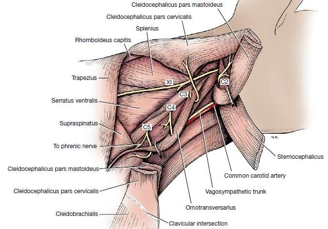
9 Start with some carefully cleaning. Be careful, the muscles are very thin and do not hurt the jugular vein. It s running on the surface of sternocephalicus and is a good landmark- Above is the cleiocephalicus. Trapezius (pars cervicalis) goes from the spina scapulae, carfully separate it from the cleiodocervicalis and you will find the omnotransversarius ventrally to it, also attached to the spina scapulae. You will see a triangle between trapezius, cleidocervicalis and omnotransversarius. Keep it as a landmark. Start clean on cleidocervicales (The fibres runs up to the neck). Clean carefully around the jugular vein and lay bare it (draw it ventrally so you get rid of it). After cleaning and moving of the jugular vein, a border between cleidocervicalis and sternocephalicus is visible (It s very thin) and transparent. Separate the cleidocervicalis from the sternocephalicus. Clean cleidocervicalis against its insertion. Stretch the leg and find clavicular intersection ( a small transparent band, put your finger under it to show) The part belox is cleidobrachialis, its insertion is deep and do not have to be shown. The parts above is the cleidocervicalis and cleidomastoideus Go to veina jugulare. Between its to branches, you will find the mandibular gland. Cut around it (but not at is cranial border) and reflect it cranially. By doing it, you will find the tendon of sternomastoideus. ( and also os cleidomastoideus but you cannot be sure yet) Go back to omnotransversarius and trace it under cleiodiocervicales (you can now clean the triangle from fat and lymph nodes). Grab cleidocervicales (Or transect it) and reflect it (but it s not transected) and you will see the not so evident separation between the clediocervicales and cleidomastoideus. Do an incision between trapezius and cleidocervicales to easier reach the deep muscles. Stop trace omnotransversarius a few centimeters from atlas. Make a distinct incision between sternocephalicus and cleidocephalicus and you will see more of omnotransversarius. (Between) Cleidomastoideus is below sternooccipital. Now is cleidomastoideus founded and you can clean the insertionpoint and see the both mastoid muscles insertions. You can now also separate the sternooccipital from the sternomastoid if you want to but it s not necessary. 1. Clean startingpoint 2. Find insertionpoints 3. Clean and trace omnotransversarius to find everything because omnotransversarius is the deepest in this topic!
Myologia Part II Objective: Students will examine the muscles of a canine in order to identify the musculature of the body.
 Okay Anatomy Anatomy I: Lesson 11 Myologia Part II Objective: Students will examine the muscles of a canine in order to identify the musculature of the body. Practical Tasks: 6) carpal flexors, pronators
16) M. quadriceps femoris, m. tensor fasciae latae, m. sartorius
 M. quadriceps femoris, m. tensor fasciae latae, m. sartorius") Anatomy 1 - Lesson XIII: Myologia Part IV Objective: Students will examine the muscles of a canine in order to identify the musculature of the body.. Practical Tasks 15-18: 15) Medial muscles of the thigh,
Anatomy 1 - Lesson XIII: Myologia Part IV Objective: Students will examine the muscles of a canine in order to identify the musculature of the body.. Practical Tasks 15-18: 15) Medial muscles of the thigh,
MCQWeek2. All arise from the common flexor origin. The posterior aspect of the medial epicondyle is the common flexor origin.
 MCQWeek2. 1. Regarding superficial muscles of anterior compartment of the forearm: All arise from the common flexor origin. The posterior aspect of the medial epicondyle is the common flexor origin. Flexor
ARM Brachium Musculature
 ARM Brachium Musculature Coracobrachialis coracoid process of the scapula medial shaft of the humerus at about its middle 1. flexes the humerus 2. assists to adduct the humerus Blood: muscular branches
Key Relationships in the Upper Limb
 Key Relationships in the Upper Limb This list contains some of the key relationships that will help you identify structures in the lab. They are organized by dissection assignment as defined in the syllabus.
The Muscular System. Chapter 10 Part C. PowerPoint Lecture Slides prepared by Karen Dunbar Kareiva Ivy Tech Community College
 Chapter 10 Part C The Muscular System Annie Leibovitz/Contact Press Images PowerPoint Lecture Slides prepared by Karen Dunbar Kareiva Ivy Tech Community College Table 10.9: Muscles Crossing the Shoulder
Practical 2 Worksheet
 Practical 2 Worksheet Upper Extremity BONES 1. Which end of the clavicle is on the lateral side (acromial or sternal)? 2. Describe the difference in the appearance of the acromial and sternal ends of the
Biceps Brachii. Muscles of the Arm and Hand 4/4/2017 MR. S. KELLY
 Muscles of the Arm and Hand PSK 4U MR. S. KELLY NORTH GRENVILLE DHS Biceps Brachii Origin: scapula Insertion: radius, fascia of forearm (bicipital aponeurosis) Action: supination and elbow flexion Innervation:
Levels of the anatomical cuts of the upper extremity RADIUS AND ULNA right
 11 CHAPTER 2 Levels of the anatomical cuts of the upper extremity AND right CUT 1 CUT 4 1 2 3 4 5 6 Isolated fixation of the radius is difficult at this level because of the anterolateral vessels and the
1/13/2013. Anatomy Guy Dissection Sheet Extensor Forearm and Hand. Eastern Virginia Medical School
 Dr. Craig Goodmurphy Anatomy Guy Superficial Extensor Muscles Complete skin removal if necessary then remove the antebrachial fascia starting at the extensor retinaculum and working proximally. Define
Muscular Nomenclature and Kinesiology - One
 Chapter 16 Muscular Nomenclature and Kinesiology - One Lessons 1-3 (with lesson 4) 1 Introduction 122 major muscles covered in this chapter Chapter divided into nine lessons Kinesiology study of human
Forearm and Wrist Regions Neumann Chapter 7
 Forearm and Wrist Regions Neumann Chapter 7 REVIEW AND HIGHLIGHTS OF OSTEOLOGY & ARTHROLOGY Radius dorsal radial tubercle radial styloid process Ulna ulnar styloid process ulnar head Carpals Proximal Row
The Clavicle Right clavicle Deltoid tubercle: Conoid tubercle, conoid ligamen Impression for the
 The Clavicle Muscle Attachment Sites in the Upper Limb Pectoralis major Right clavicle Smooth superior surface of the shaft, under the platysma muscle tubercle: attachment of the deltoid Acromial facet
Lecture 9: Forearm bones and muscles
 Lecture 9: Forearm bones and muscles Remember, the region between the shoulder and the elbow = brachium/arm, between elbow and wrist = antebrachium/forearm. Forearm bones : Humerus (distal ends) Radius
divided by the bones ( redius and ulna ) and interosseous membrane into :
 and interosseous membrane into :") fossa Cubital Has: * floor. * roof : - Skin - superficial fasica - deep fascia ( include bicipital aponeurosis ) Structures within the roof : -cephalic and basilic veins -and between them median cubital
fossa Cubital Has: * floor. * roof : - Skin - superficial fasica - deep fascia ( include bicipital aponeurosis ) Structures within the roof : -cephalic and basilic veins -and between them median cubital
The Elbow and the cubital fossa. Prof Oluwadiya Kehinde
 The Elbow and the cubital fossa Prof Oluwadiya Kehinde www.oluwadiya.com Elbow and Forearm Anatomy The elbow joint is formed by the humerus, radius, and the ulna Bony anatomy of the elbow Distal Humerus
Human Anatomy Lab #7: Muscles of the Cadaver
 Human Anatomy Lab #7: Muscles of the Cadaver Table of Contents: Expected Learning Outcomes.... 1 Introduction...... 1 Identifying Muscles on Yourself.... 2 Muscles of the Anterior Trunk and Arm.. 2 Muscles
REFERENCE DIAGRAMS OF UPPER LIMB MUSCLES: NAMES, LOCATIONS, ATTACHMENTS, FUNCTIONS MUSCLES CONNECTING THE UPPER LIMB TO THE AXIAL SKELETON
 REFERENCE DIAGRAMS OF UPPER LIMB MUSCLES: NAMES, LOCATIONS, ATTACHMENTS, FUNCTIONS MUSCLES CONNECTING THE UPPER LIMB TO THE AXIAL SKELETON A25LAB EXERCISES: UPPER LIMB MUSCLES Page 1 MUSCLES CONNECTING
Lab Activity 11: Group II
 Lab Activity 11: Group II Muscles Martini Chapter 11 Portland Community College BI 231 Origin and Insertion Origin: The place where the fixed end attaches to a bone, cartilage, or connective tissue. Insertion:
The Forearm 2. Extensor & lateral Compartments of the Forearm
 The Forearm 2 Extensor & lateral Compartments of the Forearm 1-Lateral Fascial Compartment (at the lateral side of the forearm ) *Some books mention the lateral compartment contain just the Brachioradialis
Human Anatomy Biology 351
 1 Human Anatomy Biology 351 Upper Limb Exam Please place your name on the back of the last page of this exam. You must answer all questions on this exam. Because statistics demonstrate that, on average,
Functional Anatomy of the Elbow
 Functional Anatomy of the Elbow Orthopedic Institute Daryl C. Osbahr, M.D. Chief of Sports Medicine, Orlando Health Chief Medical Officer, Orlando City Soccer Club Orthopedic Consultant, Washington Nationals
ANATOMY. Subject : Lecturer : Maher Hadidi Done by: lecture # : 11 Date :
 ANATOMY Subject : Lecturer : Maher Hadidi Done by: lecture # : 11 Date : Median nerve Meaian nerve to Pronator teres Course: Enter cubital fossa. Pierce Pronat. teres Between to FDS, FOP Below and lateral
region of the upper limb between the shoulder and the elbow Superiorly communicates with the axilla.
 1 region of the upper limb between the shoulder and the elbow Superiorly communicates with the axilla. Inferiorly, a number of important structures pass between arm & forearm through cubital fossa. 2 medial
medial half of clavicle; Sternum; upper six costal cartilages External surfaces of ribs 3-5
 MUSCLE ORIGIN INSERTION ACTION NERVE Pectoralis Major medial half of clavicle; Sternum; upper six costal cartilages Lateral lip of intertubercular groove of horizontal adduction Medial and lateral pectoral
Muscles of the Upper Limb
 Muscles of the Upper Limb anterior surface of ribs 3 5 coracoid process Pectoralis minor pectoral nerves protracts / depresses scapula Serratus anterior Subclavius ribs 1-8 long thoracic nerve rib 1 ----------------
STRUCTURAL BASIS OF MEDICAL PRACTICE EXAMINATION 5 October 6, 2006
 STRUCTURAL BASIS OF MEDICAL PRACTICE EXAMINATION 5 October 6, 2006 PART l. Answer in the space provided. (8 pts) 1. Identify the structures. (2 pts) B C A. _pisiform B. _ulnar artery A C. _flexor carpi
[[Sally Leaning Towards Peter To Take Cold Hand]]
![[[Sally Leaning Towards Peter To Take Cold Hand]]](/thumbs/84/91174469.jpg "[[Sally Leaning Towards Peter To Take Cold Hand]]") In this lecture we will talk about the bones of the hand, and the muscles and contents of the forearm. *The hand bones are: - Carpal bones. -Metacarpals. -Phalanges. *The carpal bones (wrist bones): They
In this lecture we will talk about the bones of the hand, and the muscles and contents of the forearm. *The hand bones are: - Carpal bones. -Metacarpals. -Phalanges. *The carpal bones (wrist bones): They
Connects arm to thorax 3 joints. Glenohumeral joint Acromioclavicular joint Sternoclavicular joint
 Connects arm to thorax 3 joints Glenohumeral joint Acromioclavicular joint Sternoclavicular joint Scapula Elevation Depression Protraction (abduction) Retraction (adduction) Downward Rotation Upward Rotation
# Anatomy. Upper Extremities Muscles and anatomy of axilla. Tiba Al-Ani 9/10/2015 Nabil. Page 0 of 16
 #10 25 Anatomy Upper Extremities Muscles and anatomy of axilla Tiba Al-Ani 9/10/2015 Nabil Page 0 of 16 Salam AWN Today s lecture is divided into two parts, the first part is the continuation of the upper
MUSCLES OF THE ELBOW REGION
 MUSCLES OF THE ELBOW REGION Dr Bronwen Ackermann COMMONWEALTH OF AUSTRALIA Copyright Regulation WARNING This material has been reproduced and communicated to you by or on behalf of the University of Sydney
In which arm muscle are intramuscular injections most often given? (not in text)
") AP1 Lab 9 - Muscles of the Arms and Legs Locate the following muscles on the models and on yourself. Recall anatomical position. Directional terms such as anterior, posterior, lateral, etc. all assume
AP1 Lab 9 - Muscles of the Arms and Legs Locate the following muscles on the models and on yourself. Recall anatomical position. Directional terms such as anterior, posterior, lateral, etc. all assume
forearm posterior compartment
 Quick revision: The anterior compartment of the forearm contains of 8 muscles... -4 superficial -1 intermediate -3 deep *All supplied by median nerve except 1 and 1/2 muscle (by ulnar N.) forearm posterior
10/15/2014. Wrist. Clarification of Terms. Clarification of Terms cont
 Wrist Clarification of Terms Palmar is synonymous with anterior aspect of the wrist and hand Ventral is also synonymous with anterior aspect of the wrist and hand Dorsal refers to the posterior aspect
Nerves of the upper limb Prof. Abdulameer Al-Nuaimi. E. mail:
 Nerves of the upper limb Prof. Abdulameer Al-Nuaimi E-mail: a.al-nuaimi@sheffield.ac.uk E. mail: abdulameerh@yahoo.com Brachial plexus Median nerve After originating from the brachial plexus in the axilla,
Cubital fossa and forearm
 Cubital fossa and forearm Cubital fossa is the triangular space in front of elbow joint. - The Cubital fossa has boundaries: apex, base, roof and floor and it has contents. The base: an imaginary horizontal
Peripheral Nervous Sytem: Upper Body
 Peripheral Nervous Sytem: Upper Body MSTN121 - Neurophysiology Session 10 Department of Myotherapy Cervical Plexus Accessory nerve (CN11 + C1-5) Motor: trapezius and sternocleidomastoid Greater auricular
Supplied in part by the musculocutaneous nerve. Forms the axis of rotation in movements of pronation and supination
 Anatomy: Upper limb (15 questions) 1. Latissimus Dorsi: Is innervated by the dorsal scapular nerve Lies above feres major muscle Medially rotates the humerus All of the above 2. Supinator muscle is: Deep
13 13/3/2012. Adel Muhanna
 13 13/3/2012 Adel Muhanna بسم هللا الرحمن الرحيم The Hand Extensor retinaculum: Deep fascia of anterior compartment of the wrist is thickened to form flexor retinaculum : a bridge that have 6 structures
Muscular anatomy of the forelimbs of the lesser grison (Galictis cuja), and a functional and
, and a functional and") Muscular anatomy of the forelimbs of the lesser grison (Galictis cuja), and a functional and phylogenetic overview of Mustelidae and other Caniformia Journal of Mammalian Evolution Marcos D. Ercoli, Alicia
Muscular anatomy of the forelimbs of the lesser grison (Galictis cuja), and a functional and phylogenetic overview of Mustelidae and other Caniformia Journal of Mammalian Evolution Marcos D. Ercoli, Alicia
The hand is full with sweat glands, activated at times of stress. In Slide #2 there was a mistake where the doctor mentioned lateral septum twice.
 We should only know: Name, action & nerve supply Layers - Skin - Superficial fascia - Deep fascia The hand is full with sweat glands, activated at times of stress. Deep fascia In Slide #2 there was a mistake
Main Menu. Wrist and Hand Joints click here. The Power is in Your Hands
 1 The Wrist and Hand Joints click here Main Menu K.5 http://www.handsonlineeducation.com/classes/k5/k5entry.htm[3/23/18, 1:40:40 PM] Bones 29 bones, including radius and ulna 8 carpal bones in 2 rows of
Viorel Nacu. The clinical anatomy of the Hand
 Viorel Nacu The clinical anatomy of the Hand The distal part of the upper limb is divided in to three regions: 1. The wrist (carpus) 2. The hand (metacarpus) 3. The digits (fingers) The landmarks of this
Systematic Anatomy (For international students)
") Systematic Anatomy (For international students) Department of Anatomy,Fudan University Teaching contents Muscles of abdomen & upper limbs Dr.Hongqi Zhang ( 张红旗 ) Email: zhanghq58@126.com 1 Muscles of abdomen
Systematic Anatomy (For international students) Department of Anatomy,Fudan University Teaching contents Muscles of abdomen & upper limbs Dr.Hongqi Zhang ( 张红旗 ) Email: zhanghq58@126.com 1 Muscles of abdomen
Netter's Anatomy Flash Cards Section 6 List 4 th Edition
 Netter's Anatomy Flash Cards Section 6 List 4 th Edition https://www.memrise.com/course/1577581/ Section 6 Upper Limb (66 cards) Plate 6-1 Humerus and Scapula: Anterior View 1.1 Acromion 1.2 Greater tubercle
Module 7 - The Muscular System Muscles of the Arm and Trunk
 Module 7 - The Muscular System Muscles of the Arm and Trunk This Module will cover the muscle anatomy of the arms and trunk. We have already seen the muscles that move the humerus, so this module will
Anatomy and Physiology II. Review Shoulder Girdle New Material Upper Extremities - Bones
 Anatomy and Physiology II Review Shoulder Girdle New Material Upper Extremities - Bones Anatomy and Physiology II Shoulder Girdle Review Questions From Last Lecture Can you identify the following muscles?
BRACHIAL PLEXUS. DORSAL SCAPULAR NERVE (C5) supraclavicular branch innervates rhomboids (major and minor) and levator scapulae
 supraclavicular branch innervates rhomboids (major and minor) and levator scapulae") THE BRACHIAL PLEXUS DORSAL SCAPULAR NERVE (C5) supraclavicular branch innervates rhomboids (major and minor) and levator scapulae SCHEMA OF THE BRACHIAL PLEXUS THE BRACHIAL PLEXUS PHRENIC NERVE supraclavicular
THE BRACHIAL PLEXUS DORSAL SCAPULAR NERVE (C5) supraclavicular branch innervates rhomboids (major and minor) and levator scapulae SCHEMA OF THE BRACHIAL PLEXUS THE BRACHIAL PLEXUS PHRENIC NERVE supraclavicular
The Role of Muscles in Movement
 The Role of Muscles in Movement Muscles can t push, they can only pull as they contract, so most often body movements are the result of the activity of pairs or teams of muscles acting together or against
Anatomy of the Forearm
 Anatomy of the Forearm Musculoskeletal block- Anatomy-lecture 8 Editing file Objectives List the names of the Flexors Group of Forearm (superficial & deep muscles). Identify the common flexor origin of
Wrist & Hand Ultrasonography 대구가톨릭대학교병원재활의학과 권동락
 Wrist & Hand Ultrasonography 대구가톨릭대학교병원재활의학과 권동락 Dorsal Wrist Evaluation (1 st Compartment) EPB APL Transverse View APL, abductor pollicis longus; EPB, extensor pollicis brevis Dorsal Wrist Evaluation
Fascial Compartments of the Upper Arm
 Fascial Compartments of the Upper Arm The upper arm is enclosed in a sheath of deep fascia and has two fascial septa: 1- Medial fascial septum (medial intermuscular septum): attached to the medial supracondylar
MLT Muscle(s) Patient Position Therapist position Stabilization Limb Position Picture Put biceps on slack by bending elbow.
 Patient Position Therapist position Stabilization Limb Position Picture Put biceps on slack by bending elbow.") MLT Muscle(s) Patient Position Therapist position Stabilization Limb Position Picture Put biceps on slack by bending elbow. Pectoralis Minor Supine, arm at side, elbows extended, supinated Head of Table
MLT Muscle(s) Patient Position Therapist position Stabilization Limb Position Picture Put biceps on slack by bending elbow. Pectoralis Minor Supine, arm at side, elbows extended, supinated Head of Table
Forelimb Amputation. Indications. Pre operatively 11/12/2008. Technique Preparing for surgery. Bone neoplasia. Severe trauma/paralysis
 Forelimb Amputation January 10, 2007 Amy Bringardner Becky Pentecost Indications Bone neoplasia Osteosarcoma Fibrosarcoma Chondrosarcoma Severe trauma/paralysis Brachial plexus avulsion Infection Congenital
Due in Lab weeks because of Thanksgiving Prelab #10. Homework #8. Both sides! Both sides!
 Lab 8 MUSCLES Due in Lab 10 2 weeks because of Thanksgiving Prelab #10 Both sides! Homework #8 Both sides! Refer to Muscles 22-23 Naming of muscles Origin Site of muscle attachment that doesn t move during
compartments of the forearm
 " forearm posterior compartment " compartments of the forearm Posterior Fascial compartment Muscles: ** The superficial group 1. Extensor carpi radialis brevis 2. Ex. digitorum 3. Ex. digiti minimi 4.
Anatomic Review of Eleven Important Canine Acupuncture Points Part I: Thoracic Limb
 Review Anatomic Review of Eleven Important Canine Acupuncture Points Part I: Thoracic Limb Martha A. Littlefield DVM, MS, CVA ABSTRACT Two papers (Part I and Part II) review eleven commonly used acupuncture
The Human Muscular System Required reading before beginning this lab: Saladin, KS: Human Anatomy 5th ed (2017) Chapters 10, 11, 12 INTRODUCTION
 Chapters 10, 11, 12 INTRODUCTION") Biology 322: Human Anatomy The Human Muscular System Required reading before beginning this lab: Saladin, KS: Human Anatomy 5 th ed (2017) Chapters 10, 11, 12 INTRODUCTION We will use a number of lab periods
Biology 322: Human Anatomy The Human Muscular System Required reading before beginning this lab: Saladin, KS: Human Anatomy 5 th ed (2017) Chapters 10, 11, 12 INTRODUCTION We will use a number of lab periods
Dr. Mahir Alhadidi Anatomy Lecture #9 Feb,28 th 2012
 Quick Revision: Upper arm is divided into two compartments: 1. Anterior Compartment: Contains three muscles (Biceps brachii, Coracobrachialis, Brachialis). Innervated by Musculocutaneous nerve. 2. Posterior
THE ANATOMY of the canine elbow has been fully
 Veterinary Surgery 38:135 143, 2009 INVITED REVIEW A Clinically Oriented Comprehensive Pictorial Review of Canine Elbow Anatomy GHEORGHE M. CONSTANTINESCU, DVM, PhD, mult Dr h c and ILEANA A. CONSTANTINESCU,
Al-Balqa Applied University
 Al-Balqa Applied University Faculty Of Medicine *You can use this checklist as a guide to you for the lab. the items on this checklist represent the main features of the models that you have to know for
DISSECTION 1: SKELETAL MUSCLES
 8546d_c01_1-42 6/21/02 1:34 PM Page 4 mac62 mac62:1253_ge: 4 Cat Dissection DISSECTION 1: SKELETAL MUSCLES Many skeletal muscles of the cat are similar to human muscles. This dissection will reinforce
Clinical examination of the wrist, thumb and hand
 Clinical examination of the wrist, thumb and hand 20 CHAPTER CONTENTS Referred pain 319 History 319 Inspection 320 Functional examination 320 The distal radioulnar joint.............. 320 The wrist.......................
Done By : Isra a Aweidah
 Anatomy Made Easy MSS هذا الملف يشمل تفريغ المحاضرة 10 المحاضرة 12 ابتداء من صفحة 13 وتفريغ part #9 Done By : Isra a Aweidah Edited by: AWN Academic Team Everything is included ( the drs slides in addition
Traditional Thai Acupressure Points. The anterior aspect of the body THE ANATOMICAL ATLAS
 Traditional Thai Acupressure Points The anterior aspect of the body THE ANATOMICAL ATLAS lines of the SHOULDER BLADES AND POSTERIOR ARM Scapula Line This line runs through landmarks: 1. Above the midpoint
8/25/2014. Radiocarpal Joint. Midcarpal Joint. Osteology of the Wrist
 Structure and Function of the Wrist 2 joints and 10 different bones Combine to create wrist motion Anatomical Terms: Wrist/Hand Palmar = anterior aspect of the wrist and hand Dorsal = posterior aspect
Human Anatomy and Physiology I Laboratory
 Human Anatomy and Physiology I Laboratory Gross Anatomy of the Muscular System (Two weeks) 1 This lab involves study of the laboratory exercise Gross Anatomy of the Muscular System. Complete the Review
Abduction of arm until your hand rich your head. Flexion of forearm at elbow joint. Extension of arm at elbow joint. Flexion of fingers 10.
 Num. answer 1. Medialy With the manubrium ( sternum ), and laterally with the acromion of the scapula 2. 1. Trapezius 2. Levator scapulae 3. Rhomboids 3. 1. Pectoralis major 2. Pectoralis minor 3. Latissiumus
BLUE SKY SCHOOL OF PROFESSIONAL MASSAGE AND THERAPEUTIC BODYWORK. Musculoskeletal Anatomy & Kinesiology II REVIEW
 BLUE SKY SCHOOL OF PROFESSIONAL MASSAGE AND THERAPEUTIC BODYWORK Musculoskeletal Anatomy & Kinesiology II REVIEW MSAK101-II Session 4 LEARNING OBJECTIVES: By the end of this session, the student will be
Ultrasonography of the wrist - a step-by-step approach to study protocols and normal findings
 Ultrasonography of the wrist - a step-by-step approach to study protocols and normal findings Poster No.: C-1779 Congress: ECR 2016 Type: Educational Exhibit Authors: R. R. Domingues Madaleno, A. P. Pissarra,
Nerve Injury. 1) Upper Lesions of the Brachial Plexus called Erb- Duchene Palsy or syndrome.
 Upper Lesions of the Brachial Plexus called Erb- Duchene Palsy or syndrome.") Nerve Injury - Every nerve goes to muscle or skin so if the nerve is injured this will cause paralysis in the muscle supplied from that nerve (paralysis means loss of function) then other muscles and other
Nerve Injury - Every nerve goes to muscle or skin so if the nerve is injured this will cause paralysis in the muscle supplied from that nerve (paralysis means loss of function) then other muscles and other
11/15/2018. Temporalis Elevates & retracts mandible. Masseter = Prime mover of jaw closure. Levator scapulae Supraspinatus Clavicle.
 Due in Lab 10 Lab 8 MUSCLES 2 weeks because of Thanksgiving Prelab #10 Both sides! Homework #8 Both sides! Refer to Muscles 22-23 Examples of Origin & Insertion Naming of muscles Origin Site of muscle
LIST OF STRUCTURES TO BE IDENTIFIED IN LAB: UPPER EXTREMITY REVIEW 2016
 LIST OF STRUCTURES TO BE IDENTIFIED IN LAB: UPPER EXTREMITY REVIEW 2016 BONES Ribs, sternum, clavicle Humerus: Head, greater tubercle, lesser tubercle, intertubercular sulcus, surgical neck, anatomical
MORPHOFUNCTIONAL CORRELATIONS OF THE FOREARM MUSCLE AT SHEEP AND DOG
 MORPHOFUNCTIONAL CORRELATIONS OF THE FOREARM MUSCLE AT SHEEP AND DOG Ştefănescu, S., G. Predoi, C. Belu, B. Georgescu, I. Dumitrescu, Carmen Biţoiu, Anca Şeicaru Faculty of Veterinary Medicine, Bucharest,
Muscle Anatomy Review Chart
 Muscle Anatomy Review Chart BACK Superficial (5) Trapezius Transverse cervical a. Latissimus dorsi Thoracodorsal a. Rhomboideus major Dorsal scapular a. Rhomboideus minor Levator scapulae Intermediate
Copy Right- Hongqi ZHANG-Department of Anatomy-Fudan University. Systematic Anatomy. Locomotor system - Part 6
 Systematic Anatomy Locomotor system - Part 6 Muscles of abdomen Muscles of the upper limb Dr.Hongqi Zhang ( 张红旗 ) Email: zhanghq58@126.com 1 Muscles of abdomen Muscles of the upper limb Muscles of abdomen
Muscles of the hand Prof. Abdulameer Al-Nuaimi
 Muscles of the hand Prof. Abdulameer Al-Nuaimi a.alnuaimi@sheffield.ac.uk abdulameerh@yahoo.com Thenar Muscles Thenar muscles are three short muscles located at base of the thumb. All are innervated by
Anatomy Workshop Upper Extremity David Ebaugh, PT, PhD Workshop Leader. Lab Leaders: STATION I BRACHIAL PLEXUS
 Anatomy Workshop Upper Extremity David Ebaugh, PT, PhD Workshop Leader Lab Leaders: STATION I BRACHIAL PLEXUS A. Posterior cervical triangle and axilla B. Formation of plexus 1. Ventral rami C5-T1 2. Trunks
Nerves of Upper limb. Dr. Brijendra Singh Professor & Head Department of Anatomy AIIMS Rishikesh
 Nerves of Upper limb Dr. Brijendra Singh Professor & Head Department of Anatomy AIIMS Rishikesh 1 Objectives Origin, course & relation of median & ulnar nerves. Motor & sensory distribution Carpal tunnel
8/31/2017. Objective. Canine Anatomy Differences that Make a Difference in Movement Function
 Canine Anatomy Differences that Make a Difference in Movement Function Cheryl Riegger Krugh PT, ScD, MS crieggerkrugh@gmail.com da Vinci Introduction to Canine Rehabilitation Denver, CO Sept 2017 Copyright
LECTURE 8 HANDS: BONES AND MUSCLES
 LECTURE 8 HANDS: BONES AND MUSCLES WRIST AND HAND - Human hand can do power grip and precision grip - Thumb is 90 to the rest of the hand can do fine actions - Often able to do power actions o Take tools
David G. Simpson, Ph.D.
 David G. Simpson, Ph.D. ARM & CUBITAL FOSSA Revised 7/08 Text References Moores 3 rd ed., p402 408, 436 439, 439 443, 478, 481 LEARNING OBJECTIVES: 1. Describe the humerus, indicating the sites of muscle
BIOL 4260 Human Evolu3onary Anatomy Lecture 12: Limb Development. Lecture 2: Fossil Record
 BIOL 4260 Human Evolu3onary Anatomy Lecture 12: Limb Development Lecture 2: Fossil Record Outline Limb Evolution Limb Development Limb Function A Few Definitions Appendicular skeleton girdles & limbs Pectoral
STRUCTURAL BASIS OF MEDICAL PRACTICE EXAMINATION 5. September 30, 2011
 STRUCTURAL BASIS OF MEDICAL PRACTICE EXAMINATION 5 September 30, 2011 PART l. Answer in the space provided. (12 pts) 1. Identify the structures. (2 pts) EXAM NUMBER A. Suprascapular nerve B. Axillary nerve
Deep dry needling of the arm and hand muscles
 Deep dry needling of the arm and hand s 8 César Fernández-de-las-Peñas Javier González Iglesias Christian Gröbli Ricky Weissmann CHAPTER CONTENT Introduction................... 107 Clinical relevance of
The hand. it's the most important subject of the upper limb because it has a clinical importance. the palm of the hand**
 Today at 12:48 AM The hand it's the most important subject of the upper limb because it has a clinical importance. the palm of the hand** -the palmar aponeurosis located in the palm of the hand which is
Ligaments of Elbow hinge: sagittal plane so need lateral and medial ligaments
 Ligaments of Elbow hinge: sagittal plane so need lateral and medial ligaments Ulnar Collateral ligament on medial side; arising from medial epicondyle and stops excess valgus movement (lateral movement)
Peripheral Nerve Ultrasound
 Peripheral Nerve Ultrasound Jon A. Jacobson, M.D. Professor of Radiology Director, Division of Musculoskeletal Radiology University of Michigan Normal Peripheral Nerve Ultrasound appearance: Hypoechoic
Advancement and retraction of the free limb. Raises the scapula against the trunk and lifts the limb.
 EQUINE M. trapezius It is divided into cervical and thoracic parts. Function: Innervation: Advancement and retraction of the free limb. Raises the scapula against the trunk and lifts the limb. N. accessorius
The Elbow and Radioulnar Joints Kinesiology. Dr Cüneyt Mirzanli Istanbul Gelisim University
 The Elbow and Radioulnar Joints Kinesiology Dr Cüneyt Mirzanli Istanbul Gelisim University 1 The Elbow & Radioulnar Joints Most upper extremity movements involve the elbow & radioulnar joints. Usually
Axilla and Brachial Region
 L 4 A B O R A T O R Y Axilla and Brachial Region BRACHIAL PLEXUS 5 Roots/Rami (ventral rami C5 T1) 3 Trunks Superior (C5, C6) Middle (C7) Inferior (C8, T1) 3 Cords Lateral Cord (Anterior Superior and Anterior
Elbow, Wrist & Hand Evaluation.
 Elbow, Wrist & Hand Evaluation www.fisiokinesiterapia.biz Common Injuries to the Elbow, Wrist, Hand & Fingers Lateral epicondylitis tennis elbow Medial epicondylitis golfer s s elbow, little league elbow
The Arm and Cubital Fossa
 The Arm and Cubital Fossa Dr. Andrew Gallagher School of Anatomical Sciences University of the Witwatersrand Introduction The ARM (BRACHIUM) is the most proximal segment of the upper limb musculoskeletal
Learning Objectives. 07 Aug 12. Article E-1. At the end of this section the learner will be able to:
 Module 1: Comparative Functional Anatomy and Biomechanics Article E-1 Learning Objectives At the end of this section the learner will be able to: Describe the bones of the equine thoracic Describe the
In the name of Allah, Most gracious, Most merciful
 In the name of Allah, Most gracious, Most merciful This lecture includes the following: The Palmer Oponeurosis. The Carpel tunnel. The palmaris brevis muscle. The anatomical snuffbox. The Fibrous flexor
Elbow & Forearm H O W V I T A L I S T H E E L B O W T O O U R D A I L Y L I V E S?
 Elbow & Forearm H O W V I T A L I S T H E E L B O W T O O U R D A I L Y L I V E S? Clarification of Terms The elbow includes: 3 bones (humerus, radius, and ulna) 2 joints (humeroulnar and humeroradial)
Elbow Elbow Anatomy. Flexion extension. Pronation Supination. Anatomy. Anatomy. Romina Astifidis, MS., PT., CHT
 Elbow Elbow Anatomy Romina Astifidis, MS., PT., CHT Curtis National Hand Center Baltimore, MD October 6-8, 2017 Link between the arm and forearm to position the hand in space Not just a hinge Elbow = 70%
What you should do in labs 11 & 12
 Bio 101 Laboratories 11 & 12 Muscle Histology Gross Human Skeletal Muscle Cat Muscle Dissection 1 What you should do in labs 11 & 12 Lab 11 Muscle Histology (skeletal, smooth, cardiac) Human gross skeletal
The Elbow 3/5/2015. The Elbow Scanning Sequence. * Anterior Joint (The anterior Pyramid ) * Lateral Epicondyle * Medial Epicondyle * Posterior Joint
 * Lateral Epicondyle * Medial Epicondyle * Posterior Joint") Scanning Sequence * Anterior Joint (The anterior Pyramid ) * Lateral Epicondyle * Medial Epicondyle * Posterior Joint Anterior Elbow Pyramid Courtesy of Jay Smith, MD. Vice chair PMR Mayo Clinic Rochester,
Scanning Sequence * Anterior Joint (The anterior Pyramid ) * Lateral Epicondyle * Medial Epicondyle * Posterior Joint Anterior Elbow Pyramid Courtesy of Jay Smith, MD. Vice chair PMR Mayo Clinic Rochester,
Guidelines for surgical approaches for minimally invasive plate osteosynthesis in cats
 Clinical Communication 272 Guidelines for surgical approaches for minimally invasive plate osteosynthesis in cats Philipp A. Schmierer; Antonio Pozzi Vetsuisse Faculty University of Zurich, Clinic for