Fibrous dysplasia: a case series of five cases
|
|
|
- Albert Lloyd
- 6 years ago
- Views:
Transcription
1 International Journal of Advances in Medicine Jagdale A et al. Int J Adv Med Nov;3(4): pissn eissn Case Report DOI: Fibrous dysplasia: a case series of five cases Jagdale Amol, Mittal Saurav*, Patel Krutik, Azhar Shaikh, Prasla Shopnil Department of Radio diagnosis, Dr. Vasantrao Pawar Medical College and Research Center, Maharashtra, India Received: 06 July 2016 Revised: 17 July 2016 Accepted: 06 August 2016 *Correspondence: Dr. Mittal Saurav, sauravmittal.cya@gmail.com Copyright: the author(s), publisher and licensee Medip Academy. This is an open-access article distributed under the terms of the Creative Commons Attribution Non-Commercial License, which permits unrestricted non-commercial use, distribution, and reproduction in any medium, provided the original work is properly cited. ABSTRACT Fibrous dysplasia is development anomaly in which normal bone marrow is replaced by fibro-osseous tissue which characterized by deformities of the bone, fractures, nerve compression and bone pain. It is most commonly seen in young adult. In this study we report five case of fibrous dysplasia occurring at various locations with radiological interpretation. Keywords: Bones, Cranium, Fibrous dysplasia, Mono-stotic, Poly-ostotic INTRODUCTION Fibrous dysplasia is a developmental anomaly in which normal bone marrow is replaced by fibro-osseous tissue. 1,2 It is a skeletal developmental disorder of bone forming mesenchyme that manifests as a defect in osteoblastic differentiation and maturation. 3,4 Reed defined fibrous dysplasia as an arrest of bone maturation. 3 It is a rare disorder characterized by deformities of bone, fractures, nerve compressions and bone pain. 5 It can affect a single bone, or even a small segment or the entire skeleton diffusely. 1,6 Fibrous dysplasia is a benign skeletal disorder typically seen in young adults. 1,7 it is not a true neoplasm. 1 Fibrous dysplasia as a separate entity was first described by Lichenstein and Jaffe and hence is also called as Lichtenstein Jaffe disease. 1-3,8,9 It is associated with many endocrinological diseases, most common being Albright syndrome. 7,8 There is no familial or hereditary or congenital basis to the disease. 4,9 Clinical features Bone pain is the most common feature. 5 It is mostly a disease of young age, mostly seen in first and second decade. 1,4,8 Other features are Cutaneous lesions in the form of café-au-lait spots. The borders of these lesions are typically irregular or serrated (Coast of Maine). 1 Facial asymmetry can be seen due to hemi-cranial involvement. 1 Pregnancy can exacerbate fibrous dysplasia and also cause aneurysmal bone cyst formation. 1 Sinusitis. Patho-physiology It is thought to occur as a result of abnormal activity of mesenchymal cells. 4,9 It occurs as a result of mutation in the gene that encodes the sub-unit of a stimulatory G protein (GS alpha) located on chromosome 20. 2,3,8,10 There is substitution of cysteine by arginine. Lesions of fibrous dysplasia are composed of fibrous tissue containing bone trabeculae. 1,10 Fibrous stroma is a International Journal of Advances in Medicine October-December 2016 Vol 3 Issue 4 Page 1068


 seen mainly along the anterior and part of lateral wall of right maxillary sinus.")
2 myxofibrous tissue of low vascularity while the bony trabeculae are composed of woven bone. 1 The outline of the bone trabeculae has been likened to the Chinese characters or alphabet soup. 1,2 There is no osteoblastic activity, while osteoclasts are typically seen, especially on the concave side of trabeculae. 1,10 Lesions of the fibrous dysplasia are characterized by expansion of cortical bone with gradual replacement by fibrous tissue that is firm, rubbery and gritty. 8 CASE REPORT CT examination of the brain, face, PNS and lower limbs was performed on Siemens Somatom Emo 6 machine with 6 mm and 2 mm sections. In two patients who had complaints in the limbs, MRI was also done. MRI was performed on 1.5T magnet MR system (Siemens magnetom Essenza). Five patients presented to the department with following findings CT findings Case 1 A 35 years old female came with complaints of headache, blurring of vision and focal increase in size of the left zygoma. CT scan was performed and a 2.2 x 1.5 cm sized rounded lesion arising from the left zygoma showing ground glass matrix, suggesting a diagnosis of fibrous dyplasia. Figure 1: Rounded ground glass density arising from the left zygoma. Figure 2: Diffuse osseous hyper density seen along the anterior and lateral wall of maxillary sinus. Case 2 A 12 year old female came with complaints of sinusitis, headache, persistent nasal congestion. CT scan was performed and diffuse osseous hyperdensity (ground glass opacity) seen mainly along the anterior and part of lateral wall of right maxillary sinus. The right maxillary sinus cavity was partially encroached upon. Bony walls were intact. Features were suggestive of fibrous dysplasia. Case 3 A 30 years old female came with complaints of gradual increase in size of the left half of face since past two to three years which was associated with gradual loss of hearing along with dysphagia. The patient also complained of motor symptoms in the form of hemiplegia on the right side. Other symptoms were forgetfulness. International Journal of Advances in Medicine October-December 2016 Vol 3 Issue 4 Page 1069


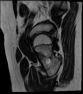


3 Figure 3: Ground glass density extending to the cranio-facial region s/o cranio-facial fibrous dysplasia. Figure 4: CT and MRI images of fibrous dysplasia arising from the neck of the left femur. International Journal of Advances in Medicine October-December 2016 Vol 3 Issue 4 Page 1070


4 CT scan was performed on this patient and it showed ground glass opacity with multiple cystic spaces with extending from the left half of mandible to left zygoma, roof of orbit on the left side, left superior and inferior nasal turbinates, left crista galli, left sided maxillary sinus entirely, basi-sphenoid, left sided frontal bone, left side squamous and petrous temporal bone. Features were suggestive of cranio-facial fibrous dysplasia. Case 4 12 years old male came to the department with complaints of H/O fall two years back with pain and swelling in the hip on left side since then. The patient also complained of gradually increasing limping on walking. CT and MRI examination was performed on this patient. CT showed ground glass opacity in the neck of left femur. MRI examination showed well demarcated ovoid lesion with narrow zone of transition in the left femoral neck anteriorly with subtle cortical irregularity in anterior cortical margin. This lesion was hypointense on T1 and heterogeneously hyperintense on T2. Post contrast study showed mild to moderate enhancement in the lesion. Case 5 A 25 years old male patient came to the department with complaints of gradually increasing pain in both lower limbs, parasthesias in both lower limbs and difficulty in walking. CT and MRI examination was performed on this patient. CT showed ground glass opacity in the femoral and tibial shaft. MRI showed expansile T2 and STIR iso to hyperintense and T1 hypointense mild heterogeneous lesions involving postero-lateral aspect of femur. Figure No 5: CT and MRI images of fibrous dysplasia arising from the femoral and tibial shaft mostly involving postero-lateral aspect of femur. International Journal of Advances in Medicine October-December 2016 Vol 3 Issue 4 Page 1071
5 RESULTS Patient one had involvement of left zygoma. Patient two had involvement anterior and part of lateral wall of right maxillary sinus Patient three had involvement of left half of mandible to left zygoma, roof of orbit on the left side, left superior and inferior nasal turbinates, left crista galli, left sided maxillary sinus entirely, basi-sphenoid, left sided frontal bone, left side squamous and petrous temporal bone. Patient four showed involvement of neck of left femur. Patient five had involvement of femoral and tibial shaft. In this study, out of the five patients, four patients presented with mono-ostotic form and only one patient had poly-ostotic form. The involvements of the facial bones were more commonly observed followed by femoral bone. DISCUSSION Fibrous dysplasia is a developmental anomaly in which normal bone marrow is replaced by fibro-osseous tissue. 1,2 It is a skeletal developmental disorder of bone forming mesenchyme that manifests as a defect in osteoblastic differentiation and maturation. 3,4 It constitutes 2 % of all bone tumors, and 7 % of all benign bony tumors. 2,9 The most common locations are ribs, femur, tibia, fibula and facial bones. 1,4,9 Skull and facial bones are affected in % of monostotic form and 50% of polyostotic form. 3,6 Amongst the facial bones, maxilla is most commonly involved. 9 Other sites are temporal bone and sphenoid bone. 8 It is of two types Monostotic It is the most common form (70%), 3,8 and it affects single bone. 1,2,4,7 Polyostotic Multiple bones are involved. Female predominance is noted in polyostotic form. 3 Polyostotic form can be associated with soft tissue myxoma. 1 The soft tissue myxoma is typically intramuscular and almost invariably multiple. Polyostotic form has three sub types- 2 Cranio-facial type - only the cranio-facial complex is involved Lichtenstein Jaffe type - In addition to cranio-facial complex involvement, there are also cutaneous lesions in the form of café-au-lait spots. Albright syndrome - It is a triad of - Polyostotic fibrous dysplasia (typically unilateral). 1 Cutaneous café-au-lait spots (Ipsilateral to bone lesions). Precocious puberty in girls. Complete triad is very rarely seen. 1 It is the most severe form and is more common in females. 8 Monostotic form is not believed to be a precursor to the polyostotic form. 8 CHEZRUBISM It is a special form of fibrous dysplasia that is autosomal dominant disorder with symmetric involvement of both maxilla and mandible. 1,4 It is more severe in males and usually regresses after adolescence. 1 Radiological features CT It is the investigation of choice for diagnosis and follow up. 7,8 It describes the extent of skeletal involvement. 1 Three main features are seen in CT Expanded bone with ground glass pattern 2,6 Homogenously dense pattern 6 Cystic variety. 6 Three radiographic standards in the cranial fibrous dysplasia and facial bones have been described 2,8 Pategoid - alternate radiodense and radiotransparent areas Sclerotic - homogenously dense Myxoid. Areas of low enhancement and cyst formation aids in differentiating the lesion from malignancy. 7 MRI Most of the lesions appear hypointense on T1WI and variable intensity on T2WI. 1,6 The variable intensity on T2WI resembles soft tissue tumor. 6 Localized fibrous dysplasia on MRI mimics a tumor because fibrous tissue enhances brilliantly after contrast administration. 6 High intensity on T2WI corresponds to non-mineralised areas and region of cystic changes seen on CT. 6 X RAY Lesion appears to be eccentric and medullary. 1 The normal architecture of bone is altered and remodelled by fibrous dysplastic lesion, which is characterized by delicate woven bone spicules (ground glass appearance). 1 International Journal of Advances in Medicine October-December 2016 Vol 3 Issue 4 Page 1072
6 Other features on X RAY 1 Endosteal scallping of the cortex Sclerotic reactive bone (rind) No clear demarcation with the surrounding bone. In calvarial fibrous dysplasia Increased density at the base of skull Obliteration of sinuses Hemi-cranial involvement In femoral fibrous dysplasia. 1 Shepherd crook deformity Marked varus deformity. This occurs because of abnormal modeling of the affected femur due to alteration of the normal bio-mechanical properties of bone. Bone scintigraphy Exquisitely sensitive especially in polyostotic fibrous dysplasia. 1 Complications Malignant transformation is the most common complication. 1,3 It may be suspected due to change in the radiologic appearance of the lesion. 1 The most common malignancy is Osteosarcoma. 1,3 Other malignancies that can occur in fibrous dysplasia are fibrosarcoma and chondrosarcoma. 1 Sudden increase in alkaline phosphatase levels in one of the indicators for malignant transformation. 3 Malignant transformation is higher in males with polyostotic fibrous dysplasia, cranio-facial lesions and monostotic fibrous dysplasia. 3 Treatment Fibrous dysplasia is usually self-limiting except in syndromic cases which require surgery. 10 Surgical correction is an effective treatment. 2,3,8,9 Radiotherapy is avoided in fibrous dysplasia as it is radioresistant 2 and also it can induce malignant changes in fibrous dysplasia. 9 Differential diagnosis 1,2 Simple bone cyst Giant cell tumor Fibroxanthoma Neurofibromatosis Hyperparathyroidism. CONCLUSION Fibrous dysplasia is a developmental anomaly in which normal bone marrow is replaced by fibro-osseous tissue. It is a skeletal developmental disorder of bone forming mesenchyme that manifests as a defect in osteoblastic differentiation and maturation. In our study, female predominance was noted and the disease presented in young females. All the three females presented with lesion in the cranium. The two males presented with lesions in the limb. The patients with cranial involvement did not have cutaneous lesions or peripheral bone involvement. There was no endocrinological abnormality in these patients. The study clearly stated the predominance of monostotic form over the polyostotic form. Funding: No funding sources Conflict of interest: None declared Ethical approval: Not required REFERENCES 1. Kransdorf M, Moser RP, GIlkey FW. From the archives of AFIP; Radiographics. 1990;10,3. 2. Tinoco P, Carlos J, Pereira O, Boechat de Carmo FS, Ruela KP. Fibrous dysplasia of maxillary sinus, International archives of otorhinolaryngology year. 2009;13:2. 3. Cholakova R, Kanasirska P, Kanasirska N, Chenchev A. Dinkova. Fibrous dysplasia in the maxilla-mandibular region: a case report. J IMAB, Annual proceedings. 2010;16:4. 4. Khwaja GA, Chaudhary N, Singla G, Saxena A, Koche S, Mehndiratta. Craniofacial fibrous dysplasia and dementia. A rare association. J Indian Academy Clin Med. 2007;8(1): Chapurlat RD, Gensburger D, Jimenez-Andrade JM, Ghilardi JR, Kelly M, Mantyh P. Pathophysiology and medical treatment of pain in patients of fibrous dysplasia; Orphan J Rar Dise. 2010;7(1). 6. Chong VFH, James B, Khoo K, Yoke-Fun F. Fibrous dysplasia involving the base of skull, American J Roentgneol. 2002;178: Subramanium V, Herle ATV. RSBO. 2010;7(3): Lustig LR, Holliday MJ, McCarthy EF, Nager GT. Fibrous dysplasia involving skull base and temporal bone; Arch otolaryngeal head neck surgery. 2001;127, Kalsotra P, Manhas M. Cranio-facial fibrous dysplasia- case report; J K Science. 1999;1(3). 10. Moshy J, Dimba E, Ocholla T, Chindia M. Characteristic radiological and histological patterns of fibrous dysplasia and ossifying fibroma of the jaws at University of Nairobi Dental teaching hospital, Surgical science. Scie Res. 2012;3: Cite this article as: Jagdale A, Saurav M, Patel K, Shaikh A, Prasla S. Fibrous dysplasia: a case series of five cases. Int J Adv Med 2016;3: International Journal of Advances in Medicine October-December 2016 Vol 3 Issue 4 Page 1073
Case McCune Albright Syndrome (MAS) - polyostotic fibrous dysplasia
 - polyostotic fibrous dysplasia") Case 14477 McCune Albright Syndrome (MAS) - polyostotic fibrous dysplasia Lukasz Augsburg 1, Filip M. Vanhoenacker 1, 2, 3, Jan Gielen1 1. University Hospital Antwerp, Department of Radiology, University
Case 14477 McCune Albright Syndrome (MAS) - polyostotic fibrous dysplasia Lukasz Augsburg 1, Filip M. Vanhoenacker 1, 2, 3, Jan Gielen1 1. University Hospital Antwerp, Department of Radiology, University
Review Article. Fibrous dysplasia: A rare bone disorder
 Available online www.jocpr.com Journal of Chemical and Pharmaceutical Research, 2015, 7(1):628-633 Review Article ISSN : 0975-7384 CODEN(USA) : JCPRC5 Fibrous dysplasia: A rare bone disorder Ashish Gawai*,
The disease process may be localized to: A single bone (monostotic fibrous dysplasia) Multiple bones (polyostotic fibrous dysplasia) [4]
![The disease process may be localized to: A single bone (monostotic fibrous dysplasia) Multiple bones (polyostotic fibrous dysplasia) [4]](/thumbs/95/124167277.jpg "The disease process may be localized to: A single bone (monostotic fibrous dysplasia) Multiple bones (polyostotic fibrous dysplasia) [4]") Scholars Journal of Applied Medical Sciences (SJAMS) Sch. J. App. Med. Sci., 2017; 5(11E):4651-4655 Scholars Academic and Scientific Publisher (An International Publisher for Academic and Scientific Resources)
Scholars Journal of Applied Medical Sciences (SJAMS) Sch. J. App. Med. Sci., 2017; 5(11E):4651-4655 Scholars Academic and Scientific Publisher (An International Publisher for Academic and Scientific Resources)
Case Report Fibrous Dysplasia versus Juvenile Ossifying Fibroma: A Dilemma
 Case Reports in Dentistry Volume 2016, Article ID 6439026, 4 pages http://dx.doi.org/10.1155/2016/6439026 Case Report Fibrous Dysplasia versus Juvenile Ossifying Fibroma: A Dilemma Sreelakshmi N. Nair,
Testicular Microlithiasis related to McCune-Albright Syndrome Joseph Junewick, MD FACR
 Testicular Microlithiasis related to McCune-Albright Syndrome Joseph Junewick, MD FACR 04/25/2010 History 12 year old with McCune-Albright syndrome. Diagnosis Testicular Microlithiasis related to Mcune-Albright
Inherited & developmental disorders:
 Inherited & developmental disorders: Osteogenesis Imperfecta: Excessive fragility of bone Defect in synthesis of type I collagen Inadequate formation of bone generalized osteoporosis Slender and fracture
Fibrous Dysplasia in Children. Professor Nick Shaw Birmingham Children s Hospital, UK
 Fibrous Dysplasia in Children Professor Nick Shaw Birmingham Children s Hospital, UK Overview What is Fibrous Dysplasia? Clinical Presentations Endocrine Problems Skeletal Problems Treatment Options Fibrous
in compact bone, large vertical canals carrying blood vessels and nerves. in compact bone, large horizontal canals carrying blood vessels and nerves.
 Carl Christensen, PhD Skeletal System (Bones`) Bio. 2304 Human Anatomy 1. Identify a term for each of the following: shaft of a long bone ends of a long bone ossified remnant of the "growth plate" connective
The Radiology Assistant : Bone tumor - well-defined osteolytic tumors and tumor-like lesions
 Bone tumor - well-defined osteolytic tumors and tumor-like lesions Henk Jan van der Woude and Robin Smithuis Radiology department of the Onze Lieve Vrouwe Gasthuis, Amsterdam and the Rijnland hospital,
MRI XR, CT, NM. Principal Modality (2): Case Report # 2. Date accepted: 15 March 2013
: Case Report # 2. Date accepted: 15 March 2013") Radiological Category: Musculoskeletal Principal Modality (1): Principal Modality (2): MRI XR, CT, NM Case Report # 2 Submitted by: Hannah Safia Elamir, D.O. Faculty reviewer: Naga R. Chinapuvvula, M.D.
Radiological Category: Musculoskeletal Principal Modality (1): Principal Modality (2): MRI XR, CT, NM Case Report # 2 Submitted by: Hannah Safia Elamir, D.O. Faculty reviewer: Naga R. Chinapuvvula, M.D.
The Radiology Assistant : Bone tumor - ill defined osteolytic tumors and tumor-like lesions
 Bone tumor - ill defined osteolytic tumors and tumor-like lesions Henk Jan van der Woude and Robin Smithuis Radiology department of the Onze Lieve Vrouwe Gasthuis, Amsterdam and the Rijnland hospital,
Original Article. Spectrum of Fibro Osseous Lesions: A Retrospective Study
 Original Article Spectrum of Fibro Osseous Lesions: A Retrospective Study Sajitha K*, Kishan Prasad H L, Netra Sajjan and Jayaprakash Shetty K Dept of Pathology, K S Hegde Medical Academy, Mangalore, India
Multifocal fibrous Dysplasia with enchondroma-like areas: Fibrocartilaginous Dysplasia
 ISPUB.COM The Internet Journal of Pathology Volume 7 Number 2 Multifocal fibrous Dysplasia with enchondroma-like areas: Fibrocartilaginous Dysplasia V Monappa, R Kudva Citation V Monappa, R Kudva. Multifocal
RADIOLOGY TEACHING CONFERENCE
 RADIOLOGY TEACHING CONFERENCE John Athas, MD Monica Tadros, MD Columbia University, College of Physicians & Surgeons Department of Otolaryngology- Head & Neck Surgery September 27, 2007 CT SCAN IMAGING
Fibrocartilaginous Dysplasia of the Bone: A Rare Variant of Fibrous Dysplasia
 Open Access Case Report DOI: 10.7759/cureus.448 Fibrocartilaginous Dysplasia of the Bone: A Rare Variant of Fibrous Dysplasia Raju Vaishya 1, Amit Kumar Agarwal 1, Nishint Gupta 2, Vipul Vijay 1 1. Department
Craniofacial Fibrous Dysplasia A Case Report and Review of Literature
 IOSR Journal of Dental and Medical Sciences (IOSR-JDMS) e-issn: 2279-0853, p-issn: 2279-0861.Volume 15, Issue 11 Ver. IV (November. 2016), PP 110-114 www.iosrjournals.org Craniofacial Fibrous Dysplasia
ISSN Supplement 2011
 ISSN 2250-0359 Supplement 2011 Fibrous dysplasia of Faciomaxillary region case reports and review of literature Dr T Balasubramanian Abstract: This article discusses the author's experience in managing
MARK D. MURPHEY MD, FACR. Physician-in-Chief, AIRP. Chief, Musculoskeletal Imaging
 ALPHABET SOUP AND CYSTIC LESIONS OF THE BONE MARK D. MURPHEY MD, FACR Physician-in-Chief, AIRP Chief, Musculoskeletal Imaging ALPHABET SOUP AND CYSTIC LESIONS OF THE BONE Giant cell tumor (GCT) Unicameral
Small lesions involving scalp and skull in pediatric age.
 Small lesions involving scalp and skull in pediatric age. Poster No.: C-1149 Congress: ECR 2013 Type: Educational Exhibit Authors: M. J. Yi, J. H. Yoo; Seoul/KR Keywords: Education and training, Education,
Radiologic Pathologic Correlation of Intraosseous Lipomas. Tim Propeck 1, Mary Anne Bullard 1, John Lin 1, Kei Doi 2, William Martel 1
 Downloaded from www.ajronline.org by 148.251.232.83 on 04/10/18 from IP address 148.251.232.83. opyright RRS. For personal use only; all rights reserved Radiologic Pathologic orrelation of Intraosseous
Benign Fibro-osseous Lesions
 Benign Fibro-osseous Lesions Plus Vision is the art of seeing things invisible. Jonathan Swift 1667-1745 Steven R. Singer, DDS srs2@columbia.edu 212.305.5674 Benign Fibro-osseous Lesions A group of lesions
Small lesions involving scalp and skull in pediatric age.
 Small lesions involving scalp and skull in pediatric age. Poster No.: C-1149 Congress: ECR 2013 Type: Educational Exhibit Authors: M. J. Yi, J. H. Yoo; Seoul/ Keywords: Education and training, Education,
COPYRIGHT 2004 BY THE JOURNAL OF BONE AND JOINT SURGERY, INCORPORATED
 84 COPYRIGHT 2004 BY THE JOURNAL BONE AND JOINT SURGERY, INCORPORATED Radiographic Evaluation of Pathological Bone Lesions: Current Spectrum of Disease and Approach to Diagnosis BY BENJAMIN G. DOMB, MD,
MANAGEMENT OF OSTEOFIBROUS DYSPLASIA OF THE ULNAAFTER RESECTION WITH ELASTIC INTRAMEDULLARY NAIL AND NON VASCULAR FIBULAR GRAFT: A CASE REPORT
 MANAGEMENT OF OSTEOFIBROUS DYSPLASIA OF THE ULNAAFTER RESECTION WITH ELASTIC INTRAMEDULLARY NAIL AND NON VASCULAR FIBULAR GRAFT: A CASE REPORT *Ujwal Ramteke and Hitesh Mangukiya Department of Orthopaedics,
Mousa Al-Abadi. Abd. Kharabsheh. Rand Abu Anzeh
 7 Mousa Al-Abadi Abd. Kharabsheh Rand Abu Anzeh 1 Recap The histological appearance of Giant cell tumor of bone shows only multi-nucleated giant cells. The histological appearance of Aneurysmal bone cyst
Bones of the skull & face
 Bones of the skull & face Cranium= brain case or helmet Copyright The McGraw-Hill Companies, Inc. Permission required for reproduction or display. The cranium is composed of eight bones : frontal Occipital
A Case of Fibrous Dysplasia with Bilateral Shepherd Crook Deformity Treated with Dynamic Hip Screw Fixation
 Case Report Journal of Orthopaedic Case Reports 2018 May-June : 8(3):Page 33-37 A Case of Fibrous Dysplasia with Bilateral Shepherd Crook Deformity Treated with Dynamic Hip Screw Fixation J K Giriraj Harshavardhan¹,
Treatment of Pathological Fracture of Femur in Mc-Cune Albright Syndrome Using A Custom Made Intramedullary Nail - A Case Report
 International Journal Dental and Medical Sciences Research (IJDMSR) ISSN: 2393-073X Volume 3, Issue 1 (Jan- 2019), PP 52-57 Treatment of Pathological Fracture of Femur in Mc-Cune Albright Syndrome Using
Disclosures. Giant Cell Rich Tumors of Bone. Outline. The osteoclast. Giant cell rich tumors 5/21/11
 Disclosures Giant Cell Rich Tumors of Bone Andrew Horvai, MD, PhD Associate Clinical Professor, Pathology This lecture discusses "off label" uses of a number of pharmaceutical agents. The speaker is describing
Multiple Synchronous Central Giant Cell Granulomas of the Maxillofacial Region: A Case Report 1
 Multiple Synchronous Central Giant Cell Granulomas of the Maxillofacial Region: A Case Report 1 Min Seok Kang, M.D., Hak Jin Kim, M.D. Multifocal central giant cell granulomas (CGCG) in the maxillofacial
Case Report An Uncommon Osseous Frontal Sinus Tumor: Monostotic Paget s Disease
 Case Reports in Otolaryngology Volume 2013, Article ID 650428, 4 pages http://dx.doi.org/10.1155/2013/650428 Case Report An Uncommon Osseous Frontal Sinus Tumor: Monostotic Paget s Disease Varant Labajian,
Musculoskeletal System (Part A-1) Module 7 -Chapter 10 Overview. Functions
 Module 7 -Chapter 10 Overview. Functions") Musculoskeletal System (Part A-1) Module 7 -Chapter 10 Overview Susie Turner, M.D. 1/8/13 Muscles Attachments Bones Bone types Surface features of bones Divisions of the skeletal system Joints or Articulations
Musculoskeletal System (Part A-1) Module 7 -Chapter 10 Overview Susie Turner, M.D. 1/8/13 Muscles Attachments Bones Bone types Surface features of bones Divisions of the skeletal system Joints or Articulations
Skeletal System. Chapter 6.1 Human Anatomy & Physiology
 Skeletal System Chapter 6.1 Human Anatomy & Physiology Overview of Skeletal System Bones Joints Skeletal System Cartilage Tendons (bone to muscle) Ligaments (bone to bone) Function of the Skeletal System
MALIGNANT TUMOURS OF THE JAWS
 MALIGNANT TUMOURS OF THE JAWS MALIGNANT TUMOURS OF THE JAWS Squamous cell carcinoma Osteogenic sarcoma Chondrosarcoma Fibrosarcoma Malignant lymphomas (incl. Burkitt s) Multiple myeloma Ameloblastoma Secondary
UNIT 4 - SKELETAL SYSTEM LECTURE NOTES
 UNIT 4 - SKELETAL SYSTEM LECTURE NOTES 4.01 FUNCTIONS OF THE SKELETAL SYSTEM A. Support 1. Provides a framework for the body 2. Supports soft tissue 3. Serves as a point of attachment for ligaments, tendons,
Pictorial Essay. CT of Calcifying Jaw Bone Diseases
 Pictorial Essay CT of Calcifying Jaw one Diseases Koichi Yonetsu 1 and Takashi Nakamura Downloaded from www.ajronline.org by 46.3.204.207 on 01/08/18 from IP address 46.3.204.207. Copyright RRS. For personal
Fibrous Dysplasia Involving The Temporal Bone: Report Of Four Cases
 ISPUB.COM The Internet Journal of Otorhinolaryngology Volume 2 Number 1 Fibrous Dysplasia Involving The Temporal Bone: Report Of Four Cases A Güngör, H Cincik, A Çolak, E Poyrazo?lu Citation A Güngör,
Skeletal system. Prof. Abdulameer Al-Nuaimi. E. mail:
 Skeletal system Prof. Abdulameer Al-Nuaimi E-mail: a.al-nuaimi@sheffield.ac.uk E. mail: abdulameerh@yahoo.com Functions of Bone and The Skeletal System Support: The skeleton serves as the structural framework
History. 33 y/o F with hx of palpable anterior tibial mass x 2 years, only painful with palpation
 History 33 y/o F with hx of palpable anterior tibial mass x 2 years, only painful with palpation Imaging Photo Album Patient also had a smaller lesion 1 cm proximal to this lesion, not seen radiographically.
Name Date Score. Skeletal System. Indicate if the following statements are true or false. Correct false statements
 Name Date Score Skeletal System True/False Indicate if the following statements are true or false. Correct false statements 1. Bones surround vital organs to protect them. 2. Bones store most of the calcium
Chapter 5 The Skeletal System
 Chapter 5 The Skeletal System The Skeletal System Parts of the skeletal system Bones (skeleton) Joints Cartilages Ligaments (bone to bone)(tendon=bone to muscle) Divided into two divisions Axial skeleton:
Monostotic Paget s Disease: A Case Report
 Chin J Radiol 2002; 27: 117-121 117 CASE REPORT Monostotic Paget s Disease: A Case Report CHI-CHEN HOU 1 CHI WEI LO 2 JINN-MING CHANG 1 CHING-CHERNG TZENG 3 Department of Diagnostic Radiology 1, Orthopedics
Case Studies in the Skull Base
 Case Studies in the Skull Base Amy C Tsai, MD Neuroradiology Fellow Department of Radiology and Imaging Sciences University of Utah Health Sciences Center Salt Lake City, Utah, USA No disclosures related
NEUROCRANIUM VISCEROCRANIUM VISCEROCRANIUM VISCEROCRANIUM
 LECTURE 4 SKULL NEUROCRANIUM VISCEROCRANIUM VISCEROCRANIUM VISCEROCRANIUM CRANIUM NEUROCRANIUM (protective case around brain) VISCEROCRANIUM (skeleton of face) NASOMAXILLARY COMPLEX MANDIBLE (DESMOCRANIUM)
Primary bone tumors > metastases from other sites Primary bone tumors widely range -from benign to malignant. Classified according to the normal cell
 Primary bone tumors > metastases from other sites Primary bone tumors widely range -from benign to malignant. Classified according to the normal cell counterpart and line of differentiation. Among the
Citation Hong Kong Practitioner, 1996, v. 18 n. 12, p
 Title Radiological conference. Frontal sinus osteoma complicated by pneumocephalus Author(s) Fung, WT; Peh, WCG Citation Hong Kong Practitioner, 1996, v. 18 n. 12, p. 658-662 Issued Date 1996 URL http://hdl.handle.net/10722/44655
Polyostotic Fibrous Dysplasia in a Young Female with McCune Albright Syndrome
 Philippine Journal of Internal Medicine Case Report Polyostotic Fibrous Dysplasia in a Young Female with McCune Albright Syndrome Marbert John T. Cardino, M.D., 1 Ceryl Cindy Tan, M.D. 2 and Cecilia Jimeno,
Case Report A Reference Finding Rarely Seen in Primary Hyperparathyroidism: Brown Tumor
 Case Reports in Medicine Volume 2012, Article ID 432676, 4 pages doi:10.1155/2012/432676 Case Report A Reference Finding Rarely Seen in Primary Hyperparathyroidism: Brown Tumor F. Mantar, 1 S. Gunduz,
Juvenile Trabecular And Psammomatoid Variant Of Ossifying Fibroma Of The Maxilla With Secondary Aneurysmal Bone Cyst.
 ISPUB.COM The Internet Journal of Otorhinolaryngology Volume 14 Number 1 Juvenile Trabecular And Psammomatoid Variant Of Ossifying Fibroma Of The Maxilla With Secondary L Emerson., M Alexander, A Job Citation
Pigmented lesions of the Oral cavity
 Oral medicine أ.م.د احسان عبد هللا كميل Pigmented lesions of the Oral cavity Pigmented oral lesions are a large group of disorders in which the dark or brown color is the essential clinical characteristic.
Clinical Guidelines for the Management of Craniofacial Fibrous Dysplasia
 University of Pennsylvania ScholarlyCommons Departmental Papers (Dental) Penn Dental Medicine 5-24-2012 Clinical Guidelines for the Management of Craniofacial Fibrous Dysplasia J S. Lee E J. FitzGibbon
Differential Diagnosis of Radiolucent Lesions of the Jaws
 Differential Diagnosis of Radiolucent Lesions of the Jaws Multilocular Multilocular Radiolucencies Odontogenic Keratocyst Botryoid Odontogenic Cyst Glandular odontogenic Cyst Invasive Ameloblastoma Central
International Journal of Research in Health Sciences ISSN: Available online at: Case Study
 International Journal of Research in Health Sciences ISSN: 2321-7251 Available online at: http://www.ijrhs.org/ Case Study Foreign body granuloma mimicking a soft tissue neoplasm *Rohan Sawant, Abhishek
Myxoma of the Vomer Bone
 Myxoma of the Vomer Bone David Besachio 1*, Edward Quigley III 1, Richard Orlandi 2, Hugh Harnsberger 1, Richard Wiggins III 1 1. Department of Radiology, University of Utah, Salt Lake City, USA 2. Department
Pathologic Fracture of the Femur in Brown Tumor Induced in Parathyroid Carcinoma: A Case Report
 CASE REPORT Hip Pelvis 28(3): 173-177, 2016 http://dx.doi.org/10.5371/hp.2016.28.3.173 Print ISSN 2287-3260 Online ISSN 2287-3279 Pathologic Fracture of the Femur in Brown Tumor Induced in Parathyroid
Chapter 7. Skeletal System
 Chapter 7 Skeletal System 1 Skull A. The skull is made up of 22 bones: 8 cranial bones, 13 facial bones, and the mandible. B. The Cranium encloses and protects the brain, provides attachments for muscles,
Pre-reading - radiolucencies
 Pre-reading - radiolucencies Multiple radiolucencies o Suggests a systemic cause o Most likely: cherubism or KCOT s of nevoid basal cell carcinoma syndrome o Sometimes: florid osseous dysplasia (if limited
Year 2003 Paper two: Questions supplied by Tricia
 question 43 A 42-year-old man presents with a two-year history of increasing right facial numbness. He has a history of intermittent unsteadiness, mild hearing loss and vertigo but has otherwise been well.
Chapter 7: Head & Neck
 Chapter 7: Head & Neck Osteology I. Overview A. Skull The cranium is composed of irregularly shaped bones that are fused together at unique joints called sutures The skull provides durable protection from
Bone tumors. RMG: jan
 Bone tumors RMG: jan 217. @Kijohs KIZZA JOHN KIJOHS Diseases arising in bone Lipoma Fibrous cortical defects Non-ossifying fibroma Bone island Benign simple cysts Enchondroma Osteochondroma Osteoid osteoma
Osteology. Dr. Carmen E. Rexach Anatomy 35 Mt San Antonio College
 Osteology Dr. Carmen E. Rexach Anatomy 35 Mt San Antonio College Functions of the Skeletal System: Support Movement Protection Hemopoiesis Electrolyte balance (Ca ++ /PO -3 4 ) Acid-base balance Storage
Aggressive Juvenile Ossifying Fibroma of the Anterior Mandible
 Case Report Aggressive Juvenile Ossifying Fibroma of the Anterior Mandible Dr.Ravikumar.R 1, Dr.Raghavendra.K 2, Dr.Santhosh Kumar 3 1 Senior Lecturer, 2 Reader Department of Oral Surgery, Sri Siddhartha
Skeletal System. Supplementary Information
 Skeletal System Supplementary Information COMMON ANATOMICAL TERMS Planes run through the body side to side and front to back eg. median plane Surfaces of the body are also named eg. anterior surface This
AMELOBLASTIC FIBROMA: A RARE CASE REPORT
 Case Report International Journal of Dental and Health Sciences Volume 04, Issue 03 AMELOBLASTIC FIBROMA: A RARE CASE REPORT Namratha Patil 1 1.Sr lecturer, dept of oral medicine and radiology, KAHES VK
Imaging in neurofibromatosis type 1: An original research article with focus on spinal lesions
 Original Research Article Imaging in neurofibromatosis type 1: An original research article with focus on spinal lesions Kalpesh Patel 1*, Siddharth Zala 2, C. Raychaudhuri 3 1 Assistant Professor, 2 1
Fluid-fluid levels in bone tumors: A pictorial review
 Fluid-fluid levels in bone tumors: A pictorial review Poster No.: C-578 Congress: ECR 2009 Type: Educational Exhibit Topic: Musculoskeletal Authors: L. Figueroa Nasra, C. Martín Hervás, M. Tapia-Viñé,
Fibrous Dysplasia an Update
 Learning Corner Journal of Bone and Soft Tissue Tumors 2016 Jan-Apr;2(1) :39-43 Abstract Fibrous Dysplasia an Update 1 2 Ashish Gulia, Pankaj Kumar Panda Background: Fibrous dysplasia (FD) belongs to a
Lab-1. Miss. Lina Al-Onazy & samar Al-Wgeet =)
") Lab-1 Introduction The human skeleton is composed of 300 bones at birth and by the time adulthood is reached, some bones have fused together to give a total of 206 bones in the body. The human skeleton
Lab-1 Introduction The human skeleton is composed of 300 bones at birth and by the time adulthood is reached, some bones have fused together to give a total of 206 bones in the body. The human skeleton
BIO 137 AXIAL SKELETON BONE STUDY THE HUMAN SKELETON
 BIO 137 THE AXIAL SKELETON MARY CATHERINE FLATH, Ph.D. THE HUMAN SKELETON AXIAL SKULL HYOID THORACIC CAGE VERTEBRAL COLUMN APPENDICULAR PECTORAL GIRDLE UPPER LIMBS PELVIC GIRDLE LOWER LIMBS AXIAL SKELETON
The SKELETAL System. The framework of bones and cartilage which protect organs, and provides a lever system that allows locomotion.
 The SKELETAL System The framework of bones and cartilage which protect organs, and provides a lever system that allows locomotion. Functions of the Skeletal System Support Protection Movement Facilitation
Cranium Facial bones. Sternum Rib
 Figure 7.1 The human skeleton. Skull Thoracic cage (ribs and sternum) Cranium Facial bones Sternum Rib Bones of pectoral girdle Vertebral column Sacrum Vertebra Bones of pelvic girdle (a) Anterior view
Bone Tumors Clues and Cues
 William Herring, M.D. 2002 Bone Tumors Clues and Cues In Slide Show mode, advance the slides by pressing the spacebar All Photos Retain the Copyright of their Authors Clues by Appearance of Lesion Patterns
Osteomas of the craniofacial region
 Imaging Science in Dentistry 2011; 41 : 107-13 http://dx.doi.org/10.5624/isd.2011.41.3.107 Department of Oral and Maxillofacial Radiology, School of Dentistry, Pusan National University, usan, Korea STRCT
The future of health is digital
 Dated: XX/XX/XXXX Name: XXXXXXXX XXXXXXXXXXX Birth Date: XX/XX/XXXX Date of scan: XX/XX/XXXX Examination of the anatomical volume: The following structures are reviewed and evaluated for bilateral symmetry,
BY Dr Farooq Khan Aman Ullah khan
 Lecture 01 General Anatomy & Classification of Bone BY Dr Farooq Khan Aman Ullah khan Dated: 22.012.2017 Skeleton The hard, supporting framework of the body is called the skeleton. Skeleton includes bones
Biology 218 Human Anatomy. Adapted from Martini Human Anatomy 7th ed. Chapter 6 The Skeletal System: Axial Division
 Adapted from Martini Human Anatomy 7th ed. Chapter 6 The Skeletal System: Axial Division Introduction The axial skeleton: Composed of bones along the central axis of the body Divided into three regions:
Skull-2. Norma Basalis Interna. Dr. Heba Kalbouneh Assistant Professor of Anatomy and Histology
 Skull-2 Norma Basalis Interna Dr. Heba Kalbouneh Assistant Professor of Anatomy and Histology Norma basalis interna Base of the skull- superior view The interior of the base of the skull is divided into
Radiologic approach to pediatric lytic bone lesions
 Radiologic approach to pediatric lytic bone lesions Poster No.: C-1177 Congress: ECR 2016 Type: Educational Exhibit Authors: J. L. LERMA GALLARDO, I. de la Pedraja, A. Lancharro 1 1 1 2 1 1 Zapata, J.
APMA 2018 Radiology Track Bone Tumors When to say Gulp!
 APMA 2018 Radiology Track Bone Tumors When to say Gulp! DANIEL P. EVANS, DPM, FACFAOM Professor, Department of Podiatric Medicine and Radiology Dr. Wm. Scholl College of Podiatric Medicine Conflict of
JOURNAL OF CASE REPORTS 2013;3(2):
:") JOURNAL OF CASE REPORTS 2013;3(2):480-484 Chondroblastic Osteosarcoma of Jaw Bone Khadse Smita Vasudeo From the Department of Oral Pathology and Microbiology, Maharaja Ganga Singh Dental College and Research
JOURNAL OF CASE REPORTS 2013;3(2):480-484 Chondroblastic Osteosarcoma of Jaw Bone Khadse Smita Vasudeo From the Department of Oral Pathology and Microbiology, Maharaja Ganga Singh Dental College and Research
Bubbly Lesions of Bone
 Residents Section Pattern of the Month w79 08.18.09 Eisenberg Residents Section Pattern of the Month Residents inradiology Ronald L. Eisenberg 1 Eisenberg RL Keywords: bubbly lesions, fegnomashic, skeletal
Skeletal System -Axial System. Chapter 7 Part A
 Skeletal System -Axial System Chapter 7 Part A Skeleton Learn: Names of the s. Identify specific landmarks that allow: Bones to fit into each other, Organs to fit into the cavities, Muscles to attach,
o Diaphysis o Area where red marrow is found o Area where yellow marrow is found o Epiphyseal plate AXIAL SKELETON Skull
 64 Anatomy & Physiology Coloring Workbook 7. Figure 5-2A is a midlevel, cross-sectional view of the diaphysis of the femur. Label the membrane that lines the cavity and the membrane that covers the outside
PRIMARY LESIONS OF THE BONY ORBIT
 USCAP 2016 Companion Meeting Primary Lesions of the Bony Orbit Milman 1 Tatyana Milman, MD Pathology Department Hospital of the University of Pennsylvania milmant@uphs.upenn.edu PRIMARY LESIONS OF THE
CASE REPORT. MANDIBULAR OSTEOSARCOMA Ashutosh Chitnis 1, Bhagirath Kandhare 2, Bhavin Patel 3, Divya Bansal 4
 MANDIBULAR OSTEOSARCOMA Ashutosh Chitnis 1, Bhagirath Kandhare 2, Bhavin Patel 3, Divya Bansal 4 HOW TO CITE THIS ARTICLE: Ashutosh Chitnis, Bhagirath Kandhare, Bhavin Patel, Divya Bansal. Mandibular osteosarcoma.
Bone/Osteoid Producing Lesions
 Chapter 2 Bone/Osteoid Producing Lesions Introduction There are many lesions that are associated with reactive new bone formation; this chapter predominantly covers those in which deposition of osteoid/bone
Chapter 7: Skeletal System: Gross Anatomy
 Chapter 7: Skeletal System: Gross Anatomy I. General Considerations A. How many bones in an average adult skeleton? B. Anatomic features of bones are based on II. Axial Skeleton A. Skull 1. Functionally
The resident will be assigned to be on call with the Oral and Maxillofacial service. Call will be set according to PARO guidelines.
 Goals and Objectives for the Otolaryngology-Head & Neck Resident on the Oral and Maxillofacial Surgery (OMFS) Rotation St. Catharines General Hospital (1 four-week rotational block) During the second year
ANATOMY & PHYSIOLOGY I Laboratory Version B Name Section. REVIEW SHEET Exercise 10 Axial Skeleton
 ANATOMY & PHYSIOLOGY I Laboratory Version B Name Section REVIEW SHEET Exercise 10 Axial Skeleton 1 POINT EACH. THE SKULL MULTIPLE CHOICE 1. The major components of the axial skeleton include the 7. The
Due in Lab. Due next week in lab - Scientific America Article Select one article to read and complete article summary
 Due in Lab 1. Skeletal System 33-34 2. Skeletal System 26 3. PreLab 6 Due next week in lab - Scientific America Article Select one article to read and complete article summary Cell Defenses and the Sunshine
University Journal of Surgery and Surgical Specialities
 University Journal of Surgery and Surgical Specialities Volume 1 Issue 1 2015 EXTRA SKELETAL MESENCHYMAL CHONDROSARCOMA :A CASE REPORT Rajaraman R Subbiah S Navin Naushad Kilpaulk Medical College Abstract:
DISCLOSURES LEARNING OBJECTIVES WE WILL NOT DISCUSS. CSB: Birdseye View MESSAGE NAVIGATING THE SELLA AND CENTRAL SKULL BASE
 NAVIGATING THE SELLA AND CENTRAL SKULL BASE Christopher P. Hess, M.D., Ph.D. DISCLOSURES Research Support, General Electric SLIDES: http://www.radiology.ucsf.edu/research/meetings/rsna LEARNING OBJECTIVES
The Skeletal System. Dr. Naim Kittana. Faculty of Medicine & Health Sciences An-Najah National University
 The Skeletal System Dr. Naim Kittana Faculty of Medicine & Health Sciences An-Najah National University 1 Declaration The content and the figures of this seminar were directly adopted from the text book
DISEASES WITH ABNORMAL MATRIX
 DISEASES WITH ABNORMAL MATRIX MSK-1 FOR 2 ND YEAR MEDICAL STUDENTS Dr. Nisreen Abu Shahin CONGENITAL DISEASES WITH ABNORMAL MATRIX OSTEOGENESIS IMPERFECTA (OI): also known as "brittle bone disease" a group
Bio 103 Skeletal System 45
 45 Lecture Outline: SKELETAL SYSTEM [Chapters 7, 8] Introduction A. Components B. Functions 1. 2. 3. 4. Classification and Parts A. Bone Shapes 1. Long: 2. Short: 3. Flat: 4. Irregular: 5. Sesamoid: B.
SKULL AS A WHOLE + ANTERIOR CRANIAL FOSSA
 SKULL AS A WHOLE + ANTERIOR CRANIAL FOSSA LEARNING OBJECTIVES At the end of this lecture, the student should be able to know: Parts of skeleton (axial and appendicular) Parts of skull Sutures of skull
CASE REPORT-NASO-ETHMOIDAL ENCEPHALOCELE Shrishail Patil 1, Tanvi Choubey 2
 -NASO-ETHMOIDAL ENCEPHALOCELE Shrishail Patil 1, Tanvi Choubey 2 HOW TO CITE THIS ARTICLE: Shrishail Patil, Tanvi Choubey. Case Report-_Naso-ethmoidal Encephalocele. Journal of Evolution of Medical and
Isolated congenital anterolateral bowing of the fibula : A case report with 24 years follow-up
 Acta Orthop. Belg., 2009, 75, 842-846 CASE REPORT Isolated congenital anterolateral bowing of the fibula : A case report with 24 years follow-up Karolien LELIEFELD, Hans VAN DER SLUIJS, Ibo VAN DER HAVEN