SWANSON. Flexible Finger Joint Implant SURGIC AL TECHNIQUE
|
|
|
- Jeffrey Clarke
- 6 years ago
- Views:
Transcription


1 SWANSON Flexible Finger Joint Implant SURGIC AL TECHNIQUE
2
3 Contents Preface 4 Chapter Chapter Appendix A 20 Appendix B 22 Design Surgeon Introduction Device Description Rationale Indications, Contraindications & Precautions Indications Contraindications Precautions Surgical Technique Metacarpophalangeal Joint Implant Arthroplasty Closure, Dressing, Postoperative Care & Bracing Explant Information PIP Joint Implant Arthroplasty Postoperative Care Explant Information Ordering Information Notes Proper surgical procedures and techniques are the responsibility of the medical professional. The following guidelines are furnished for information purposes only. Each surgeon must evaluate the appropriateness of the procedures based on his or her personal medical training and experience. Prior to use of the system, the surgeon should refer to the product package insert for complete warnings, precautions, indications, contraindications and adverse effects. Package inserts are also available by contacting the manufacturer. Contact information can be found on the back of this surgical technique and the package insert is available on the website listed. Please contact your local Wright representative for product availability. 3


4 Design Surgeon PREFACE Alfred B. SWANSON, MD Grand Rapids, MI 4 SWANSON Flexible Finger Joint Implant


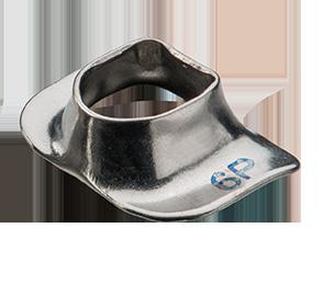
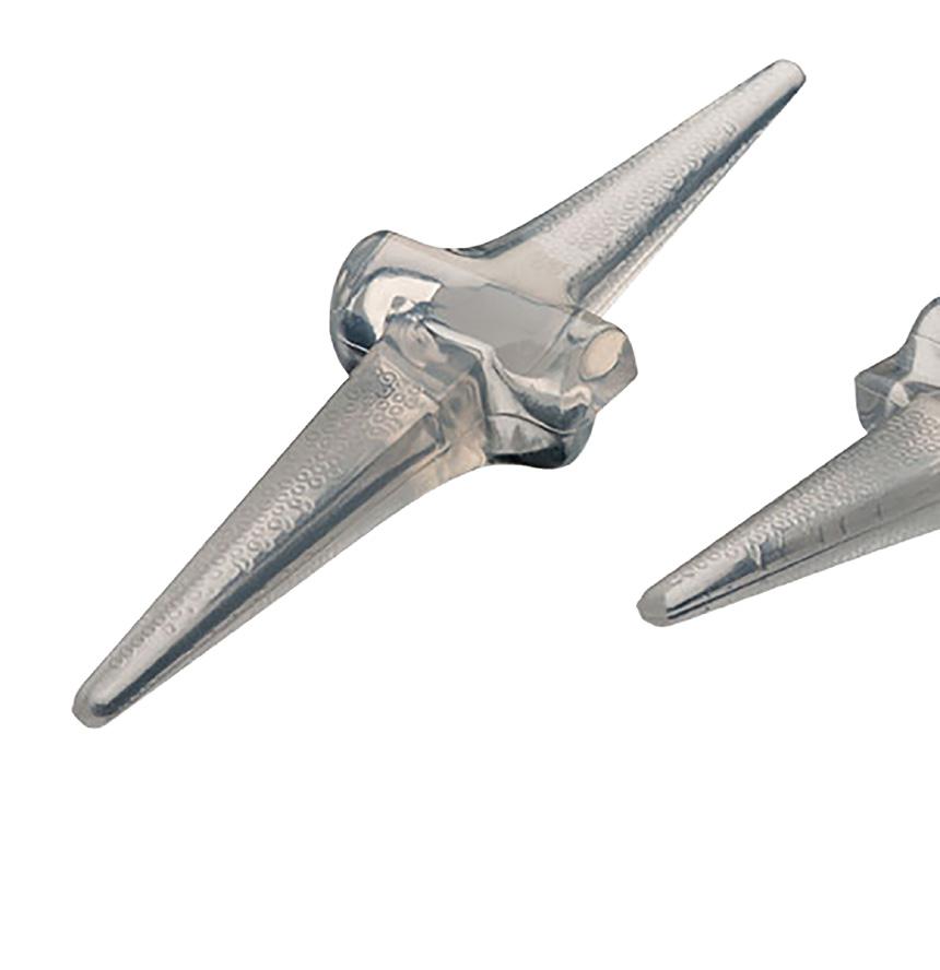
5 Introduction chapter 1 Device Description Flexible Implant The SWANSON Finger Joint Implant is a flexible intramedullary-stemmed, one-piece implant developed as an adjunct to resection arthroplasty to help restore function to hands disabled by rheumatoid, degenerative or traumatic arthritis. The midsection of the load-distributing flexible hinge has been designed to help maintain proper joint space and alignment with good lateral stability and minimal flexion-extension restriction. The implant is not fixed to bone and becomes stabilized by the encapsulation process. It acts as a dynamic spacer, internal mold and flexible hinge. The SWANSON Finger Joint Implant is available in 11 sizes to adequately meet various anatomical requirements. A color-coded sizing set (supplied non-sterile and not suitable for implantation) is available for proper size determination during surgery. Grommets The SWANSON Finger Joint Grommets are thin bone liners designed for use at the metacarpophalangeal joint level. The press-fit encircling grommet is fabricated from unalloyed titanium and its shape conforms to the contours of the implant midsection and stem junctions. The use of grommets to enhance implant durability is especially indicated in severe cases of rheumatoid arthritis where irregular and sharp bony edges can initiate tears in the implant midsection. Grommets are available in 7 sizes corresponding to sizes 3 to 9 of the SWANSON Finger Joint Implant. The outer surface of each grommet is marked with a numeral, indicating the size of finger joint implant the grommet fits, as well as the letters P or D, indicating whether it is a proximal or distal grommet. Chapter 1 Introduction 5
6 Rationale Flexible Implant The SWANSON Finger Joint implant can be used with resection arthroplasty of the metacarpophalangeal and proximal interphalangal (PIP). Insertion of the finger joint implant at two consecutive joint levels is not recommended (e.g., MP and PIP joint levels). The flexible implant resection arthroplasty method is based on the following concept: Joint Resection + Implant + Encapsulation = Functional Joint. The implant acts as a dynamic spacer to maintain internal alignment and spacing of the reconstructed joint and as an internal mold that supports the capsuloligamentous system developing around the implant while early guided motion is started. The implant becomes stabilized by this encapsulation process, and no permanent fixation is required. Joint stability is achieved from reconstruction of the ligamentous and musculotendinous systems. Because the implant is not fixed to bone, the compressive loading forces are effectively distributed to the resected end of the bone and cortical shaft. This encourages favorable bone remodeling processes as evidenced by maintenance of bone length, preserved shape of the amputated bone end, cortical thickening, and new bone formation next to the implant midsection and intramedullary stems. The slight movement of the implant stems allows distribution of forces over a broader section and allows the flexible hinge to find a better position with respect to the axis of rotation of the joint. Thus the implant life is increased and the bone is less likely to react at the implant interface when the forces are within its strain tolerance. The low modulus implant is softer than bone and has force-dampening characteristics that further protect bone and cortical shaft. Because bone removal is minimal and implants are not attached to bone, revision procedures to remove or replace an implant, or to reinforce, release, or realign capsuloligamentous structures around the implant are easily performed. Grommets The unattached, press-fit grommets effectively protect the implant midsection by lessening abrasion, wear and cutting by bone. Use of encircling grommets does not alter the function of the implant, patient indications and contraindications, nor reduce the need for careful attention to the arthroplasty technique. 6 Chapter 1 Introduction
7 Indications, Contraindications & Precautions Prior to the use of the system, the surgeon should refer to the package insert for complete warnings, precautions, indications, contraindications and adverse effects. Package inserts are also available by contacting the manufacturer. Contract information can be found on the back of this surgical technique and the package insert is available on the website listed. Indications Metacarpophalangeal Joint Implant Arthroplasty Rheumatoid or post-traumatic disabilities with: Fixed or Stiff MP Joints X-Ray Evidence of Joint Destruction or Subluxation Ulnar Drift, Noncorrectable By Surgery of Soft Tissues Alone Contracted Intrinsic and Extrinsic Musculature and Ligament System Associated Stiff Interphalangeal Joints PIP Joint Implant Arthroplasty Rheumatoid, degenerative, or post-traumatic disabilities with: Destroyed or subluxated joints Stiffened joints in which a soft tissue release alone would be inadequate. Contraindications Infection Physiologically or psychologically inadequate patient Inadequate skin, bone or neurovascular status Irreparable tendon system Possibility for conservative treatment Growing patients with open epiphyses Patients with high levels of activity NOTE: Each patient must be evaluated by the surgeon to determine the risk/benefit relationship. Chapter 1 Introduction 7
8 Precautions One of the goals of implant surgery is to minimize production of wear particles. It can never be eliminated because all moving parts (e.g., implants which articulate against bone wear) to some degree. In an implant arthroplasty, clinically significant wear can result from normal biomechanical forces. Abnormal or excessive force will further increase clinically significant wear. Abnormal force loading may be caused by: Uncorrected Instability Oversized Implant Inadequate Soft Tissue Support Implant Malposition Excessive Motion Uncorrected or Recurrent Deformity Patient Misuse or Over activity Intraoperative Fixation Some preventive measures to consider to minimize the potential for complications: Follow Guidelines for Indications and Contraindications Provided Above Identify Prior Pathology Stabilize Collapse Deformities Bone Graft Pre-Existing Cysts Use a Properly Sized Implant Avoid K-Wires and Sutures Through the Implant If complications develop, possible corrective procedures include: Implant Removal Synovectomy Bone Grafting of Cysts Limited Intercarpal Fusion Replacement of the Implant Removal of the Implant with Fusion of the Joint Clinical results depend on surgeon and technique, preoperative and post-operative care, the implant, patient pathology and daily activity. It is important that surgeons obtain appropriate informed consent and discuss the potential for complications with each patient prior to surgery. This may include a review of alternative, non-implant procedures such as soft tissue reconstruction or arthrodesis. 8 Chapter 1 Introduction
9 Surgical Technique chapter 2 Metacarpophalangeal Joint Implant Arthroplasty Incision and Exposure A transverse skin incision is made on the dorsum of the hand over the necks of the metacarpals. The dissection is carried down through subcutaneous tissue to expose the extensor tendons. The dorsal veins, which lie between the metacarpal heads are carefully released by blunt longitudinal dissection and are retracted laterally. The extensor hood is exposed to the base of the proximal phalanx. Its radial portion is usually stretched out and the extensor tendon dislocated ulnarward. In the index finger, the incision is made between the extensor digitorum communis and the indicis proprius tendons. In the middle and ring fingers, a longitudinal incision is made in the extensor hood parallel to the extensor tendon on its ulnar aspect. In the little finger, the approach is made between the extensor communis and proprius tendons. The hood fibers and capsule are carefully dissected from the underlying synovium and retracted to the radial side. The joint is exposed and the head of the metacarpal is identified. Resection of Metacarpal Head The neck of the metacarpal is exposed subperiosteally and cleanly transected with an air drill or motor saw, leaving part of the metaphyseal flare. Care should be taken to avoid splintering the bone. The head of the metacarpal is grasped and removed along with the hypertrophied synovial material. A pituitary rongeur has been found to be useful to remove further involved synovia of the joint cavity and surrounding tissues. Soft Tissue Release A comprehensive soft tissue release procedure must be done at this stage to allow the base of the proximal phalanx to be loose enough to be displaced dorsally above the metacarpal. The ulnar collateral ligament is released from its phalangeal insertion in all fingers; if severely contracted, it can be excised along with a palmar plate when necessary. At the level of all fingers, the radial collateral ligament insertions are preserved whenever possible. If it is necessary to detach this ligament, it should be reattached to the metacarpal or the base of the proximal phalanx. The important repair technique of this ligament will be described later. Chapter 2 Surgical Technique 9
 normally applies a supinatory force to this digit and should be preserved to help avoid a postoperative pronation deformity")

10 The ulnar intrinsic tendon is identified, pulled up into the wound with a blunt hook, and sectioned at the myotendinous junction if tight. However, the ulnar intrinsic of the index (first volar interosseous) normally applies a supinatory force to this digit and should be preserved to help avoid a postoperative pronation deformity tendency. In some patients who have demonstrated evidence of a flexor synovitis, the flexor sheath can be incised longitudinally in its dorsal aspect. The long flexor tendons can be identified and pulled up gently into the wound with a blunt hook. The degree of involvement of the flexor tendons can be evaluated. In some cases, a partial synovectomy and tendon sheath release or an injection of corticosteroids is done through this incision. The tendon of the abductor digiti minimi is exposed on the ulnar aspect of the fifth metacarpophalangeal joint, pulled into the wound with a blunt hook, and sectioned. Care should be taken to avoid the ulnar tendon, which will eventually reattach, but in a lengthened state. The tendon of the flexor digiti minimi is preserved because of its importance to obtain flexion at the metacarpophalangeal joint of the little finger. Furthermore, it is not an important ulnar deviator. Bone Resection and Preparation The base of the proximal phalanx is resected including marginal osteophytes which might interfere with the implant. All cartilage is removed from the base of the proximal phalanx because it is believed that progressive cartilage degeneration can eventually result in recurrent synovitis. 10 Chapter 2 Surgical Technique
11 The intramedullary canal of the metacarpal is prepared in a rectangular fashion with a rasp, curet, broach, and air drill with a special bur. These burs have a smooth leader point, which helps keep them in the canal and prevents inadvertent perforation through the cortex. The occasional construction in the intramedullary canal of the proximal third of the metacarpal can be enlarged with the bur. The intramedullary canal of the ring metacarpal is frequently quite small and requires careful preparation. Care should be taken to avoid too much reaming of the canals, especially in patients with thin bones. A trial fit is made with the appropriate color -coded sizers. The implant stem should fit well down into the canal so that the transverse midsection of the implant abuts against the bone end. The end of the implant stem must not abut the end of the intramedullary canal and the stem must be appropriately shortened. The largest implant possible should be used. Implants of sizes 4 through 9 are generally used. A rectangular hole is then made in the base of the proximal phalanx with an osteotome, knife, broach, or air drill. The intramedullary canal is reamed in the same fashion as the metacarpal to receive the distal stem of the implant selected for the metacarpal. FIGURE 1 Any sharp points or rough surfaces on the bone ends should be made completely smooth. Using the color coded sizers, proper fit of the implant is verified. With the joint extension, there should be no impingement of its midsection. If there is, soft tissue release or bone resection has not been adequate. FIGURE 2 In the index and middle fingers, the intramedullary canal is reamed in a rectangular shape, which is positioned high on the dorsal ulnar side of the base of the proximal phalanx and low on its radial palmar side. This proper rectangular and axial configuration of the canal stabilizes the implant stems and helps maintain a slight supination of the digit to prevent postoperative pronation deformity. Contrarily, in the little finger, a position of slight pronation is desired and the intramedullary rectangle is positioned high on the dorsoradial side and low on the ulnar palmar side. FIGURE 1 Bone Resection and Grommet Fitting at MP Joint FIGURE 2 Joint Relationship and Space Concept Chapter 2 Surgical Technique 11

12 In patients selected to receive SWANSON grommets, the implant sizer is removed and the bone canals are prepared to allow a press-fit of the appropriate sized grommet. The resected surfaces of the metacarpal and proximal phalanx are shaped to obtain a precise fit of the grommet sleeve and of its slightly curvilinear flanges against the resected bone ends so that contact with overlying soft tissues is avoided. Both surfaces are prepared and smoothened with a diamond bur or other appropriate fine burs. CAUTION: Fitting of the grommet requires a precise press fit. It must be accurately centered, otherwise it may impinge the intramedullary canal on one side and could cause bone absorption. Unless the shoulders of the grommet can be fitted into metaphyseal bone, rotation of the grommet could occur. In certain cases of severe metacarpophalangeal joint dislocation, more bone must be removed to obtain joint reduction and the implant should probably be used without the grommet. Occasionally, grommets are not used in the 5th MP joint and should not be used in arthritis mutilans. The grommet size corresponds to the implant size. FIGURE 3 The fitting must be exact with regard to centering and rotation. Minimal additional bone shaping is usually needed. Trial seating of the grommet is done by gentle pressure with the Grommet Seater or a flat instrument held against the exposed surface of the grommet with care to avoid bending or distorting the grommet. The grommet shoulders are seated directly against resected bone and must not protrude. If too loose, the next larger size is selected. When necessary, using a grommet one size larger than the flexible implant is permissible but a grommet smaller than the implant is never used. The implant/grommet fitting should be observed in flexion and extension. The implant must slide into the grommet and not be impinged by a too narrow joint space or an uncorrected palmar subluxation. The principal of the flexible hinge as a joint spacer must be respected. FIGURE 3: (A) FIGURE 3: (B) Encircling Finger Grommets and Flexible Finger Hinge 12 Chapter 2 Surgical Technique
13 Soft Tissue Reconstruction Soft tissue reconstruction is a critical step of the operative procedure. Proper balance of all the following structures must be attained: collateral ligaments, palmar plate, capsule, intrinsic tendons, flexor, and extensor mechanism. Rheumatoid patients often present an inadequate first dorsal interosseous muscle or have a tendency for pronation deformity of the index finger and occasionally the middle finger, which can interfere with the pinch and grasp mechanism. Reconstruction of the radial collateral ligament is important to correct the lateral and rotatory alignment and to improve lateral stability for pinch. The goal of the procedure is to place the index and middle fingers in a position of slight supination and abduction. This procedure can occasionally be indicated for the ring finger. However, it is never carried out for the little finger, as it would place this digit in supination, which would interfere with flexion of the adjacent digits. A neutral position or slight pronation is preferred. Reefing of the preserved radial collateral ligament of the index, middle, and occasionally, ring fingers, is done by passing a 3-0 Dacron suture through a 0.5 mm drill hole made in the dorsal radial side of the cut end of the metacarpal. The radial collateral ligament can require reattachment to bone proximally (metacarpal) or distally (base of proximal phalanx) or to both areas according to how much bone has been removed. The radial capsule, which has been preserved, is also included in this repair. FIGURE 4 An additional small drill hole can be made in the dorsal ulnar side of the cut end of the metacarpal to secure the radial capsule with a 4-0 Dexon suture. However it is preferred to suture the ulnar edge of the capsule to the ulnar collateral ligament to bring the repaired capsule well ulnarward over the joint. The sutures are placed before the implant is inserted and are tied as the finger is held in slight supination and abduction. Note that the first dorsal interosseous muscle fibers become dorsally relocated with this repair. A B C FIGURE 4 Measures to correct pronation deformity of index finger. Proper rectangular and axial reaming of intramedullary canal is necessary to stabilize stems and maintain slight supination. Axis of rectangle is placed high on dorsoulnar side of base of proximal phalanx and low on its radiopalmar side. Collateral ligaments and related structures are sutured with 3-0 Dacron to the dorsoradial aspect of metacarpal end through a small drill hole. (A) Preserved radial capsule is included in this repair. Preferably, the ulnar collateral ligament is sutured to the radial capsule. (B & C) Chapter 2 Surgical Technique 13
14 If the radial collateral ligament has been released from the base of the proximal phalanx it should be reattached in a manner similar to that described above. If the radial collateral ligament is inadequate, a portion of the palmar plate is used to reconstruct this ligament. A distally-based flap made of the medial half of the palmar plate is prepared and sutured in position through a drill hole made in the dorsoradial aspect of the cut end of the metacarpal, similarly as described above. Meticulous evaluation and correction of the balance of the capsuloligamentous and musculotendinous structures will be rewarded by improved results. Implant Insertion The wound is thoroughly irrigated. The proximal grommet is press-fit into the metacarpal and the distal grommet into the proximal phalanx following the procedure described for trial insertion. Both grommets are firmly seated against the resected bone by gentle pressure, taking care to avoid deforming the grommet. The flexible implant is then inserted using blunt instruments and a no-touch technique. First, the implant is inserted into the intramedullary canal of the metacarpal, and then, by slight traction on the finger, the joint is distracted and the implant is flexed so that the distal stem can easily be inserted into the proximal phalanx. With the joint in extension, there should be no impingement of the implant. If there is, soft tissue release or bone resection has been inadequate. NOTE: Handling of the implant should be done with blunt instruments to avoid surface trauma or contamination with foreign bodies. Reshaping of the implant should be avoided because it can compromise or destroy the functional integrity and the functionality of the implant. 14 Chapter 2 Surgical Technique
15 Extensor Hood Reefing The radial portion of the sagittal fibers of each extensor hood mechanism is reefed in an overlapping fashion so that the extensor tendon is brought slightly to the radial side of the center of the joint. Three to five 4-0 Dexon sutures with a buried-knot technique are used. In the index and middle fingers, it is most important not to over-correct the position of the extensor tendon too far radially to avoid deforming forces in favor of pronation. In certain cases of severe or long-standing flexion deformity, the extensor tendons may become stretched, and an extensor tendon lag may persist if not corrected. In these cases, the extensor tendon should be reefed not only transversely as described, but also longitudinally. Occasionally, the extensor tendon is tenodesed to the dorsal base of the proximal phalanx through small drill holes. Perfect balance of the extensor mechanism is essential. The juncturae tendinae which have been divided during the release of the extensor tendons are meticulously reapproximated with 4-0 Dexon sutures using inverted knots. This is done to further balance the extensor mechanism and provide all possible extensor power. Closure, Dressing, Postoperative Care & Bracing Closure, dressing, postoperative care and bracing are the responsibility of the surgeon. Explant Information If removal of the implant is required due to revision or failure of the device, the surgeon should contact the manufacturer using the contact information located on the back cover of this surgical technique to receive instruction for returning the explanted device to the manufacturer for investigation. Chapter 2 Surgical Technique 15

16 PIP Joint Implant Arthroplasty NOTE: Grommets are not used in PIP joints. Incision A C -shaped incision is made over the dorsum of the joint so that the skin suture line does not lie directly over the tendon repair. In the little and index fingers, the incision is placed away from the presenting surface. The dorsal veins are respected. If associated flexor tendon surgery is also indicated, a mid-lateral incision or palmar incision is used. This allows accessibility to both the joint and the tendon. Exposure The extensor mechanism is exposed by sharp and blunt dissection, avoiding injury to its surface. The central tendon is identified and incised longitudinally in a proximal fashion from its insertion at the base of the middle phalanx through the distal two-thirds of the proximal phalanx. FIGURE 5 In flexible deformities of the PIP joint, the extensor mechanism can be gently dislocated palmarward as the joint is flexed without disturbing the insertion of each half of the central tendon into the middle phalanx. However, in hypertrophic osteoarthritic joints, it may be necessary to section this attachment of the central tendon to excise bony spurs. A B C FIGURE 5 Repair of the central tendon: (A) The central tendon is incised longitudinally from the base of the middle phalanx through the distal two third of the proximal phalanx. (B & C) Following adequate bone preparation, a 1 mm drill hole is made in the base of the middle phalanx to receive a 3-0 Dacron suture for reinsertion of the halves of the central tendon. 16 Chapter 2 Surgical Technique
17 The tendon is later reattached to the bone with a 3-0 Dacron suture passed through a 0.5 mm drill hole made in the base of the middle phalanx. If the joint is contracted, the extensor mechanism cannot be readily dislocated and therefore, the head of the proximal phalanx is first excised by cutting it transversely at the neck with an air drill and then removing it piecemeal, or it can be removed with the burring tool of the air drill. The collateral ligaments are left intact when possible. If they are incised for joint exposure, they should be reattached to bone. FIGURE 6 A B C FIGURE 6 Repair of the collateral ligaments: (A) 4-0 Dacron sutures are placed in the dorsolateral aspect of the neck of the collateral ligament. A 3-0 Dacron suture is placed through the base of the middle phalanx for reconstruction of the central tendon. In implant arthroplasty, sutures are placed before implant insertion. Reattachment of the collateral ligament. (B & C) Joint Release Adequate release of the joint is essential for good results. If the joint is severely contracted, it may be released by removing bone from the proximal and middle phalanges. If this is inadequate, the palmar plate and collateral ligaments may be incised proximally or distally according to where more bone needs to be removed. When incised the collateral ligaments should be reattached with a 4-0 Dacron suture passed through a small drill hole made in the dorsal lateral aspect of the neck of the proximal phalanx and/or middle phalanx as indicated FIGURE 6 The sutures are placed prior to implant insertion. With the implant in position, they are tied with sufficient tension to obtain lateral stability and alignment and to allow passive motion of the joint from full extension to 70 flexion. In the presence of lateral deviation or instability, the collateral ligament should be repaired as described above. This procedure seems to limit flexion of the proximal interphalangeal joint slightly, but can be important in cases mentioned earlier. Bone Preparation After resecting the head of the proximal phalanx at the metaphyseal flare with a power saw or a side-cutting bur, the intramedullary canal is prepared to receive the implant stem. It is first penetrated with a thin broach, then reamed into a rectangular shape with an air drill and leader point bur to accept the implant stem. The base of the middle phalanx and intramedullary canal are prepared in a similar fashion. All cartilage is excised because it is believed that progressive cartilage degeneration can eventually result in recurrent synovitis. Chapter 2 Surgical Technique 17
18 Implant Selection and Insertion Using the color-coded sizing set, the largest acceptable implant is selected (size 0 through 3 and most often size 1). The midsection of the implant should seat well against the smoothed bone ends. With the joint in extension, there must be no impingement of the implant midsection by the bone ends. If this fit is not ideal, there should be additional bone resection and/or soft tissue release. If necessary, the tips of the stems can be shortened. The cut end of the stem should be 1mm shorter than the reamed canal to allow adequate gliding and prevent buckling of the implant. The joint should flex and extend like a book would open and close; the middle phalanx should move around the end of the proximal phalanx with the center of rotation through the transverse axis of the implant midsection. FIGURE 2 Extensor Mechanism in Collapse Deformities Special consideration must be given to the extensor mechanism in collapse deformities of the digits. Simply stated, in swan-neck deformity the central tendon is relatively tight as compared to the tension of the lateral tendons, and in the boutonniere deformity the central tendon is relatively loose as compared to the tension of the lateral tendons. Readjust the tension of these collapse deformities. It should be noted that implant arthroplasty is seldom indicated in swan-neck deformity. Boutonniere Deformity In a boutonniere deformity, the central tendon has usually been relatively lengthened, and the lateral tendons displaced palmarward with the connecting fibers stretched out. Implant arthroplasty is carried out as indicated (Table II). The stretched out attachment of the central tendon is sutured with a 4-0 Dacron suturing their connecting fibers or over-lapping any redundant fibers. A residual hyperextension deformity of the distal joint can be corrected by lengthening or sectioning the lateral tendons over the middle phalanx distal to the triangular ligament. Swan-Neck Deformity Flexor tendon synovitis is treated before joint reconstruction. In swanneck deformity with combined involvement of the MP and PIP joints, both joints are repaired at the same stage. Hyperextension of the PIP joint is corrected through readjustment of the joint system. The tight central tendon is lengthened, and the lateral bands are relocated palmarward. At least 10 of flexion contracture should be obtained and associated deformities of contiguous joints should be treated. In mild flexible deformity in weak hands, dermadesis of the PIP joint is indicated. In severe cases of swan-neck deformity, fusion of the joint is preferred. Implant arthroplasty is rarely indicated. 18 Chapter 2 Surgical Technique
19 The type of postoperative care varies with the preoperative deformity and the surgical reconstruction. There are three basic situations: (1) reconstruction of the stiff PIP joint, (2) reconstruction of the PIP joint with lateral deviation, (3) reconstruction of a boutonniere deformity. Postoperative Care Postoperative care is the responsibility of the medical professional. Explant Information If removal of the implant is required due to revision or failure of the device, the surgeon should contact the manufacturer using the contact information located on the back cover of this surgical technique to receive instruction for returning the explanted device to the manufacturer for investigation. Chapter 2 Surgical Technique 19
20 Ordering Information APPENDIX A How Supplied The SWANSON Flexible Finger Joint Implant has been sterilized and packaged as follows: Quantity Description Catalog Number 1 box One each, Size box One each, Size box One each, Size box One each, Size box One each, Size 3 with Grommets G box One each, Size 4 with Grommets G box One each, Size 5 with Grommets G box One each, Size 6 with Grommets G box One each, Size 7 with Grommets G box One each, Size 8 with Grommets G box One each, Size 9 with Grommets G sizing set One each, Implant Size 00, 0, 1, 2, 3, 4, 5, 6, 7, 8, 9. Numerically marked, color-coded (non-sterile) NOT FOR IMPLANTATION Typical Dimensions B D A C A B C D Millimeters 20 Appendix A Ordering Information
21 Typical Dimensions SWANSON Finger Joint Grommet F Dimension(mm) A B C D E F Size Size Size Size Size Size Size E F C D P B D A C Typical Dimensions of SWANSON Grommet on SWANSON Finger Joint Implant Proximal Stem Distal Stem Appendix A Ordering Information 21
22 Notes APPENDIX B 22 Appendix B Notes
23 Appendix B Notes 23

845 833 4435 161 Rue")
4 76 61 35 00 and denote Trademarks and Registered")

24 1023 Cherry Road Memphis, TN Kingston Road Staines-upon-Thames Surrey TW18 4NL United Kingdom +44 (0) Rue Lavoisier Montbonnot Saint Martin France +33 (0) and denote Trademarks and Registered Trademarks of Wright Medical Group N.V. or its affiliates Wright Medical Group N.V. or its affiliates. All Rights Reserved D-11-Jul-2017
SWANSON. Flexible Hinge Toe Implant SURGIC AL TECHNIQUE
 SWANSON Flexible Hinge Toe Implant SURGIC AL TECHNIQUE SWANSON flexible HINGE TOE IMPLANT with SWANSON Flexible Hinge Grommet surgical technique presented by ALFRED B. SWANSON, MD, FACS, GRAND RAPIDS,
Silicone PIP, MCP & MCP-X (PreFlex)
") Silicone PIP, MCP & MCP-X (PreFlex) Finger Joint Arthroplasty Operative Technique Silicone PIP Silicone MCP Silicone PreFlex (MCP-X) Stryker Disclaimer This publication sets forth detailed recommended
Silicone PIP, MCP & MCP-X (PreFlex) Finger Joint Arthroplasty Operative Technique Silicone PIP Silicone MCP Silicone PreFlex (MCP-X) Stryker Disclaimer This publication sets forth detailed recommended
Silicone Finger Implant
 Silicone Finger Implant Manufactured from high tear resistant implant grade silicone Surgical Technique Eleven, evenly scaled sizes for comprehensive anatomical fit Simple, precise instrumentation Designed
Ascension. Silicone MCP surgical technique. surgical technique Ascension Silicone MCP
 Ascension Silicone MCP surgical technique WW 2 Introduction This manual describes the sequence of techniques and instruments used to implant the Ascension Silicone MCP (FIGURE 1A). Successful use of this
NEUFLEX MCP/PIP FINGER JOINT IMPLANT SYSTEMS SURGICAL TECHNIQUE. This publication is not intended for distribution in the USA.
 NEUFLEX MCP/PIP FINGER JOINT IMPLANT SYSTEMS This publication is not intended for distribution in the USA. SURGICAL TECHNIQUE MCP SURGICAL TECHNIQUE Summary provided by designing surgeon Arnold-Peter C.
Integra. Silicone PIP Implant SURGICAL TECHNIQUE
 Integra Silicone PIP Implant SURGICAL TECHNIQUE Table of Contents Indications For Use...2 Contraindications...2 Warnings and Precautions...2 Surgical Technique Preoperative Assessment... 3 Step 1: Initial
ORTHOFLEX. Silicone Hammertoe Implant SURGICAL TECHNIQUE
 ORTHOFLEX Silicone Hammertoe Implant SURGICAL TECHNIQUE Contents Chapter 1 4 Product Information 4 Device Description Chapter 2 4 Intended Use 5 Indications 5 Contraindications Chapter 3 6 Surgical Technique
Integra. Silicone MCP Joint Prosthesis SURGICAL TECHNIQUE
 Integra Silicone MCP Joint Prosthesis SURGICAL TECHNIQUE Table of Contents Introduction Indications For Use...2 Contraindications...2 Warnings and Precautions... 2 System Overview... 3 Surgical Technique
Metacarpophalangeal Joint Implant Arthroplasty REHABILITATION PROTOCOL
 Andrew McNamara, MD The Orthopaedic and Fracture Clinic 1431 Premier Drive Mankato, MN 56001 507-386-6600 Metacarpophalangeal Joint Implant Arthroplasty REHABILITATION PROTOCOL Patient Name: Date: Diagnosis:
Ascension. MCP surgical technique. surgical technique Ascension MCP PyroCarbon Total Joint
 Ascension MCP surgical technique PyroCarbon Total Joint WW 1.0 Table of Contents 2.0 Introduction............................ 2 3.0 Ascension MCP Implants................ 2 4.0 Instrumentation for MCP
SWANSON. Trapezium Implant SURGIC AL TECHNIQUE
 SWANSON Trapezium Implant SURGIC AL TECHNIQUE Contents Preface 4 Chapter 1 5 5 5 5 Chapter 2 6 9 11 11 11 14 Appendix 15 Design Surgeon Introduction Device Description Indications Contraindications Surgical
DARCO. Bow 2 Plate SURGIC AL TECHNIQUE
 DARCO Bow 2 Plate SURGIC AL TECHNIQUE Contents 2 Preface 3 Chapter 1 4 Chapter 2 5 6 7 8 9 Appendix 10 10 11 Intended Use Indications/Contraindications Design Rationale Preoperative Planning Surgical Technique
SWANSON. Flexible Hinge Toe Implant with SWANSON Flexible Hinge Grommet
 Flexible Hinge Toe Implant with SWANSON Flexible Hinge Grommet flexible HINGE TOE IMPLANT with SWANSON Flexible Hinge Grommet surgical technique presented by ALFRED B. SWANSON, MD, FACS, GRAND RAPIDS,
The Rheumatoid Hand Deformities & Management. Dr. Anirudh Sharma Resident Department of Orthopedics
 + The Rheumatoid Hand Deformities & Management Dr. Anirudh Sharma Resident Department of Orthopedics + Why is Rheumatoid Arthritis important? + RA is a very debilitating disease median life expectancy
Hemi Phalangeal Implant SURGICAL TECHNIQUE
 SURGICAL TECHNIQUE SIZING AND ORDERING HEAD WIDTH A HEAD HEIGHT B HPI-0001 1 16.9 12 HPI-0002 2 19.5 13.8 HPI-0003 3 22.4 15.9 PART NO. A B SIZE DIMENSIONS ARE IN MILLIMETERS. The HPI stemmed prosthesis,
GRAVITY Plantar Plate Repair System SURGIC AL TECHNIQUE
 GRAVITY Plantar Plate Repair System SURGIC AL TECHNIQUE Contents PREFACE Preface 4 4 5 5 Chapter 1 6 6 7 8 9 11 12 13 13 Appendix 14 Introduction Positioning Indications Contraindications Surgical Technique
SWANSON BASAL THUMB IMPLANT SYSTEM. surgical technique presented by ALFRED B. SWANSON, MD, FACS, GRAND RAPIDS, MICHIGAN.
 BASAL THUB IPLANT SYSTE surgical technique presented by ALFRED B. SWANSON, D, FACS, GRAND RAPIDS, ICHIGAN. BASAL THUB IPLANT SWANSON basal thumb IPLANT as described by Alfred B. Swanson, D general PRECAUTIONS
PHALINX. Hammertoe Fixation SURGICAL TECHNIQUE
 PHALINX Hammertoe Fixation SURGICAL TECHNIQUE Contents Chapter 1 4 Product Information 4 Device Description Chapter 2 5 Intended Use 5 Indications 5 Contraindications Chapter 3 6 Surgical Technique 6
Integra. Silicone Toe System SURGICAL TECHNIQUE
 Silicone Toe System SURGICAL TECHNIQUE Silicone Toe System Table of Contents Indications...2 Contraindications...2 Warnings...2 Precautions...2 Adverse Events...2 Flexible Great Toe (FGT)...3 Surgical
LMH. Minimal Bone Resection. Lesser Metatarsal Head Implant. Thin, low-profile design for minimal bone resection
 Minimal Bone Resection LMH Lesser Metatarsal Head Implant Thin, low-profile design for minimal bone resection Stem offset dorsally for anatomically correct alignment in medullary canal Rectangular shape
HemiEDGE. Patent No. 8,845,750. Surgical Technique
 HemiEDGE Patent No. 8,845,750 Surgical Technique Contents Product Based on the clinical success of our First MPJ Hemi Implant, the HemiEDGE, incorporates an overlapping edge extending around the medial,
E-CENTRIX. Ulnar Head Replacement SURGICAL TECHNIQUE
 E-CENTRIX Ulnar Head Replacement SURGICAL TECHNIQUE E-CENTRIX ulnar head REPLACEMENT surgical technique as described by GRAHAM KING, MD University of Western Ontario London, Ontario, Canada E-CENTRIX ulnar
SWANSON. Hammertoe Implant SURGICAL TECHNIQUE
 Hammertoe Implant SURGICAL TECHNIQUE hammertoe IMPLANT surgical technique presented by LOWELL SCOTT WEIL, DPM HANDLING AND STERILIZATION This product has been sterilized and should be considered sterile
Reversing PIP Joint Contractures:
 Reversing PIP Joint Contractures: Applicability of the Digit Widget External Fixation System John M. Agee M.D. Reversing PIP Joint Contractures: Applicability of the Digit Widget External Fixation System
Swan-Neck Deformity. Introduction. Anatomy
 Swan-Neck Deformity Introduction Normal finger position and movement occur from the balanced actions of many important structures. Ligaments support the finger joints. Muscles hold and move the fingers.
Integra. PyroSphere System SURGICAL TECHNIQUE
 Integra PyroSphere System SURGICAL TECHNIQUE Table of Contents Indications For Use...02 Contraindications...02 Warnings...03 Precautions...03 Patient Positioning...03 Surgical Technique... 04 Step 1 Initial
TIE-IN. trapezium IMPLANT. surgical technique. presented by by James H. Calandruccio, MD and Mark T. Jobe, MD
 TRAPEZIUM IMPLANT TIE-IN trapezium IMPLANT surgical technique presented by by James H. Calandruccio, MD and Mark T. Jobe, MD TIE-IN TRAPEZIUM IMPLANT as described by James H. Calandruccio, MD and Mark
Fractures of the Hand in Children Which are simple? And Which have pitfalls??
 Fractures of the Hand in Children Which are simple? And Which have pitfalls?? Kaye E Wilkins DVM, MD Professor of Orthopedics and Pediatrics Departments of Orthopedics and Pediatrics University of Texas
CANNULINK. Intraossous Fixation System SURGICAL TECHNIQUE
 CANNULINK Intraossous Fixation System SURGICAL TECHNIQUE Contents Chapter 1 4 Introduction The CANNULINK Advantage Indications for Use Preoperative Planning Chapter 2 5 Surgical Technique CANNULINK Standard
LOWER EXTREMITIES SINGLE-USE INSTRUMENTATION SURGICAL IMPLANTATION TECHNIQUE. First Metatarsal Phalangeal Joint FOOT THE DIFFERENCE IS MOVING.
 LOWER EXTREMITIES SINGLE-USE INSTRUMENTATION SURGICAL IMPLANTATION TECHNIQUE First Metatarsal Phalangeal Joint FOOT THE DIFFERENCE IS MOVING. CARTIVA SYNTHETIC CARTILAGE IMPLANT SURGICAL IMPLANTATION TECHNIQUE
Contents. Chapter 1 4 Chapter Chapter Chapter Chapter 5 15
 Contents Chapter 1 4 Chapter 2 5 5 Chapter 3 6 6 7 Chapter 4 8 8 8 8 9 10 10 11 12 13 14 14 Chapter 5 15 Introduction Intended Use Indications Device Description Implant Options and Sizing Instrumentation
SURGICAL TECHNIQUE GUIDE
 DANGER indicates an imminently hazardous situation which, if not avoided, will result in death or serious injury. WARNING indicates a potentially hazardous situation which, if not avoided, could result
Lesser MPJ Hemi Implant
 Lesser MPJ Hemi Implant Surgical Technique Contents Product The BioPro Lesser MPJ Hemi Implant is a simple, durable, metallic hemiarthroplasty resurfacing prosthesis for the treatment of arthritis, Freiberg
First MPJ Hemi Implant
 First MPJ Hemi Implant Surgical Technique Contents Product The BioPro First MPJ Hemi Implant is a simple, durable, metallic hemiarthroplasty resurfacing prosthesis for the hallux metatarsophalangeal joint
Clinical examination of the wrist, thumb and hand
 Clinical examination of the wrist, thumb and hand 20 CHAPTER CONTENTS Referred pain 319 History 319 Inspection 320 Functional examination 320 The distal radioulnar joint.............. 320 The wrist.......................
OSKAR Radial Head Prosthesis
 Technique Guide OSKAR Radial Head Prosthesis DESCRIPTION The OSKAR Radial Head Implant is a pliable, onepiece intramedullary-stemmed cuffed implant designed to help preserve the joint space and relationships
MICRONAIL. Intramedullary Distal Radius System SURGICAL TECHNIQUE
 MICRONAIL II Intramedullary Distal Radius System SURGICAL TECHNIQUE Contents Introduction 3 4 Chapter 1 5 Chapter 2 6 Appendix A 18 Appendix B 19 Surgeon Design Team Introduction Product Information Surgical
DuaFit. Proximal Interphal angeal Impl ant
 DuaFit Proximal Interphal angeal Impl ant DUAFIT - TABLE OF CONTENTS PRODUCT DESCRIPTION 3 INDICATIONS 6 SURGICAL TECHNIQUE #1 7 Without guide wire (DuaFit 0 10 17 ) SURGICAL TECHNIQUE #2 14 With guide
Surgical Technique. DISCLOSURE: This device is not approved for sale in the U.S.A. Customer Service:
 DISCLOSURE: This device is not approved for sale in the U.S.A. INDICATIONS FOR USE The KinematX Modular Wrist Arthroplasty System is indicated for the replacement of a wrist joints disabled by pain, deformity,
RegJoint. Instructions for use
 RegJoint Instructions for use RegJoint is indicated for arthroplasties in small joints in hand and foot for patients suffering of rheumatoid arthritis or osteoarthritis. The specific target joints are
PIPR Proximal Interphalangeal Replacement. Operative Technique
 PIPR Proximal Interphalangeal Replacement Operative Technique Contents PIPR Proximal Interphalangeal Replacement Developed in association with Mr I Trail and The Wrightington Hospital. Contents Section
Foot & Ankle. Smart Toe II. Intramedullary Implant. Operative Technique. Foot & Ankle
 Foot & Ankle Smart Toe II Intramedullary Implant Operative Technique Foot & Ankle Smart Toe This publication sets forth detailed recommended procedures for using Stryker Osteosynthesis devices and instruments.
MICA. Minimally Invasive Foot Surgery CHEVRON OSTEOTOMY SURGIC AL TECHNIQUE
 MICA Minimally Invasive Foot Surgery CHEVRON OSTEOTOMY SURGIC AL TECHNIQUE Contents Chapter 1 4 Introduction Chapter 2 5 Indications and Warnings Chapter 3 6 Patient Positioning and Set Up Chapter 4 7
CHARLOTTE Lisfranc. Recontruction System SURGICAL TECHNIQUE
 CHARLOTTE Lisfranc Recontruction System SURGICAL TECHNIQUE Contents Preface 3 Chapter 1 4 Chapter 2 4 4 4 4 4 Chapter 3 5 5 5 6 6 6 7 7 8 Chapter 4 8 8 9 10 Appendix 10 Introduction Indications and Contraindications
Integra. Ascension MCP Joint Replacement SURGICAL TECHNIQUE
 Integra Ascension MCP Joint Replacement SURGICAL TECHNIQUE Table of Contents Introduction Indications...2 Contraindications...2 Warnings and Precautions... 2 System Overview... 3 Preoperative Assessment...
SR MCP System Metacarpophalangeal Arthroplasty. Operative technique
 SR MCP System Metacarpophalangeal Arthroplasty Operative technique SR MCP System Metacarpophalangeal Arthroplasty Operative technique SR MCP System Metacarpophalangeal Arthroplasty Contents 1. Pre-operative
Physical therapy of the wrist and hand
 Physical therapy of the wrist and hand Functional anatomy wrist and hand The wrist includes distal radius, scaphoid, lunate, triquetrum, pisiform, trapezium, trapezoid, capitate, and hamate. The hand includes
CableFIX Xpress Carpometacarpal Fixation System. Operative technique
 CableFIX Xpress Carpometacarpal Fixation System Operative technique CableFIX Xpress Carpometacarpal Fixation System CableFIX Xpress Carpometacarpal Fixation System Contents 1. Indications and contraindications...
Bipolar Radial Head System
 Bipolar Radial Head System Katalyst Surgical Technique DESCRIPTION The Katalyst Telescoping Bipolar Radial Head implant restores the support and bearing surface of the radial head in the face of fracture,
Integra. DigiFuse Cannulated Intramedullary Fusion System SURGICAL TECHNIQUE
 Integra DigiFuse Cannulated Intramedullary Fusion System SURGICAL TECHNIQUE Table of Contents Design Rationale... 2 System Features... 2 Indications... 2 Contraindications... 2 Surgical Technique...3 Step
Integra. Katalyst Bipolar Radial Head System SURGICAL TECHNIQUE
 Integra Katalyst Bipolar Radial Head System SURGICAL TECHNIQUE Surgical Technique As the manufacturer of this device, Integra does not practice medicine and does not recommend this or any other surgical
Hand injuries. The metacarpal bones may fracture through the base, shaft or the neck.
 Hand injuries Metacarpal injuries The metacarpal bones may fracture through the base, shaft or the neck. Shaft fractures; these are caused by direct trauma which may cause transverse # of one or more metacarpal
Ascension PIP Post-Operative therapy protocol
 Ascension PIP Post-Operative therapy protocol This brochure summarizes post-operative care guidelines for the Ascension PIP. HUMANITARIAN DEVICE: The Ascension PIP is authorized by Federal law for use
Hand Anatomy A Patient's Guide to Hand Anatomy
 Hand Anatomy A Patient's Guide to Hand Anatomy Introduction Few structures of the human anatomy are as unique as the hand. The hand needs to be mobile in order to position the fingers and thumb. Adequate
Revisiting the Curtis Procedure for Boutonniere Deformity Correction
 180 Letter to Editor Revisiting the Curtis Procedure for Boutonniere Deformity Correction Lee Seng Khoo*, Vasco Senna-Fernandes Ivo Pitanguy Institute, Rua Dona Mariana 65, Botafogo, Rio De Janeiro, Brazil
Integra. Spider and Mini Spider Limited Wrist Fusion System SURGICAL TECHNIQUE
 Integra Spider and Mini Spider Limited Wrist Fusion System SURGICAL TECHNIQUE Table of contents Description... 02 Indications... 02 Contraindications... 02 Surgical Technique... 03 Spider Introduction-Four
This presentation is the intellectual property of the author. Contact them for permission to reprint and/or distribute.
 The Stiff Hand: Boutonniere & Sylvia Dávila, PT, CHT San Antonio, Texas Extensor Mechanism Central slip inserts into base of the middle phalanx Lateral bands lie dorsal to the PIP joint center of rotation
Institute of Reconstructive Surgery, Sofia, Bulgaria
 TRANSPOSITION OF THE LATERAL SLIPS OF THE APONEUROSIS IN TREATMENT OF LONG-STANDING " BOUTONNIERE DEFORMITY " OF THE FINGERS By IVAN MATEV Institute of Reconstructive Surgery, Sofia, Bulgaria RUPTURE of
PIP Joint Injuries of the Finger A Patient's Guide to PIP Joint Injuries of the Finger
 PIP Joint Injuries of the Finger A Patient's Guide to PIP Joint Injuries of the Finger Introduction We use our hands constantly, placing them in harm's way continuously. Injuries to the finger joints are
Correction System. Surgical Technique
 Nextra Hammertoe Correction System Surgical Technique Maximized Bone Purchase* Stable and Secure Phalanx Optimized Screw Design Adjustable Bone-to-Bone Apposition Progressive Ratchet Tightening Mechanism
Integra. Modular Radial Head System SURGICAL TECHNIQUE
 Integra Modular Radial Head System SURGICAL TECHNIQUE Table of Contents System Overview...2 Indications and Contraindications... 3 Modular Radial Head Implant Technique...4 Component Dimensions...8 Implant
Technique Guide Hammertoe Correction System
 Technique Guide Hammertoe Correction System TM The ToeMATE Hammertoe Correction System is an easy to implant bone screw system intended for the correction of hammertoe deformity. It is provided in a complete
ORTHOLOC. 3Di. Ankle Fusion Plating System SYSTEM OVERVIEW
 ORTHOLOC with 3Di Technology Ankle Fusion Plating System SYSTEM OVERVIEW ORTHOLOC with 3Di Technology Ankle Fusion Plating System The ORTHOLOC Ankle Fusion Plating System with 3Di Technology contains plates
SJO SILICONE IMPLANTS The following languages are included in this packet:
 SJO SILICONE IMPLANTS 150823-0 The following languages are included in this packet: English (en) Deutsch (de) Nederlands (nl) Français (fr) Español (es) Italiano (it) Português (pt) 中文 - Chinese (sch)
Technique Guide. Rotation Correction Plates 1.5 and 2.0. Reposition plates for fractures and osteotomies at the metacarpals and phalanges.
 Technique Guide Rotation Correction Plates 1.5 and 2.0. Reposition plates for fractures and osteotomies at the metacarpals and phalanges. Table of Contents Introduction Rotation Correction Plates 1.5
DYNAMIC, TRANSVERSE COMPRESSION. Low Profile, Anatomic Design, Type II Anodized. Mechanical Compression Designed to Stimulate the Fusion Process
 CoLink_XP_ST_080218.pdf 1 8/2/18 8:36 AM SURGICAL TECHNIQUE CoLink XP Plates DYNAMIC, TRANSVERSE COMPRESSION C M Y CM MY CY CMY K MTP Std. Plate MTP Revision Plate Lapidus Standard +1mm and +2mm Y Plate
Modular Ulnar Head surgical technique. Transforming Extremities
 First Choice Modular Ulnar Head surgical technique Transforming Extremities instrumentation Head and Collar Trials Assembly Pad Starter Awl Trial Extractor Osteotomy Guide Stem Trials Implant Impactor
SureLock All-Suture Anchor System
 SureLock All-Suture Anchor System Guide for Bankart Repair Surgical Technique 2 SureLock All-Suture System Bankart Repair Surgical Technique Figure 1 *Curved and straight drill guides are available for
INVISION Total Ankle Replacement System with PROPHECY Preoperative Navigation Revision of a Failed Agility Total Ankle Replacement
 016625 REVISION R INVISION Total Ankle Replacement System with PROPHECY Preoperative Navigation Revision of a Failed Agility Total Ankle Replacement CASE STUDY Patient History The patient was a 65-year-old
Correction System. Surgical Technique
 Nextra Hammertoe Correction System Surgical Technique Maximized Bone Purchase* Stable and Secure Phalanx Optimized Screw Design Adjustable Bone-to-Bone Apposition Progressive Ratchet Tightening Mechanism
Interesting Case Series. Swan-Neck Deformity in Cerebral Palsy
 Interesting Case Series Swan-Neck Deformity in Cerebral Palsy Leyu Chiu, BA, a Nicholas S. Adams, MD, a,b and Paul A. Luce, MD, a,b,c a Michigan State University College of Human Medicine, Grand Rapids,
TaperFill. Surgical Technique
 TaperFill Surgical Technique Table of Contents Indications and Contraindications 3 TaperFill Hip Size Charts 4-5 DJO Surgical 9800 Metric Boulevard Austin, TX (800) 456-8696 www.djosurgical.com Preoperative
The Wrist Fusion Set. Stainless Steel and Titanium TECHNIQUE GUIDE. Instruments and implants approved by the AO Foundation
 The Wrist Fusion Set Stainless Steel and Titanium TECHNIQUE GUIDE Instruments and implants approved by the AO Foundation Three Plate Options Stainless Steel or Titanium* Standard Bend Stainless Steel [242.510]
JuggerKnot Soft Anchor 1.0 mm Mini. Scapholunate Ligament Repair/Reconstruction. Brochure and Surgical Technique
 JuggerKnot Soft Anchor 1.0 mm Mini Scapholunate Ligament Repair/Reconstruction Brochure and Surgical Technique One Surgeon. One Patient. Over 1 million times per year, Biomet helps one surgeon provide
SURGICAL TECHNIQUE CHI. Cannulated Hemi Implants 1 ST MPJ ARTHROPLASTY
 CHI Cannulated Hemi Implants 1 ST MPJ ARTHROPLASTY SURGICAL TECHNIQUE CHI Cannulated Hemi Implants Design Rationale Pain of the great toe joint often leads to altered lifestyles. The goal of MPJ implant
SURGICAL TECHNIQUE THE TENDON ANCHOR SYSTEM
 SURGICAL TECHNIQUE THE TENDON ANCHOR SYSTEM TABLE OF CONTENTS Introduction PAGE 1 Features and Benefits PAGE 1-2 Applications PAGE 3 Ordering Information PAGE 4 Indications & Contra-indications PAGE 5
Chapter 24. Arthroscopic Thumb Carpometacarpal Interposition Arthroplasty. Introduction. Operative Technique. Patient Preparation and Positioning
 Chapter 24 Arthroscopic Thumb Carpometacarpal Interposition Arthroplasty Introduction Osteoarthritis in the thumb carpometacarpal (CMC) joint is a common condition, especially in women over 60 years of
the shape, the size, the fit Ascension Modular Radial Head
 the shape, the size, the fit Ascension Modular Radial Head WW anatomicdesign stem and head sizes to fit your indications and patient anatomy articular friendly shape reduces edge loading on the capitellum
Technique Guide. 2.4 mm Variable Angle LCP Distal Radius System. For fragment-specific fracture fixation with variable angle locking technology.
 Technique Guide 2.4 mm Variable Angle LCP Distal Radius System. For fragment-specific fracture fixation with variable angle locking technology. Table of Contents Introduction 2.4 mm Variable Angle LCP
Poliomyelitis: Splints for the Upper Extremity
 Poliomyelitis: Splints for the Upper Extremity By C.E. IRWIN, M.D. Atlanta, Ga. The splints to be discussed in this presentation are designed and used for therapeutic reasons only. They are in no sense
Technique Guide. Adjustable Distal Radius Fixator. Part of the Synthes External Fixation Systems.
 Technique Guide Adjustable Distal Radius Fixator. Part of the Synthes External Fixation Systems. Table of Contents Introduction Adjustable Distal Radius Fixator 2 Indications 3 Surgical Technique Configure
Distal Radius Plate Instrument and Implant Set. Discontinued December 2017 DSUS/TRM/0916/1063(1)
") Distal Radius Plate Instrument and Implant Set Surgical Technique Discontinued December 2017 DSUS/TRM/0916/1063(1) The Distal Radius Plates Indications For fixation of fractures and osteotomies, including
Distal Radius Plate Instrument and Implant Set Surgical Technique Discontinued December 2017 DSUS/TRM/0916/1063(1) The Distal Radius Plates Indications For fixation of fractures and osteotomies, including
CrossTIE. PIP Arthrodesis Implant. surgical technique ordering information PEEK IMPLANTS PEEK IMPLANTS ALLOGRAFT IMPLANTS CROSSTIE INSERTER
 ordering information x x 0o 1443-3301 1444-3300 x x 10o 1443-3311 1444-3310 2.5mm x 2.5mm x 0o 1443-2501 1444-2500 2.5mm x 2.5mm x 10o 1443-2511 1444-2510 ALLOGRAFT IMPLANTS * Sterile Packaged in Saline,
CHARLOTTE. Snap-Off Screws SURGICAL TECHNIQUE
 CHARLOTTE Snap-Off Screws SURGICAL TECHNIQUE CHARLOTTE snap-off screws surgical technique SURGICAL ADVISORS ROBERT ANDERSON, MD BRUCE COHEN, MD W. HODGES DAVIS, MD Proper surgical procedures and techniques
INBONE II. Total Ankle System SURGICAL TECHNIQUE SUPPLEMENT TIBIAL REAMING SPECIFICATION
 INBONE II Total Ankle System SURGICAL TECHNIQUE SUPPLEMENT TIBIAL REAMING SPECIFICATION INBONE II Total Ankle System SURGICAL TECHNIQUE SUPPLEMENT Tibial Reaming Specification Contents Chapter 1 4 Product
Wright Medical Technology, Inc Cherry Road Memphis, TN
 Wright Medical Technology, Inc. 1023 Cherry Road Memphis, TN 38117 800 238 7117 901 867 9971 www.wmt.com Wright Medical EMEA Atlas Arena, Australia Building Hoogoorddreef 7 1101 BA Amsterdam the Netherlands
RHEUMATOID HAND. History Pain Loss of function Neck pain. Diminished ADL assessment:
 RHEUMATOID HAND History Pain Loss of function Neck pain Diminished ADL assessment: Using toothbrush, hairbrush, knife, fork Dressing bra, Pulling up trousers / stockings Operating remote control Hobbies
Recurrent subluxation or dislocation after surgical
 )263( COPYRIGHT 2017 BY THE ARCHIVES OF BONE AND JOINT SURGERY CASE REPORT Persistent Medial Subluxation of the Ulna with Radiotrochlear Articulation Amir R. Kachooei, MD; David Ring, MD, PhD Research
Implantable K-wire SURGIC A L T ECHNIQUE
 Implantable K-wire SURGIC A L T ECHNIQUE Contents Chapter 1 4 Product Information 4 Device Description 4 Indications 4 Contraindications Chapter 2 5 Surgical Technique 5 Hammertoe Correction 6 MTP Joint
CSS. Cannulated Screw System SURGICAL TECHNIQUE
 CSS Cannulated Screw System SURGICAL TECHNIQUE Contents Chapter 1 4 Product Information 4 Device Description Chapter 2 5 Intended Use 5 Indications 5 Contraindications Chapter 3 6 Surgical Technique Appendix
JuggerKnot Soft Anchor 1.0 mm Mini
 JuggerKnot Soft Anchor 1.0 mm Mini Ulnar Collateral Ligament (UCL) Repair of the Thumb Surgical Technique Surgical Protocols by Mark Rekant, M.D. A. Lee Osterman, M.D. One Surgeon. One Patient. Over 1
S that will permit necessary functional use but prevent most of the persistent
 CURRENT COMMENT Orthetic Devices to Prevent Deformities of the Hand in Rheumatoid Arthritis By ROBERT L. BENNETT IMPLE ORTHETIC DEVICES can be made for the fingers and thumb S that will permit necessary
Transforming Extremities
 Ascension PyroCarbon PIP Total Joint surgical technique Transforming Extremities Table of Contents Surgical Instrumentation.....................................................1 Pre-Operative Evaluation.....................................................2
CARTIVA. Synthetic Cartilage Implant SURGICAL IMPLANTATION TECHNIQUE. First Metatarsal Phalangeal Joint THE DIFFERENCE IS MOVING.
 CARTIVA Synthetic Cartilage Implant SURGICAL IMPLANTATION TECHNIQUE First Metatarsal Phalangeal Joint THE DIFFERENCE IS MOVING. CARTIVA SYNTHETIC CARTILAGE IMPLANT TABLE OF CONTENTS Introduction... 3 Cartiva
Classification of Established Volkmann s Ischemic Contracture and the Program for Its Treatment
 10 Classification of Established Volkmann s Ischemic Contracture and the Program for Its Treatment In spite of the advances made in preventive treatment of muscular ischemia at the forearm and hand, there
Wrist and Hand Anatomy
 Wrist and Hand Anatomy Bone Anatomy Scapoid Lunate Triquetrium Pisiform Trapeziod Trapezium Capitate Hamate Wrist Articulations Radiocarpal Joint Proximal portion Distal portion Most surface contact found
FINGER INJURIES. Chapter 24, pgs ,
 FINGER INJURIES Chapter 24, pgs 727 730, 741 743 1. Demonstrate mastery of anatomical references to the hand and fingers. 2. Compare and contrast Mallet Finger, Swan Neck Deformity and Boutonnière Deformity.
THE FIBULAR SESAMOID ELEVATOR: A New Instrument to Aid the Lateral Release in Hallux Valgus Surgery
 C H A P T E R 1 4 THE FIBULAR SESAMOID ELEVATOR: A New Instrument to Aid the Lateral Release in Hallux Valgus Surgery Thomas F. Smith, DPM Lopa Dalmia, DPM INTRODUCTION Hallux valgus surgery is a complex