ESMRMB. School of MRI Advanced MR Imaging of the Musculoskeletal System. November 10-12, 2016 Menton/FR
|
|
|
- Mariah Benson
- 6 years ago
- Views:
Transcription

1 School of MRI 2016 Advanced MR Imaging of the Musculoskeletal System November 10-12, 2016 Menton/FR
2 Topic 5 Knee L. Sconfienza io@lucasconfienza.it

3 THE KNEE Luca Maria Sconfienza, MD PhD Unit of Diagnostic and Interventional Radiology IRCCS Istituto Ortopedico Galeazzi, Milano Department of Biomedical Sciences for Health Università degli Studi di Milano ITALY SCHOOL OF MRI 2016 ADVANCED MRI IMAGING OF THE MSK SYSTEM Menton, 12 November 2016
4 Imaging protocol Outline Cruciate and collateral ligaments Menisci Cartilage Bone marrow edema

5 Sequence Protocol

6 Sequence Protocol
7 Sequence Protocol Int FS: is a fat suppressed sequence with a long TR and a TE between that of a traditional PD (e.g. TE= 10-20) and a traditional T2 (e.g. TE=80-100). The advantage of this sequence is that the TE is short enough to maintain sufficient signal for visualisation of the anatomy (like a PD) yet long enough to be more fluid sensitive (like a T2).


 posterolateral")
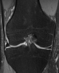
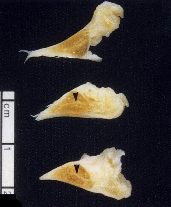
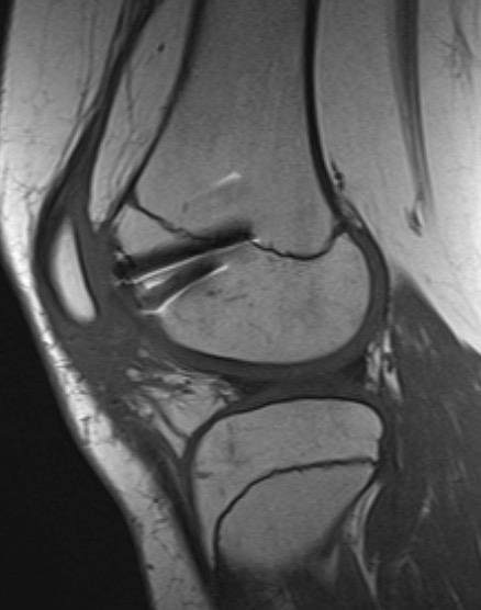
8 Intra-articular, extrasynovia Anterior Cruciate Ligament Two main fiber bundles: anteromedial bundle (AMB) posterolateral bundle (PLB)
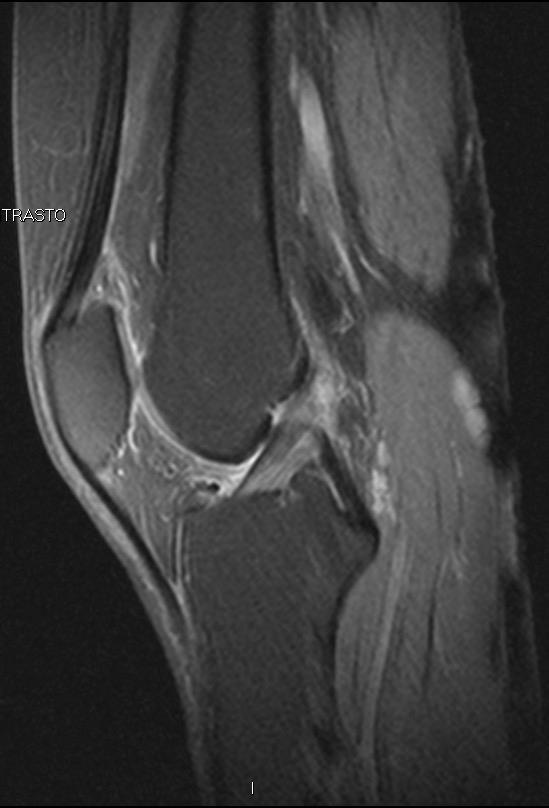
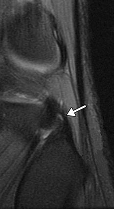
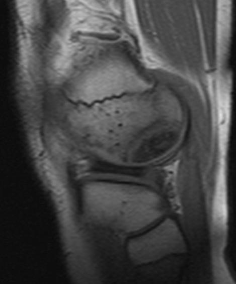
9


10 Non-contact trauma Anterior Cruciate Ligament High risk sports: Ski Soccer, american football Gymnastics LANDING, TWISTING and DECELERATION, particularly with knee in full extension ISOLATED ACL TEARS ARE VERY UNCOMMON Mamoru Niitsu. Springer 2013
11 Three main mechanisms of injury Pivot shift Hyperextension Clip injury Anterior Cruciate Ligament Sanders TG. Radiographics Oct;20 Spec No:S135-


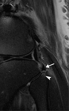
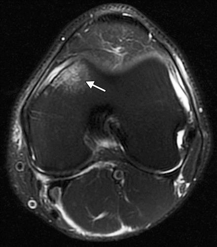
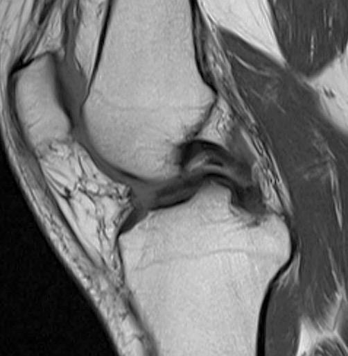
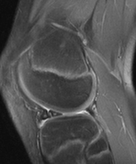
12 Anterior Cruciate Ligament Mechanism of injury: PIVOT SHIFT INJURY


13 Anterior Cruciate Ligament Associated Findings Buckling of the PCL (steep course of distal portion of PCL)


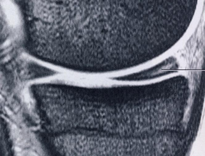
14 Anterior Cruciate Ligament (ACL) Mechanism of injury: HYPEREXTENSION INJURY If a valgus force is also applied at hyperextension, the kissing contusions will be located medially Sanders TG. Radiographics Oct;20 Spec No:S135


15 Anterior Cruciate Ligament CLIP INJURY: O Donoghue triad or unhappy triad
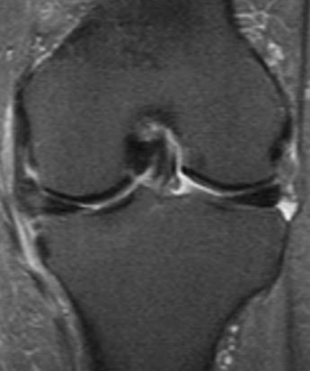
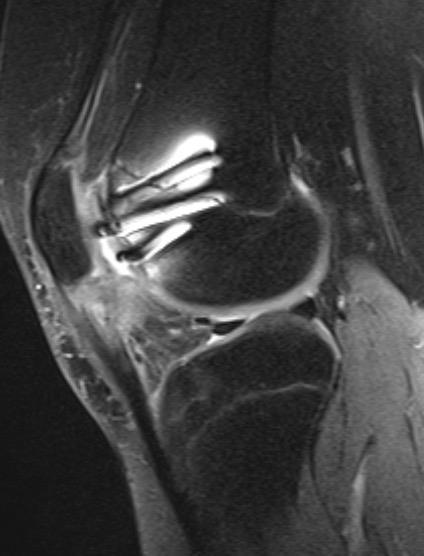
16 Anterior Cruciate Ligament (ACL) MRI FINDINGS: Partial Tears Partial tears may appear as Focal loss of the normal striated appearance (leaving intact fibers on at least one slice) Increased signal intensity on T2-W images with mild swelling of the ligament. AMB constitute the primary (96%) restraining force to anterior drawer at 30 knee flexion Rupture of AMB is believed to be functionally equivalent to complete ACL tear Distinguish partial tears from complete ruptures may be difficult in sagittal plane 40-75% sensitivity 62-89% specificity Non-specificity of signal intensity changes Chen WT. Acta Radiol Sep;43(5):511 Stoller DW. Lippincot Williams & Wilkins 2007:

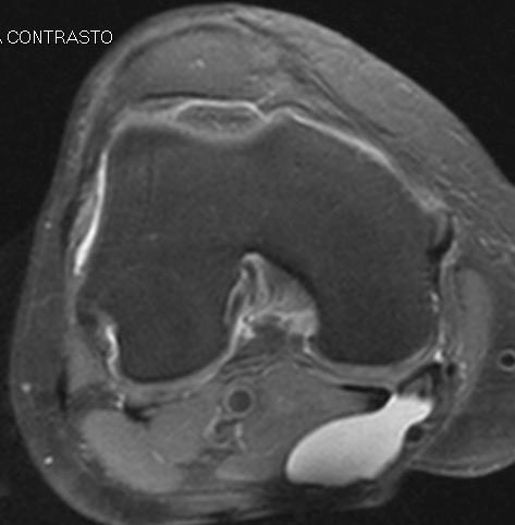
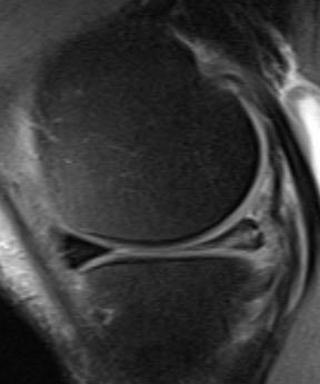
17 Anterior Cruciate Ligament (ACL) INTERPRETATION PITFALLS INTRALIGAMENTOUS GANGLION CYSTS Cysts are located in the proximal-middle portions of intact ACL Herniation of synovial tissue through capsular defect vs mucoid degeneration High signal on fluid-sensitive pulse sequences Do not enhance after intravenous contrast Roberts CC. Magn Reson Imaging Clin N Am. 2007;15 (1): 73-
 Simple anterior")

18 Anterior Cruciate Ligament (ACL) Simple anterior cruciate ligament ganglion cyst involving the whole ligament
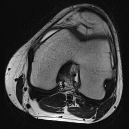
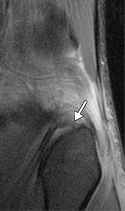
 Mucoid")

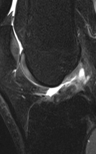
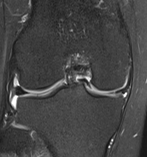
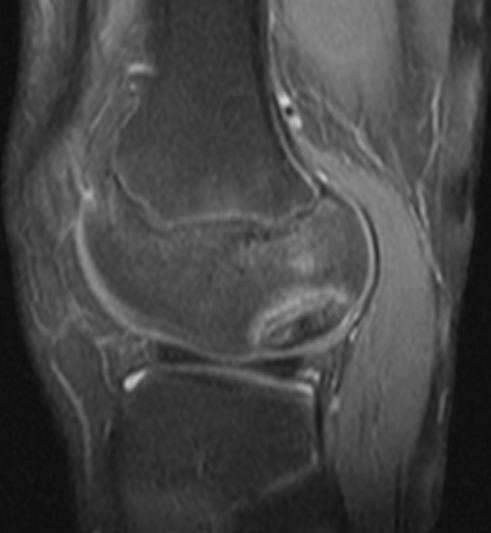
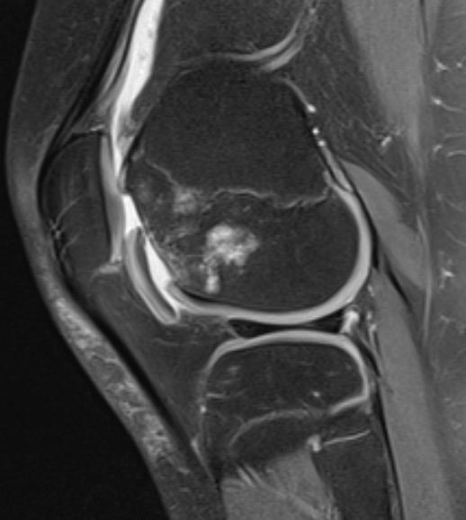
19 Anterior Cruciate Ligament (ACL) MUCOID DEGENERATION ( Celery Stalk Sign ) Mucoid degeneration/fusiform enlargement of the ligament May coesist with intraligamentous cyst
20 Anterior Cruciate Ligament (ACL) CHRONIC TEARS NON-VISUALIZATION of the ligament empty notch sign on coronal MR images ANGULATION of the ligament instead of a straight course Scarring and fibrosis BONE BRUISES typically not present LATERAL FEMORAL NOTCH SIGN Deepened sulcus Indicates a chronic insufficiency of ACL It may be seen in acute injury (impaction) Mamoru Niitsu. Springer 2013

21

22 Posterior Cruciate Ligament (PCL) Intra-articular, extra-synovial structure. Is the primary restraint against posterior subluxation of the tibia Two bundles similar to ACL, but more tightly grouped Two-to four-fold stronger than ACL uniform low signal intensity on MR Impossible to differentiate on MRI

23 Posterior Cruciate Ligament Mechanism of injury DASHBOARD INJURY It is caused by force on the anterior proximal tibia with the knee in flexion It is associated with anterior tibial and posterior patellar edema Associated with rupture of PCL and posterior joint capsule Sanders TG. Radiographics Oct;20 Spec No:S135-


24 Posterior Cruciate Ligament Chronic Partial Tear


25 Posterior Cruciate Ligament Complete Tear
26 Collateral Ligaments Classified in three grades: Grade 1: sprain or strain, mainly consisting of elongation of the ligament without any functional loss. Treated conservatively. Grade 2: partial tear. Grade 3: complete tear. Differentiating between grade 2 and 3 may be impossible (often written as grade 2 3 tear )) Mamoru Niitsu. Springer 2013 De Maeseneer M. Radiographics Oct;20 Spec No:S8
27 Medial Collateral Ligament Primary valgus + internal/external rotation stabilizer (Layer I: Thin sheet that overlies the two heads of the gastrocnemius and the structures of the popliteal fossa.) Layer II: Superficial layer of the MCL (tibial collateral ligament) Layer III: Deepest layer of the MCL called medial capsular ligament, which is continuous with the medial joint capsule. Mamoru Niitsu. Springer 2013 De Maeseneer M. Radiographics Oct;20 Spec No:S8

28 Medial Collateral Ligament

29 Medial Collateral Ligament Linear hyperintensity representing edema along the ligament s fibers due to sprain or strain can be seen. This imaging finding can also be found in medial meniscal tear and knee osteoarthritis. Coronal FS PDWI Mamoru Niitsu. Springer 2013

30 Medial Collateral Ligament Edematous changes can extend into the surrounding medial retinaculum and vastus medialis Mamoru Niitsu. Springer 2013


31 Medial Collateral Ligament Discontinuity of the fibers and signal abnormalities due to edema and hematoma Mamoru Niitsu. Springer 2013
 portion, but it can less commonly occur in the distal (tibial) portion Mamoru Niitsu.")
32 Collateral Ligaments and Posterior Corners MCL tear More than half of MCL tear occurs at the (femoral) portion, but it can less commonly occur in the distal (tibial) portion Mamoru Niitsu. Springer 2013
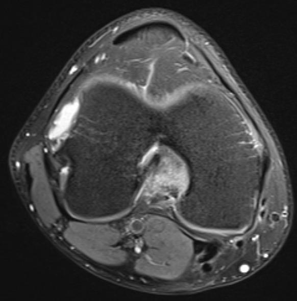
33 Posterolateral Corner Primary stabilizer for varus + external rotation Secondary stabilizer (with CLs) anterior + posterior translation during early (0-30 ) flexion Three layers LCL Popliteus tendon Popliteofibular ligament Rosas H, Radiographics 2016

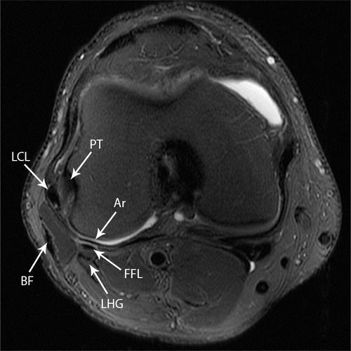
34 Normal Anatomy Rosas H, Radiographics 2016

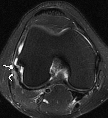
35 LCL grade I tear Rosas H, Radiographics 2016


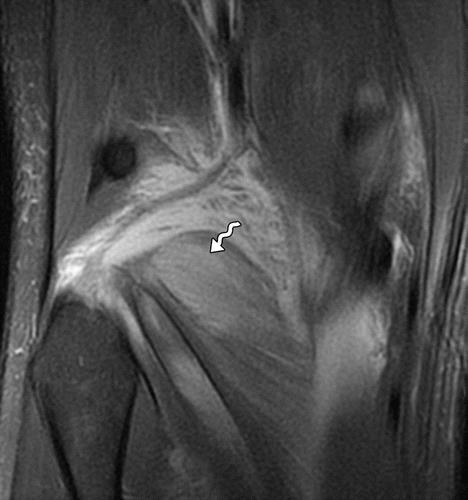
36 LCL grade tears Grade II Grade III Rosas H, Radiographics 2016

37 Popliteus strain Rosas H, Radiographics 2016

38 Popliteomeniscal fascicles Rosas H, Radiographics 2016
39 Popliteofibular ligament Normal PFL Torn PFL Avulsed PFL Rosas H, Radiographics 2016

40 Arcuate ligament injury Rosas H, Radiographics 2016

41 Arcuate + fabellofibular ligament injury Rosas H, Radiographics 2016

42 Associated findings Rosas H, Radiographics 2016


43 Menisci Menisci are comprised of fibrous cartilage, mainly of type I collagen. Collagen fibers form two zones: 1. Peripheral zone: the outer third of the meniscus. Fibers are oriented in circumferential fashion resist longitudinal stresses 2. Inner zone: internal two-third of the meniscus. Fibers are radially oriented prevent longitudinal splitting due to excessive compression Radial fibers Circumferential fibers Aagaard H. Scand J Med Sci Sports Jun;9(3):13


44 Menisci Anterior horn Medial Lateral Posterio r horn

45 Discoid Meniscus
46 Meniscal Tears May be categorized into: TRAUMATIC result from excessive force on a normal meniscus DEGENERATIVE normal force on an abnormally degenerated meniscus Contact with the articular surface on one or two or more sequential images increases confidence of a tear (sensitivity > 90%) Abnormal meniscal morphology: second diagnostic criteria for a tear De Smet AA. AJR Am J Roentgenol Jan;176(1):6 Crues JV et al. Radiology Aug;164(2):445-

47 Degenerative Tears 0 Normal meniscus 1 Focal hyperintensity 2 Linear hyperintensity 3 (3A) 4 (3B) linear hyperintensity reaching the surface = tear multiple linear hyperintensity = multiple tears Degenerative asymptomatic Pain
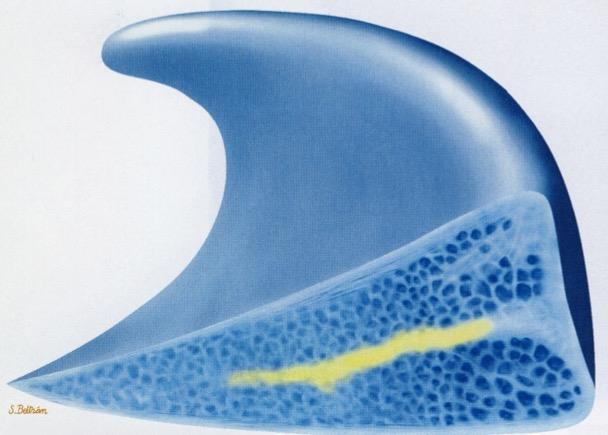
48 Degenerative Tears
49 Degenerative Tears
50 Degenerative Tears
:93 Kaplan PA. AJR Am J Roentgenol. 1991 Feb;156(2):333")
51 Horizontal tear (cleavage tear): parallel to the tibial plateau Longitudinal tear (vertical tear): perpendicular to the tibial plateau; parallel to the long axis of the meniscus Radial tear: perpendicular to the tibial plateau and the long axis of the meniscus Longitudinal Meniscal Tears Radial Flap Complex Jee WH. AJR Am J Roentgenol Jan;180(1):93 Kaplan PA. AJR Am J Roentgenol Feb;156(2):333


52 VERTICAL RADIAL TEARS traverse circumferential fibers SHEAR FORCES Meniscal Tears VERTICAL LONGITUDINAL TEA between circumferential fibers COMPRESSION FORCES


53 VERTICAL RADIAL TEARS coronal S a g i t t a l
54 DISPLACED MENISCAL TEARS VERTICAL Longitudinal tear -> Bucket-handle VERTICAL Radial tear -> Parrot-beak HORIZONTAL tear -> Flap
55 BUCKET-HANDLE TEAR
56 BUCKET-HANDLE TEAR

:1429-34.")


57 Harper KW. AJR Am J Roentgenol Dec;185(6): PARROT-BEAK TEAR Radial tear on the inner part of the meniscus and longitudinal in the peripheral aspect

58 MENISCAL ROOT TEAR

59 Superficial Zone Transitional Zone - Arc-like oriented collagen II fibrils - Chondrocytes Proteoglycans - Water Radial Zone -Vertically oriented collagen II fibrils -Chondrocytes -Proteoglycans -Water Tide Mark Calcified Cartilage - steplike Jiunction Subchondral bone plate Vascular plexus
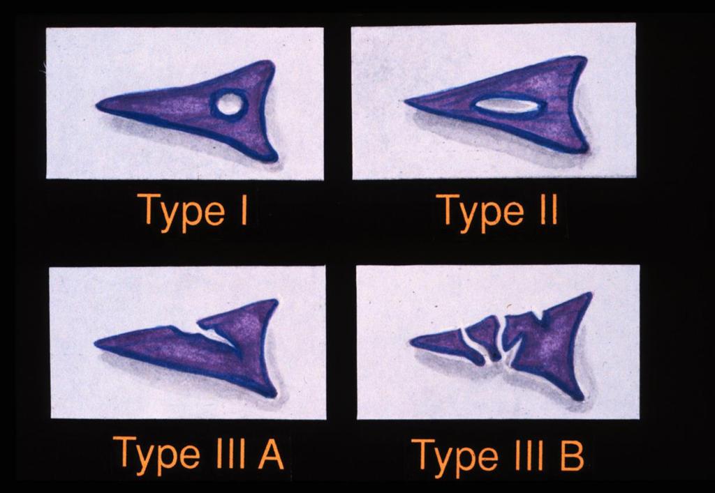
60 Arthroscopic Cartilage Lesion Classification System described by Outerbridge Grade 0 = normal cartilage Grade 1 = thickening and softening Deep disruption of the collagen framework allowing the proteoglycans to increase the hydratation of cartilage, leading to cartilage thickening and softening Grade 2 = Superficial fissuring Articular Cartilage Damage: Grading Grade 3 = Deep partial-thickness defect Grade 4 = Full-thickness cartilage defect Grades 2, 3 and 4 can be visualized with imaging These grades can be used by radiologists (Yulish et al.)

61 Grade 0 Normal articular cartilage Normal cartilage with gray-scale stratification

62 Superficial lesions, chondral softening Increase signal in articular cartilage Grade 1


63 Superficial lesions extending down to <50% of cartilage depth Fissure, fibrillation involving < 50% thickness Linear-to-ovoid foci of increased signal involving <50% thickness Grade 2 Fibrillation <50% Fissure <50%

64 Defects extending down >50% of depth but not through subchondral bone Blisters/fissures/ fibrillation involving >50% thickness Linear-to-ovoid foci increased signal involving >50% of cartilage but not extending down to bone Grade 3


65 Transverse T2 FS weighted MR of patella. Focal ulceration is seen Grade 3 chondropathy.

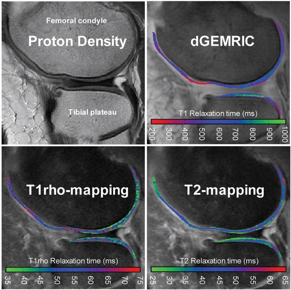
66 Ulceration to subchondral bone Exposed subchondral bone Complete loss of articular cartilage or surface flap Grade 4
67

68 Dark Cartilage Lesions True Positive Courtesy of R. Kijowski and BK Markhardt Fat-Sat T2-FSE Fat-Sat IW-FSE

69 Dark Cartilage Lesions True Positive Courtesy of R. Kijowski and BK Markhardt Grade 2b Fissuring Fat-Sat IW-FSE

70 Dark Cartilage Lesions True Positive Courtesy of R. Kijowski and BK Markhardt Fat-Sat T2-FSE Fat-Sat IW-FSE

71 Dark Cartilage Lesions True Positive Courtesy of R. Kijowski and BK Markhardt Grade 2b Fissuring Fat-Sat IW-FSE

72 Dark Cartilage Lesions True Positive Courtesy of R. Kijowski and BK Markhardt Fat-Sat IW-FSE Fat-Sat T2-FSE

73 Dark Cartilage Lesions True Positive Courtesy of R. Kijowski and BK Markhardt Grade 2b Flap Tear Fat-Sat T2-FSE

74 Dark Cartilage Lesions True Positive Courtesy of R. Kijowski and BK Markhardt IW-FSE Fat-Sat IW-FSE

75 Dark Cartilage Lesions True Positive Courtesy of R. Kijowski and BK Markhardt Grade 1b Fibrillation Fat-Sat IW-FSE

76 Dark Cartilage Lesions False Positive Courtesy of R. Kijowski and BK Markhardt IW-FSE Fat-Sat IW-FSE

77 Dark Cartilage Lesions False Positive Courtesy of R. Kijowski and BK Markhardt Normal Fat-Sat IW-FSE
78 Dark Cartilage Lesions Areas of low T2 signal within morphologically normal cartilage may be seen on all articular surfaces of knee joint Significant proportion of dark cartilage lesions were found to correspond to areas of cartilage degeneration at arthroscopy Dark cartilage lesions should be reported as areas of possible cartilage degeneration, especially in older individuals Courtesy of R. Kijowski and BK Markhardt

79 Dijkgraaf LC et al, 1995 Quantitative MR imaging Cartilage microstructure breaks down Decreased organization of matrix Loss of PG and GAG Increased water content Ability to detect changes prior to morphological degradation
![Limitations Accumulation inversely proportional to [GAG] dgemric Reduced T1 relaxation times in regions](/docs-images/77/75901385/images/80-0.jpg "with high [Gd(DTPA)2- ] T1 relaxation used as quantitative outcome, with lower T1 indicating reduced")
80 Limitations Accumulation inversely proportional to [GAG] dgemric Reduced T1 relaxation times in regions with high [Gd(DTPA)2- ] T1 relaxation used as quantitative outcome, with lower T1 indicating reduced [GAG]

81 MARATHON BEFORE 1 DAY 1 WEEK

82 Healthy cartilage traps water molecules -> lower SI on T2w Degenerated cartilage has increased free water content -> higher SI on T2w 3D multi-echo sequences Efficacy proven in vivo and in vitro Remarkable limitations T2 mapping


83 T2 mapping Cartilage degradation -> T2 prolongation

84 Pre-treatment Monitoring of the effect of intra-articular injections of hyaluronates Ferrero et al. Eur Radiol 2017 in press T2 MAPPING Post-treatment Yun Sun Choi, et al 2008
85 PROs Higher image resolution Susceptibility artifacts Isotropic 3D sequences Faster acquisition UTE-T2* mapping T2* mapping Anisotropy Joint fluid CONs

86 T2* mapping
 DWI Other")
87 Sodium MRI Ultrashort TE (UTE): T2/T2*; (T1/T1rho) GAG chemical exchange saturation transfer (gagcest) DWI Other techniques
88
89 Bone Marrow Edema Differential diagnosis Secondary to cartilage wear Osteoarhtritis Inflammatory osteoartrhopathy SONK Insufficiency fracture Stress fracture SONK Other


90 Bone Marrow Edema SONK

91 Bone Marrow Edema Insufficiency fracture

92 Bone Marrow Edema Stress fracture
93 Osteochondritis Dissecans Main features Age: years Location: lateral surface of medial condyle (55%), central portion (25%) Associated to discoid meniscus In older patients, signs may overlap to SONK Osteochondrosis characterized by necrosis of bone followed by reossification and healing History of trauma in 50% of patients Vascular theory
94
95
96
97 Imaging protocol Outline Cruciate and collateral ligaments Menisci Cartilage Bone marrow edema
98 ThankYou. Luca Maria Sconfienza, MD PhD Chair, Unit of Diagnostic and Interventional Radiology, IRCCS Istituto Ortopedico Galeazzi Associate Professor of Radiology, University of Milano Milano, Italy
MRI KNEE WHAT TO SEE. Dr. SHEKHAR SRIVASTAV. Sr.Consultant KNEE & SHOULDER ARTHROSCOPY
 MRI KNEE WHAT TO SEE Dr. SHEKHAR SRIVASTAV Sr.Consultant KNEE & SHOULDER ARTHROSCOPY MRI KNEE - WHAT TO SEE MRI is the most accurate and frequently used diagnostic tool for evaluation of internal derangement
Imaging the Knee 17/10/2017. Friction syndrome Common in runners or cyclists Fluid between ITB and Lateral femoral condyle
 17/10/2017 Imaging the Knee Alicia M. Yochum RN, DC, DACBR, RMSK Iliotibial Band Syndrome Ligamentous Tears (ACL, PCL, MCL, LCL) Meniscal Tears Cartilage Degeneration Quadriceps/Patellar tendinosis Osteochondral
MRI of the Knee: Part 2 - menisci. Mark Anderson, M.D. University of Virginia Health System
 MRI of the Knee: Part 2 - menisci Mark Anderson, M.D. University of Virginia Health System Learning Objectives At the end of the presentation, each participant should be able to: describe the normal anatomy
This presentation is the intellectual property of the author. Contact them for permission to reprint and/or distribute.
 MRI of the Knee Jennifer Swart, M.D. Musculoskeletal Radiology South Texas Radiology Group Outline Coils, Patient Positioning Acquisition Parameters, Planes and Pulse Sequences Knee Arthrography Normal
This presentation is the intellectual property of the author. Contact them at for permission to reprint and/or distribute.
 MRI of the Knee Jennifer Swart, M.D. Musculoskeletal Radiology South Texas Radiology Group Financial Disclosure Dr. Jennifer Swart has no relevant financial relationships with commercial interests to disclose.
Imaging the Athlete s Knee. Peter Lowry, MD Musculoskeletal Radiology University of Colorado
 Imaging the Athlete s Knee Peter Lowry, MD Musculoskeletal Radiology University of Colorado None Disclosures Knee Imaging: Radiographs Can be performed weight-bearing or non-weight-bearing View options
Imaging of Articular Cartilage
 Clinical Imaging of Articular Cartilage Imaging of Articular Cartilage Prof. Dr. K. Verstraete Ghent University Introduction : Articular Cartilage Histology and biochemical composition Review of Imaging
Knee Contusions and Stress Injuries. Laura W. Bancroft, M.D.
 Knee Contusions and Stress Injuries Laura W. Bancroft, M.D. Objectives Review 5 types of contusion patterns Pivot shift Dashboard Hyperextension Clip Lateral patellar dislocation Demonstrate various stress
Imaging of the Athle/c Knee: injuries associated with ACL disrup/on
 Imaging of the Athle/c Knee: injuries associated with ACL disrup/on Brian Petersen, MD Associate Professor of Radiology and Orthopaedics Chief of MSK Radiology University of Colorado CU Sports Medicine
ADVANCED IMAGING OF THE KNEE
 MENISCAL ANATOMY ADVANCED IMAGING OF THE KNEE MENISCAL ABNORMALITIES MENISCAL FUNCTION MENISCAL FUNCTION load transmission shock absorption stability The menisci DO NOT function as primary stabilizers
Differential Diagnosis
 Case 31yo M who sustained an injury to L knee while playing Basketball approximately 2 weeks ago. He describes pivoting and hyperextending his knee, which swelled over the next few days. He now presents
MRI of ligaments. Ligament biomechanics Spine Shoulder Elbow Hand/wrist Pelvis/hip Knee Foot/ankle
 MRI of ligaments Chang Ho Kang M.D. Korea University Anam Hospital Spine Shoulder Elbow Hand/wrist Pelvis/hip Knee Foot/ankle Introduction Ligament Fibrous connective tissue Attaches bone to bone Holds
Role of magnetic resonance imaging in the evaluation of traumatic knee joint injuries
 Original Research Article Role of magnetic resonance imaging in the evaluation of traumatic knee joint injuries Dudhe Mahesh 1*, Rathi Varsha 2 1 Resident, 2 Professor, Department of Radio-Diagnosis, Grant
On Field Assessment and Management of Acute Knee Injuries: A Physiotherapist s Perspective
 On Field Assessment and Management of Acute Knee Injuries: A Physiotherapist s Perspective Jessica Condliffe Physiotherapist / Clinic Manager TBI Health Wellington Presentation Outline Knee anatomy review
ORIGINAL ARTICLE. ROLE OF MRI IN EVALUATION OF TRAUMATIC KNEE INJURIES Saurabh Chaudhuri, Priscilla Joshi, Mohit Goel
 ROLE OF MRI IN EVALUATION OF TRAUMATIC KNEE INJURIES Saurabh Chaudhuri, Priscilla Joshi, Mohit Goel 1. Associate Professor, Department of Radiodiagnosis & imaging, Bharati Vidyapeeth Medical College and
Knee MRI Update Case Review 2009 Russell C. Fritz, M.D. National Orthopedic Imaging Associates San Francisco, CA
 Knee MRI Update Case Review 2009 Russell C. Fritz, M.D. National Orthopedic Imaging Associates San Francisco, CA Meniscal Tears -linear increased signal extending to an articular surface is the hallmark
MENISCAL INJURY. Meniscus. Anterior Roots. Medial Meniscus. Lateral Meniscus. Posterior Roots. MRI and Arthroscopic Findings
 Meniscus Anterior Roots MENISCAL INJURY MRI and Arthroscopic Findings Medial Meniscus AH PH PH AH Lateral Meniscus Rawiwan Pattaweerakul Naresuan University Hospital Posterior Roots Meniscus Normal Meniscus
Knee: Meniscus Back to Basics
 Knee: Meniscus Back to Basics Kyung Jin Suh kyungjin.suh@gmail.com Doctor Radiology, Daegu, KOREA Medial Lateral 7.7 10.2 11.6 9.6 10.6 mm Posterior > Anterior horn 10.6 mm Posterior = Anterior horn Medial
Multi-ligamentous knee injuries - MRI injury patterns at a glance
 Multi-ligamentous knee injuries - MRI injury patterns at a glance Poster No.: P-0068 Congress: ESSR 2015 Type: Educational Poster Authors: A. Rastogi, D. Whelan, R. Martin, W. Mak, D. Pearce ; 1 1 1 2
MRI Evaluation of Internal derangement of the Knee Joint - A PICTORIAL REVIEW
 MRI Evaluation of Internal derangement of the Knee Joint - A PICTORIAL REVIEW Poster No.: C-1386 Congress: ECR 2015 Type: Educational Exhibit Authors: V. Nadaraja, F. Abubacker Sulaiman; Chennai/IN Keywords:
Knee Injury Assessment
 Knee Injury Assessment Clinical Anatomy p. 186 Femur Medial condyle Lateral condyle Femoral trochlea Tibia Intercondylar notch Tibial tuberosity Tibial plateau Fibula Fibular head Patella Clinical Anatomy
Knee: Cruciate Ligaments
 72 Knee: Cruciate Ligaments R. Kent Sanders Sagittal oblique 2.5-mm sequences along the plane of the anterior cruciate ligament (ACL) typically yield three to four images of the ACL, with the first medial
Meniscal Tears with Fragments Displaced: What you need to know.
 Meniscal Tears with Fragments Displaced: What you need to know. Poster No.: C-1339 Congress: ECR 2015 Type: Authors: Keywords: DOI: Educational Exhibit M. V. Ferrufino, A. Stroe, E. Cordoba, A. Dehesa,
ACL AND PCL INJURIES OF THE KNEE JOINT
 ACL AND PCL INJURIES OF THE KNEE JOINT Dr.KN Subramanian M.Ch Orth., FRCS (Tr & Orth), CCT Orth(UK) Consultant Orthopaedic Surgeon, Special interest: Orthopaedic Sports Injury, Shoulder and Knee Surgery,
Why Talk About Technique? MRI of the Knee:
 Why Talk About Technique? MRI of the Knee: Part 1 - Imaging Techniques Mark Anderson, M.D. University of Virginia Health Sciences Center Charlottesville, Virginia Always had an interest teach our fellows
Post-injury painful and locked knee
 H R J Post-injury painful and locked knee, p. 54-59 Clinical Case - Test Yourself Musculoskeletal Imaging Post-injury painful and locked knee Ioannis I. Daskalakis 1, 2, Apostolos H. Karantanas 1, 2 1
Stability of Post Traumatic Osteochondritis Dissecans of the Knee: MR Imaging Findings
 Chin J Radiol 2005; 30: 199-204 199 Stability of Post Traumatic Osteochondritis Dissecans of the Knee: MR Imaging Findings YU-CHUNG HUNG 1 JON-KWAY HUANG 1,2 Department of Radiology 1, Mackay Memorial
Joints of the Lower Limb II
 Joints of the Lower Limb II Lecture Objectives Describe the components of the knee and ankle joint. List the ligaments associated with these joints and their attachments. List the muscles acting on these
The Knee. Two Joints: Tibiofemoral. Patellofemoral
 Evaluating the Knee The Knee Two Joints: Tibiofemoral Patellofemoral HISTORY Remember the questions from lecture #2? Girth OBSERVATION TibioFemoral Alignment What are the consequences of faulty alignment?
MRI grading of postero-lateral corner and anterior cruciate ligament injuries
 MRI grading of postero-lateral corner and anterior cruciate ligament injuries Poster No.: C-2533 Congress: ECR 2012 Type: Educational Exhibit Authors: J. Lopes Dias, J. A. Sousa Pereira, L. Fernandes,
JMSCR Vol 05 Issue 01 Page January
 www.jmscr.igmpublication.org Impact Factor 5.244 Index Copernicus Value: 83.27 ISSN (e)-2347-176x ISSN (p) 2455-0450 DOI: https://dx.doi.org/10.18535/jmscr/v5i1.28 Diagnostic Accuracy of Magnetic Resonance
RECENT ADVANCES IN CLINICAL MR OF ARTICULAR CARTILAGE
 In Practice RECENT ADVANCES IN CLINICAL MR OF ARTICULAR CARTILAGE By Atsuya Watanabe, MD, PhD, Director, Advanced Diagnostic Imaging Center and Associate Professor, Department of Orthopedic Surgery, Teikyo
Ligamentous and Meniscal Injuries: Diagnosis and Management
 Ligamentous and Meniscal Injuries: Diagnosis and Management Daniel K Williams, MD Franciscan Physician Network Orthopedic Specialists September 29, 2017 No Financial Disclosures INTRODUCTION Overview of
Overview Ligament Injuries. Anatomy. Epidemiology Very commonly injured joint. ACL Injury 20/06/2016. Meniscus Tears. Patellofemoral Problems
 Overview Ligament Injuries Meniscus Tears Pankaj Sharma MBBS, FRCS (Tr & Orth) Consultant Orthopaedic Surgeon Manchester Royal Infirmary Patellofemoral Problems Knee Examination Anatomy Epidemiology Very
The Meniscus. History. Anatomy. Anatomy. Blood Supply. Attachments
 History The Meniscus W. Randall Schultz, MD, MS Austin, TX January 23, 2016 Meniscus originally thought to represent vestigial tissue 1883 first reported meniscal repair (Annandale) Total menisectomy treatment
Role of Magnetic Resonance Imaging in Patients with Knee Trauma
 Original Research Article Role of Magnetic Resonance Imaging in Patients with Knee Trauma Bhautik Kapadia 1, Bhumika Suthar 2* 1 Associate Professor, 2 Assistant Professor, Department of Radiodiagnosis,
Financial Disclosure. Medial Collateral Ligament
 Matthew Murray, M.D. UTHSCSA Sports Medicine Financial Disclosure Dr. Matthew Murray has no relevant financial relationships with commercial interests to disclose. Medial Collateral Ligament Most commonly
W. Dilworth Cannon, M.D. Professor of Clinical Orthopaedic Surgery University of California San Francisco
 Knee Pain And Injuries In Adults W. Dilworth Cannon, M.D. Professor of Clinical Orthopaedic Surgery University of California San Francisco Pain Control Overview Narcotics rarely necessary after 1 st 1-2
Anterior Cruciate Ligament Surgery
 Anatomy Anterior Cruciate Ligament Surgery Roger Ostrander, MD Andrews Institute Anatomy Anatomy Function Primary restraint to anterior tibial translation Secondary restraint to internal tibial rotation
Meniscal tears on 3T MR: Patterns, pearls and pitfalls
 Meniscal tears on 3T MR: Patterns, pearls and pitfalls Poster No.: C-2221 Congress: ECR 2010 Type: Educational Exhibit Topic: Musculoskeletal Authors: J. C. Kandathil; Singapore/SG Keywords: Knee injuries,
The posterolateral corner of the knee: the normal and the pathological
 The posterolateral corner of the knee: the normal and the pathological Poster No.: P-0104 Congress: ESSR 2014 Type: Educational Poster Authors: M. Bartocci 1, C. Dell'atti 2, E. Federici 1, V. Martinelli
The Impact of Age on Knee Injury Treatment
 The Impact of Age on Knee Injury Treatment Focus on the Meniscus Dr. Alvin J. Detterline, MD Sports Medicine and Orthopaedic Surgery Towson Orthopaedic Associates University of Maryland St. Joseph Medical
The Knee. Prof. Oluwadiya Kehinde
 The Knee Prof. Oluwadiya Kehinde www.oluwadiya.sitesled.com The Knee: Introduction 3 bones: femur, tibia and patella 2 separate joints: tibiofemoral and patellofemoral. Function: i. Primarily a hinge joint,
Learning IRM. The Knee: lateral ligaments and anatomical quadrants.
 Learning IRM. The Knee: lateral ligaments and anatomical quadrants. Poster No.: C-1733 Congress: ECR 2014 Type: Educational Exhibit Authors: A. Amador Gil, M. D. C. Jurado Gómez, V. de Lara Bendahan ;
ACL Athletic Career. ACL Rupture - Warning Features Intensive pain Immediate swelling Locking Feel a Pop Dead leg Cannot continue to play
 FIMS Ambassador Tour to Eastern Europe, 2004 Belgrade, Serbia Montenegro Acute Knee Injuries - Controversies and Challenges Professor KM Chan OBE, JP President of FIMS Belgrade ACL Athletic Career ACL
Anatomy and Sports Injuries of the Knee
 Anatomy and Sports Injuries of the Knee I. Anatomy II. Assessment III. Treatment IV. Case Study V. Dissection Anatomy Not a hinge joint 6 degrees of freedom Flexion/Extension Rotation Translation Anatomy
MRI Findings of Posterolateral Corner Injury on Threedimensional
 MRI Findings of Posterolateral Corner Injury on Threedimensional Isotropic SPACE. Poster No.: C-1792 Congress: ECR 2013 Type: Scientific Exhibit Authors: S.-W. Lee, Y. M. Jeong, J. A. Sim, S. Ahn; Incheon/KR
Knee Joint Anatomy 101
 Knee Joint Anatomy 101 Bone Basics There are three bones at the knee joint femur, tibia and patella commonly referred to as the thighbone, shinbone and kneecap. The fibula is not typically associated with
and K n e e J o i n t Is the most complicated joint in the body!!!!
 K n e e J o i n t K n e e J o i n t Is the most complicated joint in the body!!!! 1-Consists of two condylar joints between: A-The medial and lateral condyles of the femur and The condyles of the tibia
Original Report. The Reverse Segond Fracture: Association with a Tear of the Posterior Cruciate Ligament and Medial Meniscus
 Eva M. Escobedo 1 William J. Mills 2 John. Hunter 1 Received July 10, 2001; accepted after revision October 1, 2001. 1 Department of Radiology, University of Washington Harborview Medical enter, 325 Ninth
What is the most effective MRI specific findings for lateral meniscus posterior root tear in ACL injuries
 What is the most effective MRI specific findings for lateral meniscus posterior root tear in ACL injuries Kazuki Asai 1), Junsuke Nakase 1), Kengo Shimozaki 1), Kazu Toyooka 1), Hiroyuki Tsuchiya 1) 1)
Meniscal Tears: Role of Axial MRI Alone and in Combination with Other Imaging Planes
 Nefise Cagla Tarhan 1,2 Christine. Chung 1 urea Valeria Rosa Mohana-orges 1 Tudor Hughes 1 Donald Resnick 1 Received September 30, 2003; accepted after revision February 2, 2004. 1 Department of Radiology,
MENISCAL INJURIES. (copyright s h palmer 2009) MENISCAL FUNCTION
 MENISCAL FUNCTION") (copyright s h palmer 2009) MENISCAL FUNCTION MENISCAL INJURIES Menisci are important for weight bearing, load distribution, joint stability and proprioception. Figure 1: A normal medial meniscus Any load
(copyright s h palmer 2009) MENISCAL FUNCTION MENISCAL INJURIES Menisci are important for weight bearing, load distribution, joint stability and proprioception. Figure 1: A normal medial meniscus Any load
MR imaging of the knee in marathon runners before and after competition
 Skeletal Radiol (2001) 30:72 76 International Skeletal Society 2001 ARTICLE W. Krampla R. Mayrhofer J. Malcher K.H. Kristen M. Urban W. Hruby MR imaging of the knee in marathon runners before and after
Ultrasound of the Knee
 Ultrasound of the Knee Jon A. Jacobson, M.D. Professor of Radiology Director, Division of Musculoskeletal Radiology University of Michigan Disclosures: Consultant: Bioclinica Book Royalties: Elsevier Advisory
Meniscus T2 Relaxation Time at Various Stages of Knee Joint Degeneration
 Meniscus T2 Relaxation Time at Various Stages of Knee Joint Degeneration Richard Kijowski, Michael Fazio, Benjamin Beduhn, and Fang Liu Department of Radiology University of Wisconsin School of Medicine
Posterolateral Corner Injuries of the Knee: Pearls and Pitfalls
 Posterolateral Corner Injuries of the Knee: Pearls and Pitfalls Robert A. Arciero,MD,Col,ret Professor, Orthopaedics University of Connecticut Incidence of PLC Injuries with ACL Tears Fanelli, 1995 12%
MY PATIENT HAS KNEE PAIN. David Levi, MD Chief, Division of Musculoskeletal l limaging Atlantic Medical Imaging
 MY PATIENT HAS KNEE PAIN David Levi, MD Chief, Division of Musculoskeletal l limaging Atlantic Medical Imaging Causes of knee pain Non traumatic Trauma Osteoarthritis Patellofemoral pain Menisci or ligaments
B one contusion is a finding substantiated by magnetic
 592 ORIGINAL ARTICLE The appearance of kissing contusion in the acutely injured knee in the athletes I P Terzidis, A G Christodoulou, A L Ploumis, S R Metsovitis, M Koimtzis, P Givissis... See end of article
SOFT TISSUE INJURIES OF THE KNEE: Primary Care and Orthopaedic Management
 SOFT TISSUE INJURIES OF THE KNEE: Primary Care and Orthopaedic Management Gauguin Gamboa Australia has always been a nation where emphasis on health and fitness has resulted in an active population engaged
Knee, Ankle, and Foot: Normal and Abnormal Features with MRI and Ultrasound Correlation. Disclosures. Outline. Joint Effusion. Suprapatellar recess
 Knee, Ankle, and Foot: Normal and Abnormal Features with MRI and Ultrasound Correlation Jon A. Jacobson, M.D. Professor of Radiology Director, Division of Musculoskeletal Radiology University of Michigan
The Knee. Tibio-Femoral
 The Knee Tibio-Femoral Osteology Distal Femur with Proximal Tibia Largest Joint Cavity in the Body A modified hinge joint with significant passive rotation Technically, one degree of freedom (Flexion/Extension)
Clinics in diagnostic imaging (177)
") Singapore Med J 2017; 58(5): 241-245 doi: 10.11622/smedj.2017038 CMEArticle Clinics in diagnostic imaging (177) Poh Lye Paul See, MBBS, FRCR Fig. 1 Sagittal proton density (PD)-weighted fast spin-echo
Singapore Med J 2017; 58(5): 241-245 doi: 10.11622/smedj.2017038 CMEArticle Clinics in diagnostic imaging (177) Poh Lye Paul See, MBBS, FRCR Fig. 1 Sagittal proton density (PD)-weighted fast spin-echo
Unlocking the locked Knee
 Unlocking the locked Knee Poster No.: P-0027 Congress: ESSR 2013 Type: Scientific Exhibit Authors: J. P. SINGH, S. Srivastava, S. S. BAIJAL ; Gurgaon, Delhi 1 1 2 1 2 NCR/IN, LUCKNOW, UTTAR PRADESH/IN
BASELINE QUESTIONNAIRE (SURGEON)
") SECTION A: STUDY INFORMATION Subject ID: - - Study Visit: Baseline Site Number: Date: / / Surgeon ID: SECTION B: INITIAL SURGEON HISTORY B1. Previous Knee Surgery: Yes No Not recorded B2. Number of Previous
SECTION A: STUDY INFORMATION Subject ID: - - Study Visit: Baseline Site Number: Date: / / Surgeon ID: SECTION B: INITIAL SURGEON HISTORY B1. Previous Knee Surgery: Yes No Not recorded B2. Number of Previous
"BONE BRUISES" OF THE KNEE: A REVIEW
 "BONE BRUISES" OF THE KNEE: A REVIEW Chad E. Mathis, M.D. Ken Noonan, M.D. Kosmas Kayes, M.D. ABSTRACT Magnetic resonance (MR) imaging is often used - to assess the location and degree of ligamentous wm.
Do Not Fall on Your Knees - Recognizing Common and Uncommon Pitfalls that May Simulate Meniscal Tears
 Do Not Fall on Your Knees - Recognizing Common and Uncommon Pitfalls that May Simulate Meniscal Tears Poster No.: C-1146 Congress: ECR 2016 Type: Educational Exhibit Authors: P. Musa Aguiar, J. Goncalves,
Medical Practice for Sports Injuries and Disorders of the Knee
 Sports-Related Injuries and Disorders Medical Practice for Sports Injuries and Disorders of the Knee JMAJ 48(1): 20 24, 2005 Hirotsugu MURATSU*, Masahiro KUROSAKA**, Tetsuji YAMAMOTO***, and Shinichi YOSHIDA****
MR Imaging Based Diagnosis of Anterior Cruciate Ligament Tears in Patients with Internal Derangement of Knee
 DOI: 10.7860/IJARS/2017/26026:2263 Radiology Section Original Article MR Imaging Based Diagnosis of Anterior Cruciate Ligament Tears in Patients with Internal Derangement of Knee Hemanth Purigali Naganna,
KNEE EXAMINATION. Tips & Tricks from an Emergency Physician Perspective. EM Physicians Less Exposed to MSK Medicine
 KNEE EXAMINATION Tips & Tricks from an Emergency Physician Perspective Dr P O CONNOR Emergency Medicine Physician EUSEM 10/09/2018 EM Physicians Less Exposed to MSK Medicine Musculoskeletal Medicine becoming
Original Research Article. Atul Bucha 1, Sunita Dashottar 2 *, Surabhi Sharma 2, Preeth Pany 2, Rushabh Bhikhanhai Suthar 2
 International Journal of Advances in Medicine http://www.ijmedicine.com pissn 2349-3925 eissn 2349-3933 Original Research Article DOI: http://dx.doi.org/10.18203/2349-3933.ijam20184747 A study to evaluate
SSSR. 1. Nov Ankle. Postoperative Imaging of Cartilage Repair. and Lateral Ligament Reconstruction
 Ankle Postoperative Imaging of Cartilage Repair and Lateral Ligament Reconstruction Andrea B. Rosskopf, MD University Hospital Balgrist Imaging of Cartilage Repair Why? To assess the technical success
In the name of god. Knee. By: Tofigh Bahraminia Graduate Student of the Pathology Sports and corrective actions. Heat: Dr. Babakhani. Nov.
 In the name of god Knee By: Tofigh Bahraminia Graduate Student of the Pathology Sports and corrective actions Heat: Dr. Babakhani Nov. 2014 1 Anatomy-Bones Bones Femur Medial/lateral femoral condyles articulate
Learning Objectives. Lecture Outline. Knee Stability. Cruciate Ligaments. Knee Stability. MRI of the Knee: Part 3: ligaments
 Learning Objectives MRI of the Knee: Part 3: ligaments Mark Anderson, M.D. University of Virginia Health System At the end of the presentation, each participant should be able to: describe the anatomy
MRI of the Knee: Part 4 - normal variants that may simulate disease. Mark Anderson, M.D. University of Virginia
 MRI of the Knee: Part 4 - normal variants that may simulate disease Mark Anderson, M.D. University of Virginia discuss the most common normal variants in the pediatric knee that may simulate pathology
UCLA UCLA Previously Published Works
 UCLA UCLA Previously Published Works Title MR-IMAGING OF TIBIAL COLLATERAL LIGAMENT INJURY - COMPARISON WITH CLINICAL EXAMINATION Permalink https://escholarship.org/uc/item/2bs9g934 Journal SKELETAL RADIOLOGY,
Comparative study of high resolusion ultrasonography and magnetic resonance imaging in diagnosing traumatic knee injuries & pathologies
 Original article: Comparative study of high resolusion ultrasonography and magnetic resonance imaging in diagnosing traumatic knee injuries & pathologies Dr. Rakesh Gujjar*, Dr. R. P. Bansal, Dr. Sandeep
CMEArticle Clinics in diagnostic imaging (141)
") Singapore Med J 2012; 53(9) : 625 CMEArticle Clinics in diagnostic imaging (141) Hollie MY Lim 1, MBBS, FRCR, Wilfred CG Peh 1, FRCP, FRCR 1a 1b 1c Fig. 1 (a & b) Contiguous sagittal fat-suppressed PD-W
Singapore Med J 2012; 53(9) : 625 CMEArticle Clinics in diagnostic imaging (141) Hollie MY Lim 1, MBBS, FRCR, Wilfred CG Peh 1, FRCP, FRCR 1a 1b 1c Fig. 1 (a & b) Contiguous sagittal fat-suppressed PD-W
ACL RECONSTRUCTION HAMSTRING METHOD. Presents ACL RECONSTRUCTION HAMSTRING METHOD. Multimedia Health Education
 HAMSTRING METHOD Presents HAMSTRING METHOD Multimedia Health Education Disclaimer Stephen J. Incavo MD This movie is an educational resource only and should not be used to make a decision on Anterior Cruciate
Your Practice Online
 Your Practice Online Disclaimer P R E S E N T S - PATELLAR TENDON This movie is an educational resource only and should not be used to make a decision on Anterior Cruciate Ligament (ACL) Reconstruction.
Knee Joint Assessment and General View
 Knee Joint Assessment and General View Done by; Mshari S. Alghadier BSc Physical Therapy RHPT 366 m.alghadier@sau.edu.sa http://faculty.sau.edu.sa/m.alghadier/ Functional anatomy The knee is the largest
Spectrum of meniscal lesions: An MR teaching atlas
 Spectrum of meniscal lesions: An MR teaching atlas Poster No.: C-2284 Congress: ECR 2010 Type: Educational Exhibit Topic: Musculoskeletal - Joints Authors: C. Leal, P. Alves, H. A. M. R. Tinto, J. Raposo,
Magnetic Resonance Imaging of Knee Trauma: Biomechanical Approach
 Topics in Magnetic Resonance Imaging 14(2): 161 178 2003 Lippincott Williams & Wilkins, Inc., Philadelphia Magnetic Resonance Imaging of Knee Trauma: Biomechanical Approach William E. Palmer, M.D. Summary:
Lateral knee injuries
 Created as a free resource by Clinical Edge Based on Physio Edge podcast episode 051 with Matt Konopinski Get your free trial of online Physio education at Orthopaedic timeframes Traditionally Orthopaedic
Original Research Article
 ROLE OF IN INTERNAL DERANGEMENT OF KNEE JOINT IN CORRELATION WITH ARTHROSCOPY Onteddu Joji Reddy 1, Jamkhana Abdul Gafoor 2, Balla Suresh 3, Polysetty Obuleswar Prasad 4 1Professor and HOD, Department
Chapter 20 The knee and related structures
 Chapter 20 The knee and related structures Athletic Training Spring 2014 Jihong Park Bones & joints Femur, tibia, fibula, & patella Femur & tibia Weight bearing & muscle attachment Patella functions Anterior
Mucoid degeneration of the posterior cruciate ligament
 Mucoid degeneration of the posterior cruciate ligament Poster No.: C-2278 Congress: ECR 2010 Type: Educational Exhibit Topic: Musculoskeletal - Joints Authors: P. Papadopoulou, I. Kalaitzoglou, I. Tsifoundoudis,
Ultrasound of the Knee Joint. Jun Sung Park,M.D. Bundang General Hospital Dept. of Rehabilitation Medicine
 Ultrasound of the Knee Joint Jun Sung Park,M.D. Bundang General Hospital Dept. of Rehabilitation Medicine Clinical History and P/E Chronic or Acute Symptoms Chronic Sx. : possible of systemic articular
RN(EC) ENC(C) GNC(C) MN ACNP *** MECHANISM OF INJURY.. MOST IMPORTANT *** - Useful in determining mechanism of injury / overuse
 ENC(C) GNC(C) MN ACNP *** MECHANISM OF INJURY.. MOST IMPORTANT *** - Useful in determining mechanism of injury / overuse") HISTORY *** MECHANISM OF INJURY.. MOST IMPORTANT *** Age of patient Sport / Occupation - Certain conditions are more prevalent in particular age groups (Osgood Schlaters in youth / Degenerative Joint Disease
HISTORY *** MECHANISM OF INJURY.. MOST IMPORTANT *** Age of patient Sport / Occupation - Certain conditions are more prevalent in particular age groups (Osgood Schlaters in youth / Degenerative Joint Disease
POSTEROLATERAL CORNER RECONSTRUCTION WHEN AND HOW?
 OTHER KNEE SURGERIES POSTEROLATERAL CORNER RECONSTRUCTION WHEN AND HOW? Written by Jacques Ménétrey, Eric Dromzée and Philippe M. Tscholl, Switzerland Injury of the posterolateral corner (PLC) is relatively
Anterior Cruciate Ligament (ACL)
") Anterior Cruciate Ligament (ACL) The anterior cruciate ligament (ACL) is one of the 4 major ligament stabilizers of the knee. ACL tears are among the most common major knee injuries in active people of
Anterior Cruciate Ligament (ACL) The anterior cruciate ligament (ACL) is one of the 4 major ligament stabilizers of the knee. ACL tears are among the most common major knee injuries in active people of
Bone Contusion Patterns of the Knee at MR Imaging: Footprint of the Mechanism of Injury
 SCIENTIFIC EXHIBIT Bone Contusion Patterns of the Knee at MR Imaging: Footprint of the Mechanism of Injury 1 S135 CME FEATURE See accompanying test at http:// www.rsna.org /education /rg_cme.html LEARNING
Anterior Cruciate Ligament (ACL) Injuries
 Injuries") Anterior Cruciate Ligament (ACL) Injuries Mark L. Wood, MD The anterior cruciate ligament (ACL) is one of the most commonly injured ligaments of the knee. The incidence of ACL injuries is currently estimated
Anterior Cruciate Ligament (ACL) Injuries Mark L. Wood, MD The anterior cruciate ligament (ACL) is one of the most commonly injured ligaments of the knee. The incidence of ACL injuries is currently estimated
Spectrum of meniscal lesions: An MR teaching atlas
 Spectrum of meniscal lesions: An MR teaching atlas Poster No.: C-2284 Congress: ECR 2010 Type: Educational Exhibit Topic: Musculoskeletal Authors: C. Leal, P. Alves, H. Tinto, J. Raposo, T. Bilhim, A.
Retinacular tear knee
 P ford residence southampton, ny Retinacular tear knee The medial patellofemoral ligament (MPFL) helps to keep the kneecap centered along the front of the knee, so that it tracks well during knee movements.
Epidemiology. Meniscal Injury & Repair. Meniscus Anatomy. Meniscus Anatomy
 Epidemiology 60-70/100,000 per year Meniscal Injury & Repair Arthroscopic Mensiscectomy One of the most common orthopaedic procedures 20% of all surgeries at some centers Male:Female ratio - 2-4:1 Younger
Knee Injuries. PSK 4U Mr. S. Kelly North Grenville DHS. Medial Collateral Ligament Sprain
 Knee Injuries PSK 4U Mr. S. Kelly North Grenville DHS Medial Collateral Ligament Sprain Result from either a direct blow from the lateral side in a medial direction or a severe outward twist Greater injury
CLASSIFICATION OF JOINTS STRUCTURAL VS FUNCTIONAL
 CHAPTER 8 JOINTS CLASSIFICATION OF JOINTS STRUCTURAL VS FUNCTIONAL The most moveable type of joint is a 1) Synarthrosis 2) Amphiarthrosis 3) Diarthrosis FIBROUS JOINTS Figure 8.1 Fibrous joints. (a) Suture
Disclosures. Outline. The Posterior Cruciate Ligament 5/3/2016
 The Posterior Cruciate Ligament Christopher J. Utz, MD Assistant Professor of Orthopaedic Surgery University of Cincinnati Disclosures I have no disclosures relevant to this topic. Outline 1. PCL Basic
Mohammad Ayati,M.D Department of Orthopaedics, Yazd University of Medical Science.
 IN THE NAME OF GOD Mohammad Ayati,M.D Department of Orthopaedics, Yazd University of Medical Science. Devastating injury resulting from : high-energy usually from MVC or fall from height commonly a dashboard