Ultrasound of the Knee Joint. Jun Sung Park,M.D. Bundang General Hospital Dept. of Rehabilitation Medicine
|
|
|
- Hugh Hancock
- 5 years ago
- Views:
Transcription


1 Ultrasound of the Knee Joint Jun Sung Park,M.D. Bundang General Hospital Dept. of Rehabilitation Medicine
2 Clinical History and P/E Chronic or Acute Symptoms Chronic Sx. : possible of systemic articular disorders rheumatoid and psoriatic arthritis : repeatitive microtrauma (sports activities ) Acute Sx. diffuse knee pain : joint? more local knee pain : tendon or local lesion?
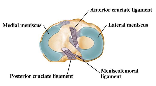
3 Inspection, Palpation, Range Both knee comparison Local areas of increased temp. Range of motion of knee joint : flexion and extension Meniscus Collateral ligaments Cruciate ligaments
4 Knee joint evaluation MRI study : best of choice MSUS study : limitation (meniscus, cruciate ligaments)
5 Probe frequency 10 ~ 15 MHz linear Probe : anterior, medial, lateral knee aspect 5 MHz Probe : Popliteal area, GCM,
6 Panoramic view 긴구조물을평가하는데큰도움이된다. Large cysts Enlarged bursa Medial collateral ligament Anterior knee aspects
7 Anterior Knee Supine position. Knee flexion of 20 ~ 30 by small pillow Avoid anisotrophy : Patellar tendon을 extension 시킨다. Examination starting from cranial to caudal.

8 Anterior Knee Supine position. Knee flexion of 20 ~ 30 by small pillow
9 Anterior Knee 검사할주요구조물들 Quadriceps tendon. Suprapatellar synovial recess Suprapatellar fat pad Prefemoral fat Femoral condyle
10 Anterior Knee : quadriceps tendon Probe position : longitudinal scanning. Proximal : quadriceps muscles Distal : edge on the patella Quadriceps tendon stretched. - US beam : perpendicular to long axis - Hyperechoic fibrillar apperance.
11 Anterior Knee : Quadriceps Multilayered appearance by high-frequency probes.(both long- and short-axis planes) 1) superficial layer : rectus femoris 2) intermediate : vastus lateralis & medialis 3) deep layer : vastus intermedius Superficial layer is always evident. 2) & 3) 은구분이가지않을경우가많다.


12 Suprapatellar tendon area Rectus femoris tendon 1. suprapatellar fat pad 2. prepatellar fat
13 Anterior Knee : Recess Suprapatellar synovial recess can be identified with US. Two fat pads (suprapatellar and prepatellar)
14 Anterior Knee : Recess Small effusion is suspected? in supine position : isometric contraction of quadriceps : ankle dorsiflexion with knee extension : check out parapatellar recesses.(med & lat) MRI 검사는 fluid 의위치가파악이되지만, MSUS 에서는직접구석구석찾아야한다.


15 Parapatellar recesses

16 Anterior Knee Suprapatellar Plica (Reminants of embyonic life) intrauterine 5 개월 : septum formation complete septum found in 12% of normal
17 Anterior Knee FEMORAL TROCHLEA ; full flexion of knee transverse plane trochlea cartilage appears as a hypoechoic line in normal condition, thicker in central portion. mean thickness ranges : 1.8 ~ 2.5 mm

18 Anterior Knee : infrapatella Supine position : 20 ~30 knee flexion Infrapatellar tendon : 4~5 mm AP thickness Infrapatellar portion V Shaped appearance Proximal 1/3 * Distal 1/3 * * thickened

19 Anterior Knee : infrapatella Increased thickness of the prox. & distal 1/3
20 Anterior Knee Deep to the patellar tendon : Hoffa Pad appears as a fatty tissue space - hyperechoic septations Deep infrapatellar bursa : small synovial cavity between distal portion of patellar tendon and ant. aspect of tibial epiphysis.
21 Bursa Diagnosis of bursa - large effusions or local pain generated by pressure with the probe Prepatellar bursa - located subcutaneous tissue. - thin wall, absence of internal fluid. - if detected minimal amout of fluid, sign of inflammation(prepatellar bursitis)
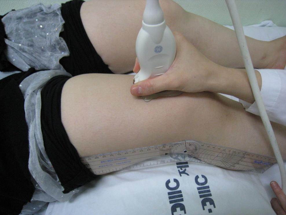
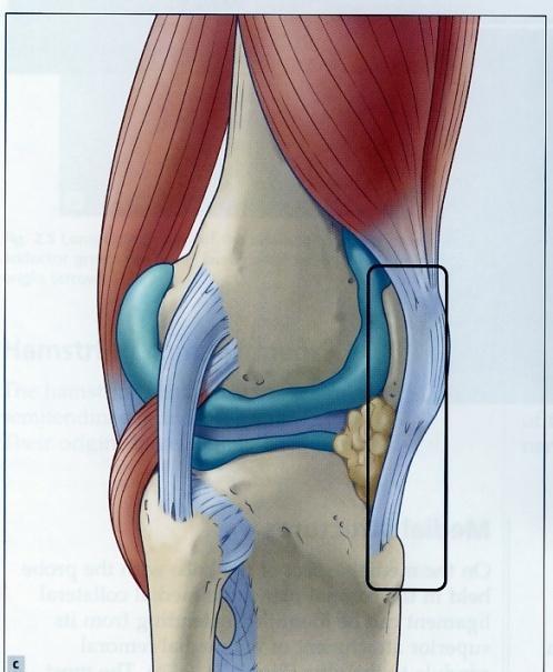
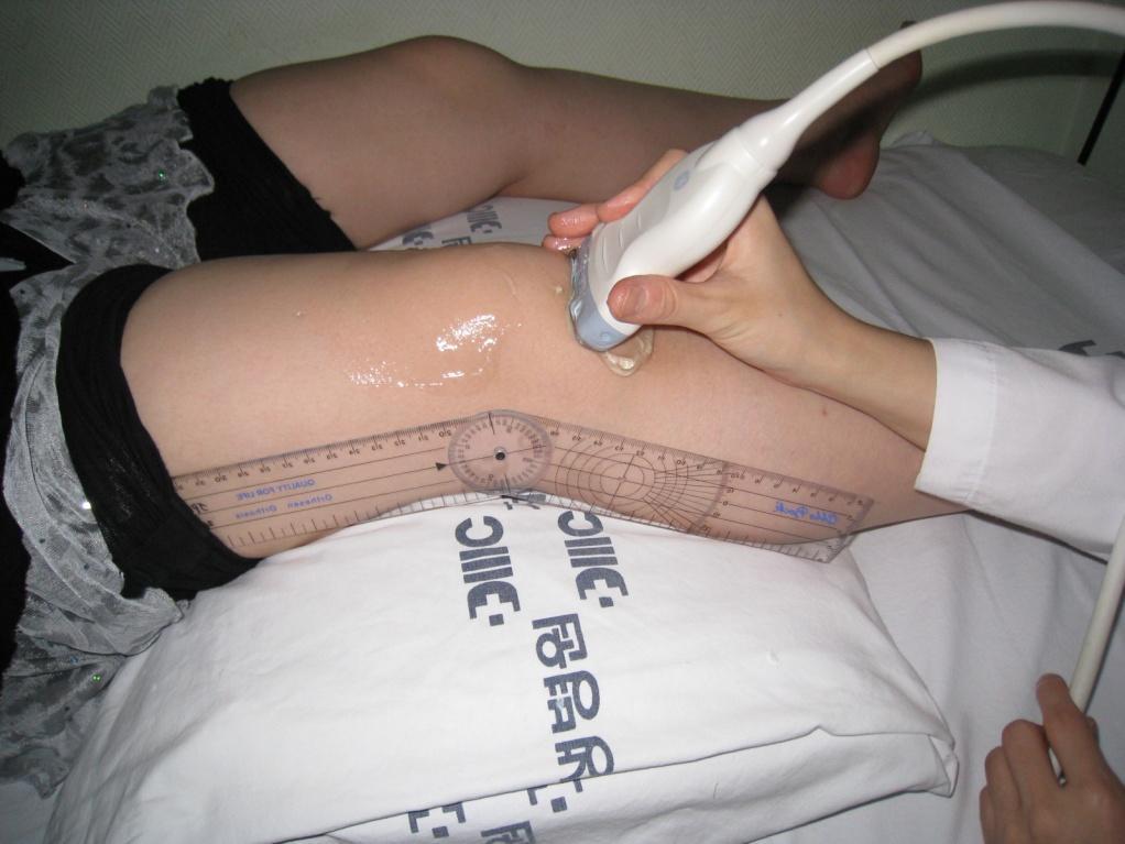
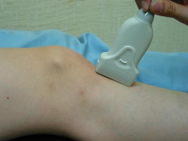
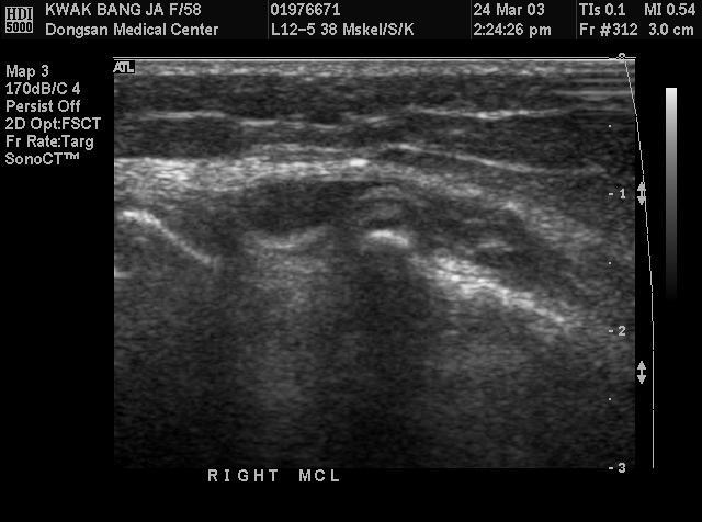
22 Medial Knee Best exam position : leg external rotated Anatomy : medial collateral ligament, medial femorotibial joint space, medial meniscus and pes anserinus complex. US assessment of the medial asepct begins with medial collateral ligament.
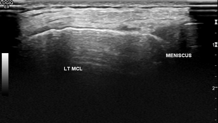
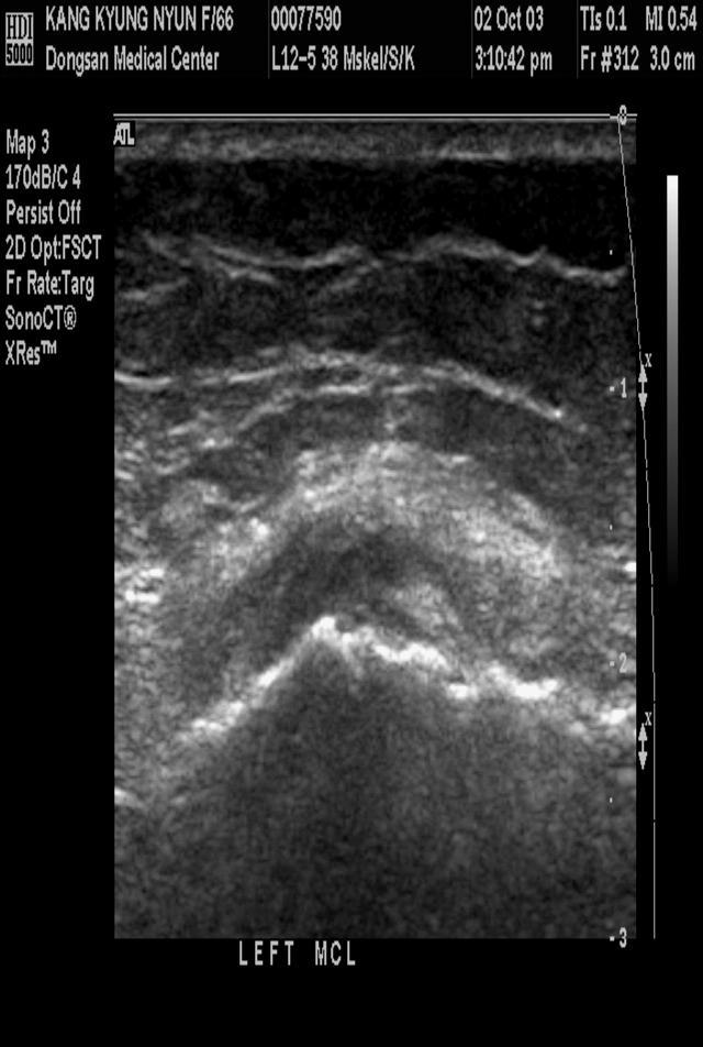
23 Medial Knee medial collateral ligment Elongated band 1~3 mm thick Formed by two hyperechoic layers Superficial part Deep meniscofemoral and meniscotibial component (separated by a slight hypoechoic line related to fatty tissue) On transverse planes, superficial comp. as a small, crescentic shaped hyperechoic structure.
24 Medial Knee Medial Meniscus Triangular hyperechoic structure. Difficult to obtain the image by US Attenuation of the US beam its small size Vagus stress by widening of medial joint space Increased visibility of the medial meniscus.
25 Medial Knee Pes Anserinus complex Sartorius, gracilis, semitendinosus Insert into the anteromedial aspect of the tibial metaphysis 5~6 cm below the joint line. More proximal level(hamstring) : 구분이가능하다. Tendon insertion level : 구분이전혀앆된다.
26 Lateral Knee Supine position to rotate the leg internally. evaluate from anterior to posterior Distal iliotibial band External femorotibial joint space Lateral meniscus Lateral collateral ligament Popliteus tendon Superior tibiofibular joint.
27 Lateral Knee Iliotibial band Best imaged on long-axis scans Insertion Land mark : Gerdy s tubercle, anterolateral aspect of the tibial epiphysis. Local compression : painful in tendinitis
28 Lateral Knee Lateral meniscus Intrisic limitation as medial meniscus Smaller than the medial meniscus Meniscal cyst : knee forceful flexion bulging of the cyst outside the joint space
29 Lateral Knee Lateral collateral ligament Cordlike fibrillar structure Located at posterolateral aspect of the joint. Inserts into the lateral femoral condyle and the fibular head. 3~4 mm thick Lies in a more anterior plane than the biceps tendon. Best evaluated on oblique longitudinal scans and knee fully extension.
30 Lateral Knee popliteus Just deep to the proximal part of the lateral collateral ligament Popliteus tendon can be imaged on longitudinal US scans. Oval structure located in its bony groove.
31 Posterior Knee Lie prone position with knee extended For Baker s cyst(pedicle opening) : take different degrees of knee flexion position. 3 zone : internal( 내측 ), central, external( 외측 ) Internal zone에서볼수있는구조물들 - Pes Anserinus complex. - semimembranosus tendon. - semimembranosus-gastrocnemius bursa
32 Posterior Knee Semimembranosus-grastrocnemius bursa Baker s Cyst medial femoral condyle Posterior central zone medial head of the GCM (triangular shape) the popliteal artery and vein tibial nerve.
33 Posterior Knee Posterior external zone muscles and tendons of the biceps femoris lateral head of the gastrocnemius common peroneal nerve.
34 MUSCULOSKELETAL KNEE ULTRASOUND EXAMINATION 2010 AUGUST
35 Anterior Knee Supine position. Knee flexion of 20 ~ 30 by small pillow Avoid anisotrophy : Patellar tendon을 extension 시킨다. Examination starting from cranial to caudal.
36 검사할주요구조물 Anterior Knee Quadriceps tendon. Suprapatellar synovial recess Suprapatellar fat pad Prefemoral fat Femoral condyle

37 Anterior

38 Anterior
39 Anterior

40 Medial Knee Best exam position : leg external rotated Anatomy : medial collateral ligament, medial femorotibial joint space, medial meniscus and pes anserinus complex. US assessment of the medial asepct begins with medial collateral ligament.


41 Medial

42 Medical Collateral Ligament


43 Medical Meniscus
44 Lateral Knee Supine position to rotate the leg internally. evaluate from anterior to posterior Distal iliotibial band External femorotibial joint space Lateral meniscus Lateral collateral ligament Popliteus tendon Superior tibiofibular joint.


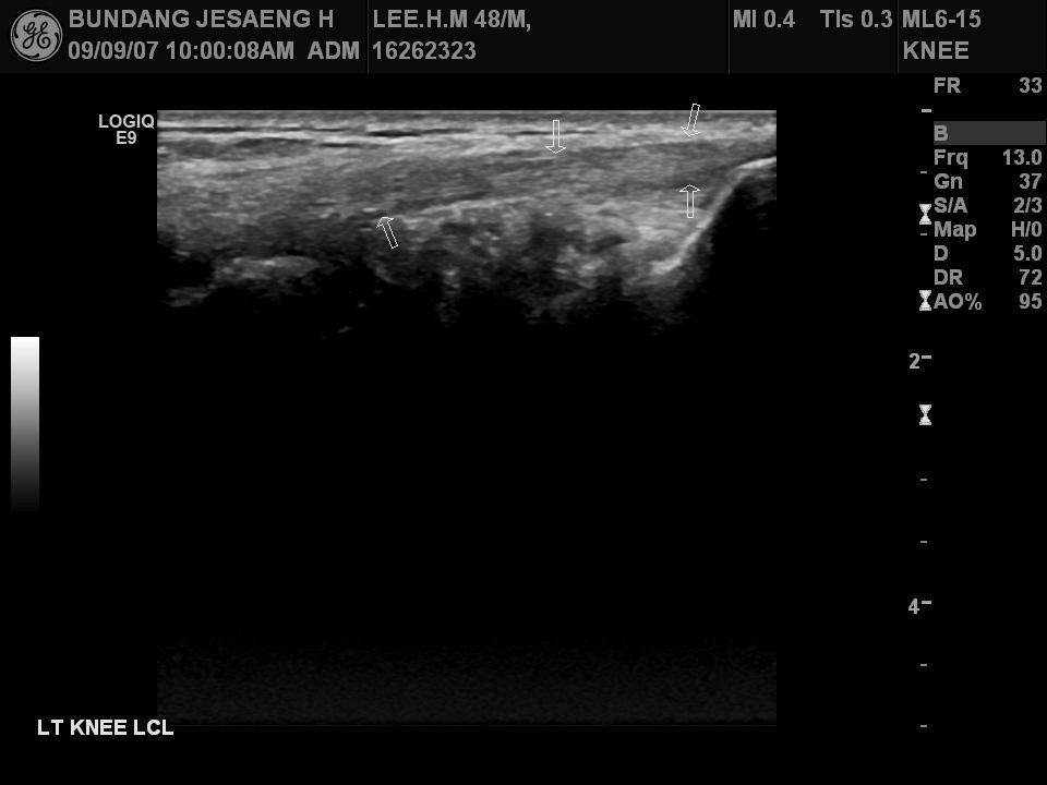
45 Lateral Collateral Ligament LCL LCL LCL appears as homogeneous, hyperechoic bands
 - popliteal tendon origin, - lateral collateral ligament on fibular head (hyperechoic and fibrillar echotexture) - distal biceps femoris tendon")
46 Lateral Lateral asepct of the knee 1) internal rotation with full extension 2) lateral decubitus position 3) lateroposterior structures, prone. - iliotibial tract on Gerdy s tubercle (proximal tibia) - popliteal tendon origin, - lateral collateral ligament on fibular head (hyperechoic and fibrillar echotexture) - distal biceps femoris tendon (less compact fibrillar echotexture & associated with hypoechoic muscle)


47 Lateral Knee Gerdy s tubercle Fibular head


48 Iliotibial band
49 Posterior Knee Lie prone position with knee extended For Baker s cyst(pedicle opening) : take different degrees of knee flexion position. 3 zone : internal( 내측 ), central, external( 외측 ) Internal zone에서볼수있는구조물들 - Pes Anserinus complex. - semimembranosus tendon. - semimembranosus-gastrocnemius bursa
50 Posterior Knee Semimembranosus-grastrocnemius bursa Posterior central zone medial head of the GCM (triangular shape) the popliteal artery and vein Posterior external zone muscles and tendons of the biceps femoris lateral head of the gastrocnemius common peroneal nerve.

51 Posterior Knee Rt. Medial GCM insertion

52 Posterior Knee semitendinosus and med. GCM head Medial femoral condyle : insertion of medial GCM head


53 Posterior Knee transverse scan Popliteal artery and vein

54 Disease of the KNEE


55 Suprapatellar Bursitis


56 Suprapatellar Bursitis


57 Villonodula Synovitis


58 Osgood-Schlatter Disease

59 Osgood-Schlatter Disease

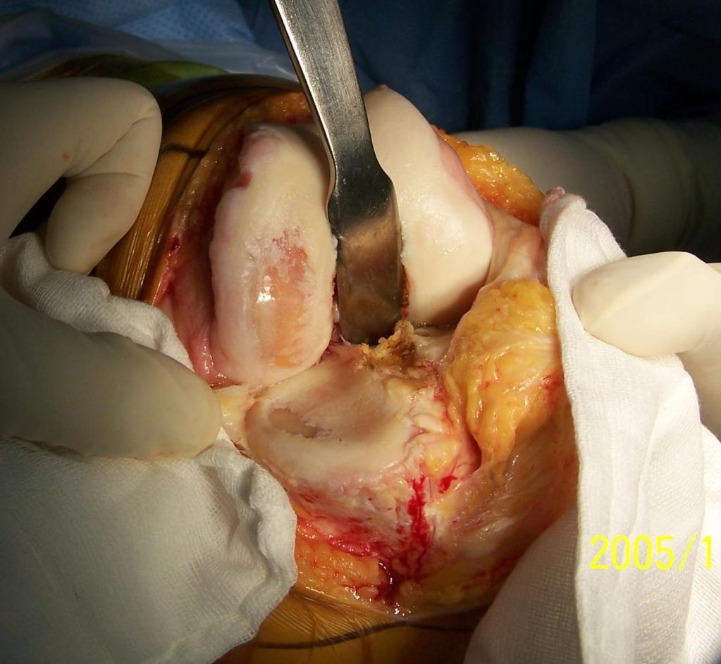
60 Collateral Ligament
61 Collateral Ligament After Acute Injury local tenderness & reflective muscle spasms difficult to gait and weight bearing Knee Instability gradually subside spontaneousn pain & tenderness weakness, giving way, muscle atrophy more severed knee instability
62 Medical Collateral Ligament Rupture tear after forceful valgus stress. If not treated, a painful granuloma / calicification. usually located at the prox. insertion. Complete rupture concurrent with ACL rupture and medial meniscal tear
63 Medical Collateral Ligament Rupture identified by an interruption in the hyperechoic bands that represent the superficial and deep components. Fluid fills the gap : either hypoechoic or anechoic Chronic injury : hypertrophic, excessive formation of granulation tissue and mucoid degeneration.

64 Medical Collateral Ligament


65

66 Medical Collateral Ligament

67 Medical Collateral Ligament

68 Medical Collateral Ligament
69 Latellar Collateral Ligament

70 Latellar Collateral Ligament
71 Meniscus


72 Meniscus Tear

73 Meniscus Tear

74
75 MR Imaging Criteria of Meniscal Tear Grade III : increased signal extending to articular surface Grade IV : several areas of high signal as well as deformity & fragmentation
76 Meniscal Tear in MSUS Hypoechoic or anechoic cleft through meniscus that extend to articular surface Defect in the hyperechoic meniscus


77 Medical Meniscus MCL Femur Tibia Tear in posterior horn of left medial meniscus.


78 Medial Meniscus Tear Femur Tibia Right knee reveals longitudial and radial tear


79 Lateral Meniscus Tear Femur Tibia Rt. Knee shows tear and lateral inferior displacement (Flap tear).


80 Lateral Meniscus Tear with Meniscal Cyst Right lateral meniscus tear, with suspected meniscal cyst posterolateral to lateral femoral condyle, about 26 x 21 x 11 mm in size.

81 Infrapatella


82 Infrapatella Tendinitis


83 Infrapatellar Tendinitis Patellar tendon shows diffuse swelling with inhomogeneous echotexture and slightly increased vascularity proximally.


84 Chondrocalcinosis Medial meniscus shows intrameniscal amorphous echogenic area, especially posterior.


85 Baker s cyst (Popliteal Cyst) Origin : from the posteromedial aspect situated between the medial head of the GCM and the semimembranosus tendon. Divided three portions anatomically. the base, superficial extent and neck.


86 Baker s cyst (Popliteal Cyst) Some limitation of flexion Cysts occur most often when the knee is damaged due to arthritis, gout, injury, or inflammation in the lining of the knee joint
87 Baker s cyst (Popliteal Cyst) not communicate at birth but developed during growth synovial proliferation in inflammatory disease. - increased intraarticular fliud & arthritis - fracture, cartilage defects, meniscal lesion and loose bodies.


88 Baker s cyst (Popliteal Cyst)
89 Bursa

90
91 Prepatellar Bursitis Prepatella Bursa The most frequent type of bursitis, especially in patients who have to kneel repeatedly in their work Patient complains of pain and swelling in front of knee Diagnosis is made on simple inspection If swelling is gross, limitation of flexion Palpation shows effusion to lie between skin and patella


92 Prepatella Bursa Fluid collection with thickened wall infrapatellar portion of right knee without definite increased vascularity


93 Prepatella Bursa Presence of heat suggests hemorrhage into bursa Heat and redness indicate possibility of sepsis


94 Infrapatella Bursa
95 Medial Collateral Liagment Bursa Usually middle aged women Complains of localized pain at medial side of knee Flexion is limited by O Painful valgus strain and lateral rotation Differential diagnosis: meniscal cyst, chronic ligament sprain

96 Medial Collateral Liagment Bursa MCL

97 Medial Collateral Liagment Bursa MCL

98 THANKS FOR YOUR ATTENTION!
Joints of the Lower Limb II
 Joints of the Lower Limb II Lecture Objectives Describe the components of the knee and ankle joint. List the ligaments associated with these joints and their attachments. List the muscles acting on these
Standardised. knee. scanning of the. Basic pathology. Nemanja Damjanov. University of Belgrade Institute of Rheumatology
 Standardised scanning of the Nemanja Damjanov University of Belgrade Institute of Rheumatology knee Basic pathology Disclosure Lecturer: Pfizer, Abbvie, Roche, MSD, Boehringer-Ingelheim, Gedeon Richter,
In the name of god. Knee. By: Tofigh Bahraminia Graduate Student of the Pathology Sports and corrective actions. Heat: Dr. Babakhani. Nov.
 In the name of god Knee By: Tofigh Bahraminia Graduate Student of the Pathology Sports and corrective actions Heat: Dr. Babakhani Nov. 2014 1 Anatomy-Bones Bones Femur Medial/lateral femoral condyles articulate
Ultrasound of the Knee
 Ultrasound of the Knee Jon A. Jacobson, M.D. Professor of Radiology Director, Division of Musculoskeletal Radiology University of Michigan Disclosures: Consultant: Bioclinica Book Royalties: Elsevier Advisory
The Knee. Prof. Oluwadiya Kehinde
 The Knee Prof. Oluwadiya Kehinde www.oluwadiya.sitesled.com The Knee: Introduction 3 bones: femur, tibia and patella 2 separate joints: tibiofemoral and patellofemoral. Function: i. Primarily a hinge joint,
RN(EC) ENC(C) GNC(C) MN ACNP *** MECHANISM OF INJURY.. MOST IMPORTANT *** - Useful in determining mechanism of injury / overuse
 ENC(C) GNC(C) MN ACNP *** MECHANISM OF INJURY.. MOST IMPORTANT *** - Useful in determining mechanism of injury / overuse") HISTORY *** MECHANISM OF INJURY.. MOST IMPORTANT *** Age of patient Sport / Occupation - Certain conditions are more prevalent in particular age groups (Osgood Schlaters in youth / Degenerative Joint Disease
HISTORY *** MECHANISM OF INJURY.. MOST IMPORTANT *** Age of patient Sport / Occupation - Certain conditions are more prevalent in particular age groups (Osgood Schlaters in youth / Degenerative Joint Disease
Sonography of Knee and Calf Pain: the differential considerations
 Sonography of Knee and Calf Pain: the differential considerations Dr. Lisa L. S.Wong Consultant Radiologist St Paul s Hospital Outline Ultrasound techniques Common pathologies in calf and posterior knee
Knee Ultrasonography step by step
 Knee Ultrasonography step by step Poster No.: C-2809 Congress: ECR 2018 Type: Educational Exhibit Authors: J. A. Torres de Abreu Macedo, N. Pereira da Silva, A. I. Aguiar, F. Alves, F. Caseiro Alves; Coimbra/PT
Knee Joint Assessment and General View
 Knee Joint Assessment and General View Done by; Mshari S. Alghadier BSc Physical Therapy RHPT 366 m.alghadier@sau.edu.sa http://faculty.sau.edu.sa/m.alghadier/ Functional anatomy The knee is the largest
Ligamentous and Meniscal Injuries: Diagnosis and Management
 Ligamentous and Meniscal Injuries: Diagnosis and Management Daniel K Williams, MD Franciscan Physician Network Orthopedic Specialists September 29, 2017 No Financial Disclosures INTRODUCTION Overview of
Knee, Ankle, and Foot: Normal and Abnormal Features with MRI and Ultrasound Correlation. Disclosures. Outline. Joint Effusion. Suprapatellar recess
 Knee, Ankle, and Foot: Normal and Abnormal Features with MRI and Ultrasound Correlation Jon A. Jacobson, M.D. Professor of Radiology Director, Division of Musculoskeletal Radiology University of Michigan
and K n e e J o i n t Is the most complicated joint in the body!!!!
 K n e e J o i n t K n e e J o i n t Is the most complicated joint in the body!!!! 1-Consists of two condylar joints between: A-The medial and lateral condyles of the femur and The condyles of the tibia
The Knee. Tibio-Femoral
 The Knee Tibio-Femoral Osteology Distal Femur with Proximal Tibia Largest Joint Cavity in the Body A modified hinge joint with significant passive rotation Technically, one degree of freedom (Flexion/Extension)
Prevention and Treatment of Injuries. Anatomy. Anatomy. Chapter 20 The Knee Westfield High School Houston, Texas
 Prevention and Treatment of Injuries Chapter 20 The Knee Westfield High School Houston, Texas Anatomy MCL, Medial Collateral Ligament LCL, Lateral Collateral Ligament PCL, Posterior Cruciate Ligament ACL,
CHAPTER 8: THE BIOMECHANICS OF THE HUMAN LOWER EXTREMITY
 CHAPTER 8: THE BIOMECHANICS OF THE HUMAN LOWER EXTREMITY _ 1. The hip joint is the articulation between the and the. A. femur, acetabulum B. femur, spine C. femur, tibia _ 2. Which of the following is
Knee Injury Assessment
 Knee Injury Assessment Clinical Anatomy p. 186 Femur Medial condyle Lateral condyle Femoral trochlea Tibia Intercondylar notch Tibial tuberosity Tibial plateau Fibula Fibular head Patella Clinical Anatomy
The Knee. Clarification of Terms. Osteology of the Knee 7/28/2013. The knee consists of: The tibiofemoral joint Patellofemoral joint
 The Knee Clarification of Terms The knee consists of: The tibiofemoral joint Patellofemoral joint Mansfield, p273 Osteology of the Knee Distal Femur Proximal tibia and fibula Patella 1 Osteology of the
Imaging the Knee 17/10/2017. Friction syndrome Common in runners or cyclists Fluid between ITB and Lateral femoral condyle
 17/10/2017 Imaging the Knee Alicia M. Yochum RN, DC, DACBR, RMSK Iliotibial Band Syndrome Ligamentous Tears (ACL, PCL, MCL, LCL) Meniscal Tears Cartilage Degeneration Quadriceps/Patellar tendinosis Osteochondral
The Lower Limb II. Anatomy RHS 241 Lecture 3 Dr. Einas Al-Eisa
 The Lower Limb II Anatomy RHS 241 Lecture 3 Dr. Einas Al-Eisa Tibia The larger & medial bone of the leg Functions: Attachment of muscles Transfer of weight from femur to skeleton of the foot Articulations
Case study #11 Rt. knee
 The patient is a 55 year old female who presents with bilateral knee pain. Patient is a collegiate softball coach and has a very active lifestyle and career that is hampered by her chronic knee pain. She
Musculoskeletal Examination Benchmarks
 Musculoskeletal Examination Benchmarks _ The approach to examining the musculoskeletal system is the same no matter what joint or limb is being examined. The affected and contralateral region should both
To describe he knee joint, ligaments, structure & To list the main features of other lower limb joints
 To describe he knee joint, ligaments, structure & neurovascular supply To demonstrate the ankle joint anatomy To list the main features of other lower limb joints To list main groups of lymph nodes in
Copyright 2012 by The McGraw-Hill Companies, Inc. All rights reserved. McGraw-Hill/Irwin
 CHAPTER 8: THE LOWER EXTREMITY: KNEE, ANKLE, AND FOOT KINESIOLOGY Scientific Basis of Human Motion, 12 th edition Hamilton, Weimar & Luttgens Presentation Created by TK Koesterer, Ph.D., ATC Humboldt State
Musculoskeletal Ultrasound of the Knee, Foot and ankle
 Musculoskeletal Ultrasound of the Knee, Foot and ankle ADVANCED TEAM PHYSICIAN COURSE SAN DIEGO, CALIFORNIA DECEMBER 11TH 2016 Jonathan S. Halperin MD Learning objec-ves: Understand the basics of knee,
Please differentiate an internal derangement from an external knee injury.
 Knee Orthopaedic Tests Sports and Knee Injuries James J. Lehman, DC, MBA, DABCO University of Bridgeport College of Chiropractic Knee Injury Strain, Sprain, Internal Derangement Anatomy of the Knee Please
Muscles of the Thigh. 6.1 Identify, describe the attachments of and deduce the actions of the muscles of the thigh: Anterior group
 Muscles of the Thigh 6.1 Identify, describe the attachments of and deduce the actions of the muscles of the thigh: Anterior group Sartorius: This is a long strap like muscle with flattened tendons at each
Exercise Science Section 4: Joint Mechanics and Joint Injuries
 Exercise Science Section 4: Joint Mechanics and Joint Injuries An Introduction to Health and Physical Education Ted Temertzoglou Paul Challen ISBN 1-55077-132-9 Types of Joints Fibrous joint Cartilaginous
UNIT 7 JOINTS. Knee and Ankle Joints DR. ABDEL-MONEM A. HEGAZY
 UNIT 7 JOINTS Knee and Ankle Joints BY DR. ABDEL-MONEM A. HEGAZY (Degree in Bachelor of Medicine and Surgery with honor 1983, Dipl."Gynaecology and Obstetrics "1989, Master "Anatomy and Embryology "1994,
The Knee Joint By Prof. Dr. Muhammad Imran Qureshi
 The Knee Joint By Prof. Dr. Muhammad Imran Qureshi Structurally, it is the Largest and the most complex joint in the body because of the functions that it performs: Allows mobility (flexion/extension)
Ultrasound Evaluation of Masses
 Ultrasound Evaluation of Masses Jon A. Jacobson, M.D. Professor of Radiology Director, Division of Musculoskeletal Radiology University of Michigan Disclosures: Consultant: Bioclinica Advisory Panel: GE,
Arthritic history is similar to that of the hip. Add history of give way and locking, swelling
 KNEE VASU PAI Arthritic history is similar to that of the hip. Add history of give way and locking, swelling INJURY MECHANISM When How Sequence Progress Disability IKDC Activity I - Strenuous activity
Recognizing common injuries to the lower extremity
 Recognizing common injuries to the lower extremity Bones Femur Patella Tibia Tibial Tuberosity Medial Malleolus Fibula Lateral Malleolus Bones Tarsals Talus Calcaneus Metatarsals Phalanges Joints - Knee
Other Culprits in Knee Dysfunction
 Unraveling the Mystery of Knee Pain #6: Other Culprits in Knee Dysfunction 1 Webinar Goals Explore the assessment and treatment of other culprits in knee dysfunction. 2 Time: 60 minutes Schedule: Logistics
ACL Athletic Career. ACL Rupture - Warning Features Intensive pain Immediate swelling Locking Feel a Pop Dead leg Cannot continue to play
 FIMS Ambassador Tour to Eastern Europe, 2004 Belgrade, Serbia Montenegro Acute Knee Injuries - Controversies and Challenges Professor KM Chan OBE, JP President of FIMS Belgrade ACL Athletic Career ACL
Sports Medicine 15. Unit I: Anatomy. The knee, Thigh, Hip and Groin. Part 4 Anatomies of the Lower Limbs
 Sports Medicine 15 Unit I: Anatomy Part 4 Anatomies of the Lower Limbs The knee, Thigh, Hip and Groin Anatomy of the lower limbs In Part 3 of this section we focused upon 11 of the 12 extrinsic muscles
CLASSIFICATION OF JOINTS STRUCTURAL VS FUNCTIONAL
 CHAPTER 8 JOINTS CLASSIFICATION OF JOINTS STRUCTURAL VS FUNCTIONAL The most moveable type of joint is a 1) Synarthrosis 2) Amphiarthrosis 3) Diarthrosis FIBROUS JOINTS Figure 8.1 Fibrous joints. (a) Suture
The Hip (Iliofemoral) Joint. Presented by: Rob, Rachel, Alina and Lisa
 Joint. Presented by: Rob, Rachel, Alina and Lisa") The Hip (Iliofemoral) Joint Presented by: Rob, Rachel, Alina and Lisa Surface Anatomy: Posterior Surface Anatomy: Anterior Bones: Os Coxae Consists of 3 Portions: Ilium Ischium Pubis Bones: Pubis Portion
The Hip (Iliofemoral) Joint Presented by: Rob, Rachel, Alina and Lisa Surface Anatomy: Posterior Surface Anatomy: Anterior Bones: Os Coxae Consists of 3 Portions: Ilium Ischium Pubis Bones: Pubis Portion
Distal Femoral Resection
 Distal Femoral Resection Annie Arteau, Bruno Fuchs Introduction This text is a general description of a distal femoral resection. Focus is on anatomical structures and muscle resection. Each femoral resection
Physical Examination of the Knee
 History: Pain Traumatic vs. atraumatic Acute vs Chronic Mechanism of injury Swelling, catching, instability Previous evaluation and treatment General Setup Examine standing, sitting and supine Evaluate
PRIMARY CARE EXAMINATION OF KEY JOINTS. Thomas M. Howard, MD, FACSM FFPC Sports Medicine
 PRIMARY CARE EXAMINATION OF KEY JOINTS Thomas M. Howard, MD, FACSM FFPC Sports Medicine General exam principles: Expose entire joint and opposite limb for comparison Have a Differential Diagnosis Exam
The examination of the painful knee. Maja K Artandi, MD, FACP Clinical Associate Professor of Medicine Stanford University
 The examination of the painful knee Maja K Artandi, MD, FACP Clinical Associate Professor of Medicine Stanford University Objectives of the talk By the end of this talk you will know The important anatomy
The Knee. Two Joints: Tibiofemoral. Patellofemoral
 Evaluating the Knee The Knee Two Joints: Tibiofemoral Patellofemoral HISTORY Remember the questions from lecture #2? Girth OBSERVATION TibioFemoral Alignment What are the consequences of faulty alignment?
Unraveling the Mystery of Knee Pain #2: Client History & The 23 Injuries Common to the Knee
 Unraveling the Mystery of Knee Pain #2: Client History & The 23 Injuries Common to the Knee Instructor: Ben Benjamin, Ph.D. Instructor: Ben Benjamin, Ph.D. 1 Webinar Goals Understand the significance of
Unraveling the Mystery of Knee Pain #2: Client History & The 23 Injuries Common to the Knee
 Unraveling the Mystery of Knee Pain #2: Client History & The 23 Injuries Common to the Knee Instructor: Ben Benjamin, Ph.D. 1 Instructor: Ben Benjamin, Ph.D. Webinar Goals Understand the significance of
Physical Examination of the Knee
 History: Pain Traumatic vs. atraumatic? Acute vs Chronic Previous procedures done on the knee? Swelling, catching, instability General Setup Examine standing, sitting and supine Evaluate gait Examine hip
Chapter 10. The Knee Joint. The Knee Joint. Bones. Bones. Bones. Bones. Knee joint. Manual of Structural Kinesiology R.T. Floyd, EdD, ATC, CSCS
 The Knee Joint Chapter 10 The Knee Joint Manual of Structural Kinesiology R.T. Floyd, EdD, ATC, CSCS 2007 McGraw-Hill Higher Education. All rights reserved. 10-1 Knee joint largest joint in body very complex
THE LOWER EXTREMITY EXAM FOR THE FAMILY PRACTITIONER
 THE LOWER EXTREMITY EXAM FOR THE FAMILY PRACTITIONER Melinda A. Scott, D.O. Orthopedic Associates of Dayton Board Certified in Primary Care Sports Medicine GOALS Identify landmarks necessary for exam of
Myology of the Knee. PTA 105 Kinesiology
 Myology of the Knee PTA 105 Kinesiology Objectives Describe the planes of motion and axes of rotation of the knee joint Visualize the origins and insertions of the muscles about the knee List the innervations
MRI KNEE WHAT TO SEE. Dr. SHEKHAR SRIVASTAV. Sr.Consultant KNEE & SHOULDER ARTHROSCOPY
 MRI KNEE WHAT TO SEE Dr. SHEKHAR SRIVASTAV Sr.Consultant KNEE & SHOULDER ARTHROSCOPY MRI KNEE - WHAT TO SEE MRI is the most accurate and frequently used diagnostic tool for evaluation of internal derangement
Lateral knee injuries
 Created as a free resource by Clinical Edge Based on Physio Edge podcast episode 051 with Matt Konopinski Get your free trial of online Physio education at Orthopaedic timeframes Traditionally Orthopaedic
DIAGNOSIS AND EARLY MANAGEMENT OF KNEE INJURIES
 DIAGNOSIS AND EARLY MANAGEMENT OF KNEE INJURIES INTRODUCTION: The knee is a common site of athletic injury. The knee injuries can be classified either into traumatic or acute and chronic, with overuse
Objectives. The BIG Joint. Case 1. Boney Architecture. Presenter Disclosure Information. Common Knee Problems
 3:30 4:15 pm Common Knee Problems SPEAKER Christopher J. Visco, MD Presenter Disclosure Information The following relationships exist related to this presentation: Christopher J. Visco, MD: Speaker s Bureau
Checklist for Physical Examination of the Knee Muscuoskeletal Block -- Chris McGrew MD, Andrew Ashbaugh DO
 Checklist for Physical Examination of the Knee Muscuoskeletal Block -- Chris McGrew MD, Andrew Ashbaugh DO This handout is for use as a rough guide and study aid. Your instructor may perform certain maneuvers
Knee MRI Update Case Review 2009 Russell C. Fritz, M.D. National Orthopedic Imaging Associates San Francisco, CA
 Knee MRI Update Case Review 2009 Russell C. Fritz, M.D. National Orthopedic Imaging Associates San Francisco, CA Meniscal Tears -linear increased signal extending to an articular surface is the hallmark
Imaging the Athlete s Knee. Peter Lowry, MD Musculoskeletal Radiology University of Colorado
 Imaging the Athlete s Knee Peter Lowry, MD Musculoskeletal Radiology University of Colorado None Disclosures Knee Imaging: Radiographs Can be performed weight-bearing or non-weight-bearing View options
Leg. Dr. Heba Kalbouneh Associate Professor of Anatomy and Histology
 Leg Dr. Heba Kalbouneh Associate Professor of Anatomy and Histology Skin of the Leg Cutaneous Nerves Medially: The saphenous nerve, a branch of the femoral nerve supplies the skin on the medial surface
W. Dilworth Cannon, M.D. Professor of Clinical Orthopaedic Surgery University of California San Francisco
 Knee Pain And Injuries In Adults W. Dilworth Cannon, M.D. Professor of Clinical Orthopaedic Surgery University of California San Francisco Pain Control Overview Narcotics rarely necessary after 1 st 1-2
Muscle Testing of Knee Extensors. Yasser Moh. Aneis, PhD, MSc., PT. Lecturer of Physical Therapy Basic Sciences Department
 Muscle Testing of Knee Extensors Yasser Moh. Aneis, PhD, MSc., PT. Lecturer of Physical Therapy Basic Sciences Department Muscle Testing of Knee Extensors othe Primary muscle Quadriceps Femoris -Rectus
Multi-ligamentous knee injuries - MRI injury patterns at a glance
 Multi-ligamentous knee injuries - MRI injury patterns at a glance Poster No.: P-0068 Congress: ESSR 2015 Type: Educational Poster Authors: A. Rastogi, D. Whelan, R. Martin, W. Mak, D. Pearce ; 1 1 1 2
Knee Joint Anatomy 101
 Knee Joint Anatomy 101 Bone Basics There are three bones at the knee joint femur, tibia and patella commonly referred to as the thighbone, shinbone and kneecap. The fibula is not typically associated with
Financial Disclosure. Medial Collateral Ligament
 Matthew Murray, M.D. UTHSCSA Sports Medicine Financial Disclosure Dr. Matthew Murray has no relevant financial relationships with commercial interests to disclose. Medial Collateral Ligament Most commonly
KNEE EXAMINATION. Tips & Tricks from an Emergency Physician Perspective. EM Physicians Less Exposed to MSK Medicine
 KNEE EXAMINATION Tips & Tricks from an Emergency Physician Perspective Dr P O CONNOR Emergency Medicine Physician EUSEM 10/09/2018 EM Physicians Less Exposed to MSK Medicine Musculoskeletal Medicine becoming
The thigh. Prof. Oluwadiya KS
 The thigh Prof. Oluwadiya KS www.oluwadiya.com The Thigh: Boundaries The thigh is the region of the lower limb that is approximately between the hip and knee joints Anteriorly, it is separated from the
Learning IRM. The Knee: lateral ligaments and anatomical quadrants.
 Learning IRM. The Knee: lateral ligaments and anatomical quadrants. Poster No.: C-1733 Congress: ECR 2014 Type: Educational Exhibit Authors: A. Amador Gil, M. D. C. Jurado Gómez, V. de Lara Bendahan ;
Pragmatic ultrasound in the diagnosis of soft tissue rheumatic pain. Plamen Todorov
 Pragmatic ultrasound in the diagnosis of soft tissue rheumatic pain Plamen Todorov INTRODUCTION Soft tissue rheumatism: nonsystemic, focal pathological syndromes involving the periarticular structures.
Hip and Knee Approaches
 Hip and Knee Approaches Professor VLADIMIR STAVREV, MD, PhD, DMSc GEORGI MINEV MD DEPARTMENT OF ORTHOPEDICS AND TRAUMATOLOGY, MEDICAL UNIVERSITY -PLOVDIV Anterior View Hip Joint Posterior View - Hip Joint
Overview Ligament Injuries. Anatomy. Epidemiology Very commonly injured joint. ACL Injury 20/06/2016. Meniscus Tears. Patellofemoral Problems
 Overview Ligament Injuries Meniscus Tears Pankaj Sharma MBBS, FRCS (Tr & Orth) Consultant Orthopaedic Surgeon Manchester Royal Infirmary Patellofemoral Problems Knee Examination Anatomy Epidemiology Very
On Field Assessment and Management of Acute Knee Injuries: A Physiotherapist s Perspective
 On Field Assessment and Management of Acute Knee Injuries: A Physiotherapist s Perspective Jessica Condliffe Physiotherapist / Clinic Manager TBI Health Wellington Presentation Outline Knee anatomy review
The Muscular System. Chapter 10 Part D. PowerPoint Lecture Slides prepared by Karen Dunbar Kareiva Ivy Tech Community College
 Chapter 10 Part D The Muscular System Annie Leibovitz/Contact Press Images PowerPoint Lecture Slides prepared by Karen Dunbar Kareiva Ivy Tech Community College Table 10.14: Muscles Crossing the Hip and
This presentation is the intellectual property of the author. Contact them for permission to reprint and/or distribute.
 MRI of the Knee Jennifer Swart, M.D. Musculoskeletal Radiology South Texas Radiology Group Outline Coils, Patient Positioning Acquisition Parameters, Planes and Pulse Sequences Knee Arthrography Normal
ASSESSMENT AND MANAGEMENT OF THE KNEE AND LOWER LIMB.
 ASSESSMENT AND MANAGEMENT OF THE KNEE AND LOWER LIMB www.fisiokinesiterapia.biz Overview History Examination X-rays Fractures and Dislocations. Soft Tissue Injuries Other Knee/Lower limb Problems Anatomy
This presentation is the intellectual property of the author. Contact them at for permission to reprint and/or distribute.
 MRI of the Knee Jennifer Swart, M.D. Musculoskeletal Radiology South Texas Radiology Group Financial Disclosure Dr. Jennifer Swart has no relevant financial relationships with commercial interests to disclose.
emoryhealthcare.org/ortho
 COMMON SOCCER INJURIES Oluseun A. Olufade, MD Assistant Professor, Department of Orthopedics and PM&R 1/7/18 GOALS Discuss top soccer injuries and treatment strategies Simplify hip and groin injuries in
American College of Physicians 2013 Ohio Chapter Scientific Meeting Columbus, OH October 11, 2013
 American College of Physicians 2013 Ohio Chapter Scientific Meeting Columbus, OH October 11, 2013 Paul J. Gubanich, MD, MPH Assistant Professor of Internal Medicine/Sports Medicine Team Physician, Ohio
Knee: Cruciate Ligaments
 72 Knee: Cruciate Ligaments R. Kent Sanders Sagittal oblique 2.5-mm sequences along the plane of the anterior cruciate ligament (ACL) typically yield three to four images of the ACL, with the first medial
Lecture 09. Popliteal Fossa. BY Dr Farooq Khan Aurakzai
 Lecture 09 Popliteal Fossa BY Dr Farooq Khan Aurakzai Dated: 14.02.2018 What is popliteus? Introduction Anything relating to, or near the part of the leg behind the knee. From New Latin popliteus the muscle
Contents of the Posterior Fascial Compartment of the Thigh
 Contents of the Posterior Fascial Compartment of the Thigh 1-Muscles: B i c e p s f e m o r i s S e m i t e n d i n o s u s S e m i m e m b r a n o s u s a small part of the adductor magnus (h a m s t
Human Anatomy Biology 351
 Human Anatomy Biology 351 Lower Limb Please place your name on the back of the last page of this exam. You must answer all questions on this exam. Because statistics demonstrate that, on average, between
BATES VISUAL GUIDE TO PHYSICAL EXAMINATION. OSCE 4: Knee Pain
 BATES VISUAL GUIDE TO PHYSICAL EXAMINATION OSCE 4: Knee Pain This video format is designed to help you prepare for objective structured clinical examinations, or OSCEs. You are going to observe and participate
Knee Contusions and Stress Injuries. Laura W. Bancroft, M.D.
 Knee Contusions and Stress Injuries Laura W. Bancroft, M.D. Objectives Review 5 types of contusion patterns Pivot shift Dashboard Hyperextension Clip Lateral patellar dislocation Demonstrate various stress
Anterior Knee Pain in Children. Joseph Chorley, MD Associate Professor, Pediatrics Baylor College of Medicine
 Anterior Knee Pain in Children Joseph Chorley, MD Associate Professor, Pediatrics Baylor College of Medicine Goals and Objectives To learn how to care for patients with chronic knee pain To be able to
1-Muscles: 2-Blood supply: Branches of the profunda femoris artery. 3-Nerve supply: Sciatic nerve
 1-Muscles: B i c e p s f e m o r i s S e m i t e n d i n o s u s S e m i m e m b r a n o s u s a small part of the adductor magnus (h a m s t r i n g p a r t o r i s c h i a l p a r t ) 2-Blood supply:
HUMAN BODY COURSE LOWER LIMB NERVES AND VESSELS
 HUMAN BODY COURSE LOWER LIMB NERVES AND VESSELS October 22, 2010 D. LOWER LIMB MUSCLES 2. Lower limb compartments ANTERIOR THIGH COMPARTMENT General lfunction: Hip flexion, knee extension, other motions
MY PATIENT HAS KNEE PAIN. David Levi, MD Chief, Division of Musculoskeletal l limaging Atlantic Medical Imaging
 MY PATIENT HAS KNEE PAIN David Levi, MD Chief, Division of Musculoskeletal l limaging Atlantic Medical Imaging Causes of knee pain Non traumatic Trauma Osteoarthritis Patellofemoral pain Menisci or ligaments
Unusual Lateral Presentation of Popliteal Cyst
 Unusual Lateral Presentation of Popliteal Cyst Tarek Hemmali,* Abstract: The most common cyst occurs in the popliteal region is the popliteal cyst and over the past years it has been received much clinical
MRI of distended bursa around the knee
 MRI of distended bursa around the knee Poster No.: C-2240 Congress: ECR 2010 Type: Educational Exhibit Topic: Musculoskeletal Authors: P. Papadopoulou, I. Kalaitzoglou, N. Michailidis, I. Tsifoundoudis,
Sports Medicine Unit 16 Elbow
 Sports Medicine Unit 16 Elbow I. Bones a. b. c. II. What movements does the elbow perform? a. Flexion b. c. Pronation d. III. Muscles in motion a. FLEXION (supinated) i Brachialis (pronated) ii (neutral)
From Childhood to Adulthood OMT for LOWER EXTREMITY Hip, Knee, Ankle, Foot. Objectives
 From Childhood to Adulthood OMT for LOWER EXTREMITY Hip, Knee, Ankle, Foot Jan Hendryx, DO, FAAO Peek n Peak CME March 1, 2019 Objectives 1. Demonstrate knowledge of the anatomy of the lower extremity-
Anatomy. ACL PCL MCL LCL Meniscus. Medial Lateral
 Skis for Knees Anatomy ACL PCL MCL LCL Meniscus Medial Lateral Knee Anatomy THE KNEE HISTORY Pain (PQRST) Contact vs noncontact Effusions Mechanical symptoms Locking Instability (falls) Initial treatment
Joints: Part B 10/30/14. Classification of Synovial Joints. Six types, based on shape of articular surfaces: Plane Joints
 PowerPoint Lecture Slides prepared by Janice Meeking, Mount Royal College C H A P T E R 8 Joints: Part B Classification of Synovial Joints Six types, based on shape of articular surfaces: Plane Hinge Pivot
Mastering the Musculoskeletal Exam UCSF Essentials of Women s Health July 7, 2016 Carlin Senter, M.D. Henry Crevensten, M.D.
 Mastering the Musculoskeletal Exam UCSF Essentials of Women s Health July 7, 2016 Carlin Senter, M.D. Henry Crevensten, M.D. I have nothing to disclose Outline Knee exam Shoulder exam Knee Anatomy The
Chapter 20 The knee and related structures
 Chapter 20 The knee and related structures Athletic Training Spring 2014 Jihong Park Bones & joints Femur, tibia, fibula, & patella Femur & tibia Weight bearing & muscle attachment Patella functions Anterior
Goals &Objectives. 1. Review the anatomy of the knee 2. Practice your hands-on skills 3. By the end of the workshop:
 Clinical Knee Exam Goals &Objectives 1. Review the anatomy of the knee 2. Practice your hands-on skills 3. By the end of the workshop: Be able to categorize knee injuries Understand the significance of
Client History & The 23 Injuries Common to the Knee
 Unraveling the Mystery of Cervical Pain #2: Client History & The 23 Injuries Common to the Knee Instructor: Ben Benjamin, Ph.D. 1 Instructor: Ben Benjamin, Ph.D. Webinar Goals Understand the significance
MCL Injuries: When and How to Repair Scott D. Mair, MD
 MCL Injuries: When and How to Repair Scott D. Mair, MD Professor and Team Physician: Orthopaedic Surgery University of Kentucky School of Medicine Disclosure Institution: Research/Education Smith-Nephew
MRI of the Knee: Part 2 - menisci. Mark Anderson, M.D. University of Virginia Health System
 MRI of the Knee: Part 2 - menisci Mark Anderson, M.D. University of Virginia Health System Learning Objectives At the end of the presentation, each participant should be able to: describe the normal anatomy
Case study #12 Left knee
 The patient is a 55 year old female who presents with bilateral knee pain. Patient is a collegiate softball coach and has a very active lifestyle and career that is hampered by her chronic knee pain. She
Differential Diagnosis
 Case 31yo M who sustained an injury to L knee while playing Basketball approximately 2 weeks ago. He describes pivoting and hyperextending his knee, which swelled over the next few days. He now presents
MRI grading of postero-lateral corner and anterior cruciate ligament injuries
 MRI grading of postero-lateral corner and anterior cruciate ligament injuries Poster No.: C-2533 Congress: ECR 2012 Type: Educational Exhibit Authors: J. Lopes Dias, J. A. Sousa Pereira, L. Fernandes,