Dr. Nabil Khouri MD MSc Ph.D
|
|
|
- Berenice Evans
- 6 years ago
- Views:
Transcription

1 Dr. Nabil Khouri MD MSc Ph.D
2 Appendicular Musculature
3 Muscles of the Shoulders and Upper Limbs Position the pectoral girdle Move the arm Move the forearm and hand Move the hand and fingers
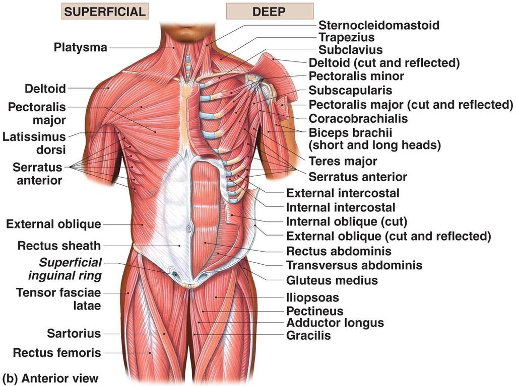
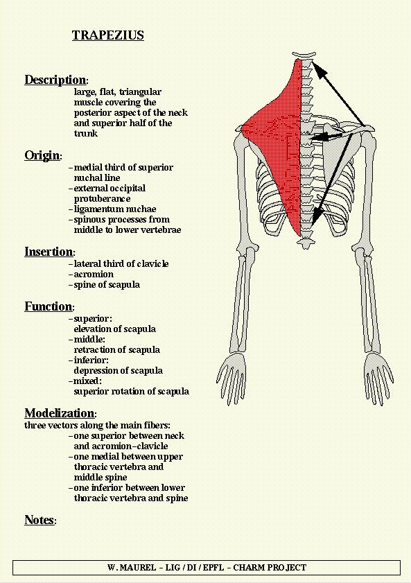
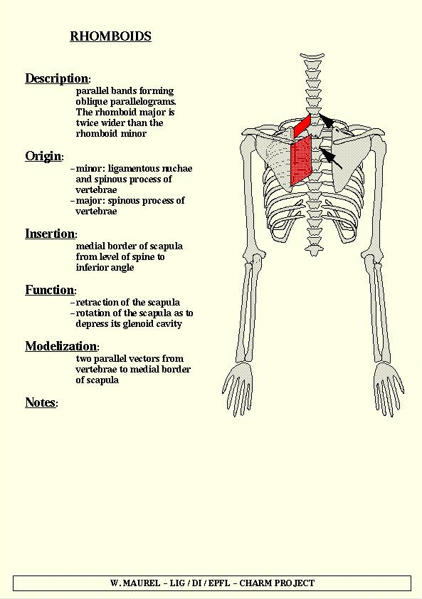
4 Muscles That Position the Pectoral Girdle Trapezius Superficial Covers back and neck to base of skull Inserts onto clavicles and scapular spines Rhomboid and levator scapulae Deep to trapezius Attach to cervical and thoracic vertebrae Insert on scapular border Serratus anterior On the chest Originates along ribs Inserts on anterior scapular margin

5 Muscles That Position the Pectoral Girdle.
6 Latissimus dorsi
7
8

9 Antaerior Muscles That Position the Pectoral Girdle Subclavius Originates on ribs Inserts on clavicle

10 Anterior thoracic Muscles (Pictorials Muscle Dose not Crossing the Shoulder)


11 Musles crossing the shoulder

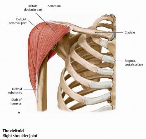
12 Deltoid muscle
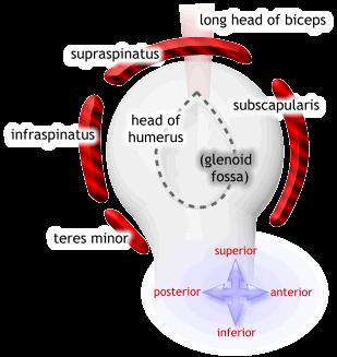
13 Muscles Crossing the Shoulder Rotator cuff muscles supraspinatus, infraspinatus, teres minor, and subscapularis Function mainly to reinforce the capsule of the shoulder Secondarily act as synergists and fixators The coracobrachialis and teres major: Act as synergists Do not contribute to reinforcement of the shoulder joint
14 Rotator calf muscles
 & lat. Rotation N.SUPPLY: Suprascapular.")
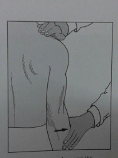
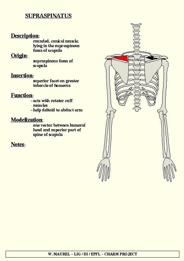
15 SUPRASPINATUS ORIGIN: supraspinatous fossa of the scapula INSERTION: superior facet on the gr.tubercle of the humerus ACTION: initiation of abduction (15*) & lat. Rotation N.SUPPLY: Suprascapular.N From B.P (C5,C6)
16 SUPRASPINATUS

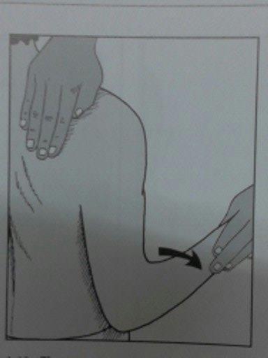
17 INFRASPINATUS ORIGIN: infraspinatous fossa of the scapula INSERTION: middle facet on the gr.tubercle of the humerus ACTION: lat.rotation of shoulder, strengthen the shoulder by bracing the head of humerus. N.SUPPLY: Suprascapular.N of B.P (C5,c6)
18 INFRASPINATUS
")
19 TERES MINOR ORIGIN: lat.borderof the scapula INSERTION: inf. facet on the grt. tubercle of the humerus ACTION: ext. rotator & weak adductor N.SUPPLY: Axillary.N.(C5,C6)
20
 N.SUPPLY: Upper &Lower Subscapular.")
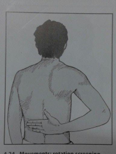
21 SUBSCAPULARIS ORIGIN: medial 2/3 subscapular fossa, lat border of tha scapula INSERTION: lesser tubercle of the humerus ACTION: stabilise the shoulder & prevents anterior displacement of the shoulder (med. rotation, adduction of arm) N.SUPPLY: Upper &Lower Subscapular.N (c5,c6)
22 Sub-scapularis Muscle
23 SUBSCAPULARIS

24 TERES MAJOR ORIGIN: dorsal surface of the inf.angle of the scapula INSERTION: med.lip of the intetubercular groove of the humerus ACTION: extends the arm from flexd.position & it adducts, med.rotates the arm N.SUPPLY: Lower Subscapular (C6,C7)
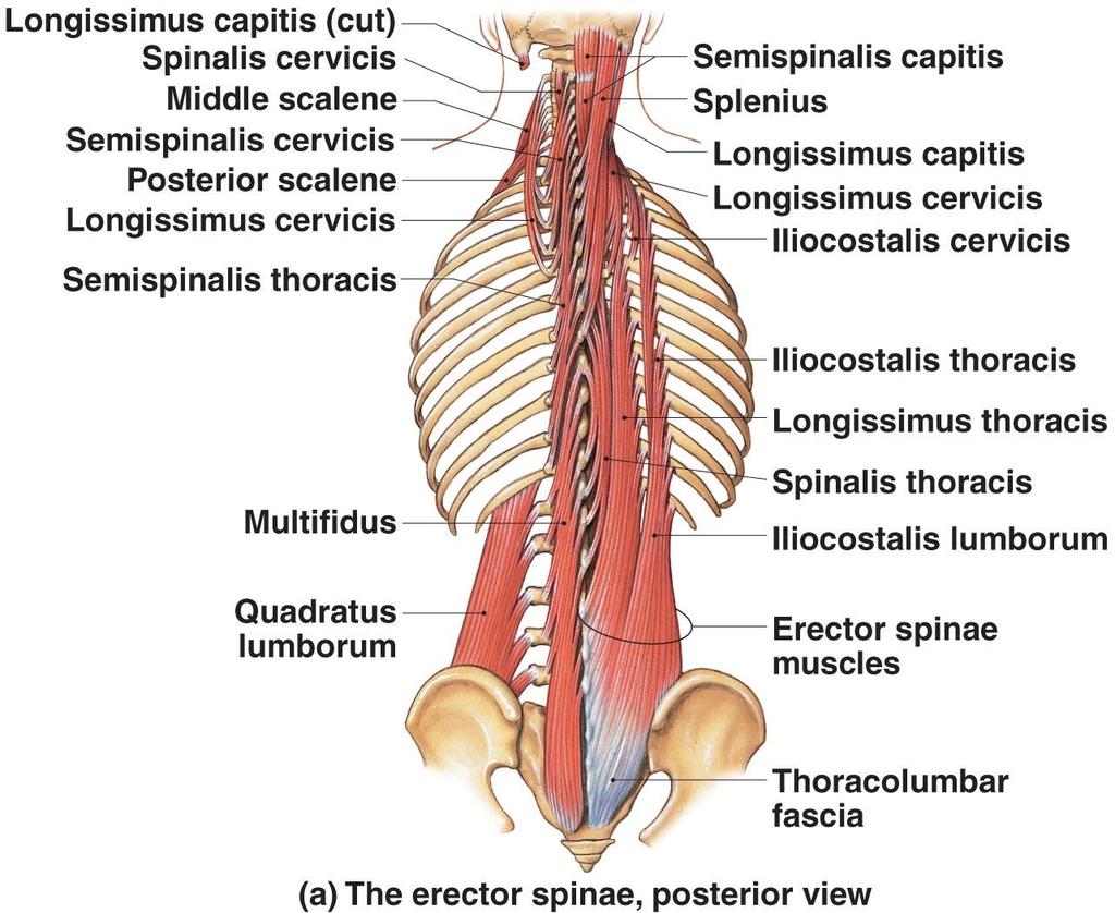
25 Superficial Spinal Extensors Spinalis group Longissimus group Iliocostalis group Deep Spinal Extensors Semispinalis group Multifidus muscle Interspinalis muscles Intertransversarii muscles Rotatores muscles
26
27 Trunk Movements: Deep Back Muscles The prime mover of back extension is the erector spinae Erector spinae, or sacrospinalis, muscles consist of three columns on each side of the vertebrae iliocostalis, longissimus, and spinalis Lateral bending of the back is accomplished by unilateral contraction of these muscles Other deep back extensors include the semispinalis muscles and the quadratus lumborum

28 [INSERT Table (top portion)]

29 [INSERT Table (middle portion)]
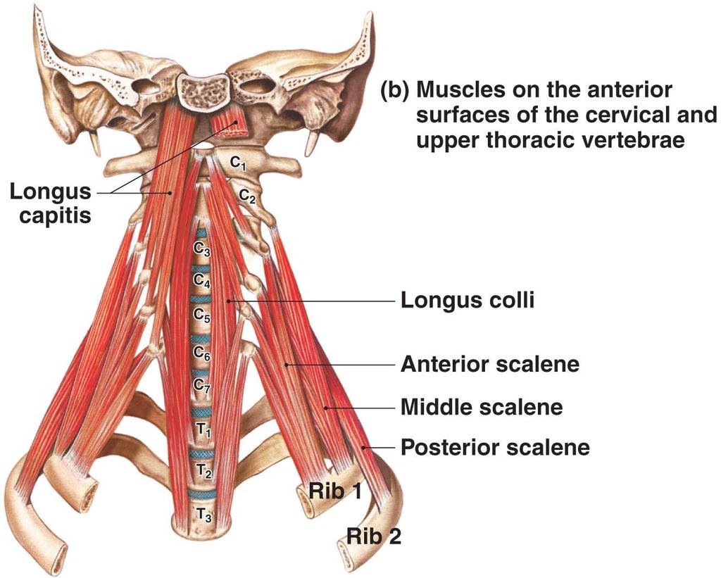
30 Anterior Muscles of the Vertebral Column Spinal flexors Neck Longus capitis and longus colli Rotate and flex the neck Lumbar Quadratus lumborum muscles Flex spine and depress ribs
31
32 Muscles of the upper extremities
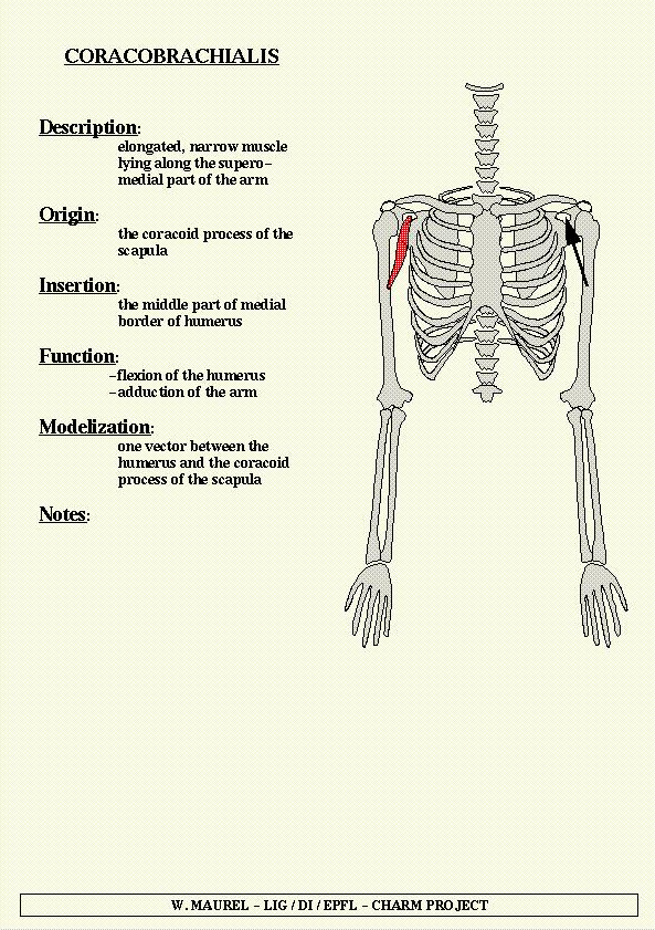
33 coracobrachialis
34 Muscles Crossing the Elbow Forearm flexion Brachialis and biceps brachii are the chief forearm flexion The brachioradialis acts as a synergist and helps stabilize the elbow Forearm extension The triceps brachii is the prime mover of forearm extension The anconeus is a weak synergist

35 Biceps Brachi Muscle

36 Brachalis muscle

37 Triceps Brachi Muscle ORIGIN: Long head: infraglenoid tubercle of the scapula Lat.head: oblique ridge on upper surface above the radial groove Medial head: posterior surface of the humerus inf.to the radial groove INSERTION: proximal olecrenon of the ulna ACTION:extends the forearm N.SUPPLY: Radial.N.(C7,c8)


38 The ARM An aponeurotic sheet separating various muscles of the upper limbs, including lateral and medial humeral septa. The lateral and medial intermuscular septa divide the distal part of the arm into two compartments: Anterior compartments also known as the flexor compartment Posterior compartments also known as the extensor compartment ski n Fasci a Humeru s Lateral intermuscul ar septum Medial intermuscul ar septum Neurovascul ar bundle
39 MUSCLES OF FOREARM Anterior Compartment: common flxor origin SUPERFICIAL 1.Pronator Teres 2.Flx Carpi Ulnaris 3.Palmaris Longus 4.Flx.Carpi Radialis 5.Flx.Digitorum Superficialis (sublimus) DEEP: 1.Flx.Digitorum Profundus 2.Flx.Pollicis Longus 3.Pronator Quadratus
40 Forearm Muscles Superficial Compartment Flexor Carpi Radialis Flexor Carpi Ulnaris Flexor Digitorum Superficialis - all Palmaris Longus Pronator Teres

41 Forearm Muscles Superficial Compartment Superficial layer These muscles are primarily flexors of the wrist and fingers

42 PRONATOR TERES Insertion: midway long the lateral surface of the radius Action: pronation,flexion of forearm N.Supply:Median.N (C6c7)
43 FLX.CARPI RADIALIS INSERTION:base of the 2 nd mc bone and slip to the bace of the 3 rd MC bone ACTION: flexion and abduction of wrist N.SUPPLY: Median.N (c6,c7)
44 PALMARIS LONGUS INSERTION: anterior aspect of the distal flx.retinaculum and palmar aponeurosis ACTION: flx.the wrist, and tightens the palmar.aponeurosis N.SUPPLY: Median. N (c6,c7)
45 FLX CARPI ULNARIS ORIGIN: humoral head: med epicondyle Ulnar head:med.margin of the olecrenon,posterior border of the ulna INSERTION: pisiform, hook of hamate, base of the 5 th MC & flx.retinaculum ACTION:flexes and adducts the hand N.SUPPLY: Ulnar.N (C7,C8)
46 Forearm Muscles Deep Compartment Flexor Digitorum Profundus - all Flexor Pollicis Longus Pronator Quadratus

47 Muscles of the Forearm: Anterior Compartment Meddle and Deep Layers
48 FLX.DIGITORUM SUPERFICIALIS ORIGIN: humeroulnar head: med.epicondyle of the humerus, coronoid process Radial head: sup.half of anterior aspect of the radius INSERTION : bodies of the Middle phalanges of the medial 4 digits ACTION: flx.of all joints it crosses N.SUPPLY :Median.N (C7,C8,T1)

49 FLX.DIGITORUM PROFUNDUS ORIGIN:prox.3/4 of the medial and anterior aspect of the ulna and from interosseous memb. INSERTION:base of The Diatal phalanges of the medial 4 digits ACTION: flx.dip,,pip,mp, wrist N.SUPPLY: Medial-ulnar.N (C8,t1) Lateral-AIN Of Median.N (C8,t1)
50 FLX.POLLICIS LONGUS ORIGIN: upper 3/4 of anterior surface of radius INSERTION: base of distal phalanx of the thumb ACTION: flexion of proximal & distal phalnx of the thumb N.SUPPLY:AIN (C7,C8,T1)

51 PRONATOR QUADRATUS ORIGIN: lower 1/4 th of anterior surface of ulna INSERTION: lower 1/4 th of anterior surface of radius ACTION :pronation N.SUPPLY:AIN (C8,T1)
52 POSTERIOR COMPARTMENT OF FOREARM SUPERFICIAL 1.anconeus 2.Brachioradialis 3.Ext.Carpi Radialis Longus 4.Ext.Carpi Radialis Brevis 5.Ext.Digitorum 6.Ext.Digiti Minimi 7.Ext.Carpi Ulnaris DEEP 1.Abductor Pollicis Longus 2.Ext.Pollicis longus 3.Ext.Pollicis brevis 4.Ext.Indicis 5.supinator
53 Forearm Muscles superficial Posterior Compartment Anconeus Brachioradialis Extensor Carpi Radialis Brevis Extensor Carpi Radialis Longus Extensor Carpi Ulnaris Extensor Digiti Minimi Extensor Digitorum - All

54 Forearm Muscles Posterior Compartment These muscles are primarily extensors of the wrist and fingers

55 ANCONEUS ORIGIN: posterior surface of the lat.epicondyle INSERTION:lateral surface of olecranon,body of ulna ACTION: extension of forearm N.SUPPLY: radial.n

56 BRACHIORADIALIS ORIGIN: prox.2/3 rd of lat.supracondylar ridge of humerus, lateral intermuscular septum INSERTION: lat.aspect of distal radius just prox.to the styloid process ACTION: acc.flexor of elbow N.SUPPLY: Radial.N (C5,C6)

57 EXT.CARPI RADIALIS LONGUS ORIGIN: lat.supracondylar ridge of humerus INSERTION: base of the 2 nd MC bone ACTION: extends and abducts the hand N.SUPPLY: Radial.N (C6,C7)

58 EXTENSOR CARPI RADIALIS BREVIS ORIGIN: lat.epicondyle of the humerus INSERTION: base of the 3 rd MC bone ACTION: extends and abducts the wrist N.SUPPLY: deep br.of radial.n(c7c8)

59 EXTENSOR DIGITORUM ORIGIN: lat epicondyle of the humerus INSERTION: Extensor Expansions Of The Medial 4 digits ACTION: extension at MCP, IP joints, ext of wrist when the fingers are extended N.SUPPLY: PIN (c7,c8)

60 EXTENSOR DIGITI MINIMI ORIGIN:lat epicondyle of the humerus INSERTION:extensor expansion of the 5 th digit ACTION: ext.of 5 th digit at MC, PIP, ext of wrist when little finger in extension N.SUPPLY:PIN (c7,c8)
61 EXT.CARPI ULNARIS ORIGIN:2 heads lat epicondyle of humerus, Posterior border of ulna INSERTION:medial side of the base of the 5 th MC ACTION: extends and adducts the hand N.SUPPLY:PIN (c7c8)

62 SUPINATOR ORIGIN: lat epicondyle of humerus, radial collateral lig., annular lig.,supinator fossa, crest of ulna INSERTION: lateral,posterior,anterior surface of prox 1/ 3 rd 0f radius ACTION:supination N.Supply: deep Br.Of Radial.N (C5,c6)
63 Forearm Muscles Deep Posterior Compartment Abductor Pollicis Longus Extensor Indicis Extensor Pollicis Brevis Extensor Pollicis Longus Flexor Digitorum Profundus Supinator

64 ABDUCTOR POLLICIS LONGUS ORIGIN:upper parts of the posterior surface of the radius & ulna and interosseous mem. INSERTION:base of the 1 st MC bone ACTION: abducts,extends, lat.rotates the thumb at carpometacarpal joint, & abducts the wrist. N.SUPPLY:PIN (c7,c8)
65 EXTENSOR POLLICIS BREVIS ORIGIN:posterior surface of the radius below the origin ofabductor Pollicis longusand from interosseous mem. INSERTION:base of the prox.phalanx of the thumb ACTION:extends pox.phalanx of the thumb at MC joint, ext.at 1 st carpometacarpal joint N.SUPPLY:PIN (c7,c8)
66 EXT.POLLICIS LONGUS ORIGIN: posterior surface of middle 1/3 rd of ulna And interrosseous mem. INSERTION:base of the diastal phalanx of the thumb ACTION:extends distal phalanx of the thumb at MP joint,ip joint.and it can contribute abduction of the thumb N.SUPPLY:PIN (c7,c8).

67 EXTENSOR INDICIS Origin:posterior surface of ulna m/3 rd below the EPL Insertion:extensor expansion of the 2 nd finger Action: ext of index finger and wrist. N.supply:PIN (c7,c8)

68 Forearm Muscles Posterior Compartment These muscles are primarily extensors of the wrist and fingers

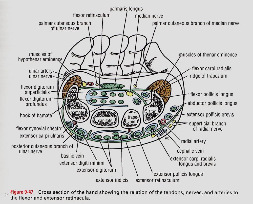
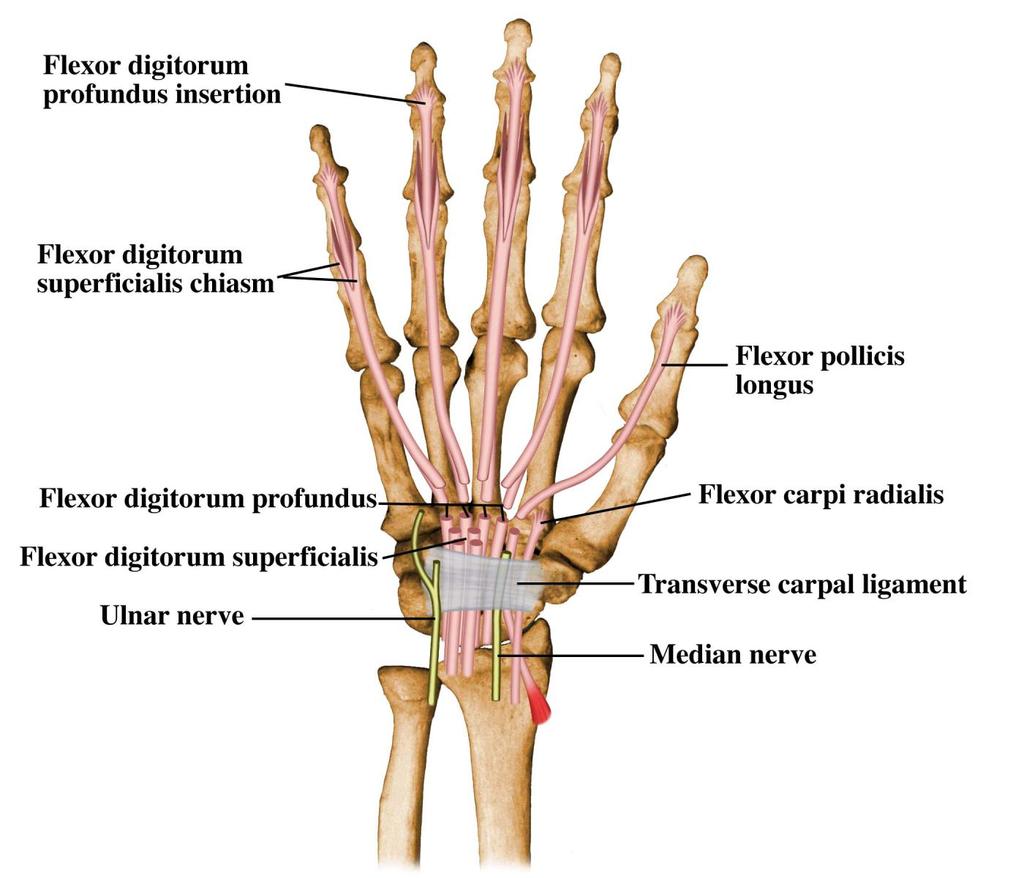
69 Surface Anatomy of Upper Limb Carpal Tunnel Carpals concave anteriorly Carpal ligament covers it Contains: long tendons, Median nerve Inflammation of tendons = compression of Median nerve Anatomical Snuffbox Lateral = E.pollicis brevis Medial = E. pollicis longus Floor = scaphoid, styloid of radius Contains Radial Artery (pulse) Frolich, Human Anatomy,UpprLimb
70 CARPAL TUNNEL TUNNEL FORMED BETWEEN THE CONCAVITY OF THE CARPAL BONES AND A LIGAMENT THAT COVERS THIS( FLEXOR RETINACULAM) TENDONS OF THE FLEXORS PASS THROUGH MEDIAN NERVE ALSO PASSES THROUGH CROWDED TUNNEL CARPAL TUNNEL SYNDROME - CAUSED DUE TO COMPRESSION OF THE NERVE IN THE TUNNEL - CAUSES SWELLING OF THE TEDONS( OVERUSE) - 2. PREGNANCY( EDEMA) - 3. ARTHRITIS SYMPTOMS- TINGLING OR NUMBNESS-LATERAL PART OF HAND, WEAKNESS IN THUMB MOVEMENT TREATMENT- REST, SPLINTING,ANTI-INFLAMMATORY DRUGS, SURGERY
71
72
73
74
75
76
77
78
79
80
81
# Anatomy. Upper Extremities Muscles and anatomy of axilla. Tiba Al-Ani 9/10/2015 Nabil. Page 0 of 16
 #10 25 Anatomy Upper Extremities Muscles and anatomy of axilla Tiba Al-Ani 9/10/2015 Nabil Page 0 of 16 Salam AWN Today s lecture is divided into two parts, the first part is the continuation of the upper
Lab Activity 11: Group II
 Lab Activity 11: Group II Muscles Martini Chapter 11 Portland Community College BI 231 Origin and Insertion Origin: The place where the fixed end attaches to a bone, cartilage, or connective tissue. Insertion:
ARM Brachium Musculature
 ARM Brachium Musculature Coracobrachialis coracoid process of the scapula medial shaft of the humerus at about its middle 1. flexes the humerus 2. assists to adduct the humerus Blood: muscular branches
Muscles of the Upper Limb
 Muscles of the Upper Limb anterior surface of ribs 3 5 coracoid process Pectoralis minor pectoral nerves protracts / depresses scapula Serratus anterior Subclavius ribs 1-8 long thoracic nerve rib 1 ----------------
Muscular Nomenclature and Kinesiology - One
 Chapter 16 Muscular Nomenclature and Kinesiology - One Lessons 1-3 (with lesson 4) 1 Introduction 122 major muscles covered in this chapter Chapter divided into nine lessons Kinesiology study of human
The Muscular System. Chapter 10 Part C. PowerPoint Lecture Slides prepared by Karen Dunbar Kareiva Ivy Tech Community College
 Chapter 10 Part C The Muscular System Annie Leibovitz/Contact Press Images PowerPoint Lecture Slides prepared by Karen Dunbar Kareiva Ivy Tech Community College Table 10.9: Muscles Crossing the Shoulder
Human Anatomy Biology 351
 1 Human Anatomy Biology 351 Upper Limb Exam Please place your name on the back of the last page of this exam. You must answer all questions on this exam. Because statistics demonstrate that, on average,
The Clavicle Right clavicle Deltoid tubercle: Conoid tubercle, conoid ligamen Impression for the
 The Clavicle Muscle Attachment Sites in the Upper Limb Pectoralis major Right clavicle Smooth superior surface of the shaft, under the platysma muscle tubercle: attachment of the deltoid Acromial facet
REFERENCE DIAGRAMS OF UPPER LIMB MUSCLES: NAMES, LOCATIONS, ATTACHMENTS, FUNCTIONS MUSCLES CONNECTING THE UPPER LIMB TO THE AXIAL SKELETON
 REFERENCE DIAGRAMS OF UPPER LIMB MUSCLES: NAMES, LOCATIONS, ATTACHMENTS, FUNCTIONS MUSCLES CONNECTING THE UPPER LIMB TO THE AXIAL SKELETON A25LAB EXERCISES: UPPER LIMB MUSCLES Page 1 MUSCLES CONNECTING
medial half of clavicle; Sternum; upper six costal cartilages External surfaces of ribs 3-5
 MUSCLE ORIGIN INSERTION ACTION NERVE Pectoralis Major medial half of clavicle; Sternum; upper six costal cartilages Lateral lip of intertubercular groove of horizontal adduction Medial and lateral pectoral
Practical 2 Worksheet
 Practical 2 Worksheet Upper Extremity BONES 1. Which end of the clavicle is on the lateral side (acromial or sternal)? 2. Describe the difference in the appearance of the acromial and sternal ends of the
Module 7 - The Muscular System Muscles of the Arm and Trunk
 Module 7 - The Muscular System Muscles of the Arm and Trunk This Module will cover the muscle anatomy of the arms and trunk. We have already seen the muscles that move the humerus, so this module will
Netter's Anatomy Flash Cards Section 6 List 4 th Edition
 Netter's Anatomy Flash Cards Section 6 List 4 th Edition https://www.memrise.com/course/1577581/ Section 6 Upper Limb (66 cards) Plate 6-1 Humerus and Scapula: Anterior View 1.1 Acromion 1.2 Greater tubercle
Anatomy and Physiology II. Review Shoulder Girdle New Material Upper Extremities - Bones
 Anatomy and Physiology II Review Shoulder Girdle New Material Upper Extremities - Bones Anatomy and Physiology II Shoulder Girdle Review Questions From Last Lecture Can you identify the following muscles?
Key Relationships in the Upper Limb
 Key Relationships in the Upper Limb This list contains some of the key relationships that will help you identify structures in the lab. They are organized by dissection assignment as defined in the syllabus.
forearm posterior compartment
 Quick revision: The anterior compartment of the forearm contains of 8 muscles... -4 superficial -1 intermediate -3 deep *All supplied by median nerve except 1 and 1/2 muscle (by ulnar N.) forearm posterior
Muscles in the Shoulder, Chest, Arm, Stomach, and Back
 Muscles in the Shoulder, Chest, Arm, Stomach, and Back Shoulder Muscles Deltoid Supraspinatus Infraspinatus Teres Major Teres Minor Subscapularis Deltoid (Delts) Function: Raises the upper arm Origin:
compartments of the forearm
 " forearm posterior compartment " compartments of the forearm Posterior Fascial compartment Muscles: ** The superficial group 1. Extensor carpi radialis brevis 2. Ex. digitorum 3. Ex. digiti minimi 4.
Al-Balqa Applied University
 Al-Balqa Applied University Faculty Of Medicine *You can use this checklist as a guide to you for the lab. the items on this checklist represent the main features of the models that you have to know for
Systematic Anatomy (For international students)
") Systematic Anatomy (For international students) Department of Anatomy,Fudan University Teaching contents Muscles of abdomen & upper limbs Dr.Hongqi Zhang ( 张红旗 ) Email: zhanghq58@126.com 1 Muscles of abdomen
Systematic Anatomy (For international students) Department of Anatomy,Fudan University Teaching contents Muscles of abdomen & upper limbs Dr.Hongqi Zhang ( 张红旗 ) Email: zhanghq58@126.com 1 Muscles of abdomen
The Forearm 2. Extensor & lateral Compartments of the Forearm
 The Forearm 2 Extensor & lateral Compartments of the Forearm 1-Lateral Fascial Compartment (at the lateral side of the forearm ) *Some books mention the lateral compartment contain just the Brachioradialis
Biceps Brachii. Muscles of the Arm and Hand 4/4/2017 MR. S. KELLY
 Muscles of the Arm and Hand PSK 4U MR. S. KELLY NORTH GRENVILLE DHS Biceps Brachii Origin: scapula Insertion: radius, fascia of forearm (bicipital aponeurosis) Action: supination and elbow flexion Innervation:
Connects arm to thorax 3 joints. Glenohumeral joint Acromioclavicular joint Sternoclavicular joint
 Connects arm to thorax 3 joints Glenohumeral joint Acromioclavicular joint Sternoclavicular joint Scapula Elevation Depression Protraction (abduction) Retraction (adduction) Downward Rotation Upward Rotation
Axilla and Brachial Region
 L 4 A B O R A T O R Y Axilla and Brachial Region BRACHIAL PLEXUS 5 Roots/Rami (ventral rami C5 T1) 3 Trunks Superior (C5, C6) Middle (C7) Inferior (C8, T1) 3 Cords Lateral Cord (Anterior Superior and Anterior
MUSCLES OF THE ELBOW REGION
 MUSCLES OF THE ELBOW REGION Dr Bronwen Ackermann COMMONWEALTH OF AUSTRALIA Copyright Regulation WARNING This material has been reproduced and communicated to you by or on behalf of the University of Sydney
Upper limb Arm & Cubital region 黃敏銓
 Upper limb Arm & Cubital region 黃敏銓 1 Arm Lateral intermuscular septum Anterior (flexor) compartment: stronger Medial intermuscular septum Posterior (extensor) compartment 2 Coracobrachialis Origin: coracoid
Copy Right- Hongqi ZHANG-Department of Anatomy-Fudan University. Systematic Anatomy. Locomotor system - Part 6
 Systematic Anatomy Locomotor system - Part 6 Muscles of abdomen Muscles of the upper limb Dr.Hongqi Zhang ( 张红旗 ) Email: zhanghq58@126.com 1 Muscles of abdomen Muscles of the upper limb Muscles of abdomen
STRUCTURAL BASIS OF MEDICAL PRACTICE EXAMINATION 5 October 6, 2006
 STRUCTURAL BASIS OF MEDICAL PRACTICE EXAMINATION 5 October 6, 2006 PART l. Answer in the space provided. (8 pts) 1. Identify the structures. (2 pts) B C A. _pisiform B. _ulnar artery A C. _flexor carpi
The Elbow and the cubital fossa. Prof Oluwadiya Kehinde
 The Elbow and the cubital fossa Prof Oluwadiya Kehinde www.oluwadiya.com Elbow and Forearm Anatomy The elbow joint is formed by the humerus, radius, and the ulna Bony anatomy of the elbow Distal Humerus
Upper Limb Muscles Muscles of Axilla & Arm
 Done By : Saleh Salahat Upper Limb Muscles Muscles of Axilla & Arm 1) Muscles around the axilla A- Muscles connecting the upper to thoracic wall (4) 1- pectoralis major Origin:- from the medial half of
BLUE SKY SCHOOL OF PROFESSIONAL MASSAGE AND THERAPEUTIC BODYWORK. Musculoskeletal Anatomy & Kinesiology II REVIEW
 BLUE SKY SCHOOL OF PROFESSIONAL MASSAGE AND THERAPEUTIC BODYWORK Musculoskeletal Anatomy & Kinesiology II REVIEW MSAK101-II Session 4 LEARNING OBJECTIVES: By the end of this session, the student will be
Abduction of arm until your hand rich your head. Flexion of forearm at elbow joint. Extension of arm at elbow joint. Flexion of fingers 10.
 Num. answer 1. Medialy With the manubrium ( sternum ), and laterally with the acromion of the scapula 2. 1. Trapezius 2. Levator scapulae 3. Rhomboids 3. 1. Pectoralis major 2. Pectoralis minor 3. Latissiumus
MCQWeek2. All arise from the common flexor origin. The posterior aspect of the medial epicondyle is the common flexor origin.
 MCQWeek2. 1. Regarding superficial muscles of anterior compartment of the forearm: All arise from the common flexor origin. The posterior aspect of the medial epicondyle is the common flexor origin. Flexor
Muscle Anatomy Review Chart
 Muscle Anatomy Review Chart BACK Superficial (5) Trapezius Transverse cervical a. Latissimus dorsi Thoracodorsal a. Rhomboideus major Dorsal scapular a. Rhomboideus minor Levator scapulae Intermediate
Thank You for Your Support! Hosford Muscle Tables
 Thank You for Your Support! This PDF document has been placed online for your enjoyment and I hope you find it useful. These tables are both a teaching tool, and a study / review tool. I created these
Synergist Muscles. Shoulder (glenohumeral joint) Flexion Deltoid (anterior fibers) Pectoralis major (upper fibers) Biceps Brachii Coracobrachialis
 Flexion Deltoid (anterior fibers) Pectoralis major (upper fibers) Biceps Brachii Coracobrachialis") Synergist Muscles Dr Gene Desepoli DrGeneLMT@gmail.com Shoulder (glenohumeral joint) Deltoid (anterior fibers) Pectoralis major (upper fibers) Biceps Brachii Coracobrachialis Deltoid (posterior fibers)
Synergist Muscles Dr Gene Desepoli DrGeneLMT@gmail.com Shoulder (glenohumeral joint) Deltoid (anterior fibers) Pectoralis major (upper fibers) Biceps Brachii Coracobrachialis Deltoid (posterior fibers)
divided by the bones ( redius and ulna ) and interosseous membrane into :
 and interosseous membrane into :") fossa Cubital Has: * floor. * roof : - Skin - superficial fasica - deep fascia ( include bicipital aponeurosis ) Structures within the roof : -cephalic and basilic veins -and between them median cubital
fossa Cubital Has: * floor. * roof : - Skin - superficial fasica - deep fascia ( include bicipital aponeurosis ) Structures within the roof : -cephalic and basilic veins -and between them median cubital
BRACHIAL PLEXUS. DORSAL SCAPULAR NERVE (C5) supraclavicular branch innervates rhomboids (major and minor) and levator scapulae
 supraclavicular branch innervates rhomboids (major and minor) and levator scapulae") THE BRACHIAL PLEXUS DORSAL SCAPULAR NERVE (C5) supraclavicular branch innervates rhomboids (major and minor) and levator scapulae SCHEMA OF THE BRACHIAL PLEXUS THE BRACHIAL PLEXUS PHRENIC NERVE supraclavicular
THE BRACHIAL PLEXUS DORSAL SCAPULAR NERVE (C5) supraclavicular branch innervates rhomboids (major and minor) and levator scapulae SCHEMA OF THE BRACHIAL PLEXUS THE BRACHIAL PLEXUS PHRENIC NERVE supraclavicular
LIST OF STRUCTURES TO BE IDENTIFIED IN LAB: UPPER EXTREMITY REVIEW 2016
 LIST OF STRUCTURES TO BE IDENTIFIED IN LAB: UPPER EXTREMITY REVIEW 2016 BONES Ribs, sternum, clavicle Humerus: Head, greater tubercle, lesser tubercle, intertubercular sulcus, surgical neck, anatomical
Main Menu. Wrist and Hand Joints click here. The Power is in Your Hands
 1 The Wrist and Hand Joints click here Main Menu K.5 http://www.handsonlineeducation.com/classes/k5/k5entry.htm[3/23/18, 1:40:40 PM] Bones 29 bones, including radius and ulna 8 carpal bones in 2 rows of
Peripheral Nervous Sytem: Upper Body
 Peripheral Nervous Sytem: Upper Body MSTN121 - Neurophysiology Session 10 Department of Myotherapy Cervical Plexus Accessory nerve (CN11 + C1-5) Motor: trapezius and sternocleidomastoid Greater auricular
Due in Lab weeks because of Thanksgiving Prelab #10. Homework #8. Both sides! Both sides!
 Lab 8 MUSCLES Due in Lab 10 2 weeks because of Thanksgiving Prelab #10 Both sides! Homework #8 Both sides! Refer to Muscles 22-23 Naming of muscles Origin Site of muscle attachment that doesn t move during
Prime movers provide the major force for producing a specific movement Antagonists oppose or reverse a particular movement Synergists
 Dr. Gary Mumaugh Prime movers provide the major force for producing a specific movement Antagonists oppose or reverse a particular movement Synergists Add force to a movement Reduce undesirable or unnecessary
MLT Muscle(s) Patient Position Therapist position Stabilization Limb Position Picture Put biceps on slack by bending elbow.
 Patient Position Therapist position Stabilization Limb Position Picture Put biceps on slack by bending elbow.") MLT Muscle(s) Patient Position Therapist position Stabilization Limb Position Picture Put biceps on slack by bending elbow. Pectoralis Minor Supine, arm at side, elbows extended, supinated Head of Table
MLT Muscle(s) Patient Position Therapist position Stabilization Limb Position Picture Put biceps on slack by bending elbow. Pectoralis Minor Supine, arm at side, elbows extended, supinated Head of Table
Forearm and Wrist Regions Neumann Chapter 7
 Forearm and Wrist Regions Neumann Chapter 7 REVIEW AND HIGHLIGHTS OF OSTEOLOGY & ARTHROLOGY Radius dorsal radial tubercle radial styloid process Ulna ulnar styloid process ulnar head Carpals Proximal Row
Anatomage Table Instructors Guide- Upper Limb
 The Upper Limb Anatomage Table Instructors Guide- Upper Limb Table of Contents Upper Limb 1- The Skeletal System...3 1: Clavicle...3 2: Scapula...5 3: Shoulder (Glenohumeral) and Proximal Humerus...7 4:
MUSCLES. Anconeus Muscle
 LAB 7 UPPER LIMBS MUSCLES Anconeus Muscle anconeus origin: distal end of dorsal surface of humerus insertion: lateral surface of ulna from distal margin of the semilunar notch to proximal end of the olecranon
Lecture 9: Forearm bones and muscles
 Lecture 9: Forearm bones and muscles Remember, the region between the shoulder and the elbow = brachium/arm, between elbow and wrist = antebrachium/forearm. Forearm bones : Humerus (distal ends) Radius
Supplied in part by the musculocutaneous nerve. Forms the axis of rotation in movements of pronation and supination
 Anatomy: Upper limb (15 questions) 1. Latissimus Dorsi: Is innervated by the dorsal scapular nerve Lies above feres major muscle Medially rotates the humerus All of the above 2. Supinator muscle is: Deep
11/15/2018. Temporalis Elevates & retracts mandible. Masseter = Prime mover of jaw closure. Levator scapulae Supraspinatus Clavicle.
 Due in Lab 10 Lab 8 MUSCLES 2 weeks because of Thanksgiving Prelab #10 Both sides! Homework #8 Both sides! Refer to Muscles 22-23 Examples of Origin & Insertion Naming of muscles Origin Site of muscle
Dr. Mahir Alhadidi Anatomy Lecture #9 Feb,28 th 2012
 Quick Revision: Upper arm is divided into two compartments: 1. Anterior Compartment: Contains three muscles (Biceps brachii, Coracobrachialis, Brachialis). Innervated by Musculocutaneous nerve. 2. Posterior
Nerves of Upper limb. Dr. Brijendra Singh Professor & Head Department of Anatomy AIIMS Rishikesh
 Nerves of Upper limb Dr. Brijendra Singh Professor & Head Department of Anatomy AIIMS Rishikesh 1 Objectives Origin, course & relation of median & ulnar nerves. Motor & sensory distribution Carpal tunnel
Anatomy Upper Limb Muscles
 Anatomy Upper Limb Muscles Rotator cuff/scapulohumeral muscles 4 muscles (SITS) form musculotendinous rotator cuff around glenohumeral joint, provide stability of joint Supraspinatus Course: med 2/3 supraspinatous
Levels of the anatomical cuts of the upper extremity RADIUS AND ULNA right
 11 CHAPTER 2 Levels of the anatomical cuts of the upper extremity AND right CUT 1 CUT 4 1 2 3 4 5 6 Isolated fixation of the radius is difficult at this level because of the anterolateral vessels and the
Lectures Muscular System 10-1
 Lectures 12-14 Muscular System 10-1 Properties of Muscle Ability of a muscle to shorten with force Capacity of muscle to respond to a stimulus Muscle can be stretched to its normal resting length and beyond
Functional Anatomy of the Elbow
 Functional Anatomy of the Elbow Orthopedic Institute Daryl C. Osbahr, M.D. Chief of Sports Medicine, Orlando Health Chief Medical Officer, Orlando City Soccer Club Orthopedic Consultant, Washington Nationals
region of the upper limb between the shoulder and the elbow Superiorly communicates with the axilla.
 1 region of the upper limb between the shoulder and the elbow Superiorly communicates with the axilla. Inferiorly, a number of important structures pass between arm & forearm through cubital fossa. 2 medial
Figure 27: The synovial membrane of the shoulder joint (anterior view)
") The coracoacromial ligament; is an accessory ligament that protects the superior aspect of the joint extending from the coracoid process to the acromion over the tendon of supraspinatus. The synovial membrane
The coracoacromial ligament; is an accessory ligament that protects the superior aspect of the joint extending from the coracoid process to the acromion over the tendon of supraspinatus. The synovial membrane
Fascial Compartments of the Upper Arm
 Fascial Compartments of the Upper Arm The upper arm is enclosed in a sheath of deep fascia and has two fascial septa: 1- Medial fascial septum (medial intermuscular septum): attached to the medial supracondylar
Anatomy of the Shoulder Girdle. Prof Oluwadiya Kehinde FMCS (Orthop)
") Anatomy of the Shoulder Girdle Prof Oluwadiya Kehinde FMCS (Orthop) www.oluwadiya.com Bony Anatomy Shoulder Complex: Sternum(manubrium) Clavicle Scapula Proximal humerus Manubrium Sterni Upper part of
Anatomy of the Shoulder Girdle Prof Oluwadiya Kehinde FMCS (Orthop) www.oluwadiya.com Bony Anatomy Shoulder Complex: Sternum(manubrium) Clavicle Scapula Proximal humerus Manubrium Sterni Upper part of
Human Anatomy and Physiology I Laboratory
 Human Anatomy and Physiology I Laboratory Gross Anatomy of the Muscular System (Two weeks) 1 This lab involves study of the laboratory exercise Gross Anatomy of the Muscular System. Complete the Review
Elbow, Wrist & Hand Evaluation.
 Elbow, Wrist & Hand Evaluation www.fisiokinesiterapia.biz Common Injuries to the Elbow, Wrist, Hand & Fingers Lateral epicondylitis tennis elbow Medial epicondylitis golfer s s elbow, little league elbow
Anatomy of the Forearm
 Anatomy of the Forearm Musculoskeletal block- Anatomy-lecture 8 Editing file Objectives List the names of the Flexors Group of Forearm (superficial & deep muscles). Identify the common flexor origin of
Wrist and Hand Anatomy
 Wrist and Hand Anatomy Bone Anatomy Scapoid Lunate Triquetrium Pisiform Trapeziod Trapezium Capitate Hamate Wrist Articulations Radiocarpal Joint Proximal portion Distal portion Most surface contact found
TABLES OF MUSCLE ACTIONS, INNERVATIONS, AND ATTACHMENTS
 TABLES OF MUSCLE ACTIONS, INNERVATIONS, AND ATTACHMENTS Table 1-1 ERECTOR SPINAE MUSCLES Intrinsic muscles producing extension and/or lateral of the spine Muscle Joint and Action Innervation Inferior Attachment
Human Anatomy Lab #7: Muscles of the Cadaver
 Human Anatomy Lab #7: Muscles of the Cadaver Table of Contents: Expected Learning Outcomes.... 1 Introduction...... 1 Identifying Muscles on Yourself.... 2 Muscles of the Anterior Trunk and Arm.. 2 Muscles
The Free Upper Limb. Bone of the Arm. aus: Platzer, Locomotor System (ISBN ), 2009 Georg Thieme Verlag KG
, 2009 Georg Thieme Verlag KG") : ones, Ligaments, Joints The Free The bones of the free upper limb are The humerus The radius and ulna The carpal bones The metacarpal bones The phalanges one of the Arm Humerus (A H) The humerus articulates
: ones, Ligaments, Joints The Free The bones of the free upper limb are The humerus The radius and ulna The carpal bones The metacarpal bones The phalanges one of the Arm Humerus (A H) The humerus articulates
Scapula Spine Lateral edge of clavicle. Medial border Scapula. Medial border of Scapula, between superior angle and root of spine. Scapula.
 Muscle attachments and actions answer sheet Muscle Origins insertions Movements Joints crossed Trapezius Base of skull Spinous process of C7 Thoracic Spine Lateral edge of clavicle Elevation Retraction
Kinesiology of The Wrist and Hand. Cuneyt Mirzanli Istanbul Gelisim University
 Kinesiology of The Wrist and Hand Cuneyt Mirzanli Istanbul Gelisim University Bones The wrist and hand contain 29 bones including the radius and ulna. There are eight carpal bones in two rows of four to
Anatomy of the Upper Limb
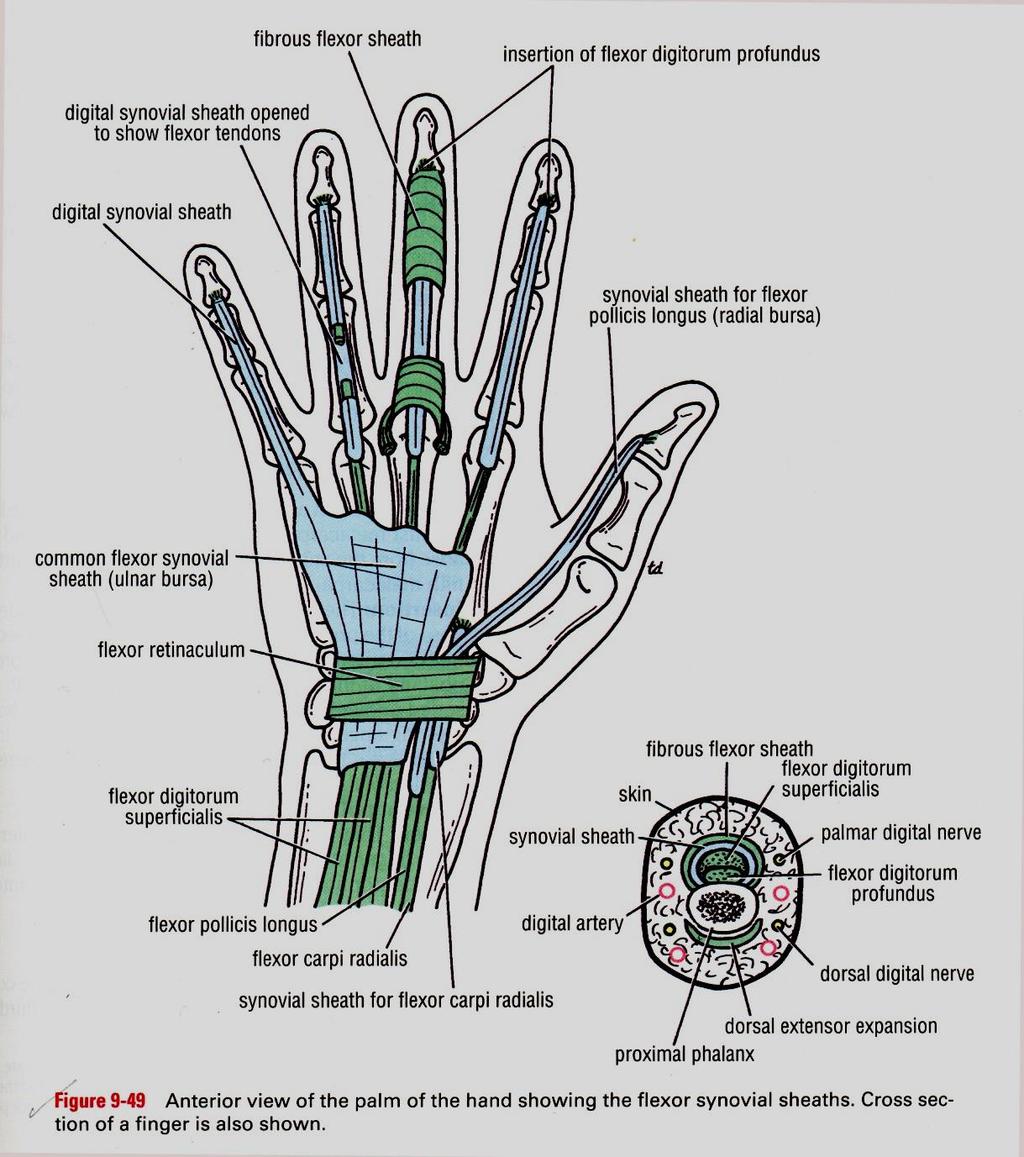
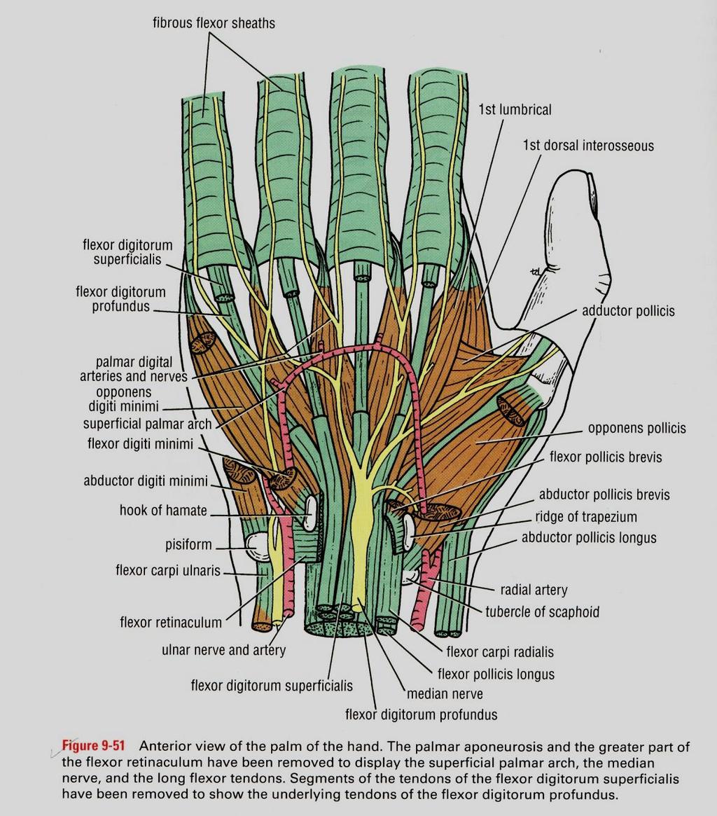
 Anatomy of the Upper Limb Figure 53: The thenar & midpalmar spaces. The synovial (tendon) sheaths of the long flexors [Figure.54] These sheaths surround the tendons of the long flexors; flexor digitorum
This figure (of humerus) is from Dr. Maher's newest slides. -Its added here just for consideration-
 is from Dr. Maher's newest slides. -Its added here just for consideration-") This figure (of humerus) is from Dr. Maher's newest slides. -Its added here just for consideration- Slides of Anatomy Please note : These slides are Dr. Maher Hadidi s slides of spring 2016 and were edited
This figure (of humerus) is from Dr. Maher's newest slides. -Its added here just for consideration- Slides of Anatomy Please note : These slides are Dr. Maher Hadidi s slides of spring 2016 and were edited
[[Sally Leaning Towards Peter To Take Cold Hand]]
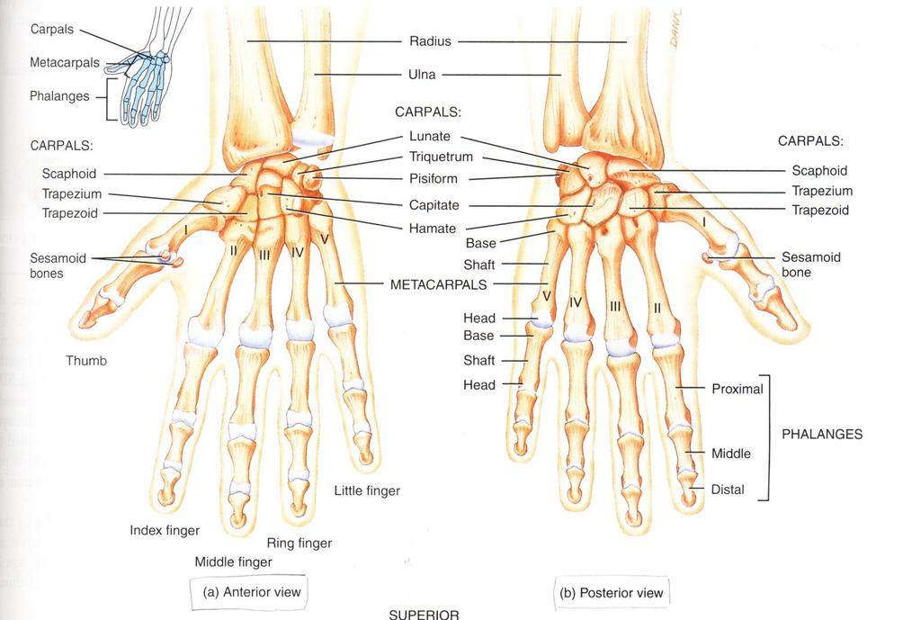
![[[Sally Leaning Towards Peter To Take Cold Hand]]](/thumbs/84/91174469.jpg "[[Sally Leaning Towards Peter To Take Cold Hand]]") In this lecture we will talk about the bones of the hand, and the muscles and contents of the forearm. *The hand bones are: - Carpal bones. -Metacarpals. -Phalanges. *The carpal bones (wrist bones): They
In this lecture we will talk about the bones of the hand, and the muscles and contents of the forearm. *The hand bones are: - Carpal bones. -Metacarpals. -Phalanges. *The carpal bones (wrist bones): They
Region of upper limb attachment to the trunk Proximal segment of limb overlaps parts of the trunk (thorax and back) and lower lateral neck.
 and lower lateral neck.") Region of upper limb attachment to the trunk Proximal segment of limb overlaps parts of the trunk (thorax and back) and lower lateral neck. includes Pectoral Scapular Deltoid regions of the upper limb
Region of upper limb attachment to the trunk Proximal segment of limb overlaps parts of the trunk (thorax and back) and lower lateral neck. includes Pectoral Scapular Deltoid regions of the upper limb
The arm: *For images refer back to the slides
 The arm: *For images refer back to the slides Muscles of the arm: deltoid, triceps (which is located at the back of the arm), biceps and brachialis (it lies under the biceps), brachioradialis (it lies
STRUCTURAL BASIS OF MEDICAL PRACTICE EXAMINATION 5. September 30, 2011
 STRUCTURAL BASIS OF MEDICAL PRACTICE EXAMINATION 5 September 30, 2011 PART l. Answer in the space provided. (12 pts) 1. Identify the structures. (2 pts) EXAM NUMBER A. Suprascapular nerve B. Axillary nerve
THE SHOULDER JOINT T H E G L E N O H U M E R A L ( G H ) J O I N T
 J O I N T") THE SHOULDER JOINT T H E G L E N O H U M E R A L ( G H ) J O I N T CLARIFICATION OF TERMS Shoulder girdle = scapula and clavicle Shoulder joint (glenohumeral joint) = scapula and humerus Lippert, p115
THE SHOULDER JOINT T H E G L E N O H U M E R A L ( G H ) J O I N T CLARIFICATION OF TERMS Shoulder girdle = scapula and clavicle Shoulder joint (glenohumeral joint) = scapula and humerus Lippert, p115
Chapter 8. The Pectoral Girdle & Upper Limb
 Chapter 8 The Pectoral Girdle & Upper Limb Pectoral Girdle pectoral girdle (shoulder girdle) supports the arm consists of two on each side of the body // clavicle (collarbone) and scapula (shoulder blade)
Pectoral girdle, SUPERIEUR ARM AND HAND. Danil Hammoudi.MD
 Pectoral girdle, SUPERIEUR ARM AND HAND Danil Hammoudi.MD The pectoral girdle is the set of bones which connect the upper limb to the axial skeleton on each side. It consists of the clavicle scapula in
BIOH111. o Cell Module o Tissue Module o Skeletal system o Integumentary system o Muscle system o Nervous system o Endocrine system
 BIOH111 o Cell Module o Tissue Module o Skeletal system o Integumentary system o Muscle system o Nervous system o Endocrine system TEXTBOOK AND REQUIRED/RECOMMENDED READINGS o Principles of anatomy and
Sports Medicine Part II : ANATOMY OF THE SPINE, ABDOMEN AND SHOULDER COMPLEX
 Sports Medicine 25 1.1 Part II : ANATOMY OF THE SPINE, ABDOMEN AND SHOULDER COMPLEX c.w.p. Wagner High School, Sports Medicine, A. Morgan, T. Morgan & A. Eastlake, 2008 Muscles of the Upper Limbs In this
Nerve Injury. 1) Upper Lesions of the Brachial Plexus called Erb- Duchene Palsy or syndrome.
 Upper Lesions of the Brachial Plexus called Erb- Duchene Palsy or syndrome.") Nerve Injury - Every nerve goes to muscle or skin so if the nerve is injured this will cause paralysis in the muscle supplied from that nerve (paralysis means loss of function) then other muscles and other
Nerve Injury - Every nerve goes to muscle or skin so if the nerve is injured this will cause paralysis in the muscle supplied from that nerve (paralysis means loss of function) then other muscles and other
Done By : Isra a Aweidah
 Anatomy Made Easy MSS هذا الملف يشمل تفريغ المحاضرة 10 المحاضرة 12 ابتداء من صفحة 13 وتفريغ part #9 Done By : Isra a Aweidah Edited by: AWN Academic Team Everything is included ( the drs slides in addition
SUPERIEUR ARM AND HAND
 Pectoral girdle, SUPERIEUR ARM AND HAND Danil Hammoudi.MD The pectoral girdle is the set of bones which connect the upper limb to the axial skeleton on each side. It consists of the clavicle scapula in
7/31/2012 THE SHOULDER JOINT CLARIFICATION OF TERMS OSTEOLOGY OF THE GH JOINT(BONES)
") THE SHOULDER JOINT T H E G L E N O H U M E R AL ( G H ) J O I N T CLARIFICATION OF TERMS Shoulder girdle = scapula and clavicle Shoulder joint (glenohumerual joint) = scapula and Lippert, p115 OSTEOLOGY
THE SHOULDER JOINT T H E G L E N O H U M E R AL ( G H ) J O I N T CLARIFICATION OF TERMS Shoulder girdle = scapula and clavicle Shoulder joint (glenohumerual joint) = scapula and Lippert, p115 OSTEOLOGY
Joints of the upper limb II
 Joints of the upper limb II Prof. Abdulameer Al-Nuaimi E-mail: a.al-nuaimi@sheffield.ac.uk E. mail: abdulameerh@yahoo.com Elbow joint The elbow joint is connecting the upper arm to the forearm. It is classed
Cubital fossa and forearm
 Cubital fossa and forearm Cubital fossa is the triangular space in front of elbow joint. - The Cubital fossa has boundaries: apex, base, roof and floor and it has contents. The base: an imaginary horizontal
*the Arm* -the arm extends from the shoulder joint (proximal), to the elbow joint (distal) - it has one bone ; the humerus which is a long bone
, to the elbow joint (distal) - it has one bone ; the humerus which is a long bone") *the Arm* -the arm extends from the shoulder joint (proximal), to the elbow joint (distal) - it has one bone ; the humerus which is a long bone - muscles in the arm : *brachialis muscle *Biceps brachii
*the Arm* -the arm extends from the shoulder joint (proximal), to the elbow joint (distal) - it has one bone ; the humerus which is a long bone - muscles in the arm : *brachialis muscle *Biceps brachii
M C P T. "Excellence in Education" SHOULDER AND UPPER LIMB. Part 1. Advanced Anatomy 2 Part 2. Assessment
 M C P T Melbourne College of Professional Therapists "Excellence in Education" SHOULDER AND UPPER LIMB Part 1. Advanced Anatomy 2 Part 2. Assessment 2006 MCPT Advanced Anatomy and Assessment 1 Diploma
The Arm and Cubital Fossa
 The Arm and Cubital Fossa Dr. Andrew Gallagher School of Anatomical Sciences University of the Witwatersrand Introduction The ARM (BRACHIUM) is the most proximal segment of the upper limb musculoskeletal
The Elbow and Radioulnar Joints Kinesiology. Dr Cüneyt Mirzanli Istanbul Gelisim University
 The Elbow and Radioulnar Joints Kinesiology Dr Cüneyt Mirzanli Istanbul Gelisim University 1 The Elbow & Radioulnar Joints Most upper extremity movements involve the elbow & radioulnar joints. Usually
Elbow & Forearm H O W V I T A L I S T H E E L B O W T O O U R D A I L Y L I V E S?
 Elbow & Forearm H O W V I T A L I S T H E E L B O W T O O U R D A I L Y L I V E S? Clarification of Terms The elbow includes: 3 bones (humerus, radius, and ulna) 2 joints (humeroulnar and humeroradial)
Appendix. Useful Anatomical Data of Clinical Significance
 Appendix Useful Anatomical Data of Clinical Significance Appendix Outline Respiratory System 426 Table I. Important Airway Distances (Adult) 426 Table II. Important Data Concerning the Trachea 426 Musculoskeletal
BOGOMOLETS NATIONAL MEDICAL UNIVERSITY. Department of human anatomy. GUIDELINES Student's independent work during the preparation to practical lesson
 BOGOMOLETS NATIONAL MEDICAL UNIVERSITY Department of human anatomy GUIDELINES Student's independent work during the preparation to practical lesson Academic discipline HUMAN ANATOMY Module 1 Content module
LEVEL II MUSCLE CHART NB: Needle length varies with tissue depth, this chart acts as a guide only. Side lye or prone.25 x 30-50mm Inferior to ilium
 LUMBAR SPINE LEVEL II MUSCLE CHART NB: Needle length varies with tissue depth, this chart acts as a guide only Muscle/ Innervation Comments Position Quadratus Lumborum T12-L3/4 segmentally PSIS Comments.
Biology 2401 Muscles List for CPC models
 Biology 2401 List for CPC models Italicized muscles are dissect and similar in the cat = Dissect and note the differences in human and cat Major of the Human Head Facial Expression Epicranius frontalis
Scapular and Deltoid Regions
 M1 Gross and Developmental Anatomy Scapular and Deltoid Regions Dr. Peters 1 Outline I. Skeleton of the Shoulder and Attachment of the Upper Extremity to Trunk II. Positions and Movements of the Scapula
Deep dry needling of the arm and hand muscles
 Deep dry needling of the arm and hand s 8 César Fernández-de-las-Peñas Javier González Iglesias Christian Gröbli Ricky Weissmann CHAPTER CONTENT Introduction................... 107 Clinical relevance of
Elbow Elbow Anatomy. Flexion extension. Pronation Supination. Anatomy. Anatomy. Romina Astifidis, MS., PT., CHT
 Elbow Elbow Anatomy Romina Astifidis, MS., PT., CHT Curtis National Hand Center Baltimore, MD October 6-8, 2017 Link between the arm and forearm to position the hand in space Not just a hinge Elbow = 70%
Epicranius (frontal belly) Zygomaticus minor. Zygomaticus major Buccinator
 Zygomaticus minor. Zygomaticus major Buccinator") Epicranius (frontal belly) Zygomaticus minor Zygomaticus major Buccinator Masseter Digastric (posterior belly) Stylohyoid Sternocleidomastoid Trapezius Scalenus Omohyoid (inferior belly) Orbicularis oris
Epicranius (frontal belly) Zygomaticus minor Zygomaticus major Buccinator Masseter Digastric (posterior belly) Stylohyoid Sternocleidomastoid Trapezius Scalenus Omohyoid (inferior belly) Orbicularis oris
Learning Objectives. 07 Aug 12. Article E-1. At the end of this section the learner will be able to:
 Module 1: Comparative Functional Anatomy and Biomechanics Article E-1 Learning Objectives At the end of this section the learner will be able to: Describe the bones of the equine thoracic Describe the