Surgical. Technique CRF II. Radial Head Prosthesis
|
|
|
- Carmella Hill
- 5 years ago
- Views:
Transcription

1 Surgical Technique CRF II
2 CONTENTS CONTENTS 1. OVERVIEW 2. INDICATIONS 1. Acute Trauma 2. Trauma Sequalae 3. DESIGN RATIONALE 4. SURGICAL TECHNIQUE 1. Preoperative assessment 2. Patient positioning 3. Surgical approach and Incision of the annular ligament 4. Radial head/neck osteotomy 5. Associated fracture of the coronoid 6. Use of the end-cutting mill 7. Use of the resection gauge 8. Preparation of the radial canal 9. Selection and insertion of the trial stem 10. Selection and insertion of the trial radial head 11. Distal bone plug 12. Use of a modern cementing technique for the final components 13. Bipolar radial head assembly 14. Assessment of stability and range of motion - Soft tissue repair p. 3 p. 4 p. 5 p Wound closure 5. POSTOPERATIVE MANAGEMENT p Radiographic evaluation 2. Rehabilitation program INSTRUMENTS IMPLANTS p. 13 p. 16 2
. 5.")
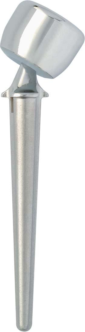
 Two stem sizes and two radial head sizes are offered to anatomically match each individual patient: 6 mm diameter ball 15 neck-shaft angle Radial stems: - 55 mm stem: 6.")
3 OVERVIEW 1. OVERVIEW 1. The long conical radial stem can be inserted using a modern cementing technique with pressurization of acrylic cement in the medullary canal. 2. The 15 neck-shaft angle restores anatomy of the proximal radius. 3. The 6 mm diameter ball creates a ball-and-socket joint with the bipolar radial head. 4. Metal-backed radial head (CoCr/PE). 5. Maximum PROM : 35 (bipolar radial head/ball articulation) (Fig. 1) (fig. 1) Two stem sizes and two radial head sizes are offered to anatomically match each individual patient: 6 mm diameter ball 15 neck-shaft angle Radial stems: - 55 mm stem: 6.5 mm diameter under the collar - 60 mm stem: 8 mm diameter under the collar. Ø 6,5 mm Ø 8 mm Radial heads: - Ø 19 mm x H 14.7 mm - Ø 22 mm x H 14.7 mm. 55 mm 60 mm Distance between the base of the prosthetic neck and the top of the bipolar radial head is 22.5 mm. As the prosthetic neck and ball have a fixed size, the small and large radial heads offer mix-and-match capabilities with the short and long radial stems. 14,7 mm Ø 19 mm Ø 22 mm 14,7 mm 3

")
. I II III IV (fig.")

4 INDICATIONS 2. INDICATIONS Restoration of a sound lateral bone platform is mandatory in specific indications: 1 Acute Trauma Radial head fracture not suitable for internal fixation (Mason grade III with significant displacement) associated with another destabilizing injury (elbow dislocation, coronoid fracture or rupture of the internal ligament). I II III IV (fig. 2) 2 Trauma Sequalae a/ Wrist pain syndrom following radial head resection after inferior radio-cubital dislocation or internal hyper-pressure of the carpus. b/ Elbow instability after radial head resection (fig. 3) 4


5 DESIGN RATIONALE 3. DESIGN RATIONALE The head of the radius works together with the adjacent bony, ligamentous, and tendinous structures to stabilize the elbow joint. The head of the radius acts as a stabilizer in three planes: Coronal plane: the synergistic action of the ulnar collateral ligament and the head of the radius resists valgus forces. Sagittal plane: the head of the radius works together with the medial and posterolateral ligamentous structures, and the coronoid process to prevent posterior dislocation. Axial plane: acting together with the interosseous membrane, it prevents upward migration of the radial shaft which has a detrimental effect on the inferior radioulnar joint. Therefore, the head of the radius can be considered as an "additional multifunctional stabilizer" of the elbow joint. However, to perform all these functions, the prosthesis must be able to withstand the forces exerted on the humeroradial joint, which implies: high mechanical strength deformation and fatigue, strong, reliable long-term fixation. Good wear characteristics are necessary to maintain high mechanical performance in the long run with no generation of pathogenic wear debris. There must be a tight fit between the bipolar radial head and the humerus capitulum during flexion, extension, and pronation/supination (Fig. 4) Point contacts should be avoided to eliminate the potential for hyperpressure and osteocartilaginous lesions of the capitulum. (fig. 4) 5

6 SURGICAL TECHNIQ 4. SURGICAL TECHNIQUE 1 Preoperative assessment Preoperative assessment requires the use of: AP and lateral views of the head of the radius. x-ray of the ipsilateral wrist, if painful. 2 Patient positioning The patient is supine on the operating table, and a tourniquet is placed at the root of the limb. The elbow is flexed and allowed to rest on a short arm board that extends 25 cm away from the table and stops just past the olecranon. This way, the surgeon faces the affected joint, and a slight rotation of the patient's shoulder provides him easy access to the elbow through any approach. The forearm is placed and held in pronation, so that: the radial nerve is medialized, the radial shaft is oriented in the axis of the operative field. 3 Surgical approach and Incision of the annular ligament With the elbow flexed, make one single angled incision centered over the tip of the epicondyle. It should extend proximally for a distance of 1 to 2 cm along the axis of the arm, and distally for a distance of 6 to 7 cm toward the ulnar crest. The most popular approach is the lateral one, between the extensor carpi radialis brevis and the extensor digitorum communis. This interval is easier to identify at the distal end of the incision. Once the radial muscles have been retracted anteriorly and the head of the radius has been removed, it affords access to the radial notch, the humeroulnar joint line, and the coronoid process (Fig. 5a) (fig. 5) b a The other option is to use a posterolateral approach between the anconeus, posteriorly, and the remaining epicondylar muscles, anteriorly. This approach affords a more distal access to the radius (Fig. 5b) 6
7 UE 4. SURGICAL TECHNIQUE Incise the annular ligament (if intact). Anyway, whether it was torn or incised, it should be repaired whenever possible. An oblique incision will greatly facilitate the repair. 4 Radial head/neck osteotomy Insertion of two double bent retractors provides adequate exposure. Not only the anterior retractor must be inserted with great caution, but throughout the radial preparation, the forearm must be held in full pronation to avoid the potential risk of elongation of the radial nerve. The correct position for the osteotomy is 23 mm distal to the capitulum. This position is easy to determine, using the resection gauge included in the instrument set (Fig. 6) The cut is performed with the oscillating saw. Avoid using cutting pliers which tend to cause bone splinters and propagation of fracture lines (Fig. 6 bis) (fig. 6) 5 Associated fracture of the coronoid Fracture of the coronoid is often seen when the radial head fracture is associated with dislocation. If present, the coronoid fracture should be managed at this stage of the procedure. Depending on the size of the bone fragment, simple resection or guided reduction is performed, followed by lag screw fixation (from the ulnar crest). (fig. 6 bis) 7


8 SURGICAL TECHNIQ 4. SURGICAL TECHNIQUE 6 Use of the end-cutting mill Use the end-cutting mill to smooth off the cut surface. Smooth off the cut surface to achieve a perfect fit between the prosthetic neck and the cut surface (fig. 7) 7 Use of the resection gauge Make a final check of the resection level using the resection gauge. Perfect humeroulnar congruity must be achieved at the superior border of the radial notch. (fig. 7) 8 Preparation of the radial canal Ream the medullary canal using the reamers. Work the small diameter reamer back and forth and, if necessary, use the large diameter reamer. The reaming step allows determination of the appropriate size stem according to the radius size (fig. 8) 9 Selection and insertion of the trial stem Insert the trial stem into the prepared cavity. The neck of the trial stem must be flush against the resected surface. Orientation of the prosthetic neck is determined relative to the bistyloid plane (fig. 9) (fig. 8) (fig. 9) 8


 11 Distal bone plug Make a bone plug using bone fragments from the resected head or use")
9 E 4. SURGICAL TECHNIQUE 10 Selection and insertion of the trial radial head Once the trial stem has been properly seated, determine the size of the bipolar radial head. The trial radial head should allow for 0.5 to 1 mm clearance (figs 10 & 10 bis). The size of the bipolar radial head should correspond to the size of the removed head after it has been reassembled. In the few cases where the head of the radius is absent, use the trial radial head. (fig. 10) (fig. 10 bis) 11 Distal bone plug Make a bone plug using bone fragments from the resected head or use a distal canal occluder, and insert it into the medullary canal with the help of the cylindrical rasp. Place the bone plug/occluder 10 mm distal to the stem end (figs 11 & 11 bis) (fig. 11) (fig. 11 bis) 9
.")


10 SURGICAL TECHNIQ 4. SURGICAL TECHNIQUE 12 Use of a modern cementing technique for the final components Thoroughly dry the prepared cavity using a small diameter drain. Then, inject low viscosity cement while in the liquid phase with a large-nozzle syringe, using a retrograde cement filling technique (fig. 12). Pressurize the cement until formation of the doughy phase. Then, insert the radial stem using the positioner/impactor and firmly hold it in place until the cement is hard. The stem can now be impacted with the positioner/ impactor. Carefully remove any excess cement. The orientation mark on the convex side of the prosthetic neck assists in correct orientation of the concave side of the neck, laterally. Confirm proper position of the stem by palpation (fig. 12 bis) (fig. 12) (fig. 12 bis) 13 Bipolar radial head assembly Assemble the bipolar head. Place the bipolar head on the ball ; positive capture of the bipolar head is confirmed by a clean audible click (fig. 13) (fig. 13) 10
.")

11 E 4. SURGICAL TECHNIQUE 14 Assessment of stability and range of motion - Soft tissue repair After reduction, assess range of motion in flexion/ extension and in various pronation/supination positions, before and after suture repair of the annular ligament (if present). Assess stability of the bipolar radial head relative to the capitulum, good fit, and stability in the coronal plane (figs 14 & 14 bis). In fresh fractures, severe lesions of the soft tissues (i.e. rupture of the annular ligament, detachment of its posterolateral fibers and part of the muscle insertions) are often seen. This causes anterior-posterior instability of the bipolar radial head and requires anatomical repair of the injured soft tissues. Implantation of the CRF II is seldom associated with a residual valgus instability that requires repair of the medial collateral ligaments. However, should this be the case, the posterolateral capsule and ligaments should be repaired using transverse sutures. Place a Redon drain and close the wound. (fig. 14) (fig. 14 bis) 15 Wound closure Repair the annular ligament (whenever possible), place a suction drain, and close the wound in a routine fashion. 11


12 POSTOPERATIVE MANAGEMENT 5. POSTOPERATIVE MANAGEMENT 1 Radiographic evaluation Take x-rays to check the position of the prosthesis and proper restoration of the humeroulnar joint line. In case of incongruity that indicates the presence of a foreign body within the joint or high positioning of the prosthesis, immediate revision should be considered. 2 Rehabilitation program Early rehabilitation is essential to prevent joint stiffening which frequently occurs after management of a radial head fracture. The rehabilitation program (i.e. all motions or a specific range of motion that was initially restricted) and the use of a splint depend on whether soft tissue injuries were associated with the radial head fracture. In any case, gentle active exercises are always recommended. The use of an elbow CPM machine may be considered. Application of an ice bag and use of antiinflammatories are recommended. 12
13 INSTRUMENTS INSTRUMENTS Reamers Ø 6,5 mm Ref. MTJ 026 Ø 8 mm Ref. MTJ 028 End-cutting mill Ref. MTJ 010 Trial stems Ø 6,5 mm Ref. MTJ 036 Ø 8 mm Ref. MTJ 038 Instrument set Ref. YKAD 048 Rasps Ø 6,5 mm Ref. MTJ 016 Ø 8 mm Ref. MTJ 018 Trial radial heads Ø 19 mm Ref. MTJ 020 Ø 22 mm Ref. MTJ 021 Positioner/impactor Ref. MTJ 013 Resection gauge Ref. MTJ
14 NOTES NOTES 14
15 NOTES NOTES 15
16 IMPLANTS IMPLANTS Stems Ø 6,5 mm Ref. DTJ 006 Ø 8 mm Ref. DTJ 008 Bipolar radial heads Ø 19 mm Ref. DTJ 019 Ø 22 mm Ref. DTJ , rue Lavoisier. Montbonnot Saint-Ismier Cedex. France. Tél. : 33 (0) Fax : 33 (0) UDCT06.2
UDHT08.1.qxd:UDHT /03/08 17:14 Page 1. Surgical. Technique. Elbow Prosthesis. RHS Radial Head System.
 UDHT08.1.qxd:UDHT08.1 13/03/08 17:14 Page 1 Surgical Technique Elbow Prosthesis RHS Radial Head System www.tornier.com UDHT08.1.qxd:UDHT08.1 13/03/08 17:14 Page 2 TABLE OF CONTENTS DESIGN RATIONALE INDICATIONS
Integra. Katalyst Bipolar Radial Head System SURGICAL TECHNIQUE
 Integra Katalyst Bipolar Radial Head System SURGICAL TECHNIQUE Surgical Technique As the manufacturer of this device, Integra does not practice medicine and does not recommend this or any other surgical
Bipolar Radial Head System
 Bipolar Radial Head System Katalyst Surgical Technique DESCRIPTION The Katalyst Telescoping Bipolar Radial Head implant restores the support and bearing surface of the radial head in the face of fracture,
Surgical. Technique. AEQUALIS Spherical Base Glenoid. Shoulder Prosthesis.
 Surgical Technique Shoulder Prosthesis AEQUALIS Spherical Base Glenoid www.tornier.com CONTENTS CONTENTS 1. Subscapularis 2. Anterior capsule 3. Humeral protector 4. Inserting retractors 1. DESIGN FEATURES
The Elbow and the cubital fossa. Prof Oluwadiya Kehinde
 The Elbow and the cubital fossa Prof Oluwadiya Kehinde www.oluwadiya.com Elbow and Forearm Anatomy The elbow joint is formed by the humerus, radius, and the ulna Bony anatomy of the elbow Distal Humerus
the shape, the size, the fit Ascension Modular Radial Head
 the shape, the size, the fit Ascension Modular Radial Head WW anatomicdesign stem and head sizes to fit your indications and patient anatomy articular friendly shape reduces edge loading on the capitellum
modular RADIAL HEAD E VOLVE
 E VOLVE modular RADIAL HEAD surgical technique 1-5 in situ assembly 6-7 alternate O.R. back table assembly 8 Proper surgical procedures and techniques are the responsibility of the medical professional.
Integra. Modular Radial Head System SURGICAL TECHNIQUE
 Integra Modular Radial Head System SURGICAL TECHNIQUE Table of Contents System Overview...2 Indications and Contraindications... 3 Modular Radial Head Implant Technique...4 Component Dimensions...8 Implant
Elbow Elbow Anatomy. Flexion extension. Pronation Supination. Anatomy. Anatomy. Romina Astifidis, MS., PT., CHT
 Elbow Elbow Anatomy Romina Astifidis, MS., PT., CHT Curtis National Hand Center Baltimore, MD October 6-8, 2017 Link between the arm and forearm to position the hand in space Not just a hinge Elbow = 70%
Elbow & Forearm H O W V I T A L I S T H E E L B O W T O O U R D A I L Y L I V E S?
 Elbow & Forearm H O W V I T A L I S T H E E L B O W T O O U R D A I L Y L I V E S? Clarification of Terms The elbow includes: 3 bones (humerus, radius, and ulna) 2 joints (humeroulnar and humeroradial)
Fractures and dislocations around elbow in adult
 Lec: 3 Fractures and dislocations around elbow in adult These include fractures of distal humerus, fracture of the capitulum, fracture of the radial head, fracture of the olecranon & dislocation of the
The Biomechanics of the Human Upper Extremity-The Elbow Joint C. Mirzanli Istanbul Gelisim University
 The Biomechanics of the Human Upper Extremity-The Elbow Joint C. Mirzanli Istanbul Gelisim University Structure of The Elbow Joint A simple hinge joint, actually categorized as a trochoginglymus joint
Modular Ulnar Head surgical technique. Transforming Extremities
 First Choice Modular Ulnar Head surgical technique Transforming Extremities instrumentation Head and Collar Trials Assembly Pad Starter Awl Trial Extractor Osteotomy Guide Stem Trials Implant Impactor
Osteology of the Elbow and Forearm Complex. The ability to perform many activities of daily living (ADL) depends upon the elbow.
 depends upon the elbow.") Osteology of the Elbow and Forearm Complex The ability to perform many activities of daily living (ADL) depends upon the elbow. Activities of Daily Living (ADL) Can you think of anything that you do to
Osteology of the Elbow and Forearm Complex The ability to perform many activities of daily living (ADL) depends upon the elbow. Activities of Daily Living (ADL) Can you think of anything that you do to
rhead System Extended stems Operative technique
 rhead System Extended stems rhead System rhead System Extended stems Contents 1. Indications and contraindications... 3 2..... 4 The initial incision...4 Capsular exposure...5 Using the radial head resection
rhead System Radial Head Arthroplasty Operative technique
 rhead System Radial Head Arthroplasty Operative technique rhead System Operative technique rhead System Radial Head Arthroplasty Contents 1. Indications and contraindications... 3 2. Operative technique....
KATALYST. Bipolar Radial Head System. Surgical Technique. orthopedics. KATALYST English. PRODUCTS FOR SALE IN EUROPE, MIDDLE-EAST and AFRICA ONLY
 KATALYST Bipolar Radial Head System KATALYST English Surgical Technique orthopedics UPPER extremity PRODUCTS FOR SALE IN EUROPE, MIDDLE-EAST and AFRICA ONLY KATALYST Introduction Description The Katalyst
Connects arm to thorax 3 joints. Glenohumeral joint Acromioclavicular joint Sternoclavicular joint
 Connects arm to thorax 3 joints Glenohumeral joint Acromioclavicular joint Sternoclavicular joint Scapula Elevation Depression Protraction (abduction) Retraction (adduction) Downward Rotation Upward Rotation
Integra. Modular Radial Head System SURGICAL TECHNIQUE
 Integra Modular Radial Head System SURGICAL TECHNIQUE Table of Contents System Overview...2 Indications and Contraindications... 3 Modular Radial Head Implant Technique...4 Component Dimensions...8 Implant
The Elbow and Radioulnar Joints Kinesiology. Dr Cüneyt Mirzanli Istanbul Gelisim University
 The Elbow and Radioulnar Joints Kinesiology Dr Cüneyt Mirzanli Istanbul Gelisim University 1 The Elbow & Radioulnar Joints Most upper extremity movements involve the elbow & radioulnar joints. Usually
Functional Anatomy of the Elbow
 Functional Anatomy of the Elbow Orthopedic Institute Daryl C. Osbahr, M.D. Chief of Sports Medicine, Orlando Health Chief Medical Officer, Orlando City Soccer Club Orthopedic Consultant, Washington Nationals
Joints of the upper limb II
 Joints of the upper limb II Prof. Abdulameer Al-Nuaimi E-mail: a.al-nuaimi@sheffield.ac.uk E. mail: abdulameerh@yahoo.com Elbow joint The elbow joint is connecting the upper arm to the forearm. It is classed
URSA HEMI-SHOULDER ARTHROPLASTY B I O T E K
 URSA HEMI-SHOULDER ARTHROPLASTY SURGICAL TECHNIQUE B I O T E K 2 Surgical Position Once general anesthesia has been satisfactorily induced, or a supraclavicular nerve block has been given, the patient
Ascension. Silicone MCP surgical technique. surgical technique Ascension Silicone MCP
 Ascension Silicone MCP surgical technique WW 2 Introduction This manual describes the sequence of techniques and instruments used to implant the Ascension Silicone MCP (FIGURE 1A). Successful use of this
Motion of Left Upper Extremity During A Right- Handed Golf Swing
 Motion of Left Upper Extremity During A Right- Handed Golf Swing Description of Movement While the movement required for a golf swing requires many muscles, joints, & ligaments throughout the body, the
E-CENTRIX. Ulnar Head Replacement SURGICAL TECHNIQUE
 E-CENTRIX Ulnar Head Replacement SURGICAL TECHNIQUE E-CENTRIX ulnar head REPLACEMENT surgical technique as described by GRAHAM KING, MD University of Western Ontario London, Ontario, Canada E-CENTRIX ulnar
Practical 2 Worksheet
 Practical 2 Worksheet Upper Extremity BONES 1. Which end of the clavicle is on the lateral side (acromial or sternal)? 2. Describe the difference in the appearance of the acromial and sternal ends of the
Ligaments of Elbow hinge: sagittal plane so need lateral and medial ligaments
 Ligaments of Elbow hinge: sagittal plane so need lateral and medial ligaments Ulnar Collateral ligament on medial side; arising from medial epicondyle and stops excess valgus movement (lateral movement)
Aequalis -Glenoid. Keeled and Pegged. Surgical Technique
 Aequalis -Glenoid Keeled and Pegged Surgical Technique TABLE OF CONTENTS COMMON OPERATIVE TECHNIQUES FOR THE KEELED AND PEGGED AEQUALIS-GLENOIDS p. 1-3 IMPLANTATION OF THE AEQUALIS KEELED GLENOID p. 4-5
Terrible triad of the elbow
 Terrible triad of the elbow Vasu Pai ? Terrible triad of the elbow" Posterior dislocation of the elbow + Fractures of the radial head + Fracture of coronoid process Uncommon injury 5% of dislocation Problems
Rehabilitation after Total Elbow Arthroplasty
 Rehabilitation after Total Elbow Arthroplasty Total Elbow Atrthroplasty Total elbow arthroplasty (TEA) Replacement of the ulnohumeral articulation with a prosthetic device. Goal of TEA is to provide pain
Main Menu. Elbow and Radioulnar Joints click here. The Power is in Your Hands
 1 The Elbow and Radioulnar Joints click here Main Menu K.4 http://www.handsonlineeducation.com/classes//k4entry.htm[3/23/18, 1:29:53 PM] Bones Ulna is much larger proximally than radius Radius is much
Technique. Aequalis Resurfacing Humeral Head
 S u r g i c a l Technique Aequalis Resurfacing Humeral Head 1 The Aequalis Resurfacing Humeral Head has been developed in conjunction with Drew Miller, MD - Atlanta, GA. The Aequalis Resurfacing Humeral
WEEKEND 2 Elbow. Elbow Range of Motion Assessment
 Virginia Orthopedic Manual Physical Therapy Institute - 2016 Technique Manual WEEKEND 2 Elbow Elbow Range of Motion Assessment - Patient Positioning: Sitting or supine towards the edge of the bed - Indications:
Chapter 8. The Pectoral Girdle & Upper Limb
 Chapter 8 The Pectoral Girdle & Upper Limb Pectoral Girdle pectoral girdle (shoulder girdle) supports the arm consists of two on each side of the body // clavicle (collarbone) and scapula (shoulder blade)
SURGICAL TECHNIQUE GUIDE
 SURGICAL TECHNIQUE GUIDE As described by: Jorge L. Orbay, M.D. Miami Hand & Upper Extremity Institute Miami, Florida. 1 ELBOW LANDMARKS With the elbow flexed 90 0, palpate and mark the lateral epicondyle.
S H O U L D E R Solutions by Tornier. BIO-RSA TM ANGled SURGICAL TECHNIQUE. BIO-RSA Angled. surgical technique
 S H O U L D E R Solutions by Tornier BIO-RSA TM ANGled SURGICAL TECHNIQUE BIO-RSA Angled Bony increased offset - reversed shoulder arthroplasty surgical technique BIO-RSA TM ANGled SURGICAL TECHNIQUE BIO-RSA
Osteology of the Elbow and Forearm Complex
 Osteology of the Elbow and Forearm Complex The ability to perform m any activities of daily living (ADL) d epends upon the elbow. Activities of Daily Living (ADL) Can you think of anything that you do
RADIOGRAPHY OF THE ELBOW & HUMERUS
 RADIOGRAPHY OF THE ELBOW & HUMERUS Patient Position: ELBOW AP Projection in same plane Part Position: Hand in ; patient Centered to Humeral epicondyles Central Ray: Structures Shown: AP Elbow Criteria
The Elbow Scanning Protocol
 The Elbow Scanning Protocol Diagnostic Imaging of the Elbow: Introduction The elbow maybe considered as consisting of four quadrants, anterior, medial, lateral and posterior. Ultrasound would normally
1/19/2018. Winter injuries to the shoulder and elbow. Highgate Private Hospital (Whittington Health NHS Trust)
") Winter injuries to the shoulder and elbow Omar Haddo Consultant Orthopaedic Surgeon, Shoulder, Elbow, Hand & Wrist Specialist MBBS, BmedSci, FRCS(Orth) Highgate Private Hospital (Whittington Health NHS
Winter injuries to the shoulder and elbow Omar Haddo Consultant Orthopaedic Surgeon, Shoulder, Elbow, Hand & Wrist Specialist MBBS, BmedSci, FRCS(Orth) Highgate Private Hospital (Whittington Health NHS
MEDIAL EPICONDYLE FRACTURES
 MEDIAL EPICONDYLE FRACTURES Demographic 20% of elbow fractures 60% of which are associated with elbow dislocation. 75% in boys between 6-12 years 20% of elbow dislocation with ME fracture, the ME is incarcerated
Olecranon Osteotomy Nail. For simple fractures and osteotomies of the olecranon.
 Olecranon Osteotomy Nail. For simple fractures and osteotomies of the olecranon. Technique Guide Discontinued June 2016; AVAILABLE FOR IMPLANT REMOVAL PURPOSES ONLY DSEM/TRM/0517/0843 Table of Contents
SURGICAL TECHNIQUE GUIDE
 DANGER indicates an imminently hazardous situation which, if not avoided, will result in death or serious injury. WARNING indicates a potentially hazardous situation which, if not avoided, could result
Elbow Joint Anatomy ELBOW ANATOMY, BIOMECHANICS. Bone Anatomy. Bone Anatomy. Property of VOMPTI, LLC
 ELBOW ANATOMY, BIOMECHANICS AND PATHOLOGY Kristin Kelley, DPT, OCS, FAAOMPT Elbow Joint Anatomy Joint articulations Humeroulnar Radiohumeral Radioulnar (proximal and distal) Orthopaedic Manual Physical
Distal Radius Plate Instrument and Implant Set. Discontinued December 2017 DSUS/TRM/0916/1063(1)
") Distal Radius Plate Instrument and Implant Set Surgical Technique Discontinued December 2017 DSUS/TRM/0916/1063(1) The Distal Radius Plates Indications For fixation of fractures and osteotomies, including
Distal Radius Plate Instrument and Implant Set Surgical Technique Discontinued December 2017 DSUS/TRM/0916/1063(1) The Distal Radius Plates Indications For fixation of fractures and osteotomies, including
Chapter 6 The Elbow and Radioulnar Joints
 The Elbow & Radioulnar Chapter 6 The Elbow and Radioulnar Manual of Structural Kinesiology R.T. Floyd, EdD, ATC, CSCS Most upper extremity movements involve the elbow & radioulnar joints Usually grouped
Ascension. MCP surgical technique. surgical technique Ascension MCP PyroCarbon Total Joint
 Ascension MCP surgical technique PyroCarbon Total Joint WW 1.0 Table of Contents 2.0 Introduction............................ 2 3.0 Ascension MCP Implants................ 2 4.0 Instrumentation for MCP
The Elbow 3/5/2015. The Elbow Scanning Sequence. * Anterior Joint (The anterior Pyramid ) * Lateral Epicondyle * Medial Epicondyle * Posterior Joint
 * Lateral Epicondyle * Medial Epicondyle * Posterior Joint") Scanning Sequence * Anterior Joint (The anterior Pyramid ) * Lateral Epicondyle * Medial Epicondyle * Posterior Joint Anterior Elbow Pyramid Courtesy of Jay Smith, MD. Vice chair PMR Mayo Clinic Rochester,
Scanning Sequence * Anterior Joint (The anterior Pyramid ) * Lateral Epicondyle * Medial Epicondyle * Posterior Joint Anterior Elbow Pyramid Courtesy of Jay Smith, MD. Vice chair PMR Mayo Clinic Rochester,
#12. Joint نبيل خوري
 #12 30 Anatomy Joint هيام الر جال 9/10/2015 نبيل خوري Salam Awn Some notes before starting : ** Not all slides are included, so I recommend having a look at the slides beside this sheet ** If you find
Elbow Anatomy, Growth and Physical Exam. Donna M. Pacicca, MD Section of Sports Medicine Division of Orthopaedic Surgery Children s Mercy Hospital
 Elbow Anatomy, Growth and Physical Exam Donna M. Pacicca, MD Section of Sports Medicine Division of Orthopaedic Surgery Children s Mercy Hospital Contributing Factors to Elbow Injury The elbow is affected
Anatomical Shoulder Glenoid. Surgical Technique
 Anatomical Shoulder Glenoid Surgical Technique Anatomical Shoulder Glenoid Surgical Technique 3 Table of Contents Glenoid Preparation Surgical Steps 4 Anatomical Shoulder Glenoid 4 Glenoid Components
Terrible Triad: Tricks for Dealing with the Unstable Elbow
 Terrible Triad: Tricks for Dealing with the Unstable Elbow Mark A. Mighell, MD Kaitlyn N. Christmas, BS Disclosure Paid Consultation Research Support Speakers Bureau Paid Consultation Speakers Bureau The
A Patient s Guide to Adult Radial Head (Elbow) Fractures
 Fractures") A Patient s Guide to Adult Radial Head (Elbow) Fractures 2321 Coronado Idaho Falls, ID 83404 Phone: 208-227-1100 jpond@summitortho.net 1 DISCLAIMER: The information in this booklet is compiled from a variety
A Patient s Guide to Adult Radial Head (Elbow) Fractures 2321 Coronado Idaho Falls, ID 83404 Phone: 208-227-1100 jpond@summitortho.net 1 DISCLAIMER: The information in this booklet is compiled from a variety
Anterior Elbow Capsulodesis
 7(1):72 76, 2006 m R E V I E W m Anterior Elbow Capsulodesis Donald H. Lee, MD, Douglas R. Weikert, and Jeffry T. Watson Department of Orthopaedic Surgery Vanderbilt Orthopaedic Institute Nashville, TN
Radial Head Prosthesis System
 For Primary and Revision Joint Replacement of the Radial Head Radial Head Prosthesis System Surgical Technique TABLE OF CONTENTS INTRODUCTION Radial Head Prosthesis System 2 Indications and Contraindications
Posterolateral elbow dislocation with entrapment of the medial epicondyle in children: a case report Juan Rodríguez Martín* and Juan Pretell Mazzini
 Open Access Case report Posterolateral elbow dislocation with entrapment of the medial epicondyle in children: a case report Juan Rodríguez Martín* and Juan Pretell Mazzini Address: Department of Orthopaedic
3.5 mm LCP Olecranon Plates
 Part of the DePuy Synthes Locking Compression Plate (LCP ) System 3.5 mm LCP Olecranon Plates Surgical Technique Table of Contents Introduction 3.5 mm LCP Olecranon Plates 2 AO Principles 3 Indications
Recurrent subluxation or dislocation after surgical
 )263( COPYRIGHT 2017 BY THE ARCHIVES OF BONE AND JOINT SURGERY CASE REPORT Persistent Medial Subluxation of the Ulna with Radiotrochlear Articulation Amir R. Kachooei, MD; David Ring, MD, PhD Research
GLENOID SURGICAL TECHNIQUE
 UP. EXTREMITY Dual-Platform Shoulder Prosthesis GLENOID SURGICAL TECHNIQUE ANATOMICAL REVERSE SURGICAL TECHNIQUE REFERENCE NUMBERS HUMERAL STEM REFERENCE DIAMETER HEIGHT 267 360 Ø 06 100 265 102 Ø 08 120
S H O U L D E R Solutions by Tornier. Aequalistm. Pyrocarbon Humeral Head. surgical technique
 S H O U L D E R Solutions by Tornier Aequalis TM Pyrocarbon humeral head SURGICAL TECHNIQUE Aequalistm Pyrocarbon Humeral Head surgical technique Aequalis TM Pyrocarbon humeral head surgical TECHNIQUE
Sports Medicine Unit 16 Elbow
 Sports Medicine Unit 16 Elbow I. Bones a. b. c. II. What movements does the elbow perform? a. Flexion b. c. Pronation d. III. Muscles in motion a. FLEXION (supinated) i Brachialis (pronated) ii (neutral)
Forearm and Wrist Regions Neumann Chapter 7
 Forearm and Wrist Regions Neumann Chapter 7 REVIEW AND HIGHLIGHTS OF OSTEOLOGY & ARTHROLOGY Radius dorsal radial tubercle radial styloid process Ulna ulnar styloid process ulnar head Carpals Proximal Row
RADIAL HEAD PROSTHESIS SYSTEM For primary and revision joint replacement of the radial head
 RADIAL HEAD PROSTHESIS SYSTEM For primary and revision joint replacement of the radial head This publication is not intended for distribution in the USA. SURGICAL TECHNIQUE Warning This description alone
region of the upper limb between the shoulder and the elbow Superiorly communicates with the axilla.
 1 region of the upper limb between the shoulder and the elbow Superiorly communicates with the axilla. Inferiorly, a number of important structures pass between arm & forearm through cubital fossa. 2 medial
Integral 180 Surgical Technique
 Integral 180 Surgical Technique The Integral 180 and 225 are part of the Alliance Family Total Hip System. The Integral 225 femoral component is marketed for use with bone cement in the United States.
Chapter 4: Forearm 4.3 Forearm shaft fractures, transverse (12-D/4)
") AO Manual of ESIN in children s fractures Chapter 4: Forearm 4.3 Forearm shaft fractures, transverse (12-D/4) Title AO Manual of ESIN in children Subtitle Elastic stable intramedullary nailing (ESIN) Author
AO Manual of ESIN in children s fractures Chapter 4: Forearm 4.3 Forearm shaft fractures, transverse (12-D/4) Title AO Manual of ESIN in children Subtitle Elastic stable intramedullary nailing (ESIN) Author
UNI-Elbow System Radio-Capitellar Replacement. Operative technique
 UNI-Elbow System Radio-Capitellar Replacement Operative technique 0798-2016 UniElbow Op tech (EMS-ST-1) - proof 3 UNI-Elbow System Operative technique UNI-Elbow System Radio-Capitellar Replacement Contents
TORNIER BIO-RSA. Bony Increased Offset - Reversed Shoulder Arthroplasty SURGICAL TECHNIQUE
 TORNIER BIO-RSA Bony Increased Offset - Reversed Shoulder Arthroplasty SURGICAL TECHNIQUE 2 Table of Contents: Concept...4 Bony Increased Offset Reversed Shoulder Arthroplasty (BIO-RSA ) Concept...4 Surgical
OSKAR Radial Head Prosthesis
 Technique Guide OSKAR Radial Head Prosthesis DESCRIPTION The OSKAR Radial Head Implant is a pliable, onepiece intramedullary-stemmed cuffed implant designed to help preserve the joint space and relationships
Preoperative Planning. The primary objectives of preoperative planning are to:
 Preoperative Planning The primary objectives of preoperative planning are to: - Determine preoperative leg length discrepancy. - Assess acetabular component size and placement. - Determine femoral component
Levels of the anatomical cuts of the upper extremity RADIUS AND ULNA right
 11 CHAPTER 2 Levels of the anatomical cuts of the upper extremity AND right CUT 1 CUT 4 1 2 3 4 5 6 Isolated fixation of the radius is difficult at this level because of the anterolateral vessels and the
Muscular Nomenclature and Kinesiology - One
 Chapter 16 Muscular Nomenclature and Kinesiology - One Lessons 1-3 (with lesson 4) 1 Introduction 122 major muscles covered in this chapter Chapter divided into nine lessons Kinesiology study of human
TABLE OF CONTENTS SURGICAL TECHNIQUE 1 POST-OPERATIVE REHABILITATION 18. pages 1 RADIOLOGICAL ASSESSMENT 1 2 PATIENT POSITIONING 1
 TABLE OF CONTENTS SURGICAL TECHNIQUE 1 1 RADIOLOGICAL ASSESSMENT 1 2 PATIENT POSITIONING 1 3 DELTO-PECTORAL APPROACH 2 4 HUMERAL HEAD OSTEOTOMY 6 5 CHOICE OF HUMERAL INCLINATION AND RETROVERSION 7 pages
OPERATIVE TECHNIQUE. Intermediate. elbow prosthesis. mobile radial cup cemented radial stem. Orthopaedic Implants
 OPERATIVE TECHNIQUE Intermediate elbow prosthesis mobile radial cup cemented radial stem www.aston-medical.com Orthopaedic Implants I ntroduc tion The Evolutive prosthesis has been designed as the culmination
THE HUMERUS 20 THE HUMERUS* CROSS SECTION CROSS SECTION SUPERIOR VIEW
 20 THE HUMERUS* CROSS SECTION CROSS SECTION SUPERIOR VIEW The marrow canal of the humerus is funnel-shaped. Its successful pinning is influenced by many factors. With a few exceptions, the entire humerus
Lecture 9: Forearm bones and muscles
 Lecture 9: Forearm bones and muscles Remember, the region between the shoulder and the elbow = brachium/arm, between elbow and wrist = antebrachium/forearm. Forearm bones : Humerus (distal ends) Radius
David G. Simpson, Ph.D.
 David G. Simpson, Ph.D. ARM & CUBITAL FOSSA Revised 7/08 Text References Moores 3 rd ed., p402 408, 436 439, 439 443, 478, 481 LEARNING OBJECTIVES: 1. Describe the humerus, indicating the sites of muscle
Figure 1: Bones of the upper limb
 BONES OF THE APPENDICULAR SKELETON The appendicular skeleton is composed of the 126 bones of the appendages and the pectoral and pelvic girdles, which attach the limbs to the axial skeleton. Although the
Hands PA; Obl. Lat.; Norgaard s Thumb AP; Lat. PA. PA; Lat.: Obls.; Elongated PA with ulnar deviation
 Projections Region Basic projections Additional / Modified projections Upper Limbs Hands PA; Obl. Lat.; Norgaard s Thumb ; Lat. PA Fingers PA; Lat. Wrist PA; Lat. Obls. Scaphoid Lunate Trapezium Triquetral
Elbow. Chapter 2 LISTEN. Mechanism of Injury (If Applicable) Pain
 Pain") Chapter 2 Elbow LISTEN Mechanism of Injury (If Applicable) Patient usually remembers their position at the time of injury Certain mechanisms of injury result in characteristic patterns Fall on outstretched
Chapter 2 Elbow LISTEN Mechanism of Injury (If Applicable) Patient usually remembers their position at the time of injury Certain mechanisms of injury result in characteristic patterns Fall on outstretched
Locking Radial Head Plates
 Locking Radial Head Plates Locking Radial Head Plates Since 1988, Acumed has been designing solutions to the demanding situations facing orthopaedic surgeons, hospitals and their patients. Our strategy
PIPR Proximal Interphalangeal Replacement. Operative Technique
 PIPR Proximal Interphalangeal Replacement Operative Technique Contents PIPR Proximal Interphalangeal Replacement Developed in association with Mr I Trail and The Wrightington Hospital. Contents Section
LCP Distal Humerus Plates
 The anatomic fixation system for the distal humerus with angular stability Surgical technique LCP Locking Compression Plate Contents Indications and contraindications 2 Implants 3 Instruments 5 Preparation
Proximal radioulnar translocation associated with elbow dislocation and radial neck fracture in child: a case report and review of literature
 DOI 10.1007/s00402-013-1820-8 TRAUMA SURGERY Proximal radioulnar translocation associated with elbow dislocation and radial neck fracture in child: a case report and review of literature Hong Kee Yoon
Distal Cut First Femoral Preparation
 Surgical Technique Distal Cut First Femoral Preparation Primary Total Knee Arthroplasty LEGION Total Knee System Femoral preparation Contents Introduction...3 DCF femoral highlights...4 Preoperative planning...6
Technique Guide. 3.5 mm LCP Olecranon Plates. Part of the Synthes locking compression plate (LCP) system.
 system.") Technique Guide 3.5 mm LCP Olecranon Plates. Part of the Synthes locking compression plate (LCP) system. Table of Contents Introduction 3.5 mm LCP Olecranon Plates 2 AO Principles 3 Indications 3 Clinical
Technique Guide 3.5 mm LCP Olecranon Plates. Part of the Synthes locking compression plate (LCP) system. Table of Contents Introduction 3.5 mm LCP Olecranon Plates 2 AO Principles 3 Indications 3 Clinical
PT, CHT, FAAOMPT. What is Hand Therapy? Manual therapy 10/23/2013. Pam Kikillus PT, DHSc, OCS, CHT, FAAOMPT NSC 2013
 Manual therapy www.naiomt.com Pam Kikillus PT, DHSc, OCS, CHT, FAAOMPT NSC 2013 60 minute GOALS Simplify and de mystify the distal arm and hand therapy Highlight biomechanics of the proximal RU joint Exposure
Anatomy of the Shoulder Girdle. Prof Oluwadiya Kehinde FMCS (Orthop)
") Anatomy of the Shoulder Girdle Prof Oluwadiya Kehinde FMCS (Orthop) www.oluwadiya.com Bony Anatomy Shoulder Complex: Sternum(manubrium) Clavicle Scapula Proximal humerus Manubrium Sterni Upper part of
Anatomy of the Shoulder Girdle Prof Oluwadiya Kehinde FMCS (Orthop) www.oluwadiya.com Bony Anatomy Shoulder Complex: Sternum(manubrium) Clavicle Scapula Proximal humerus Manubrium Sterni Upper part of
Surgical. Technique. Aequalis Press-Fit. Shoulder Prosthesis.
 Surgical Technique Shoulder Prosthesis www.tornier.com TABLE OF CONTENTS 1. Preoperative planning 2. Patient positioning 3. Delto-pectoral approach 4. Humeral head osteotomy 5. Reaming the humeral shaft
MANAGEMENT OF INTRAARTICULAR FRACTURES OF ELBOW JOINT. By Dr B. Anudeep M. S. orthopaedics Final yr pg
 MANAGEMENT OF INTRAARTICULAR FRACTURES OF ELBOW JOINT By Dr B. Anudeep M. S. orthopaedics Final yr pg INTRAARTICULAR FRACTURES Intercondyar fracture Elbow dislocation Capitellum # Trochlea # Radial head
Technique Guide. 2.4 mm Variable Angle LCP Distal Radius System. For fragment-specific fracture fixation with variable angle locking technology.
 Technique Guide 2.4 mm Variable Angle LCP Distal Radius System. For fragment-specific fracture fixation with variable angle locking technology. Table of Contents Introduction 2.4 mm Variable Angle LCP
THE NANCY NAIL. The End Caps ADVANTAGES OF NANCY NAIL
 NANCY NAIL THE NANCY NAIL Nancy nails are manufactured from a specific titanyum alloy with proprietary surface treatment, which provides increased fatigue resistance. Six nail diameters (1.5 mm 2.0 mm
Wright Medical Technology, Inc Cherry Road Memphis, TN
 Wright Medical Technology, Inc. 1023 Cherry Road Memphis, TN 38117 800 238 7117 901 867 9971 www.wmt.com Wright Medical EMEA Atlas Arena, Australia Building Hoogoorddreef 7 1101 BA Amsterdam the Netherlands
A Locking IM Rod that won't back out. Simple and straight to the point! SURGICAL TECHNIQUE
 A Locking IM Rod that won't back out. Simple and straight to the point! SURGICAL TECHNIQUE The SLIM (Simple Locking IntraMedullary) System is a new generation of pediatric orthopedic nails specifically
JOINT RULER. Surgical Technique For Knee Joint JRReplacement
 JR JOINT RULER Surgical Technique For Knee Joint JRReplacement INTRODUCTION The Joint Ruler * is designed to help reduce the incidence of flexion, extension, and patellofemoral joint problems by allowing