Ultrasound Physics & Doppler
|
|
|
- Emily Franklin
- 5 years ago
- Views:
Transcription


1 Ultrasound Physics & Doppler Endocrine University 2018 Mark Lupo, MD, FACE, ECNU
2 Objectives Review the essential components of ultrasound physics in neck sonography Demonstrate the importance of ultrasound artifacts in clinical imaging interpretation Highlight the utility of Doppler flow as a complement to B-mode ultrasound

3 Natural Ability to see with sound
4 Imaging with Sound Create a visual image based on sound waves instead of light Mechanical versus Electromagnetic Energy Passive Sonar - Just listening Active Sonar - Acoustic Echo: Ping a target Reflection of sound waves Creation of a 2-D or 3-D image from reflected sound waves Requires technology and understanding of physics
5 Sound Waves Physics of sound Role of the propagating medium Acoustic Impedance Density, Stiffness, and Speed of Sound Reflection Occurs at Boundaries of Impedance Attenuation
6 Principles of Sound Waves Sound is the Propagation of mechanical energy through matter. No sound in a vacuum Qualities of the transmitting medium influence sound transmission Density Stiffness Acoustic Impedance Speed of Sound (SOS)
7 Sound Waves Sound waves propagate by compression and rarefaction of molecules in space. Molecules of the medium vibrate around their resting position and transfer their energy to neighboring molecules. Waves carry energy, not matter.
8 Characteristics of waves Frequency: # cycles of compression & rarefaction in a sound wave per second (Hz) Wavelength: Distance travelled in one second Amplitude, power, intensity db
9 Pulse of Sound Waves Spatial Pulse Length: Cycle length * # cycles per pulse time spent emitting echoes Determinant of axial resolution (1/2 SPL) Higher Frequency: shorter SPL Better Resolution Lower Frequency: Better Depth Penetration, Lower Resolution Duty Factor (%): Time spent generating pulse (usually 1% - while 99% of the time is spent listening for signal return)
10 Properties of Transmitting Material Speed of Sound = Propagation Speed Constant for a given tissue Not affected by frequency or wavelength Increases with stiffness Decreases with density
11 Propagation Velocity of Common Tissues
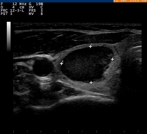
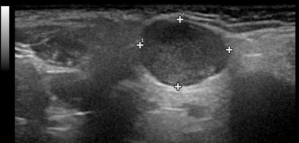
12 Properties of Transmitting Material Acoustic Impedance (AI) Reflection (echo) of sound occurs at interfaces of Acoustic Impedance (density * propagation speed) Most tissues have heterogeneous impedance If two tissues have same AI no echo If similar weak echo If very different AIs strong echo Homogeneous medium does not reflect sound Cyst (Anechoic NO echo)
13
14 Reflection Reflection is the redirection of a portion of a sound beam back at the interface of tissues of unequal acoustic impedance. The greater the difference in impedance, the greater the reflection. Categories of Reflection Specular Mirror like (e.g., diaphragm) Reflection dependent on angle of incidence Diffuse (Soft Tissue e.g., neck ultrasound) Adjacent structures smaller than wavelength
 Solid Cystic Complex")
15 1960s - A-mode of Thyroid A = Amplitude (strength of echo) Solid Cystic Complex Blum, 1971

16 From One Dimensional A-Mode to Two Dimensional B-Mode

17 Mid 1960s - B Mode

18 B Mode Scanner Fujimoto, 1967


19 B-Mode - Image Formation Meritt, 1998

20 B-Mode Thijs, 1971

21 Compound Arm B Mode Scanner


22 Gray Scale High Resolution 1979 and 2010 st e s h sh s t sh A e SM st V A e Scheible 1979 Legend: st = sternothyroid muscle, sh = sternohyoid muscle, arrows = posterior border of thyroid, A = carotid artery, V = jugular vein, sm = sternocleidomastoid muscle, e = esophagus

23 Key elements of an Ultrasound Scanner Sound emitter-sends sound toward the object of interest Sound detector-receives the returning reflected echoes Computer - analyze the detected signal Display shows an image of the object

24 Transducer Transducers both emit sound and receive the reflected echoes necessary for imaging 1% time emitting sound, 99% listening Linear transducer Curvilinear transducer
25 Piezoelectric Crystal Transducers have a naturally occurring or manmade crystal which can convert electrical energy to sound energy and sound energy to electric energy crystal Alternating electric current Sound wave
26 US Makes Assumptions Echo originates w/in main US beam Echo returns after a single reflection Depth defined by round trip time of echo SOS constant in human tissue (1540m/s) Sound beam and echo travel in straight path Acoustic energy is uniformly attenuated
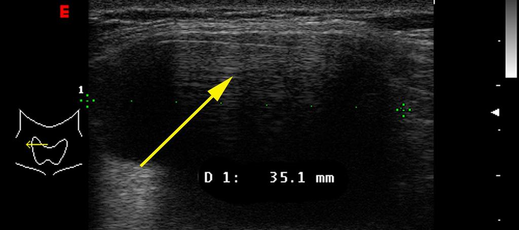
27 Illustrating Physics by Understanding Artifacts Speed of Sound Propagation Artifacts Reflection Artifacts Shadowing and Enhancement Edge Artifact Reverberation Artifact Comet Tail Artifact

28 Speed Displacement (Propagation) Artifact 2009 by Radiological Society of North America Feldman M K et al. Radiographics 2009;29:

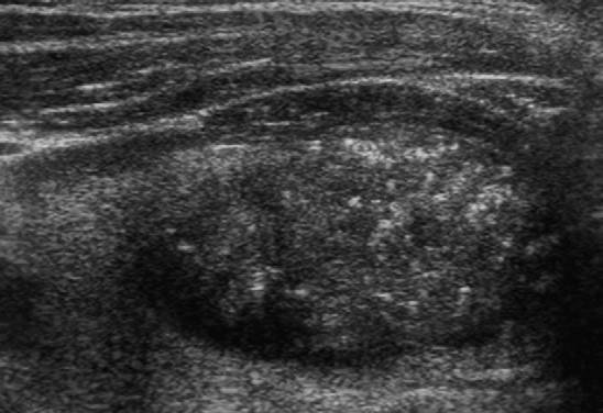
29 Speed of Sound - Propagation Artifact Meritt, 1998
 Adjusts for loss of amplitude from echoes returning from the far-field (i.e., compensates for attenuation)")
30 Attenuation Attenuation of acoustic energy results from Reflection Scatter Absorption Attenuation is frequency dependent Higher frequencies have greater attenuation Less depth of imaging with higher frequencies Artifacts due to attenuation Shadowing Enhancement Time Gain Compensation (TGC) Adjusts for loss of amplitude from echoes returning from the far-field (i.e., compensates for attenuation)
31 Attenuation of sound correction Time Gain Compensation T C G Returning sound

32 Normal Settings


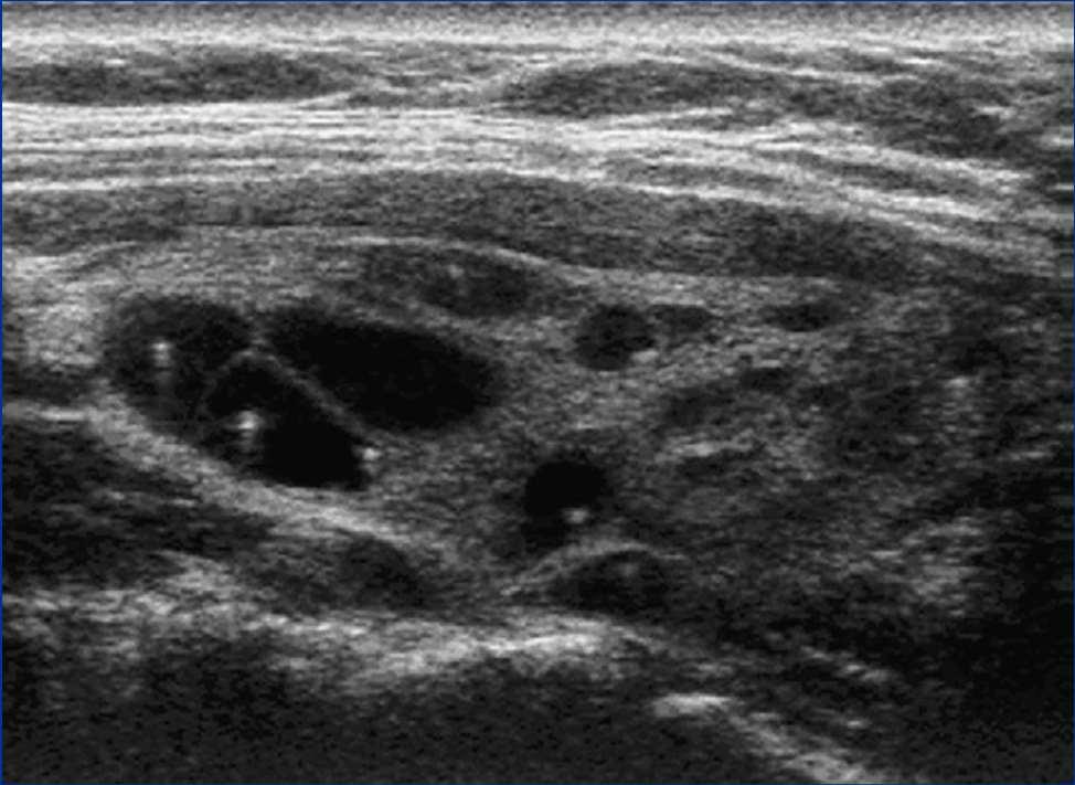
33 Far-Field Not Seen Due to Lack of TGC for Attenuation

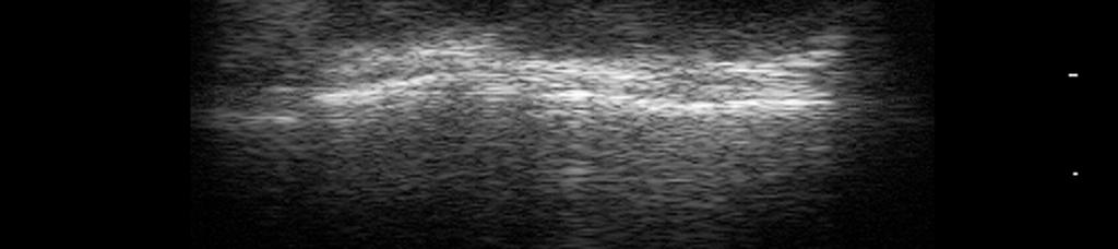
34 Increased through transmission Feldman M K et al. Radiographics 2009;29: BEAM ATTENUATED LESS THAN EXPECTED leads to POSTERIOR ENHANCEMENT 2009 by Radiological Society of North America

35 Posterior acoustic enhancement Occurs when the sound beam is attenuated less than expected; i.e. if it travels through fluid (or other medium) rather than tissue The TGC amplifies the returning echo using time (depth) as the only parameter, assuming homogeneous soft tissue in the path
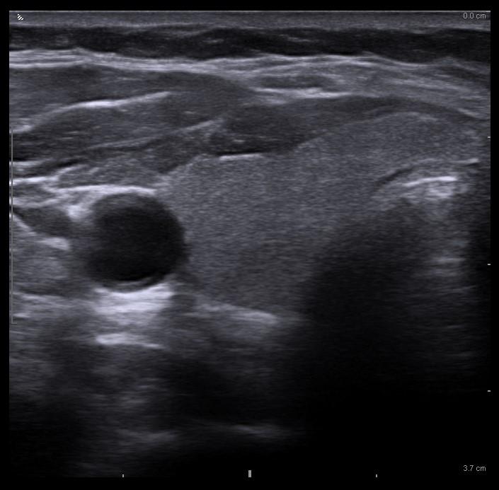
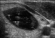
36 Superficial cyst


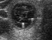
37 Posterior Acoustic Enhancement Seen behind cysts, blood vessels and often lymph nodes Can occur with solid nodules, particularly if hypervascular Turn on Doppler to confirm a hypoechoic structure w/ posterior enhancement is a cyst or solid nodule

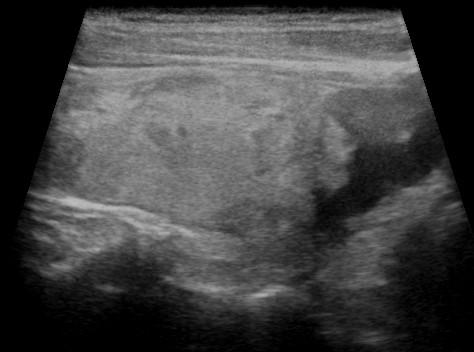
38 Enhancement is seen behind cysts - But can see it behind homogeneous solid objects as well Benign hyperechoic nodule seen in Hashimoto s Thyroiditis

39 Shadowing LARGE AI MISMATCH 2009 by Radiological Society of North America Feldman M K et al. Radiographics 2009;29:

40 Reflection Artifacts Eggshell Calcification

41 Shadowing and Enhancement
42 Reflection Artifacts Edge Artifact
43 Reverberation Occurs when sound bounces back and forth between two highly reflective surfaces multiple times before returning to the transducer The effect is to produce a step ladder of echoes in longitudinal orientation, each a little smaller Image

44 Reverberation Artifact Meritt, 1998
45 Reverberation Artifact

46 Reverb artifact Adding harmonics

47 Tunneling R IJ Catheter Reverberation Artifact

")
48 Comet Tail (Ring Down) Artifact
49 Microcalcification vs Comet Tail
 Intensity more uniform Pulse duration determines Axial Resolution (axial resolution = 1/2 SPL) Practical Considerations As frequency increases, axial resolution improves, but")
50 Focus and Resolution Focused beam width determines Lateral and Azimuthal Resolution Near field (Fresnel Zone) Large variations of intensity Focal Zone - Area of maximal narrowing (Adjustable) Far field (Fraunhofer Zone) Intensity more uniform Pulse duration determines Axial Resolution (axial resolution = 1/2 SPL) Practical Considerations As frequency increases, axial resolution improves, but depth of imaging decreases. The depth of the focal zone is adjustable and indicated on the display


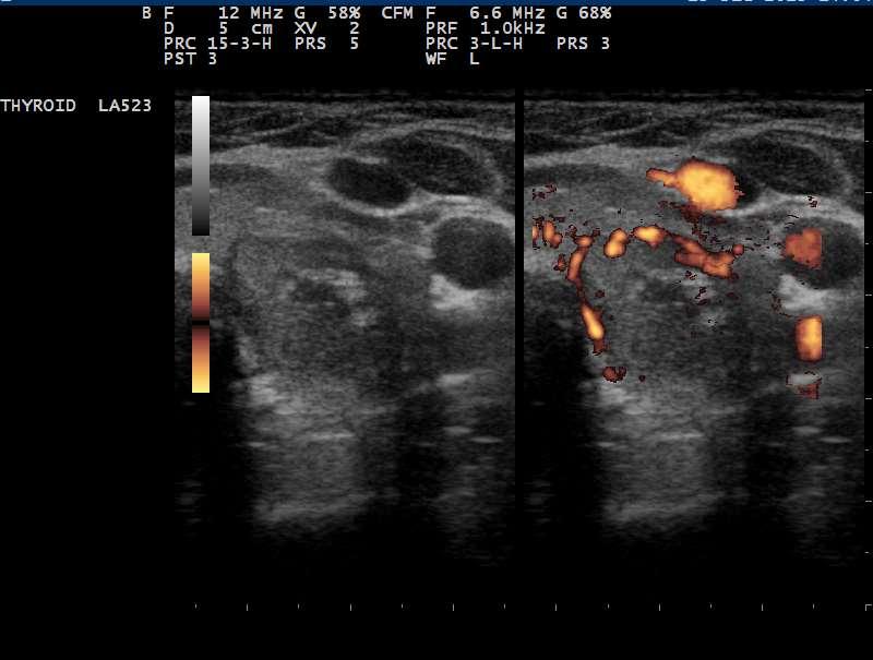
51 Frequency and Resolution With higher frequency the resolution of the near field is improved, but the deep structures are better visualized with the lower frequency.
52 Lateral resolution Ability to discriminate two objects perpendicular to the sound beam The narrower the beam, the better the lateral resolution. Higher frequencies have narrower beams Image 3 MHz 10 MHz
53 Focusing the Beam: Lateral resolution All beams have a focal zone, the narrowest part of the beam which has the best quality sound Near field Far field Focused Focal zone

54 Focal zone
55 Image Optimization Create the sharpest image to allow tissue discrimination. Equipment factors: Quality of Transducer Quality of Electronics Image Enhancement and Compound Imaging User Adjustments: Depth, Gain, Frequency Focal zones Number and Location Compound Imaging Tissue Harmonic Imaging Dynamic range Time Gain Compensation

56 Optimal Gain

57 Image Optimization Dynamic range May increase conspicuity of subtle lesions


58 Adjustment of number and position of focal zones
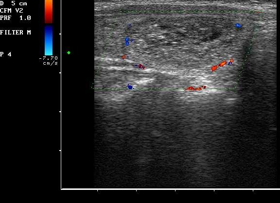
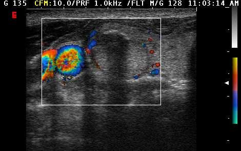
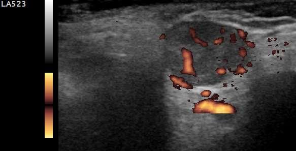
59 Doppler Physics When an interface is moving with respect to the reflected sound wave, the frequency of the reflected sound is altered. Frequency shifted up by approaching target Frequency shifted down by receding target Degree of frequency shift proportional to velocity Doppler Signal Processing and Display Doppler Frequency Spectrum Color Flow Power Mode
60 Illustration of Doppler Shift
61 Uses of Doppler Vascular Imaging and flow analysis of blood vessels Vascularity of tissue Probability of Malignancy Graves v. Thyroiditis Amiodarone Thyrotoxicosis Image Clarification
62 Color and Power Doppler Meritt, 1998

63 Color and Power Doppler Meritt, 1998
64 Color and Power Doppler Color Doppler Provides information regarding direction and velocity. More useful in vascular studies Power Doppler No information regarding velocity Less angle dependence Less noise Increased sensitivity for detection of flow
65 Doppler Issues High Sensitivity vs. Conventional Power User Variability Grading Scales (for thyroid nodules) 1 (absent vascularity) 2 (mostly peripheral vascularity) 3 (peripheral greater than intranodular) 4 (predominately intranodular)

66 Grade 1 No flow to nodule
67 Grade 2 Peripheral Flow


68 Grade 3 Moderate Central Flow

69 Grade 3 - Moderate Central Flow Suspicious for cystic papillary carcinoma
70 Grade 4 High Velocity Penetrating Flow

71 Increased Intranodular Flow Predictive of Malignancy? B-Mode Power Doppler NO LONGER CONSIDERED AN INDEPENDENT RISK
72 Vascularity - What to do? Consider whether features are suspicious for papillary or follicular cancers. Hypoechoic or features of papillary: Vascularity may be less important Iso or hyperechoic with variable thickness halo: Consider intranodular vascularity as possible risk. AACE & 2009 ATA guidelines do consider vascularity. Korean guidelines do not consider vascularity ATA 2015 guidelines do not consider vascularity **Don t be reassured by absence of vascularity.**
73 Doppler in the diagnosis of Thyrotoxicosis Graves Thyroid inferno Peak systolic velocity (PSV) 8-20 cm/sec Thyroiditis Various vascular patterns absent to hypervascular Thyrotoxicosis Factitia Minimal intrathyroidal vascular flow Peak systolic velocity (PSV) 3-5 cm/sec Amiodarone Induced Type 1 (Grave s like) increased or normal flow Type 2 (Destructive) decreased flow


74 Graves Disease Thyroid Inferno

75 Doppler of Lymph Nodes Suspicious Peripheral Normal Central/Hilar Kim et al, JUM 2013


76 Doppler for the Clarification of interpretation Central Compartment Lymph Nodes? Blood Vessels, Not Nodes
77 Clarification of Interpretation - Cyst?

78 Doppler Before Biopsy

79 Doppler Parathyroid Glands
80 Summary Sound transmission is dependent on the conducting medium. Sound is reflected at interfaces of acoustic impedance mismatch. Resolution is dependent on frequency and beam focal width. Artifacts such as shadowing and enhancement provide useful clinical imaging information. Doppler complements B-mode sonography in the evaluation of nodules, parathyroid, lymph nodes and thyrotoxicosis etiology
Thyroid Ultrasound Physics and Doppler
 Thyroid Ultrasound Physics and Doppler Advanced AACE-ACE US training course 2017 Dev Abraham MD, MRCP(UK), ECNU, FACE Professor of Medicine, University of Utah No Disclosures Natural Ability to see with
Principles of Ultrasound. Cara C. Prideaux, M.D. University of Utah PM&R Sports Medicine Fellow March 14, 2012
 Principles of Ultrasound Cara C. Prideaux, M.D. University of Utah PM&R Sports Medicine Fellow March 14, 2012 None Disclosures Outline Introduction Benefits and Limitations of US Ultrasound (US) Physics
Ultrasonography of the Neck as an Adjunct to FNA. Nicole Massoll M.D.
 Ultrasonography of the Neck as an Adjunct to FNA Nicole Massoll M.D. Basic Features of Head and Neck Ultrasound and Anatomy Nicole Massoll M.D. University of Arkansas for Medical Sciences, Little Rock
Ultrasound Physics & Terminology
 Ultrasound Physics & Terminology This module includes the following: Basic physics terms Basic principles of ultrasound Ultrasound terminology and terms Common artifacts seen Doppler principles Terms for
Ultrasound Principles cycle Frequency Wavelength Period Velocity
 ! Teresa S. Wu, MD, FACEP Director, EM Ultrasound Program & Fellowship Co-Director, Simulation Based Training Program & Fellowship Associate Program Director, EM Residency Program Maricopa Medical Center
The Physics of Ultrasound. The Physics of Ultrasound. Claus G. Roehrborn. Professor and Chairman. Ultrasound Physics
 The Physics of Ultrasound Pipe Organ 10-8000 Emission Dog 452-1080 Man 85-1100 Spectrum Bat 10,000-120,000 Porpoise 7000-120,000 Claus G. Roehrborn Professor and Chairman 10 20 Cycles per second Reception
Preamble (disclaimer)
") Preamble (disclaimer) PHYSICS AND PRINCIPLES OF HEAD/NECK ULTRASOUND Joseph C. Sniezek, MD FACS LTC, MC, USA Otolaryngology/H&N Surgery Tripler Army Medical Center 1. I am not a physicist 2. ACS has recommended
Preamble (disclaimer) PHYSICS AND PRINCIPLES OF HEAD/NECK ULTRASOUND Joseph C. Sniezek, MD FACS LTC, MC, USA Otolaryngology/H&N Surgery Tripler Army Medical Center 1. I am not a physicist 2. ACS has recommended
Physical Principles of Ultrasound
 Physical Principles of Ultrasound Grateful appreciation to Richard A. Lopchinsky, MD, FACS and Nancy H. Van Name, RDMS, RTR, and MarleneKattaron, RDMS 2000 UIC All Rights Reserved. Course Objectives Identify
Basic Physics of Ultrasound and Knobology
 WELCOME TO UTMB Basic Physics of Ultrasound and Knobology By Daneshvari Solanki, FRCA Laura B. McDaniel Distinguished Professor Anesthesiology and Pain Medicine University of Texas Medical Branch Galveston,
Endocrine University, 2016 AACE-ACE-MAYO CLINIC
 Endocrine University, 2016 AACE-ACE-MAYO CLINIC Dev Abraham MD, MRCP (UK), ECNU Professor of Medicine (clinical), Division of Endocrinology Adjunct Professor of Surgery and Pathology Medical Director,
Chapter 14. Imaging Artifacts
 Chapter 14 Image Artifacts The complex physical interactions that occur between an ultrasound beam and human anatomy and the intricate and sophisticated technological components of a sonographic imaging
Ultrasound. Principles of Medical Imaging. Contents. Prof. Dr. Philippe Cattin. MIAC, University of Basel. Oct 17th, 2016
 Ultrasound Principles of Medical Imaging Prof. Dr. Philippe Cattin MIAC, University of Basel Contents Abstract 1 Image Generation Echography A-Mode B-Mode M-Mode 2.5D Ultrasound 3D Ultrasound 4D Ultrasound
Dr Emma Chung. Safety first - Physical principles for excellent imaging
 Safety first - Physical principles for excellent imaging Dr Emma Chung Lecturer in Medical Physics, University of Leicester Clinical Scientist, University Hospitals of Leicester NHS Trust Thanks to Caroline
Basic of Ultrasound Physics E FAST & Renal Examination. Dr Muhammad Umer Ihsan MBBS,MD, DCH CCPU,DDU1,FACEM
 Basic of Ultrasound Physics E FAST & Renal Examination Dr Muhammad Umer Ihsan MBBS,MD, DCH CCPU,DDU1,FACEM What is Sound? Sound is Mechanical pressure waves What is Ultrasound? Ultrasounds are sound waves
What is Ultrasound? Resolution Image production Attenuation Imaging modes Ultrasound artifacts... 7
 What is Ultrasound?... 1 Resolution... 3 Image production... 3 Attenuation... 4 Imaging modes... 5 Ultrasound artifacts... 7 0 What is Ultrasound? High frequency sound of frequencies 2-50 MHz is used in
1 Fundamentals. Basic Definitions and Physics Principles. Fundamentals
 1 To become versed in the language of ultrasonography, it is necessary to review some of the basic principles of physics. The wave physics principles of ordinary (i.e., audible) sound apply to ultrasound
Ultrasound Physics and Knobology Alan Macfarlane. Consultant Anaesthetist Glasgow Royal Infirmary
 Ultrasound Physics and Knobology Alan Macfarlane Consultant Anaesthetist Glasgow Royal Infirmary RAPM 2009; 34: 40-46 Ultrasound Proficiency Understanding US image generation and device operation Image
Terminology Tissue Appearance
 By Marc Nielsen, MD Advantages/Disadvantages Generation of Image Ultrasound Machine/Transducer selection Modes of Ultrasound Terminology Tissue Appearance Scanning Technique Real-time Portable No ionizing
CONTENTS. Test Number cpd Tanya Reynolds (Nat. Dip. Diag. Rad., B. Tech. Diag. Rad., B. Tech. Ultrasound)
") CONTENTS page 1-15 page 16 BASIC 2-DIMENSIONAL ULTRASOUND PRINCIPLES Multiple Choice Test Test Number cpd 41640 Tanya Reynolds (Nat. Dip. Diag. Rad., B. Tech. Diag. Rad., B. Tech. Ultrasound) Tanya is
CONTENTS page 1-15 page 16 BASIC 2-DIMENSIONAL ULTRASOUND PRINCIPLES Multiple Choice Test Test Number cpd 41640 Tanya Reynolds (Nat. Dip. Diag. Rad., B. Tech. Diag. Rad., B. Tech. Ultrasound) Tanya is
Ultrasound in Anesthesia: Applying Scientific Principles to Clinical Practice
 AANA Journal Course Update for Nurse Anesthetists 3 6 CE Credits* Ultrasound in Anesthesia: Applying Scientific Principles to Clinical Practice Christian R. Falyar, CRNA, DNAP The use of ultrasound as
Thyroid Nodules: US Risk Stratification. Alex Tessnow, MD, FACE, ECNU University of Texas Southwestern Associate Professor of Medicine Dallas, Texas
 Thyroid Nodules: US Risk Stratification Alex Tessnow, MD, FACE, ECNU University of Texas Southwestern Associate Professor of Medicine Dallas, Texas Which of the following is true? A. All echogenic foci
Diagnostic Ultrasound. Sutiporn Khampunnip, M.D.
 Diagnostic Ultrasound Sutiporn Khampunnip, M.D. Definition of Ultrasound Ultrasound is simply sound waves, like audible sound. High-frequency sound and refers to mechanical vibrations above 20 khz. Human
Contents. Basic Ultrasound Principles and Terminology. Ultrasound Nodule Characteristics
 Contents Basic Ultrasound Principles and Terminology Basic Ultrasound Principles... 1 Ultrasound System... 2 Linear Transducer for Superficial Images and Ultrasound-Guided FNA... 3 Scanning Planes... 4
Lesson 03: Sound Wave Propagation and Reflection. This lesson contains 15 slides plus 14 multiple-choice questions.
 Lesson 03: Sound Wave Propagation and Reflection This lesson contains 15 slides plus 14 multiple-choice questions. Accompanying text for the slides in this lesson can be found on pages 8 through 14 in
Introduction & Physics of ED Ultrasound. Objectives. What? - Limited Studies. Who? - ED Docs
 Introduction & Physics of ED Ultrasound Martine Sargent, MD Ultrasound Director, Assistant Professor UCSF Department of Emergency Medicine San Francisco General Hospital & Trauma Center Objectives Who?
Basic Ultrasound Physics Board Review Questions
 Basic Ultrasound Physics Board Review Questions Sidney K. Edelman, PhD ESP Ultrasound The Woodlands, TX Question 1 What is the wavelength of 2 MHz sound in soft tissue? 1. 1.54 mm 2. 0.75 mm 3. 0.75 cm
of Thyroid Lesions Comet Tail Crystals
 2 Ultrasound Features of Thyroid Lesions There are many different features indicating a certain benign or malignant tumor type, but many of these are overlapping signs. Combining several features is considered
Ultrasound Knobology
 Ultrasound Knobology Raj Dasgupta MD, FACP, FCCP, FASSM Assistant Professor of Clinical Medicine Pulmonary / Critical Care / Sleep Medicine University of Southern California (USC) Objectives Physics of
ULTRASOUND IMAGING EE 472 F2018. Prof. Yasser Mostafa Kadah
 ULTRASOUND IMAGING EE 472 F2018 Prof. Yasser Mostafa Kadah www.k-space.org Recommended Textbook Diagnostic Ultrasound: Physics and Equipment, 2nd ed., by Peter R. Hoskins (Editor), Kevin Martin (Editor),
DIGITAL IMAGE PROCESSING IN ULTRASOUND IMAGES
 DIGITAL IMAGE PROCESSING IN ULTRASOUND IMAGES Kamaljeet Kaur Computer Science & Engineering Department Guru Nanak Dev Engg. College, Ludhiana. Punjab-India meetk.89@gmail.com ABSTRACT-- Image processing
What is Ultrasound? What is Ultrasound? B A. Basic Principles of Ultrasound. Basic Principles of Ultrasound. Basic Principles of Ultrasound
 Introduction to Ultrasound Principles Mani Montazemi, RDMS Baylor College of Medicine Division of Maternal-Fetal Medicine Department of Obstetrics and Gynecology Manager, Maternal Fetal Center Imaging
Breast Imaging Essentials
 Breast Imaging Essentials Module 9 Transcript 2016 ASRT. All rights reserved. Breast Imaging Essentials Module 9 Breast Ultrasound 1. ASRT Animation 2. Welcome Welcome to Module 9 of Breast Imaging Essentials
Thyroid Nodules: US Risk Stratification and FNA Guidelines
 Thyroid Nodules: US Risk Stratification and FNA Guidelines Mark A. Lupo, MD, FACE, ECNU Thyroid & Endocrine Center of Florida Assistant Clinical Professor of Medicine Florida State University, College
ULTRASOUND NOMENCLATURE
 Chapter 1: Ultrasound Nomenclature, Image Orientation, and Basic Instrumentation CYNTHIA SIKOWSKI Ultrasound waves are sound waves that have a frequency exceeding 20,000 Hz. When sound waves are transmitted
Introduction to Ultrasound Guided Region Anesthesia
 Introduction to Ultrasound Guided Region Anesthesia Brian D. Sites, MD Dept of Anesthesiology Dartmouth-Hitchcock Medical Center INTRODUCTION Welcome to Introduction to Ultrasound Guided Regional Anesthesia.
ULTRASOUND. OB/Gyn (Core) Ultrasound PIEZOELECTRIC EFFECT. Principles of Ultrasound Physics and Instrumentation. Nathan Pinkney, BS, CDOS
 Ultrasound PIEZOELECTRIC EFFECT. Principles of Ultrasound Physics and Instrumentation. Nathan Pinkney, BS, CDOS") 1 OB/Gyn (Core) Ultrasound Principles of Ultrasound Physics and Instrumentation Nathan Pinkney, BS, CDOS Philadelphia College of Osteopathic Medicine 2016 ULTRASOUND CATEGORIES OF SOUND INFRASOUND = below
1 OB/Gyn (Core) Ultrasound Principles of Ultrasound Physics and Instrumentation Nathan Pinkney, BS, CDOS Philadelphia College of Osteopathic Medicine 2016 ULTRASOUND CATEGORIES OF SOUND INFRASOUND = below
Diploma of Medical Ultrasonography (DMU) Physical Principles of Ultrasound and Instrumentation Syllabus
 Physical Principles of Ultrasound and Instrumentation Syllabus") Diploma of Medical Ultrasonography (DMU) Physical Principles of Ultrasound and Instrumentation Syllabus Page 1 of 7 11/18 Candidates are expected to cover all of the content of this syllabus when preparing
Diploma of Medical Ultrasonography (DMU) Physical Principles of Ultrasound and Instrumentation Syllabus Page 1 of 7 11/18 Candidates are expected to cover all of the content of this syllabus when preparing
Sonographic Features of Thyroid Nodules & Guidelines for Management
 Sonographic Features of Thyroid Nodules & Guidelines for Management Mark A. Lupo, MD, FACE, ECNU Thyroid & Endocrine Center of Florida Assistant Clinical Professor of Medicine Florida State University,
Point-of-Care Ultrasound: An Introduction
 Point-of-Care Ultrasound: An Introduction Delegation Teaching Package for Registered Respiratory Therapists and Anesthesia Assistants Developed by: Rob Bryan RRT, AA Edited by: Kelly Hassall RRT, FCSRT,
Ultrasound guidance in regional anesthesia has
 Ultrasound and Regional Anesthesia Artifacts and Pitfall Errors Associated With Ultrasound-Guided Regional Anesthesia. Part I: Understanding the Basic Principles of Ultrasound Physics and Machine Operations
Sound in medicine. CH.12. Dr.Rajaa أ.م.د. رجاء سهيل جنم جامعة تكريت كلية طب االسنان. General Properties of Sound
 CH.12. Dr.Rajaa Sound in medicine أ.م.د. رجاء سهيل جنم جامعة تكريت كلية Sound : It is the audible waves of frequency between 20 Hz and 20 khz. Infrasound : refers to the sound of frequency below the normal
WELCOME! Introduction to Bedside Ultrasound
 WELCOME! Introduction to Bedside Ultrasound TEACHERS University of California-Irvine School of Medicine Nathan Molina nathan.d.molina@gmail.com Trevor Plescia taplescia90@gmail.com Jack Silva jpsilva42@gmail.com
Ultrasonic Testing Level I:
 Ultrasonic Testing Level I: 1- Sound Wave - Introduction - ASNT Level I - Sound Wave Propagation - Velocity / Frequency / Wave Length - Acoustic Impedance - Energy / Intensity 2- Ultrasound Wave Modes
Thyroid Nodule Risk Stratification and FNA Guidelines
 Thyroid Nodule Risk Stratification and FNA Guidelines Mark A. Lupo, MD, FACE, ECNU Thyroid & Endocrine Center of Florida Assistant Clinical Professor of Medicine Florida State University, College of Medicine
Ultrasound: Past and Present. Lecturer: Dr. John M Hudson, PhD
 Ultrasound: Past and Present Lecturer: Dr. John M Hudson, PhD Disclosures 2 No conflicts of interest to declare Course Outline 3 1. Survey of ultrasound physics & applications 2. (Sep 21) 3. (Sep 28) 4.
Introduction to Biomedical Imaging
 Alejandro Frangi, PhD Computational Imaging Lab Department of Information & Communication Technology Pompeu Fabra University www.cilab.upf.edu Basic principles. Comparison to X-rays Ultrasound > 20kHz
Basic Physics of Ultrasound in Transesophageal Echocardiography
 SPECIAL ARTICLE IJUTPC Basic Physics of Ultrasound in Transesophageal Echocardiography Basic Physics of Ultrasound in Transesophageal Echocardiography 1 Mary Korula, 2 Ravi Hebballi 1 Senior Consultant,
Chapter 3. Sonographic Image Interpretation
 Chapter 3 Sonographic Image Interpretation Sonograms are two-dimensional gray-scale images that allow assessment and diagnosis of many anatomic and pathologic changes that can occur in the human body.
Lesson 07: Ultrasound Transducers. This lesson contains 73 slides plus 16 multiple-choice questions.
 Lesson 07: Ultrasound Transducers This lesson contains 73 slides plus 16 multiple-choice questions. This lesson was derived from pages 33 through 42 in the textbook: Ultrasound Transducers Ultrasound Transducers
Ultrasound 5/1/2017. Ultrasound in the FNA Clinic. Uses of Ultrasound Outside of. Cardiology
 Ultrasound in the FNA Clinic Martha Bishop Pitman, M.D. Director, Cytopathology Massachusetts General Hospital Professor of Pathology Harvard Medical School Boston, MA Uses of Ultrasound Outside of Radiology
Thyroid and Parathyroid Ultrasound Protocol
 Thyroid and Parathyroid Ultrasound Protocol Reviewed By: Anna Ellermeier, MD Last Reviewed: December 2017 Contact: (866) 761-4200, Option 1 **NOTE for all examinations: 1. If documenting possible flow
Physical Principles of Ultrasound
 Physical Principles of Ultrasound Pat F. Fulgham 2 Introduction The use of ultrasound is fundamental to the practice of urology. In order for urologists to best use this technology on behalf of their patients,
Endocrinology and Metabolic Disorder Unit Regina Apostolorum Hospital
 Enrico Papini Endocrinology and Metabolic Disorder Unit Regina Apostolorum Hospital Albano Laziale, Italy The Following Faculty have provide no information regarding significant relationship with commercial
Application of Phased Array Radar Theory to Ultrasonic Linear Array Medical Imaging System
 Application of Phased Array Radar Theory to Ultrasonic Linear Array Medical Imaging System R. K. Saha, S. Karmakar, S. Saha, M. Roy, S. Sarkar and S.K. Sen Microelectronics Division, Saha Institute of
Supplement (videos)
") Supplement (videos) Ruben s tube (sound): http://www.youtube.com/watch?v=gpcquuwqayw Doppler US (diagnostic use): http://www.youtube.com/watch?v=fgxzg-j_hfw http://www.youtube.com/watch?v=upsmenyoju8 High
Supplement (videos) Ruben s tube (sound): http://www.youtube.com/watch?v=gpcquuwqayw Doppler US (diagnostic use): http://www.youtube.com/watch?v=fgxzg-j_hfw http://www.youtube.com/watch?v=upsmenyoju8 High
Advanced Anatomy of the Neck
 AACE 2018 Advanced Anatomy of the Neck Alex Tessnow, MD, MBA, FACE, ECNU University of Texas Southwestern Dallas, TX Content contributed by: H. Jack Baskin, Daniel Duick, Diana Dean, Robert A. Levine,
Confident and Conclusive Diagnosis with Flexible Solutions
 Internal Medicine Confident and Conclusive Diagnosis with Flexible Solutions ALPINION MEDICAL SYSTEMS We are Ultrasound Professionals Ultrasound is widely used as the gold-standard for internal medicine
Chapter 3 Physical Principles of Ultrasound of the Male Genitalia
 Chapter 3 Physical Principles of Ultrasound of the Male Genitalia Bruce R. Gilbert and Pat Fox Fulgham Introduction The value of ultrasound evaluation of the male genitalia depends, in large part, on the
Table of contents. Foreword. Preface. 1 Introduction Historical Perspective 00
 Table of contents Foreword Preface 1 Introduction 00 1.1 Historical Perspective 00 2 Fundamentals of musculoskeletal ultrasound 00 2.1 Frequency and wavelength 00 2.2 Generating ultrasound waves 00 2.3
Background & Indications Probe Selection
 Teresa S. Wu, MD, FACEP Director, EM Ultrasound Program & Fellowship Co-Director, Simulation Based Training Program & Fellowship Associate Program Director, EM Residency Program Maricopa Medical Center
Tissue Strain Analytics Virtual Touch Tissue Imaging and Quantification
 Whitepaper Tissue Strain Analytics Virtual Touch Tissue Imaging and Quantification ACUSON S2000 Ultrasound System Answers for life. Page 1 Tissue Strain Analytics: Virtual Touch Tissue Imaging and Quantification
Physics and Principles of Ultrasound
 Physics and Principles of Ultrasound 2 Robert A. Sofferman Ultrasound has a storied history which achieved reality during World War II in finding German submarines in the protection of North Atlantic convoys.
US Artifacts 1 EDUCATION EXHIBIT. Myra K. Feldman, MD Sanjeev Katyal, MD Margaret S. Blackwood, MS
 Note: This copy is for your personal, non-commercial use only. To order presentation-ready copies for distribution to your colleagues, use the RadioGraphics Reprints form at the end of this article. EDUCATION
Emergency Medicine Interest Group (EMIG) 2016
 2016") Emergency Medicine Interest Group (EMIG) 2016 Welcome to the flipped classroom (learning objectives summary) for the 2016 Emergency Medicine Interest Group (EMIG) Procedures Workshop. Overview - Tuesday
Emergency Medicine Interest Group (EMIG) 2016 Welcome to the flipped classroom (learning objectives summary) for the 2016 Emergency Medicine Interest Group (EMIG) Procedures Workshop. Overview - Tuesday
Pulse-Echo Ultrasound Imaging. Resolution in Ultrasound Imaging. Doppler Ultrasound. Resolution vs Penetration. Medical Imaging (EL582/BE620/GA4426)
") Medical Imaging (EL582/BE620/GA4426) Pulse-Echo Ultrasound Imaging Ultrasound Imaging Lecture 2 Daniel (Dan) Turnbull, Ph.D. Skirball Institute and Dept of Radiology NYU School of Medicine (daniel.turnbull@med.nyu.edu)
Medical Imaging (EL582/BE620/GA4426) Pulse-Echo Ultrasound Imaging Ultrasound Imaging Lecture 2 Daniel (Dan) Turnbull, Ph.D. Skirball Institute and Dept of Radiology NYU School of Medicine (daniel.turnbull@med.nyu.edu)
4.17. RESEARCHING MODELS WITH AN ULTRASONIC ECHOSCOPE
 4.17. RESEARCHING MODELS WITH AN ULTRASONIC ECHOSCOPE Purpose of experiment Determine the main characteristics of ultrasound waves, and the distances and positions of models using an ultrasonic echoscope.
Underwater Acoustic Measurements in Megahertz Frequency Range.
 Underwater Acoustic Measurements in Megahertz Frequency Range. Current State and Prospects of Development in Russia Alexander M. Enyakov,, Many medical applications of underwater acoustic measurements
GUNDERSEN/LUTHERAN ULTRASOUND DEPARTMENT POLICY AND PROCEDURE MANUAL
 GUNDERSEN/LUTHERAN ULTRASOUND DEPARTMENT POLICY AND PROCEDURE MANUAL SUBJECT: Carotid Duplex Ultrasound SECTION: Vascular Ultrasound ORIGINATOR: Deborah L. Richert, BSVT, RDMS, RVT DATE: October 15, 2015
The Essentials Tissue Characterization and Knobology
 The Essentials Tissue Characterization and Knobology Randy E. Moore, DC, RDMS RMSK No relevant financial relationships Ultrasound The New Standard of Care Musculoskeletal sonography has become the standard
Concepts of Imaging and Knobology
 Concepts of Imaging and Knobology Pravin Patil, MD FACC FASE Associate Professor of Medicine Director, Cardiovascular Disease Training Program Lewis Katz School of Medicine at Temple University Disclosures
Human Systems. Technology - Ultrasounds
 Human Systems Technology - Ultrasounds What is General Ultrasound Imaging? Ultrasound imaging, also called ultrasound scanning or sonography, involves exposing part of the body to high-frequency sound
Chapter 2 Pitfalls in Musculoskeletal Ultrasound
 Chapter 2 Pitfalls in Musculoskeletal Ultrasound Violeta Maria Vlad MD, PhD Introduction Taking a good ultrasound (US) picture is an art. Interpreting it is a science. This is in fact everything US is
4.17. RESEARCHING MODELS WITH AN ULTRASONIC ECHOSCOPE
 4.17. RESEARCHING MODELS WITH AN ULTRASONIC ECHOSCOPE Purpose of experiment Determine the main characteristics of ultrasound waves, and the distances and positions of models using an ultrasonic echoscope.
Ultrasound Applied Physics
 Ultrasound Applied Physics University of Toronto Department of Medical Imaging Applied Physics Mini-Course #3 2016 Ultrasound Laboratory Manual and Examination Booklet 1/21/2016 Ultrasound Applied Physics
Linear Ultrasonic Wave Propagation in Biological Tissues
 Indian Journal of Biomechanics: Special Issue (NCBM 7-8 March 29) Linear Ultrasonic Wave Propagation in Biological Tissues Narendra D Londhe R. S. Anand 2, 2 Electrical Engineering Department, IIT Roorkee,
Critical Care Ultrasound Study Notes
 Critical Care Ultrasound Study Notes Compiled by David Tripp October 2014 Ultrasound Physics 2 Ultrasound in Tissue 2 Ultrasound Interaction with Tissue 2 Pulsed Ultrasound and Imaging 3 Image Formation
AACE/ACE Advanced Endocrine Neck Ultrasound Training Course 2016
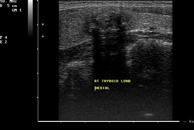
 AACE/ACE Advanced Endocrine Neck Ultrasound Training Course 2016 This 9mm left inferior nodule should remind us all why we re here! There is no absolute number of images required for documentation
Roaa M.Hussein Professor, Department of Physics, College of Science, Ramadi, Iraq. Abstract:
 Abstract: Ultrasound Waves Employment in the Medical Diagnostic for Lıver and Gallbladder Faik H. Antar Professor, Department of Physics, College of Science, AL- Anbar University, Ramadi, Iraq. Ultrasound
Physics. Norman McDicken Tom Anderson CHAPTER ULTRASOUND. Ultrasound Propagation
 CHPTER 2 Physics Norman McDicken Tom nderson This chapter provides an introduction to the physics of medical ultrasound (US). Several books exist that can be consulted to extend the material presented
Case-based discussion:
 Case-based discussion: Pailin Kongmebhol, M.D. Department of Radiology Faculty of Medicine Chiang Mai University There are many guidelines for managing thyroid nodules Two important guidelines: 2015 American
Pre-operative Ultrasound of Lymph Nodes in Thyroid Cancer
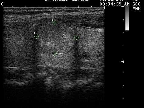
 Pre-operative Ultrasound of Lymph Nodes in Thyroid Cancer AACE - Advances in Medical and Surgical Management of Thyroid Cancer - 2018 Robert A. Levine, MD, FACE, ECNU Thyroid Center of New Hampshire Geisel
High resolution ultrasound scanner for skin imaging
 High resolution ultrasound scanner for skin imaging Christine Turlat Sales Director Atys medical 17 Parc d Arbora 69510 SOUCIEU EN JARREST Atys company Principle of ultrasound imaging DERMCUP Normal image
Ultrasound 10/1/2014. Basic Echocardiography for the Internist. Mechanical (sector) transducer Piezoelectric crystal moved through a sector sweep
 transducer Piezoelectric crystal moved through a sector sweep") Ultrasound Basic Echocardiography for the Internist Carol Gruver, MD, FACC UT Erlanger Cardiology Mechanical wave of compression and rarefaction Requires a medium for transmission Ultrasound frequency
Ultrasound Basic Echocardiography for the Internist Carol Gruver, MD, FACC UT Erlanger Cardiology Mechanical wave of compression and rarefaction Requires a medium for transmission Ultrasound frequency
The 2 nd Cambridge Advanced Emergency Ultrasound Course
 The 2 nd Cambridge Advanced Emergency Ultrasound Course Addenbrooke s Hospital Cambridge Sept 2008 1 2 Faculty! UK! USA! Australia! Toshiba! Emergency Medicine! Radiology 3 Programme! Day 1 Introduction
3/20/2017. Disclosures. Ultrasound Fundamentals. Ultrasound Fundamentals. Bone Anatomy. Tissue Characteristics
 Disclosures Images of ultrasound equipment in this presentation are not an endorsement Fundamentals of Musculoskeletal Ultrasound Physics and Knobology Shane A. Shapiro, M.D. Assistant Professor Orthopedic
Ultrasound in Medicine
 Ultrasound in Medicine Experimental Equipment for Medical Education Universities Colleges Medical Schools Medical and Med-Technical Training Education can befun! WELCOME TO GAMPT Devices and accessories
INTRODUCTION. Getting the best scan. Choosing a probe. Choosing the frequency
 Getting the best scan Choosing a probe Select the most appropriate probe for the particular scan required. s vary in their: operating frequency range higher ultrasound frequencies provide better discrimination
Strain Assessment in Echo
 Strain Assessment in Echo Joe M. Moody, Jr, MD UTHSCSA and STVHCS 2010 Acknowledging many illustrations from Weyman s text and others. Echo-Doppler Basic Principles Background: Ultrasound physics (resolution,
Basic Training Programme. 16 Februrary 2018, ROTTERDAM. Pre and Post-Course Test Answers
 Basic Training Programme 16 Februrary 2018, ROTTERDAM Pre and Post-Course Test Answers Your details: Name: Conference registration number/ BT delegate number: Email address: Are you already performing
Optimising your Doppler settings for an accurate PI. Alison McGuinness Mid Yorks Hospitals
 Optimising your Doppler settings for an accurate PI Alison McGuinness Mid Yorks Hospitals Applications Both maternal uterine and fetal circulations can be studied with doppler sonography Uterine arteries
Development of Ultrasound Based Techniques for Measuring Skeletal Muscle Motion
 Development of Ultrasound Based Techniques for Measuring Skeletal Muscle Motion Jason Silver August 26, 2009 Presentation Outline Introduction Thesis Objectives Mathematical Model and Principles Methods
The table below shows the density and velocity of waves in two different substances. Density / kg m 3 Velocity / m s 1
 Q1.(a) When ultrasound is incident at an interface between two different media some energy is transmitted and some is reflected. The ratio of the reflected energy intensity I r to the incident energy intensity
Chapter 1. Principles of medical ultrasound. Overview. Background history: first steps to the piezo-electric effect.
 Chapter 1 Principles of medical ultrasound GRAHAM ARTHURS, PATRICK HILL AND TREVOR FRANKEL Overview This chapter provides an introduction to the ultrasound process for trainees in anesthesia and other
An Overview of Ultrasound Testing For Lesion Detection in Human Kidney
 Journal of Tomography System & Sensors Application Vol.1, Issue 1, June 2018 An Overview of Ultrasound Testing For Lesion Detection in Human Kidney Aina Fadhilah Abd Rahim 1, Zawin Najah Abd Halim 1, Jaysuman
Real Time Spatial Compound Imaging in breast ultrasound: technology and early clinical experience
 R. Entrekin 1, P. Jackson 1, J.R. Jago 1 and B.A. Porter 2 Real Time Spatial Compound Imaging in breast ultrasound: technology and early clinical experience In current clinical practice, high-resolution
Category Term Definition Comments 1 Major Categories 1a
 Working Lexicon Categories, Terms & Definitions Category Term Definition Comments 1 Major Categories 1a Physiologic Category (consistent with normal ovarian physiology) Follicle Simple 3 cm in premenopausal
ACRIN 6666 IM Additional Evaluation: Additional Views/Targeted US
 Additional Evaluation: Additional Views/Targeted US For revised or corrected form check box and fax to 215-717-0936. Instructions: The form is completed based on recommendations (from ID form) for additional
Abdominal Ultrasound
 Abdominal Ultrasound Imaging Control Buttons Depth The organ imaged should take up 3/4 of the screen Frequency = Penetration Use high frequencies (harmonics) for fluid filled and superficial structures
Abdominal Ultrasound
 Abdominal Ultrasound What is Ultrasound Imaging of the Abdomen? What are some common uses of the procedure? How should I prepare? What does the equipment look like? How does the procedure work? How is
Aixplorer MultiWave Ultrasound System An Innovation in Breast Ultrasound Imaging
 b r e a s t i m a g i n g S O L U T I O N S Aixplorer MultiWave Ultrasound System An Innovation in Breast Ultrasound Imaging The new wave in breast ultrasound Aixplorer is a next-generation ultrasound