WELCOME! Introduction to Bedside Ultrasound
|
|
|
- Franklin Boyd
- 5 years ago
- Views:
Transcription

1 WELCOME! Introduction to Bedside Ultrasound
2 TEACHERS University of California-Irvine School of Medicine Nathan Molina Trevor Plescia Jack Silva
3 COURSE SUMMARY 7 topics Knobology/FAST Cardiac Abdominal Pulmonary Musculoskeletal Head and Neck Procedures For each topic Training with live models Training with SonoSim ultrasound simulator for randomly selected participants Final exam and survey at the end of the course

4 INTRODUCTION TO ULTRASOUND: KNOBOLOGY


5 WHAT IS ULTRASOUND? Ultrasound, or diagnostic sonography, is an imaging technique that uses high frequency sound waves to create images of internal body structures
6 ULTRASOUND PHYSICS Sound waves are made of high pressure and low pressure pulses traveling through a medium High pressure areas (compression) air particles are squeezed together Low pressure areas (rarefaction) air particles are spread apart Sonography is also called ultrasound because it uses sound frequencies that are higher than the normal hearing range
7 ULTRASOUND PHYSICS The ultrasound transducer: 1. Produces a sound wave, which 2. Bounces off structures creating an echo, which 3. Is received by the transducer and converted to an electrical signal, which 4. Is transformed into a digital image Higher frequency sound waves produce sharper images (high resolution), but are absorbed by tissue more easily (poor penetrance) Ideal for superficial structures Sound waves are reflected (produce an echo) wherever there is a change in density of the medium Soft tissue, bone, air, urine, blood, water, etc.
8 MODES AND SETTINGS Modes 2D or B-Mode Doppler, Color Doppler M-Mode Settings Depth Gain Presets Cardiac Abdominal more

9 2D or B-Mode B = Brightness This is for standard ultrasound use, where you will spend most of your time Press 2D to reset all your settings back to normal

10 M-Mode Motion mode Shows how the tissue in one slice moves over time

11 COLOR FLOW DOPPLER Shows fluid moving toward or away from the transducer BART: Blue Away, Red Towards
12 DEPTH The most important setting in ultrasound! You should try to maximize the space on the screen Put the structure you re looking at in the middle of the screen, and make it as big as you can!
13 TOO MUCH DEPTH Wasted Space

14 PERFECT DEPTH Less Wasted Space Much Better!
15 GAIN The strength of the sound waves coming back to the transducer The brightness of the screen Increasing gain brighter image Decreasing gain darker image Auto-gain is a good place to start
16 UNDER-GAINED Too Dark
17 OVER-GAINED Too Bright

18 PERFECT GAIN Just Right

19 LANGUAGE OF ECHOGENICITY Hyperechoic More echogenic than surrounding tissue Dense, hard structures

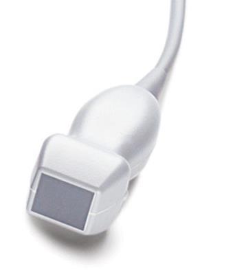
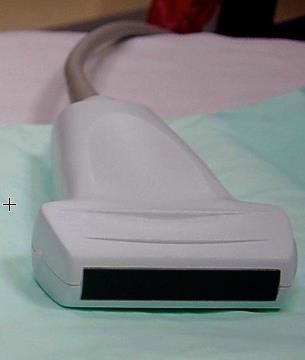
20 LANGUAGE OF ECHOGENICITY Hypoechoic Less echogenic than surrounding tissue
21 LANGUAGE OF ECHOGENICITY Anechoic Devoid of echoes
22 ATTENUATION The energy loss as sound travels through a medium Attenuating can describe The sound wave: High frequency sounds are high attenuating. They lose energy more quickly than low frequency sounds. The medium: Water is low attenuating. Very little energy is lost as sound travels through water. High attenuating: appears hyperechoic (white) Low attenuating: appears hypoechoic (black)
23 ATTENUATION Material Attenuation Coefficient Water Black Blood 0.2 Marrow 0.5 Soft tissue (average) 0.5 Muscle 1.1 Air 1.6 Tendon 4.7 Bone, cortical 6.9 Bone, trabecular 9.9 Enamel 120 White


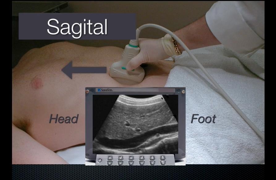
24 High Attenuating Structure Gallstone Echoes are diminished behind high attenuating structures, creating an acoustic shadow Shadow

25 Low Attenuating Structure Echoes are enhanced behind low attenuating structures May be used as a window to visualize anatomy Posterior Acoustic Enhancement

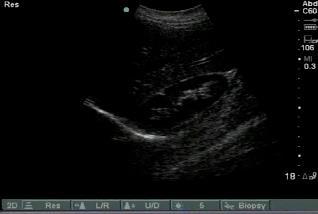
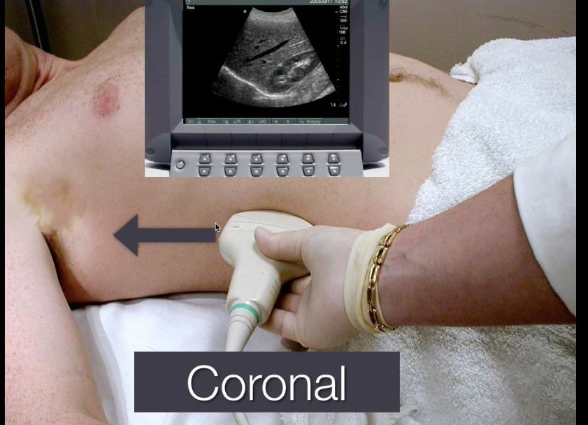
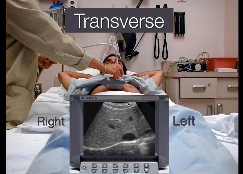
26 Increasing frequency = better resolution but lower penetration ICT C60 L38 P21

27 TRANSDUCER BASICS Convex Array C60

28 TRANSDUCER BASICS Phased Array P21


29 TRANSDUCER BASICS Linear L38
30 QUESTION How does increasing the frequency of the sound waves affect resolution and penetration?

31 HOW TO HOLD THE TRANSDUCER Like a pencil (usually) Anchor part of your hand or your forearm on the patient for stability Indicator
32 MOVING THE TRANSDUCER Scanning or Sliding or Sweeping Fanning or Tilting Rocking or Heel-toe Rotating Compressing


33 TRANSDUCER INDICATOR Position the indicator toward the patient s right or toward their head (there are some exceptions) The indicator on the transducer corresponds to the dot on screen
34
35
36

37


38 ARTIFACTS: MIRROR IMAGE Sound glances off diaphragm, returning to probe with a longer time of flight Machine misinterprets this as more liver tissue further afield Liver Mirror image of liver Diaphragm

39 ARTIFACTS: MIRROR IMAGE Liver Liver and its Mirror Image Mirror image of liver Diaphragm

40 ARTIFACTS: REVERB Recurrent bright arcs at equidistant intervals from the transducer

41 ARTIFACTS: GAS SCATTER Sound waves scatter when traveling through air. Causes a loss of signal and poor visualization of structures behind the air pocket

42 QUESTION Which artifact can be seen in a normal patient near the liver/diaphragm?
43 BENEFITS OF ULTRASOUND Non-Invasive Diagnostic tool Portable Dependent on proficiency of handler
44 Thank you!
Ultrasound Knobology
 Ultrasound Knobology Raj Dasgupta MD, FACP, FCCP, FASSM Assistant Professor of Clinical Medicine Pulmonary / Critical Care / Sleep Medicine University of Southern California (USC) Objectives Physics of
Introduction & Physics of ED Ultrasound. Objectives. What? - Limited Studies. Who? - ED Docs
 Introduction & Physics of ED Ultrasound Martine Sargent, MD Ultrasound Director, Assistant Professor UCSF Department of Emergency Medicine San Francisco General Hospital & Trauma Center Objectives Who?
Terminology Tissue Appearance
 By Marc Nielsen, MD Advantages/Disadvantages Generation of Image Ultrasound Machine/Transducer selection Modes of Ultrasound Terminology Tissue Appearance Scanning Technique Real-time Portable No ionizing
Principles of Ultrasound. Cara C. Prideaux, M.D. University of Utah PM&R Sports Medicine Fellow March 14, 2012
 Principles of Ultrasound Cara C. Prideaux, M.D. University of Utah PM&R Sports Medicine Fellow March 14, 2012 None Disclosures Outline Introduction Benefits and Limitations of US Ultrasound (US) Physics
Basic of Ultrasound Physics E FAST & Renal Examination. Dr Muhammad Umer Ihsan MBBS,MD, DCH CCPU,DDU1,FACEM
 Basic of Ultrasound Physics E FAST & Renal Examination Dr Muhammad Umer Ihsan MBBS,MD, DCH CCPU,DDU1,FACEM What is Sound? Sound is Mechanical pressure waves What is Ultrasound? Ultrasounds are sound waves
3/20/2017. Disclosures. Ultrasound Fundamentals. Ultrasound Fundamentals. Bone Anatomy. Tissue Characteristics
 Disclosures Images of ultrasound equipment in this presentation are not an endorsement Fundamentals of Musculoskeletal Ultrasound Physics and Knobology Shane A. Shapiro, M.D. Assistant Professor Orthopedic
Ultrasound Principles cycle Frequency Wavelength Period Velocity
 ! Teresa S. Wu, MD, FACEP Director, EM Ultrasound Program & Fellowship Co-Director, Simulation Based Training Program & Fellowship Associate Program Director, EM Residency Program Maricopa Medical Center
The Essentials Tissue Characterization and Knobology
 The Essentials Tissue Characterization and Knobology Randy E. Moore, DC, RDMS RMSK No relevant financial relationships Ultrasound The New Standard of Care Musculoskeletal sonography has become the standard
Preamble (disclaimer)
") Preamble (disclaimer) PHYSICS AND PRINCIPLES OF HEAD/NECK ULTRASOUND Joseph C. Sniezek, MD FACS LTC, MC, USA Otolaryngology/H&N Surgery Tripler Army Medical Center 1. I am not a physicist 2. ACS has recommended
Preamble (disclaimer) PHYSICS AND PRINCIPLES OF HEAD/NECK ULTRASOUND Joseph C. Sniezek, MD FACS LTC, MC, USA Otolaryngology/H&N Surgery Tripler Army Medical Center 1. I am not a physicist 2. ACS has recommended
The Physics of Ultrasound. The Physics of Ultrasound. Claus G. Roehrborn. Professor and Chairman. Ultrasound Physics
 The Physics of Ultrasound Pipe Organ 10-8000 Emission Dog 452-1080 Man 85-1100 Spectrum Bat 10,000-120,000 Porpoise 7000-120,000 Claus G. Roehrborn Professor and Chairman 10 20 Cycles per second Reception
Image optimization for critical care US
 Image optimization for critical care US 1 Although we assume you are already familiar with focused US in the ED, it might not hurt to revise the basics: Machines & transducers US appearance of normal tissues
Introduction to Ultrasound Guided Region Anesthesia
 Introduction to Ultrasound Guided Region Anesthesia Brian D. Sites, MD Dept of Anesthesiology Dartmouth-Hitchcock Medical Center INTRODUCTION Welcome to Introduction to Ultrasound Guided Regional Anesthesia.
Physical Principles of Ultrasound
 Physical Principles of Ultrasound Grateful appreciation to Richard A. Lopchinsky, MD, FACS and Nancy H. Van Name, RDMS, RTR, and MarleneKattaron, RDMS 2000 UIC All Rights Reserved. Course Objectives Identify
Basic Physics of Ultrasound and Knobology
 WELCOME TO UTMB Basic Physics of Ultrasound and Knobology By Daneshvari Solanki, FRCA Laura B. McDaniel Distinguished Professor Anesthesiology and Pain Medicine University of Texas Medical Branch Galveston,
Ultrasound basics Part 1
 Ultrasound basics Part 1 'Ultrasound enhanced critical care medicine' Rohit Patel, MD University of Florida Health Director, Critical Care Ultrasound Surgical ICU Center for Intensive Care Gainesville,
Diagnostic Ultrasound. Sutiporn Khampunnip, M.D.
 Diagnostic Ultrasound Sutiporn Khampunnip, M.D. Definition of Ultrasound Ultrasound is simply sound waves, like audible sound. High-frequency sound and refers to mechanical vibrations above 20 khz. Human
Ultrasound Physics and Knobology Alan Macfarlane. Consultant Anaesthetist Glasgow Royal Infirmary
 Ultrasound Physics and Knobology Alan Macfarlane Consultant Anaesthetist Glasgow Royal Infirmary RAPM 2009; 34: 40-46 Ultrasound Proficiency Understanding US image generation and device operation Image
Abdominal Ultrasound
 Abdominal Ultrasound Imaging Control Buttons Depth The organ imaged should take up 3/4 of the screen Frequency = Penetration Use high frequencies (harmonics) for fluid filled and superficial structures
1 Fundamentals. Basic Definitions and Physics Principles. Fundamentals
 1 To become versed in the language of ultrasonography, it is necessary to review some of the basic principles of physics. The wave physics principles of ordinary (i.e., audible) sound apply to ultrasound
Ultrasound Physics & Terminology
 Ultrasound Physics & Terminology This module includes the following: Basic physics terms Basic principles of ultrasound Ultrasound terminology and terms Common artifacts seen Doppler principles Terms for
ULTRASOUND NOMENCLATURE
 Chapter 1: Ultrasound Nomenclature, Image Orientation, and Basic Instrumentation CYNTHIA SIKOWSKI Ultrasound waves are sound waves that have a frequency exceeding 20,000 Hz. When sound waves are transmitted
Ultrasound Physics & Doppler
 Ultrasound Physics & Doppler Endocrine University 2018 Mark Lupo, MD, FACE, ECNU Objectives Review the essential components of ultrasound physics in neck sonography Demonstrate the importance of ultrasound
Concepts of Imaging and Knobology
 Concepts of Imaging and Knobology Pravin Patil, MD FACC FASE Associate Professor of Medicine Director, Cardiovascular Disease Training Program Lewis Katz School of Medicine at Temple University Disclosures
The 2 nd Cambridge Advanced Emergency Ultrasound Course
 The 2 nd Cambridge Advanced Emergency Ultrasound Course Addenbrooke s Hospital Cambridge Sept 2008 1 2 Faculty! UK! USA! Australia! Toshiba! Emergency Medicine! Radiology 3 Programme! Day 1 Introduction
Background & Indications Probe Selection
 Teresa S. Wu, MD, FACEP Director, EM Ultrasound Program & Fellowship Co-Director, Simulation Based Training Program & Fellowship Associate Program Director, EM Residency Program Maricopa Medical Center
Dr Emma Chung. Safety first - Physical principles for excellent imaging
 Safety first - Physical principles for excellent imaging Dr Emma Chung Lecturer in Medical Physics, University of Leicester Clinical Scientist, University Hospitals of Leicester NHS Trust Thanks to Caroline
What is Ultrasound? What is Ultrasound? B A. Basic Principles of Ultrasound. Basic Principles of Ultrasound. Basic Principles of Ultrasound
 Introduction to Ultrasound Principles Mani Montazemi, RDMS Baylor College of Medicine Division of Maternal-Fetal Medicine Department of Obstetrics and Gynecology Manager, Maternal Fetal Center Imaging
Intro to Bedside Ultrasound. Cardiac Ultrasound
 Intro to Bedside Ultrasound Cardiac Ultrasound TEACHERS University of California-Irvine School of Medicine Nathan Molina nathan.d.molina@gmail.com Trevor Plescia taplescia90@gmail.com Jack Silva jpsilva42@gmail.com
Introduction to Biomedical Imaging
 Alejandro Frangi, PhD Computational Imaging Lab Department of Information & Communication Technology Pompeu Fabra University www.cilab.upf.edu Basic principles. Comparison to X-rays Ultrasound > 20kHz
Learning Objectives. Ultrasound for the Primary Care Provider. Portable Ultrasound: Laptops, Tablets, Plug-in Probes, and Pocket devices
 Learning Objectives Ultrasound for the Primary Care Provider Richard Hoppmann, MD, FACP University of South Carolina School of Medicine Assess the main components and functions of a portable ultrasound
CONTENTS. Test Number cpd Tanya Reynolds (Nat. Dip. Diag. Rad., B. Tech. Diag. Rad., B. Tech. Ultrasound)
") CONTENTS page 1-15 page 16 BASIC 2-DIMENSIONAL ULTRASOUND PRINCIPLES Multiple Choice Test Test Number cpd 41640 Tanya Reynolds (Nat. Dip. Diag. Rad., B. Tech. Diag. Rad., B. Tech. Ultrasound) Tanya is
CONTENTS page 1-15 page 16 BASIC 2-DIMENSIONAL ULTRASOUND PRINCIPLES Multiple Choice Test Test Number cpd 41640 Tanya Reynolds (Nat. Dip. Diag. Rad., B. Tech. Diag. Rad., B. Tech. Ultrasound) Tanya is
Extended FAST Exam. Goal of Trauma Care. Golden Hour of Trauma
 Extended FAST Exam Goal of Trauma Care Golden Hour of Trauma Best INITIAL screening modality in trauma efast 2014 LLSA Article (ACEP Policy Statement) Level B Recommendation: In hemodynamically unstable
Point-of-Care Ultrasound: An Introduction
 Point-of-Care Ultrasound: An Introduction Delegation Teaching Package for Registered Respiratory Therapists and Anesthesia Assistants Developed by: Rob Bryan RRT, AA Edited by: Kelly Hassall RRT, FCSRT,
Ultrasonography of the Neck as an Adjunct to FNA. Nicole Massoll M.D.
 Ultrasonography of the Neck as an Adjunct to FNA Nicole Massoll M.D. Basic Features of Head and Neck Ultrasound and Anatomy Nicole Massoll M.D. University of Arkansas for Medical Sciences, Little Rock
ULTRASOUND. OB/Gyn (Core) Ultrasound PIEZOELECTRIC EFFECT. Principles of Ultrasound Physics and Instrumentation. Nathan Pinkney, BS, CDOS
 Ultrasound PIEZOELECTRIC EFFECT. Principles of Ultrasound Physics and Instrumentation. Nathan Pinkney, BS, CDOS") 1 OB/Gyn (Core) Ultrasound Principles of Ultrasound Physics and Instrumentation Nathan Pinkney, BS, CDOS Philadelphia College of Osteopathic Medicine 2016 ULTRASOUND CATEGORIES OF SOUND INFRASOUND = below
1 OB/Gyn (Core) Ultrasound Principles of Ultrasound Physics and Instrumentation Nathan Pinkney, BS, CDOS Philadelphia College of Osteopathic Medicine 2016 ULTRASOUND CATEGORIES OF SOUND INFRASOUND = below
Ultrasound in Sports Medicine
 Ultrasound in Sports Medicine CASES AND USES T I F FA N Y T S AY, M D T O W S O N O R T H O PA E D I C A S S O C I AT E S T H E P R I M A R Y C A R E A P P R O A C H T O T R E AT I N G T H E I N J U R
DIGITAL IMAGE PROCESSING IN ULTRASOUND IMAGES
 DIGITAL IMAGE PROCESSING IN ULTRASOUND IMAGES Kamaljeet Kaur Computer Science & Engineering Department Guru Nanak Dev Engg. College, Ludhiana. Punjab-India meetk.89@gmail.com ABSTRACT-- Image processing
Focused Assessment with Sonography in Trauma (FAST) UC Irvine School of Medicine
 UC Irvine School of Medicine") Focused Assessment with Sonography in Trauma (FAST) UC Irvine School of Medicine Purpose of FAST exam Quickly evaluate patient s status in emergency situations Blunt or penetrating trauma Visualize fluid
Focused Assessment with Sonography in Trauma (FAST) UC Irvine School of Medicine Purpose of FAST exam Quickly evaluate patient s status in emergency situations Blunt or penetrating trauma Visualize fluid
Learning Objectives. Frequency: resolution and depth. The Evolution of Ultrasound Technology. Systems are smaller and portable
 9:45 10:45am Ultrasound for the PCP SPEAKER Richard Hoppmann, MD, FACP Presenter Disclosure Information The following relationships exist related to this presentation: Richard Hoppmann, MD, FACP, has no
Session 2: Ultrasonography for Primary Care Clinicians Learning Objectives
 Session 2: Ultrasonography for Primary Care Clinicians Learning Objectives 1. Assess the main components and functions of a portable ultrasound unit. 2. Identify three clinical applications of portable
Critical Care Ultrasound Seeing is Believing
 Critical Care Ultrasound Seeing is Believing Philip D Lumb, M.B., B.S., MCCM Los Angeles, California USA Objectives Integrate ultrasound into routine critical care practice Introduce HOLA (Holistic Approach)
Table of contents. Foreword. Preface. 1 Introduction Historical Perspective 00
 Table of contents Foreword Preface 1 Introduction 00 1.1 Historical Perspective 00 2 Fundamentals of musculoskeletal ultrasound 00 2.1 Frequency and wavelength 00 2.2 Generating ultrasound waves 00 2.3
INTRODUCTION. Getting the best scan. Choosing a probe. Choosing the frequency
 Getting the best scan Choosing a probe Select the most appropriate probe for the particular scan required. s vary in their: operating frequency range higher ultrasound frequencies provide better discrimination
Lesson 07: Ultrasound Transducers. This lesson contains 73 slides plus 16 multiple-choice questions.
 Lesson 07: Ultrasound Transducers This lesson contains 73 slides plus 16 multiple-choice questions. This lesson was derived from pages 33 through 42 in the textbook: Ultrasound Transducers Ultrasound Transducers
Diploma of Medical Ultrasonography (DMU) Physical Principles of Ultrasound and Instrumentation Syllabus
 Physical Principles of Ultrasound and Instrumentation Syllabus") Diploma of Medical Ultrasonography (DMU) Physical Principles of Ultrasound and Instrumentation Syllabus Page 1 of 7 11/18 Candidates are expected to cover all of the content of this syllabus when preparing
Diploma of Medical Ultrasonography (DMU) Physical Principles of Ultrasound and Instrumentation Syllabus Page 1 of 7 11/18 Candidates are expected to cover all of the content of this syllabus when preparing
Ultrasound in Medicine
 Ultrasound in Medicine Experimental Equipment for Medical Education Universities Colleges Medical Schools Medical and Med-Technical Training Education can befun! WELCOME TO GAMPT Devices and accessories
Emergency Medicine Interest Group (EMIG) 2016
 2016") Emergency Medicine Interest Group (EMIG) 2016 Welcome to the flipped classroom (learning objectives summary) for the 2016 Emergency Medicine Interest Group (EMIG) Procedures Workshop. Overview - Tuesday
Emergency Medicine Interest Group (EMIG) 2016 Welcome to the flipped classroom (learning objectives summary) for the 2016 Emergency Medicine Interest Group (EMIG) Procedures Workshop. Overview - Tuesday
Sound in medicine. CH.12. Dr.Rajaa أ.م.د. رجاء سهيل جنم جامعة تكريت كلية طب االسنان. General Properties of Sound
 CH.12. Dr.Rajaa Sound in medicine أ.م.د. رجاء سهيل جنم جامعة تكريت كلية Sound : It is the audible waves of frequency between 20 Hz and 20 khz. Infrasound : refers to the sound of frequency below the normal
Development of Ultrasound Based Techniques for Measuring Skeletal Muscle Motion
 Development of Ultrasound Based Techniques for Measuring Skeletal Muscle Motion Jason Silver August 26, 2009 Presentation Outline Introduction Thesis Objectives Mathematical Model and Principles Methods
Needle visualization with ZONARE ultrasound systems
 Needle visualization with ZONARE ultrasound systems This material provides a general overview of ultrasound guided needle imaging and techniques and is not intended to replace formal training or education
Abdominal Ultrasound
 Abdominal Ultrasound What is Ultrasound Imaging of the Abdomen? What are some common uses of the procedure? How should I prepare? What does the equipment look like? How does the procedure work? How is
Tissue Strain Analytics Virtual Touch Tissue Imaging and Quantification
 Whitepaper Tissue Strain Analytics Virtual Touch Tissue Imaging and Quantification ACUSON S2000 Ultrasound System Answers for life. Page 1 Tissue Strain Analytics: Virtual Touch Tissue Imaging and Quantification
ULTRASOUND IMAGING EE 472 F2018. Prof. Yasser Mostafa Kadah
 ULTRASOUND IMAGING EE 472 F2018 Prof. Yasser Mostafa Kadah www.k-space.org Recommended Textbook Diagnostic Ultrasound: Physics and Equipment, 2nd ed., by Peter R. Hoskins (Editor), Kevin Martin (Editor),
Musculoskeletal Ultrasound: Basics, Utility, and Clinical Applications
 Musculoskeletal Ultrasound: Basics, Utility, and Clinical Applications Andrew Lavigne, MD, FRCPC Physical Medicine and Rehabilitation CSCN Diplomat (EMG) Dip Sport Medicine Eugene Maida, MD, PGY-4 Resident
Ultrasound. Principles of Medical Imaging. Contents. Prof. Dr. Philippe Cattin. MIAC, University of Basel. Oct 17th, 2016
 Ultrasound Principles of Medical Imaging Prof. Dr. Philippe Cattin MIAC, University of Basel Contents Abstract 1 Image Generation Echography A-Mode B-Mode M-Mode 2.5D Ultrasound 3D Ultrasound 4D Ultrasound
Lesson 03: Sound Wave Propagation and Reflection. This lesson contains 15 slides plus 14 multiple-choice questions.
 Lesson 03: Sound Wave Propagation and Reflection This lesson contains 15 slides plus 14 multiple-choice questions. Accompanying text for the slides in this lesson can be found on pages 8 through 14 in
Breast Imaging Essentials
 Breast Imaging Essentials Module 9 Transcript 2016 ASRT. All rights reserved. Breast Imaging Essentials Module 9 Breast Ultrasound 1. ASRT Animation 2. Welcome Welcome to Module 9 of Breast Imaging Essentials
Ultrasound 10/1/2014. Basic Echocardiography for the Internist. Mechanical (sector) transducer Piezoelectric crystal moved through a sector sweep
 transducer Piezoelectric crystal moved through a sector sweep") Ultrasound Basic Echocardiography for the Internist Carol Gruver, MD, FACC UT Erlanger Cardiology Mechanical wave of compression and rarefaction Requires a medium for transmission Ultrasound frequency
Ultrasound Basic Echocardiography for the Internist Carol Gruver, MD, FACC UT Erlanger Cardiology Mechanical wave of compression and rarefaction Requires a medium for transmission Ultrasound frequency
for the Veterinary Technician
 An Overview of Abdominal Ultrasound for the Veterinary Technician Valerie Gates, CVT, VTS (ECC) Learning Objective: The reader should gain a basic understanding of ultrasound, including physics, terminology,
Features and Benefits
 Features and Benefits DC7 Significant Features HIPAA Compliant Frequency Compound Imaging Standard i-clear Standard i-beam Standard SCI Digital Processing Channels 2048 Dynamic Range > 160 Db 10 Bit A/D
GE Healthcare. LOGIQ E9 XDclear 2.0. Complete Ultrasound HEAD TO TOE OBESE TO THIN NEONATE TO GERIATRIC
 GE Healthcare LOGIQ E9 XDclear 2.0 Complete Ultrasound HEAD TO TOE OBESE TO THIN NEONATE TO GERIATRIC In addition to extraordinary image quality, the system has been further enhanced to meet the needs
ULTRASOUND QA SOLUTIONS. Ensure Accurate Screening, Diagnosis and Monitoring DOPPLER FLOW PHANTOMS MULTI-PURPOSE PHANTOMS TRAINING PHANTOMS
 ULTRASOUND QA SOLUTIONS Ensure Accurate Screening, Diagnosis and Monitoring DOPPLER FLOW PHANTOMS MULTI-PURPOSE PHANTOMS TRAINING PHANTOMS INNOVATORS IN ADVANCED ULTRASOUND TECHNIQUES Gammex is the only
High resolution ultrasound scanner for skin imaging
 High resolution ultrasound scanner for skin imaging Christine Turlat Sales Director Atys medical 17 Parc d Arbora 69510 SOUCIEU EN JARREST Atys company Principle of ultrasound imaging DERMCUP Normal image
Abdominal ultrasound:
 Abdominal ultrasound: Non-traumatic acute abdomen Wittanee Na-ChiangMai, MD Department of Radiology ChiangMai University 26/04/2017 Contents Technique of examination Normal anatomy Emergency conditions
Underwater Acoustic Measurements in Megahertz Frequency Range.
 Underwater Acoustic Measurements in Megahertz Frequency Range. Current State and Prospects of Development in Russia Alexander M. Enyakov,, Many medical applications of underwater acoustic measurements
Brachial plexus blockade within the interscalene groove involves local anesthetic
 Interscalene Brachial Plexus Block- How I do it. Part 1 of a 2 part discussion on technique. Stuart Grant Professor of Anesthesiology Duke University Medical Center Durham NC Brachial plexus blockade within
Probe Selection A high frequency (7-12 MHz) linear array transducer should be used to visualize superficial structures (Image 1).
 linear array transducer should be used to visualize superficial structures (Image 1).") ! Teresa S. Wu, MD, FACEP Director, Emergency Ultrasound Program & Fellowships Co-Director, Women s Imaging Fellowship Maricopa Medical Center Associate Professor, Emergency Medicine Director, Simulation
! Teresa S. Wu, MD, FACEP Director, Emergency Ultrasound Program & Fellowships Co-Director, Women s Imaging Fellowship Maricopa Medical Center Associate Professor, Emergency Medicine Director, Simulation
Focused Assessment Sonography of Trauma (FAST) Scanning Protocol
 Scanning Protocol") Focused Assessment Sonography of Trauma (FAST) Scanning Protocol Romolo Gaspari CHAPTER 3 GOAL OF THE FAST EXAM Demonstrate free fluid in abdomen, pleural space, or pericardial space. EMERGENCY ULTRASOUND
Focused Assessment Sonography of Trauma (FAST) Scanning Protocol Romolo Gaspari CHAPTER 3 GOAL OF THE FAST EXAM Demonstrate free fluid in abdomen, pleural space, or pericardial space. EMERGENCY ULTRASOUND
What is Ultrasound? Resolution Image production Attenuation Imaging modes Ultrasound artifacts... 7
 What is Ultrasound?... 1 Resolution... 3 Image production... 3 Attenuation... 4 Imaging modes... 5 Ultrasound artifacts... 7 0 What is Ultrasound? High frequency sound of frequencies 2-50 MHz is used in
ULTRASOUND QA SOLUTIONS. Ensure Accurate Screening, Diagnosis & Monitoring DOPPLER FLOW PHANTOMS MULTI-PURPOSE PHANTOMS TRANSDUCER TEST PHANTOMS
 ULTRASOUND QA SOLUTIONS Ensure Accurate Screening, Diagnosis & Monitoring DOPPLER FLOW PHANTOMS MULTI-PURPOSE PHANTOMS TRANSDUCER TEST PHANTOMS INNOVATORS IN ADVANCED ULTRASOUND TECHNIQUES Gammex is the
Ultrasound - Musculoskeletal
 Ultrasound - Musculoskeletal What is Ultrasound Imaging of the Musculoskeletal System? Ultrasound imaging, also called ultrasound scanning or sonography, involves exposing part of the body to high-frequency
FAST Focused Assessment with Sonography in Trauma
 FAST Focused Assessment with Sonography in Trauma Wilma Rodriguez Mojica,MD,FACR Professor of Radiology UPR School of Medicine Ultrasound Section - Radiological Sciences Department OBJECTIVES Understand
Chapter 14. Imaging Artifacts
 Chapter 14 Image Artifacts The complex physical interactions that occur between an ultrasound beam and human anatomy and the intricate and sophisticated technological components of a sonographic imaging
Medical Imaging. By: Engr. Joseph Ronald Canedo
 Medical Imaging By: Engr. Joseph Ronald Canedo Medical Sonography (Ultrasound) is an ultrasound-based diagnostic imaging technique used to visualize muscles and internal organs, their size, structures
Routine Quality Assurance Cookbook
 This Cookbook is a companion guide to the AIUM Routine Quality Assurance (QA) for Diagnostic Ultrasound Equipment document, which outlines the basic QA requirements for AIUM-accredited practices. The Guide
Children's (Pediatric) Ultrasound - Abdomen
 Ultrasound - Abdomen") Scan for mobile link. Children's (Pediatric) Ultrasound - Abdomen Children s (pediatric) ultrasound imaging of the abdomen is a safe, noninvasive test that uses sound waves to produce a clear picture of
Scan for mobile link. Children's (Pediatric) Ultrasound - Abdomen Children s (pediatric) ultrasound imaging of the abdomen is a safe, noninvasive test that uses sound waves to produce a clear picture of
ULTRASOUND QA SOLUTIONS. Ensure Accurate Screening, Diagnosis & Monitoring DOPPLER FLOW PHANTOMS MULTI-PURPOSE PHANTOMS TRANSDUCER TEST PHANTOMS
 ULTRASOUND QA SOLUTIONS Ensure Accurate Screening, Diagnosis & Monitoring DOPPLER FLOW PHANTOMS MULTI-PURPOSE PHANTOMS TRANSDUCER TEST PHANTOMS INNOVATORS IN ADVANCED ULTRASOUND TECHNIQUES Gammex is the
CSB 046 Complementary Imaging Techniques
 CSB 046 Complementary Imaging Techniques - Quizzes are only ultrasound, final includes nuc med and ultrasound Week 1 Intro to Ultrasound Physics - Uses 1 to 20 MHz frequencies, which is way above the sound
Confident Diagnosis, Confident Decisions
 Confident Diagnosis, Confident Decisions ALPINION MEDICAL SYSTEMS We are Ultrasound Professionals The E-CUBE Value Creation Optimal Imaging Suite Image Quality Enhancements Optimal Imaging Suite, ALPINION
High performance compact ultrasound
 High performance compact ultrasound Philips CX30 CompactXtreme ultrasound system Philips CX30 CompactXtreme Philips CX30 CompactXteme ultrasound system goes wherever you need it, bringing ultrasound excellence
NCVH. Ultrasongraphy: State of the Art Vein Forum 2015 A Multidisciplinary Approach to Otptimizing Venous Circulation From Wounds to WOW
 Ultrasongraphy: State of the Art 2015 NCVH New Cardiovascular Horizons Vein Forum 2015 A Multidisciplinary Approach to Otptimizing Venous Circulation From Wounds to WOW Anil K. Chagarlamudi, M.D. Cardiovascular
Ultrasound Guidance Needle Techniques
 Ultrasound Guidance Needle Techniques Dr TANG Ho-ming AED/UCH USG Guidance Needle Techniques Commonly used in EM 1. Vessel cannulation-peripheral & central 2. Foreign body removal 3. Peripheral nerve/plexus
An Overview of Ultrasound Testing For Lesion Detection in Human Kidney
 Journal of Tomography System & Sensors Application Vol.1, Issue 1, June 2018 An Overview of Ultrasound Testing For Lesion Detection in Human Kidney Aina Fadhilah Abd Rahim 1, Zawin Najah Abd Halim 1, Jaysuman
Abdomen Sonography Examination Content Outline
 Abdomen Sonography Examination Content Outline (Outline Summary) # Domain Subdomain Percentage 1 2 3 Anatomy, Perfusion, and Function Pathology, Vascular Abnormalities, Trauma, and Postoperative Anatomy
Basic Training Programme. 16 Februrary 2018, ROTTERDAM. Pre and Post-Course Test Answers
 Basic Training Programme 16 Februrary 2018, ROTTERDAM Pre and Post-Course Test Answers Your details: Name: Conference registration number/ BT delegate number: Email address: Are you already performing
Ultrasound imaging is a noninvasive medical test that helps physicians diagnose and treat medical conditions.
 CAROTID ULTRASOUND What is Carotid Ultrasound Imaging? Ultrasound imaging, also called ultrasound scanning or sonography, involves exposing part of the body to highfrequency sound waves to produce pictures
Basic Ultrasound Physics Board Review Questions
 Basic Ultrasound Physics Board Review Questions Sidney K. Edelman, PhD ESP Ultrasound The Woodlands, TX Question 1 What is the wavelength of 2 MHz sound in soft tissue? 1. 1.54 mm 2. 0.75 mm 3. 0.75 cm
Contents. Basic Ultrasound Principles and Terminology. Ultrasound Nodule Characteristics
 Contents Basic Ultrasound Principles and Terminology Basic Ultrasound Principles... 1 Ultrasound System... 2 Linear Transducer for Superficial Images and Ultrasound-Guided FNA... 3 Scanning Planes... 4
An abdominal ultrasound produces a picture of the organs and other structures in the upper abdomen.
 Scan for mobile link. Ultrasound - Abdomen Ultrasound imaging of the abdomen uses sound waves to produce pictures of the structures within the upper abdomen. It is used to help diagnose pain or distention
High performance compact ultrasound
 High performance compact ultrasound Philips CX30 CompactXtreme ultrasound system Philips CX30 CompactXtreme Philips CX30 CompactXtreme ultrasound system goes wherever you need it, bringing ultrasound excellence
Ultrasound Guided Peripheral Intravenous Access
 Ultrasound Guided Peripheral Intravenous Access J. Christian Fox, MD, RDMS, FACEP, FAAEM, FAIUM Professor and Interim Chair of Emergency Medicine Director of Instructional Ultrasound University of California,
Knobology for Dummies
 Knobology for Dummies Power On/Off Preset button Patient Information Entry Choose preset Transducer probes Connect and disconnect transducer Approach to the patient (machine placement, comfort, draping,
Ultrasound in Peripheral Nerve Interventions
 Ultrasound in Peripheral Nerve Interventions John L. Lin, M.D. Shepherd Center Assistant Clinical Professor Emory University, School of Medicine Outline Ultrasound basics Nerve blocks in physiatric setting
Ultrasound in Anesthesia: Applying Scientific Principles to Clinical Practice
 AANA Journal Course Update for Nurse Anesthetists 3 6 CE Credits* Ultrasound in Anesthesia: Applying Scientific Principles to Clinical Practice Christian R. Falyar, CRNA, DNAP The use of ultrasound as
L o o k L i s t e n F e e l S c a n. Your Pocus Cards For Your Every Day Scanning.
 L o o k L i s t e n F e e l S c a n Your Pocus Cards For Your Every Day Scanning E-FAST Extended Focused Assessment by Sonography in Trauma Subcostal Heart View Pleural Sliding on M-mode (Sea-shore sign)
Design Your Performance
 Samsung Medison is a global leading medical devices company. Founded in 1985, the company now sells cutting-edge medical devices including diagnostic ultrasound, digital X-ray and blood analyzer, in 110
Supplement (videos)
") Supplement (videos) Ruben s tube (sound): http://www.youtube.com/watch?v=gpcquuwqayw Doppler US (diagnostic use): http://www.youtube.com/watch?v=fgxzg-j_hfw http://www.youtube.com/watch?v=upsmenyoju8 High
Supplement (videos) Ruben s tube (sound): http://www.youtube.com/watch?v=gpcquuwqayw Doppler US (diagnostic use): http://www.youtube.com/watch?v=fgxzg-j_hfw http://www.youtube.com/watch?v=upsmenyoju8 High
Focused Musculoskeletal Ultrasound
 Focused Musculoskeletal Ultrasound David Lewis Consultant Emergency Medicine Ipswich (Club Doctor, Ipswich Town FC) Advanced Emergency Ultrasound Objectives! General principles! Musculoskeletal anatomy!
Endobronchial Ultrasound
 Endobronchial Ultrasound l Armin Ernst Editors Felix J.F. Herth Endobronchial Ultrasound An Atlas and Practical Guide 13 Editors Armin Ernst, MD Chief, Section of Interventional Pulmonology; Director,
GE Healthcare. LOGIQ E9 XDclear 2.0. Complete Ultrasound HEAD TO TOE OBESE TO THIN NEONATE TO GERIATRIC
 GE Healthcare LOGIQ E9 XDclear 2.0 Complete Ultrasound HEAD TO TOE OBESE TO THIN NEONATE TO GERIATRIC Complete Ultrasound HEAD TO TOE OBESE TO THIN NEONATE TO GERIATRIC OUR BEST JUST GOT BETTER. With the