Vascular Portfolio: Carotid Reflection. Paige Fabre
|
|
|
- Jason Williams
- 6 years ago
- Views:
Transcription


1 Vascular Portfolio: Carotid Reflection Paige Fabre
2 Carotid Reflection For this portfolio I produced three pieces of work; a case study, a PowerPoint of study protocol and a poster of stenosis and plaque grading according to ASUM. Though the case study was the last study I physically produced it was the inspiration for the other two pieces. Evidence Piece 1: Case Study Carotid Artery Plaque and Stenosis This piece was intended to meet all three criteria. By presenting a case study I had intended to display my imaging ability, identification of pathology and patient care were at a beginner level for carotid imaging. It was originally my intention to present a case that displayed the best of my ability but upon reflection, in the latter half of the semester, I decided that it would be more beneficial for me to review a case from my early experience to highlight where I needed improvement. The case presented here I encountered early in my training. At that stage I did not have a very good grasp on the protocol followed by my practice. As part of our training, we are sent to work alongside different sonographers of varying techniques and personal protocols. After researching our department protocols, I came to realise that the scanning protocol in our practice allowed for too much variation, making it very difficult learn a specific routine and sometimes to reproduce the results from previous scans. After attending an in-house training session with our head tutors I gained a better idea of what was expected in the imaging that I produce. This gave rise to the protocol description and images indicated in Evidence Piece 2 and in the protocol section of Evidence Piece 1. Images produced in this study I believe were of diagnostic quality however in reviewing this piece I can see that there is room for improvement particularly in the spectral Doppler traces. These traces are an important part to stenosis grading and can ultimately effect the treatment that the patient receives. Since this examination I have learnt several techniques, outlined in the piece, that have assisted me to produce images of a higher quality (as seen in Evidence Piece 2). It is our duty of care to our patients to provide the best imaging service that we can by improving our skills were possible. To complement our improvement of skills it is also important to improve our understanding of pathology. I believe that the imaging, subsequent worksheet and resulting report would have all benefited from a better understanding of pathology. At the time of the scan I had yet to see stenosis or significant plaque. As I was unsure about the correct technique and protocol, conversations that I had with supervising sonographers rarely addressed pathology and how to correctly document it. This had not really become an issue until this case. As a result I failed to check the report and previous worksheet thoroughly enough to notice that the previous sonographer had documented the previous ICA stenosis by direct measurement and not by velocity measurement as I had. Though this can be corrected it made it difficult to make a direct comparison between studies. Evidence Piece 3 is a direct result of my lack of understanding of stenosis grading and plaque descriptions as highlighted to me by this case. I believe that this piece helps to demonstrate my progression from novice to beginner in Carotid imaging. At the time of the examination I believe I demonstrated a novice/beginner level of imaging, however, my reflection on this case study shows that I recognise the need to improve my
3 understanding of imaging techniques, protocols and pathology as they relate to carotid imaging and thus I am at a beginner level in all three statements. Evidence Piece 2 Carotid Artery Patient Preparation and Protocol Throughout the semester I have increased my knowledge in patient care and imaging protocol as it relates to carotid artery scanning. As previously mentioned, this piece came about as a result of my confusion with scanning protocols and imaging. The patient preparation aspect of this piece aims to address statement 3. As this type of scan is different to other examination in which I am more proficient I compiled the techniques and considerations imparted on me by my tutors and supervising sonographers. Though there were slight variations in techniques, the ideas presented here are employed by the majority of those who have assisted me in my training. Though I believe that patient care level in most examinations is competent, when it comes to obtaining a good clinical history for carotid patients there is still some work to be done. Currently I still require some prompting when it comes to follow up questions. To improve on this I intend to talk more with my fellow sonographers and radiologist to better understand what information is important and in which situations. I am a lot more confident in the protocol required in my department after completing this piece. The information here came primarily from my tutors at an in-house training session and the department protocol. The images displayed here, I believe, are of better quality than those shown in Evidence Piece 1. These images were obtained by consolidating the techniques described in Piece 1. Unlike Piece 1, I believe that these images demonstrate that I am at a beginning level for Competence Statement 1. In practice it still takes me more time to achieve these images than others, but with continued training I hope to improve on this. Evidence Piece 3 This piece is primarily to demonstrate that I am at a beginner level in Competence 2. In Evidence Piece 1 I demonstrated my lack of understanding of pathology found in the carotid artery system, in particular plaques and stenosis grading. I designed this poster for those at a similar level to myself to better assist my understanding of pathology. At the time the afore mentioned case study, I had a basic understanding of what plaque appearance on ultrasound and stenosis grading. This piece required me to further investigate the criteria for each. I chose to make this into a poster, adding images, to act as a quick reference guide. During my investigations for this I learnt that the NASCET criteria used by ASUM and by our practice does not apply to the ECA or CCA and therefore I ensured only ICA images were used. After completing this piece I revised Piece 1 and realised that some of my confusion may have come from previous reports and examinations using the NASCET criteria for vessels other than the ICA. In the future I therefore aim to be consistent and correct with my measurements and comments.
4 Vascular Portfolio: Carotid Ultrasound Case Study Paige Fabre Paige Fabre
5 Case Study: Carotid Artery Stenosis Introduction Thrombus or plaques are a common pathology found in venous sonography. In the carotid artery, stenotic plaques can have serious cerebrovascular consequences if they are not diagnosed and monitored/treated correctly. The following case I performed with the assistance of one of my supervising sonographers in the early stages of my training. Upon reflection of this case I have noted several areas where I may make improvements to my technique and understanding of carotid ultrasound. Patient Presentation 78 year old female Previous history of right sided ICA stenosis with subsequent endarterectomy in 2010 Previous imaging was available for comparison Examination Request The patient presented to the department with the request below. She had been sent from her general practitioner who was managing her case post operatively.
6 Examination Preparation As per department protocol the previous imaging and reports were reviewed before the examination was performed. The most recent scan available was performed 5/12/11 post surgery. The report from this scan is seen below.
7 Room Preparation Prior to collecting the patient from the waiting area, the room was set up ready for the examination. This meant that The ultrasound machine was moved to the head of the bed to ensure that good ergonomics were maintained throughout the scan A variety of probes were made readily available to allow for adequate imaging. For this case we utilised the linear multiple frequency 11L4 transducer on a Toshiba Aplio 500. The pillow was placed at the head of the bed Thick examination gel was made available. Towels were also made available for cleaning up post examination Patient Preparation The patient was called from the waiting are with their name, date of birth and the examination verified. I introduced myself, informed that I was training in this type of scan and would completing the scan with assistance from my supervising sonographer. The patient said that this was okay and this consent was obtained. The patient was then asked to change into a gown for ease of access and to avoid getting gel on their clothing. Upon entering the room, I introduced the patient to the supervising sonographer. The patient was asked to lie on the bed with their head on the pillow. At this point the patient s identification was again checked before the examination began. Explanation of Examination Having had experience with the scans the patient seemed very at easy with the procedure. Still, it was explained to the patient that the examination would look at both sides of the neck, that during the scan the she may hear some strange sounds and that she should try not to move or talk too much but that swallowing was allowed. Additional Clinical Information Given the limited information provided on the referral I proceeded to ask the patient questions to gain a better understanding of their past and present history. From further questioning it was determined that No imaging had been conducted since 2011 The endarterectomy was definitely performed in 2010 and was without complication. No further surgery had been undertaken No current symptoms of current high grade stenosis e.g.: o No episode of dizziness, headaches or fainting o No recent of recent CVA or TIA The examination was just to check up as no other investigations had been recently conducted
8 Imaging Protocol An initial scout scan of the right carotid system was done in transverse. First colour Doppler images and spectral images were not taken. All colour and spectral images had the gain, angle and position altered to ensure that the most accurate information was taken. ECA and ICA were determined by position and spectral waveform. Images required as per our department protocol: 1. Longitudinal of the proximal CCA with Spectral waveform with velocity measurement. Waveform should be a mix of ICA and ECA patterns rapid upstroke with brief reversal and a favourable diastolic component 2. Measure distal CCA peak velocity and enter into IC/CC equation 3. Identify the ECA by detecting waveform, the branches, using a temporal tap. Waveform rapid upstroke, brief systole, post systolic notch and minimal diastolic flow 4. Measure peak ICA velocity and enter it IC/CC systolic equation 5. Colour Image of the bifurcation 6. Vertebral artery in long with waveform and velocity reading. 7. CCA in Transverse 8. CCA in Transverse at BIF 9. Bifurcation in transverse with ICA and ECA Modification to protocol Our protocol defines the minimum number of images taken. It is up to the discretion of the sonographer if they wish to make additional images to better visualise pathology or a lack there of. For this study, I followed the protocol as taught to me by supervising sonographer in addition to the department protocol.


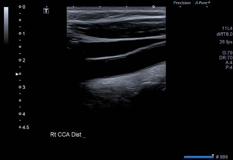
9 Images Imaging of the Right


10


11 RT


12 Image Interpretation of Right After the initial imaging on the right it was determined that: There was no recurrence of significant stenosis Flow throughout the ICA, CCA, ECA and Vertebral Artery was adequate An area of plaque was visualised in the right ECA approximately 1.7mm in AP diameter that had not been previously identified. An area of the distal CCA displayed slight intimal thickening


13 Left Carotid Scan


14


15


16 Image Interpretation of Left Image of the left side when compared to the previous imaging suggested: A region of possible mixed echogenicity plaque was seen in the mid CCA with an AP measurement of 2mm. This area was not apparent on the previous 2011 scan. There are regions of plaque seen at: o Distal CCA 2.7mm o At the bifurcation Proximal ECA 53% stenosis by direct measurement with a maximum velocity of 288cm/s o Previously >90% by direct measurement with maximum velocity 413cm/s Possible 50-59% stenosis seen in the ICA by peak velocity measurement of 151cm/s close to the bifurcation As there was a stenosis of >50% at the ICA, additional it was decided that images of the orbits should be done.


17 Orbital Views Image Interpretation Orbits Examination of the orbits showed patent forward flow to both orbits. When compared to the previous examination this was unchanged.


18 Worksheet As per department protocol an electronic and written worksheet were completed. The electronic work sheet is generated by the machine at the completion of the scan and is as demonstrated.
19
20 Report At the completion of the examination the images were shown to the radiologist on site who prepared the following report.
21 Discussion At the completion of this scan I realised that there were many areas in which I needed to improve. In the areas of patient communication and management I believe that the patient understood my instructions well and felt comfortable throughout the scan. At the commencement of the scan however, I felt that I did not fully understand what follow up questions were needed to obtain a better history, particularly after conducting some additional research and study. My supervising sonographer often prompted me with what information was needed. Though uncertain of what images needed to be taken, due to the variable nature of our protocol, I felt I adapted well to the instruction given to me at the time, producing images of diagnostic quality. At times I did require assistance particularly in imaging all three vessels together in longitudinal. In subsequent scans with other sonographers and tutors, I believe I have gained a better understanding of what is required particularly in area of spectral traces. One of the most prominent lessons that I have learnt is not to take the first waveform trace. Many of my tutors recommend that always trying to improve on the first trace by adjusting factors and position of the gate to ensure that you obtain the most accurate trace possible. Another technique that I have learnt is to decrease the scale of the colour to induce aliasing within non stenotic vessels (taking advantage of laminar flow physics) to identify the correct position for sampling. To reduce any confusion for those reviewing and reporting, factors should then be correctly adjusted to improve image quality before the image is stored. In addition to this, I have also been encouraged to increase the volume of the machines as an audible understanding of wall thump and other artefacts, as well as normal flow helps with my comprehension of vascular studies. Upon further review of these images I have found additional areas of improvement including the information entered into the electronic worksheet. At the time of the examination was not entirely familiar with how it worked and did not realise that it needed user adjustment. According to our department protocol the ICA/CCA ratio should be taken using the NASCET protocol. According to the machine used, the ICA/CCA ratio is taken using the highest ICA measurement rather than the distal ICA measurement. This therefore makes the numbers that appear on the worksheet incorrect. Recently steps have been initiated to change the protocol on the machine to correct this issue. In addition to this change, there has recently been an initiative to adjust the current protocols to create a better uniformity and reproducibility in carotid imaging. When reviewing this piece after further training I have come to recognise that even though learning a variation of techniques from various people gives you a range of tools to work with, it makes it difficult to learn the basic standard of imaging with no two normal scans being the same. Conclusion Overall, the scan was completed to a diagnostic quality with assistance from my supervising sonographer. As this scan was done early in my experience I have had the chance to review my work and identify areas for improvement in terms of patient communication and image quality.
22 Paige Fabre
23 Carotid Artery Scanning It is important as professionals that we manage our patients effectively before, during and at the completion of our scans. When examining the Carotid Artery System increased patient age, body habitus and the position needed for the scan make it increasingly difficult to balance patient comfort and diagnostic image quality. In this piece I intend to demonstrate the techniques and tips given to me by my tutors and colleagues to ensure that an effective study be completed. Some of the techniques displayed in this piece I have learnt while attending an in-house, hands on tutorial.
24 Before the Examination As per our department protocol, previous imaging should be reviewed if available. The room should be set up ready for the patient to enter and the examination to begin. The request should be read thoroughly to ensure that the correct examination is performed on the correct patient for the correct reason.

25
26 Room Set Up Before the patient is collected from the waiting area the room should be set up so that: The machine is located at the head of the bed, set at an appropriate height (whether sitting or standing) to ensure that sonographer posture and overall good ergonomics are maintained. An appropriate variety of probes need to be readily available either connected to the machine or very close by. These should include a vascular transducer that will allow for adequate penetration and resolution. At our practice we primarily utilise a Toshiba 11L4, Phillips L11 and a GE 9L6. It is also important to have lower and higher frequency probes available for more challenging patients and more superficial structures respectively.


27 Room Set Up Room set up with the machine at the head of the bed An appropriate array of transducers and gels
28 Room Set Up The bed should be moved into an appropriate approximate location. This is to reduce any need to more the bed once the patient is positioned. Not only can this be uncomfortable for the patient but can cause undue stress on the sonographer and potentially cause injury. Dependant on sonographer preference a pillow may be positioned at the head of the bed. Its use and position is critical to the examinations success. Too much extension in the neck may cause the to be uncomfortable and make it difficult for them to maintain the position. Too little extension and it may become difficult for the sonographer to acquire the necessary imaging. In room preparation pillows and sponges should at least be readily available so that patient position can be adjusted easily.
29
30 Greeting the Patient After the patient has been called from the waiting area they should be taken to a private changing area or location where their identity can be verified. Correct patient, correct examination, correct location should all be checked at this point. Once this has been done, the patient should be changed in to a gown if their clothing is not appropriate for the examination.

31 Changing the Patient If the patient requires changing they should be given an appropriate gown and instructed on how it is to be worn. It is part of our duty of care that the patient feel comfortable and is covered appropriately. Consideration should be made for patients who are usually covered for religious or cultural reasons. If the patient is wearing a head dress they may feel uncomfortable with their arms, neck or shoulders bare. In these cases, to ensure patient comfort, if possible the patient should be allowed to change in the examination room so that they are not uncovered in the waiting areas.
32 Explanation of Exam To ensure that patient consent is obtained, the examination should be explained prior to the examination. An interpreter should be used if necessary. It should be explained that: The examination may take a length of time The patient must remain as still as possible throughout the examination The patient must not talk throughout the examination but they may swallow as normal The patient will hear noises throughout the scan from the machine (it is important to inform the patient of this so they are not startled when they hear the Doppler) The scan will look at both sides of the neck The final reporting will be done by the radiologist and then transmitted to the referring doctor
33 Obtaining Patient History As with all imaging examinations it is important that a thorough patient history be obtained. Questions that may be asked include but are not limited to: Have you previously had an examination of the carotid vessels? Is there any previous history of stenosis? Is there any previous history of surgery to the carotid vessels or surrounding vascular system? What symptoms have they experienced? How long have they experienced symptoms for? Has there been any recent change in these symptoms? Are they on any blood thinning medications?
34 Obtaining Patient History Care should be taken to use laymen terminology where necessary to allow a more accurate history. Terms such as: blood thinning rather than anticoagulant clot or blockage rather than stenosis
35
36 Patient Positioning The patient should be positioned so that: The neck is slightly extended to a comfortable degree and slightly rotated to the contralateral side to the one being scanned. A blanket may be placed over the patient if they are feeling cold. If the patient is still in their own clothes, towels should placed in the collar to avoid contact from the gel.

37 Patient Positioning
38 The following protocol has been taken from the Vascular Protocol Manual
39 Carotid Artery Protocol Before images are taken the carotid should be scanned in B-mode to generally assess for plaque. The same should then briefly done covering the CCA, ICA and ECA.

40 Carotid Artery Protocol 1. Longitudinal of the proximal CCA with Spectral waveform with velocity measurement. Waveform should be a mix of ICA and ECA patterns rapid upstroke with brief reversal and a favourable diastolic component

41 Carotid Artery Protocol 2. Measure distal CCA peak velocity and enter into IC/CC equation
42 Carotid Artery Protocol 3. Identify the ECA by detecting waveform, the branches, using a temporal tap. Waveform rapid upstroke, brief systole, post systolic notch and minimal diastolic flow


43 Carotid Artery Protocol Waveform Temporal Tap
44 Carotid Artery Protocol 4. Measure peak ICA velocity and enter it IC/CC systolic equation

45 Carotid Artery Protocol 5. Colour Image of the bifurcation

46 Carotid Artery Protocol 6. Vertebral artery in long with waveform and velocity reading.
47 Though not specified in our practice protocol, I have been trained to also take images in the transverse plane as follows.

48 Carotid Artery Protocol CCA in Transverse

49 Carotid Artery Protocol CCA in Transverse at BIF

50 Carotid Artery Protocol Bifurcation in transverse with ICA and ECA
51 The following is the additional information needed in the presence of plaque or stenosis
52 Additional Imaging Measurement of ICA end diastolic flow if stenosis is detected

53 Additional Imaging Image of bifurcation with plaque labelled Ratio data should be included on image Colour and B-mode image needed
54 Additional Imaging Stenosis at proximal ICA, CCA and ECA direct measurement

55 Additional Imaging Transverse image to show eccentric plaques

56 Additional Imaging Retro-Orbital waveforms may also be examined

57 Additional Imaging In the presence of vertebral reversal, the proximal subclavian should be assessed for stenosis. Brachial artery waveforms may also be examined bilaterally.

58 Carotid Portfolio: Carotid Artery Disease A Trainee Guide to Plaque types and stenosis grading The following is a poster aimed at trainee sonographers so assist with the ultrasound appearance and descriptions of carotid artery disease according to the ASUM Guidelines. This piece is sized as per an A2 poster Paige Fabre Paige Fabre


59 Carotid Artery Disease A Trainee Guide to Plaque Types and Stenosis Grading Echogenic Hypoechoic 1 Transverse image of the Distal CCA with low echogenicity plaque 2 Echogenic Plaque in the ICA Echogenic plaque lining the wall of the vessel 1 Usually of a uniform density Commonly known as a Fatty Streak Carries an increased risk of embolization 1 1 Mixed Calcified Intermediate Plaque of Mixed echogenicity with intraplaque haemorrhage in the CCA bifurcation 1 Calcified Plaque with posterior shadowing 4 Intermediate Plaque in the CCA 2 Focal or diffuse areas of hypoechogenicity within the plaque formation 1 Sonolucent areas may be indicative of intraplaque 3 haemorrhage 1 Carries an increased risk of embolization Echogenic focus along the vessel wall with 1,3 posterior acoustic shadowing 1 May be focal or diffuse 1 Carries a decreased risk of embolization If on the superficial wall may make interrogation 1 with Colour Doppler difficult Intermediate plaque is a combination of soft and hard plaque (partially calcified) or echolucent and echorich plaque 5 Intermediate plaque is more stable then 5 hypoechoic plaques 0 ICA Stenosis Grading <15 % Diameter Reduction 16-49% Diameter Reduction Normal ICA Waveform and Colour filling of the ICA Minor reduction in Diameter 30% Stenosis Normal waveform and Image Laminar Flow 6 Deceleration Spectral Broadening 6 PSV <125 cm/sec EDV <40cm/sec Uniform intraluminal colour flow Pansystolic Spectral Broadening 6 PSV <125 cm/sec 1 EDV <40cm/sec Uniform intraluminal colour flow % Diameter Reduction 70-79% Diameter Reduction 80-90% Diameter Reduction Occluded % Stenosis in the ICA 70-79% Diameter reduction in proximal ICA 8 Significant Stenosis 9 Occlusion 9 Pansystolic Spectral Broadening 6 PSV >125 cm/sec and EDV < 110 cm/sec ICA/CCA >2 6 Mild disturbance in intraluminar colour hues at 1 the site of and distal to stenosis 6 Pansystolic Spectral Broadening 6 PSV >270 cm/sec or EDV > 110 cm/sec 6 ICA/CCA >4 Aliasing seen at the stenosis with post stenotic 1 turbulence 6 Pansystolic Spectral Broadening 6 6 PSV >270 cm/sec or EDV > 140 cm/sec 6 ICA/CCA >4 Aliasing seen at the stenosis with post stenotic 1 turbulence No colour flow seen 2,6 2,6 Terminal thump in systole 6 PSV and EDV absent Echogenic material will completely fill the vessel 1-2 lumen 2 Ipsilateral ECA may demonstrate internal flow pattern 2 Reduction in overall size with chronic occlusion
60 References 1. Ahuja, A. Diagnostic Imaging: Ultrasound. Canada: Amirsys; Lee, Whal. General principles of carotid Doppler ultrasonography. Ultrasonography. 2014: 33(1): doi: /usg Henningson, C. Clinical Guide to Ultrasonography. St Louis (Missouri): Mosby Elsevier; Crimmins, Timothy. Carotid Plaque Appearance [Internet]. Columbia University [cited 2014 October 26]. Available from 5. Gronholdt, Marie-Louise Moes, Borge G. Nordestgaard, Tina Nielson, and Henrick Sillesen. Echolucent Carotid Artery Plaques Are Associated With Elevated Levels of Fasting and Postprandial Triglyceride-Rich Lipoproteins. Stroke (12): Australasian Society for Ultrasound in Medicine. Policies and Statements: D14 Colour Duplex Doppler Ultrasound Extracranial Carotid Artery Disease [Internet]. Australia: Australasian Society for Ultrasound in Medicine: 2006 [updated 2008; cited 2014]. Available from: 7. Australasian Society for Ultrasound in Medicine. ASUM Ultrasound Clinical Handbook: Carotid Duplex [Internet] [cited 2014 October 26]. Available from: 8. Quality Vascular Imaging. Case of the Month [Internet] Florida; 2013 [cited 2014 September 28]. Available from: 9. Radswiki. Carotid Artery Stenosis [Internet]. Radiopaedia; n.d. [cited 2014 October 26]. Available from:
Carotid Abnormalities Coils, Kinks and Tortuosity David Lorelli M.D., RVT, FACS Michigan Vascular Association Conference Saturday, October 20, 2012
 Carotid Abnormalities Coils, Kinks and Tortuosity David Lorelli M.D., RVT, FACS Michigan Vascular Association Conference Saturday, October 20, 2012 Page 1 Table of Contents Carotid Anatomy Carotid Duplex
GUNDERSEN/LUTHERAN ULTRASOUND DEPARTMENT POLICY AND PROCEDURE MANUAL
 GUNDERSEN/LUTHERAN ULTRASOUND DEPARTMENT POLICY AND PROCEDURE MANUAL SUBJECT: Carotid Duplex Ultrasound SECTION: Vascular Ultrasound ORIGINATOR: Deborah L. Richert, BSVT, RDMS, RVT DATE: October 15, 2015
Disclosure Statement:
 Marsha M. Neumyer, BS, RVT, FSVU, FSDMS, FAIUM International Director Vascular Diagnostic Educational Services Vascular Resource Associates Harrisburg, PA Disclosure Statement: CME Calendar QR Code Marsha
Carotid Artery Doppler
 Carotid Artery Doppler Patient Position supine or semisupine head slightly hyper extended rotated 45 away from the side being examined. Higher frequency linear transducers (7 MHz) Vessels should be imaged
Protokollanhang zur SPACE-2-Studie Neurology Quality Standards
 Protokollanhang zur SPACE-2-Studie Neurology Quality Standards 1. General remarks In contrast to SPACE-1, the neurological center participating in the SPACE-2 trial will also be involved in the treatment
Carotid Duplex: Beyond Stenosis Ido Weinberg, MD Vascular Medicine Massachusetts General Hospital Assistant Professor of Medicine Harvard Medical
 Carotid Duplex: Beyond Stenosis Ido Weinberg, MD Vascular Medicine Massachusetts General Hospital Assistant Professor of Medicine Harvard Medical School Boston, Massachusetts Disclosures I do not have
Carotid Ultrasound: Improving Ultrasound
 Carotid Ultrasound: Improving Ultrasound Edward I. Bluth, M.D., F.A.C.R. Chairman Emeritus, Department of Radiology, Ochsner Clinic Foundation, New Orleans, Louisiana Professor, Ochsner Clinical School,
Certificate in Clinician Performed Ultrasound (CCPU) Syllabus. Above Knee Deep Vein Thrombosis (DVT)
 Syllabus. Above Knee Deep Vein Thrombosis (DVT)") Certificate in Clinician Performed Ultrasound (CCPU) Syllabus Above Knee Deep Vein Thrombosis (DVT) Deep Vein Thrombosis (DVT) Purpose: Prerequisites: Training: Assessments: This unit is designed to cover
Certificate in Clinician Performed Ultrasound (CCPU) Syllabus Above Knee Deep Vein Thrombosis (DVT) Deep Vein Thrombosis (DVT) Purpose: Prerequisites: Training: Assessments: This unit is designed to cover
Certificate in Clinician Performed Ultrasound (CCPU) Syllabus
 Syllabus") Certificate in Clinician Performed Ultrasound (CCPU) Syllabus Proximal Deep Vein Thrombosis (DVT) Page 1 of 6 03/17 Deep Vein Thrombosis (DVT) Syllabus Purpose: This unit is designed to cover the theoretical
Certificate in Clinician Performed Ultrasound (CCPU) Syllabus Proximal Deep Vein Thrombosis (DVT) Page 1 of 6 03/17 Deep Vein Thrombosis (DVT) Syllabus Purpose: This unit is designed to cover the theoretical
Pre-and Post Procedure Non-Invasive Evaluation of the Patient with Carotid Disease
 Pre-and Post Procedure Non-Invasive Evaluation of the Patient with Carotid Disease Michael R. Jaff, D.O., F.A.C.P., F.A.C.C. Assistant Professor of Medicine Harvard Medical School Director, Vascular Medicine
Ultrasound Imaging of The Posterior Circulation
 Ultrasound Imaging of The Posterior Circulation Michigan Sonographers Society 2 Nd Annual Fall Vascular Conference Larry N. Raber RDMS-RVT Clinical Manager General Ultrasound/Neurovascular Laboratory Cleveland
Deb Coghlan AMS (Vascular and General ) Brisbane, Australia
 Brisbane, Australia") Deb Coghlan AMS (Vascular and General ) Brisbane, Australia ANEURYSMAL DIISEASE The infrarenal aorta enlarges with age, and is the commonest site for arterial aneurysms. An aneurysm is a permanent focal
Deb Coghlan AMS (Vascular and General ) Brisbane, Australia ANEURYSMAL DIISEASE The infrarenal aorta enlarges with age, and is the commonest site for arterial aneurysms. An aneurysm is a permanent focal
Policies and Statements D16. Intracranial Cerebrovascular Ultrasound
 Policies and Statements D16 Intracranial Cerebrovascular Ultrasound SECTION 1: INSTRUMENTATION Policies and Statements D16 Intracranial Cerebrovascular Ultrasound May 2006 (Reaffirmed July 2007) Essential
Beyond Stenosis Severity: Top 5 Important Duplex Characteristics to Identify in a Patient with Carotid Disease
 Beyond Stenosis Severity: Top 5 Important Duplex Characteristics to Identify in a Patient with Carotid Disease Jan M. Sloves RVT, RCS, FASE Technical Director New York Cardiovascular Associates Disclosures
8/20/18. The Doppler Effect. Objectives. What is the Doppler Effect. Doppler principles. Spectral Waveform. Image recognition. Vascular Ultrasound
 Vascular Ultrasound: Physics and Haemodynamics Objectives Doppler principles Spectral Waveform Key factors Haemodynamics: Stenosis Waveforms Image recognition Vascular Ultrasound: A flawed paradigm What
Neurovascular Ultrasound Course
 Neurovascular Ultrasound Course William M. McKinney (6/6/30-10/24/03) Father of Neurosonology Founder, Neurosonology Course, WFUSM Welcome to Winston-Salem, NC, Wake Forest School of Medicine, and the
No financial or commercial relationships to disclose
 Deanna New, RVT No financial or commercial relationships to disclose IAC REQUIREMENTS: The main duty of a sonographer is to make the physician or radiologists job easier by capturing images and doing
Carotid Imaging IT S ABOUT MORE THAN JUST OBTAINING THE IMAGES
 Carotid Imaging IT S ABOUT MORE THAN JUST OBTAINING THE IMAGES No financial or commercial relationships to disclose Carotid artery disease: Stroke is one of the most serious causes of mortality and morbidity
Certificate in Clinician Performed Ultrasound (CCPU) Syllabus. Vascular Access (venous (peripheral and central) and arterial)
 Syllabus. Vascular Access (venous (peripheral and central) and arterial)") Certificate in Clinician Performed Ultrasound (CCPU) Syllabus Vascular Access (venous (peripheral and central) and arterial) Page 1 of 8 04/16 Vascular Access (venous (peripheral and central) and arterial)
Certificate in Clinician Performed Ultrasound (CCPU) Syllabus Vascular Access (venous (peripheral and central) and arterial) Page 1 of 8 04/16 Vascular Access (venous (peripheral and central) and arterial)
Optimising your Doppler settings for an accurate PI. Alison McGuinness Mid Yorks Hospitals
 Optimising your Doppler settings for an accurate PI Alison McGuinness Mid Yorks Hospitals Applications Both maternal uterine and fetal circulations can be studied with doppler sonography Uterine arteries
Measure #195 (NQF 0507): Radiology: Stenosis Measurement in Carotid Imaging Reports National Quality Strategy Domain: Effective Clinical Care
: Radiology: Stenosis Measurement in Carotid Imaging Reports National Quality Strategy Domain: Effective Clinical Care") Measure #195 (NQF 0507): Radiology: Stenosis Measurement in Carotid Imaging Reports National Quality Strategy Domain: Effective Clinical Care 2017 OPTIONS FOR INDIVIDUAL MEASURES: CLAIMS ONLY MEASURE TYPE:
Measure #195 (NQF 0507): Radiology: Stenosis Measurement in Carotid Imaging Reports National Quality Strategy Domain: Effective Clinical Care 2017 OPTIONS FOR INDIVIDUAL MEASURES: CLAIMS ONLY MEASURE TYPE:
Guidelines, Policies and Statements D20 Statement on Peripheral Venous Ultrasound
 Guidelines, Policies and Statements D20 Statement on Peripheral Venous Ultrasound Disclaimer and Copyright The ASUM Standards of Practice Board have made every effort to ensure that this Guideline/Policy/Statement
DISCLOSURE TEST YOUR WAVEFORM IQ. Partial volume artifact. 86 yo female with right arm swelling, picc line. AVF on left? Dx?
 Deborah Rubens University of Rochester Rochester, NY DISCLOSURE Neither I nor my immediate family have a financial relationship with a commercial organization that may have a direct or indirect interest
Quality ID #195 (NQF 0507): Radiology: Stenosis Measurement in Carotid Imaging Reports National Quality Strategy Domain: Effective Clinical Care
: Radiology: Stenosis Measurement in Carotid Imaging Reports National Quality Strategy Domain: Effective Clinical Care") Quality ID #195 (NQF 0507): Radiology: Stenosis Measurement in Carotid Imaging Reports National Quality Strategy Domain: Effective Clinical Care 2018 OPTIONS FOR INDIVIDUAL MEASURES: REGISTRY ONLY MEASURE
Quality ID #195 (NQF 0507): Radiology: Stenosis Measurement in Carotid Imaging Reports National Quality Strategy Domain: Effective Clinical Care 2018 OPTIONS FOR INDIVIDUAL MEASURES: REGISTRY ONLY MEASURE
NCVH. Ultrasongraphy: State of the Art Vein Forum 2015 A Multidisciplinary Approach to Otptimizing Venous Circulation From Wounds to WOW
 Ultrasongraphy: State of the Art 2015 NCVH New Cardiovascular Horizons Vein Forum 2015 A Multidisciplinary Approach to Otptimizing Venous Circulation From Wounds to WOW Anil K. Chagarlamudi, M.D. Cardiovascular
BEDSIDE ULTRASOUND BEDSIDE ULTRASOUND. Deep Vein Thrombosis. Probe used
 BEDSIDE ULTRASOUND Part 2 Diagnosis of deep vein thrombosis Kishore Kumar Pichamuthu, Professor, Department of Critical Care, CMC, Vellore Summary: Deep vein thrombosis (DVT) is a problem encountered in
NON-ATHEROSCLEROTIC PATHOLOGY OF THE CAROTID ARTERIES
 NON-ATHEROSCLEROTIC PATHOLOGY OF THE CAROTID ARTERIES Leslie M. Scoutt, MD, FACR Professor of Diagnostic Radiology & Surgery Vice Chair, Dept of Radiology & Biomedical Imaging Chief, Ultrasound Section
Pelvic Ultrasound.
 Pelvic Ultrasound Before Your Exam: Drink 32 oz. of water one hour before your examination time. Try to drink all the liquid within 30 minutes. Do not urinate before the exam. Arrive for your exam with
Duplex Ultrasound. A Detailed Look at Your Blood Vessels
 Duplex Ultrasound A Detailed Look at Your Blood Vessels What Is Duplex Ultrasound? Ultrasound is a test that uses sound waves to create detailed pictures of the inside of your body. Duplex ultrasound is
Carotid Imaging. Dr Andrew Farrall. Consultant Neuroradiologist
 20121123 SSCA http://www.neuroimage.co.uk/network Andrew Farrall Carotid Imaging Dr Andrew Farrall Consultant Neuroradiologist SFC Brain Imaging Research Centre (www.sbirc.ed.ac.uk), SINAPSE Collaboration
Upper Extremity Venous Duplex Evaluation
 VASCULARTECHNOLOGY PROFESSIONAL PERFORMANCE GUIDELINES Upper Extremity Venous Duplex Evaluation This Guideline was prepared by the Professional Guidelines Subcommittee of the Society for Vascular Ultrasound
STRUCTURED EDUCATION REQUIREMENTS IMPLEMENTATION DATE: JULY 1, 2016
 STRUCTURED EDUCATION REQUIREMENTS Vascular Sonography The purpose of structured education is to provide the opportunity for individuals to develop mastery of discipline-specific knowledge that, when coupled
Vascular Sonography Examination
 Vascular Sonography Examination The purpose of The American Registry of Radiologic Technologists (ARRT ) Vascular Sonography Examination is to assess the knowledge and cognitive skills underlying the intelligent
Diploma of Medical Ultrasonography (DMU) Physical Principles of Ultrasound and Instrumentation Syllabus
 Physical Principles of Ultrasound and Instrumentation Syllabus") Diploma of Medical Ultrasonography (DMU) Physical Principles of Ultrasound and Instrumentation Syllabus Page 1 of 7 11/18 Candidates are expected to cover all of the content of this syllabus when preparing
Diploma of Medical Ultrasonography (DMU) Physical Principles of Ultrasound and Instrumentation Syllabus Page 1 of 7 11/18 Candidates are expected to cover all of the content of this syllabus when preparing
Background & Indications Probe Selection
 Teresa S. Wu, MD, FACEP Director, EM Ultrasound Program & Fellowship Co-Director, Simulation Based Training Program & Fellowship Associate Program Director, EM Residency Program Maricopa Medical Center
Image Formation (10) 2 Evaluation and Selection of Representative Images (10)
 2 Evaluation and Selection of Representative Images (10)") STRUCTURED SELF ASSESSMENT CONTENT SPECIFICATIONS SSA LAUNCH DATE: JANUARY 1, 2018 Vascular Sonography The purpose of continuing qualifications requirements (CQR) is to assist registered technologists
STRUCTURED SELF ASSESSMENT CONTENT SPECIFICATIONS SSA LAUNCH DATE: JANUARY 1, 2018 Vascular Sonography The purpose of continuing qualifications requirements (CQR) is to assist registered technologists
Certificate in Clinician Performed Ultrasound (CCPU) Syllabus
 Syllabus") Certificate in Clinician Performed Ultrasound (CCPU) Syllabus Abdominal Aortic Aneurysm (AAA) Page 1 of 6 12/18 Abdominal Aortic Aneurysm (AAA) Syllabus Purpose: This unit is designed to cover the theoretical
Certificate in Clinician Performed Ultrasound (CCPU) Syllabus Abdominal Aortic Aneurysm (AAA) Page 1 of 6 12/18 Abdominal Aortic Aneurysm (AAA) Syllabus Purpose: This unit is designed to cover the theoretical
Lower Extremity Arterial Doppler
 Lower Extremity Arterial Doppler 1. Spectral Doppler waveform should be taken in distal aorta and common iliac arteries. 2. R/L common femoral artery (CFA) color Doppler with velocity and B-mode. 3. R/L
Performance Evaluation of Ultrasound Systems: Advanced Methods and Applications
 Performance Evaluation of Ultrasound Systems: Advanced Methods and Applications NJ Hangiandreou PhD, Z Long PhD, DJ Tradup RDMS, SF Stekel BS, A Ferrero PhD, AT Tao PhD, W Zhou PhD, JE Browne PhD*, N Strissel
(Department of Radiology, Beylikdüzü State Hospital, İstanbul, Turkey) Corresponding Author: Dr. Mete Özdikici
 Corresponding Author: Dr. Mete Özdikici") Quest Journals Journal of Medical and Dental Science Research Volume 5~ Issue 6 (2018) pp: 61-65 ISSN(Online) : 2394-076X ISSN (Print):2394-0751 www.questjournals.org Research Paper Quantitative Measurements
Quest Journals Journal of Medical and Dental Science Research Volume 5~ Issue 6 (2018) pp: 61-65 ISSN(Online) : 2394-076X ISSN (Print):2394-0751 www.questjournals.org Research Paper Quantitative Measurements
Section II: Patient Interview Grade: 5
 Only written competency completed with this EXACT form will be accepted for grading. No modifications to the LAYOUT of the form will be accepted for a written competency. Failure to comply will result
Neuro Quiz 29 Transcranial Doppler Monitoring
 Verghese Cherian, MD, FFARCSI Penn State Hershey Medical Center, Hershey Quiz Team Shobana Rajan, M.D Suneeta Gollapudy, M.D Angele Marie Theard, M.D Neuro Quiz 29 Transcranial Doppler Monitoring This
Recommendations for documentation of neurosonographic examinations
 Recommendations for documentation of neurosonographic examinations The documentation of ultrasound examinations is subject to a dynamic development particularly as regards newer applications. The present
Duplex ultrasound is first-line imaging for all
 Our Protocol for Transabdominal Pelvic Vein Duplex Ultrasound A summary of s protocol for pelvic vein duplex ultrasonography, including equipment, patient positioning, ultrasound settings, and technique.
HD Scanning: Velocities and Volume Flow
 HD Scanning: Velocities and Volume Flow Non-Invasive Lab Symposium West Orange, NJ April 27, 2018 Volume Flow Cindy Sturt, MD, FACS, RVT 500,000 Americans on dialysis 20-25% annual mortality 65% 5 year
Non-invasive Imaging of Carotid Artery Atherosclerosis
 Non-invasive Imaging of Carotid Artery Atherosclerosis 최연현 성균관의대삼성서울병원영상의학과 Noninvasive Techniques US with Doppler CT MRI Ultrasonography Techniques of Carotid US US Anatomy (ICA vs ECA) Gray scale and
Routine Quality Assurance Cookbook
 This Cookbook is a companion guide to the AIUM Routine Quality Assurance (QA) for Diagnostic Ultrasound Equipment document, which outlines the basic QA requirements for AIUM-accredited practices. The Guide
Transcranial Doppler (Basic Step) Dae-il Chang, M.D., Sung Sang Yoon, M.D. Department of Neurology, College of Medicine, Kyunghee university
 Dae-il Chang, M.D., Sung Sang Yoon, M.D. Department of Neurology, College of Medicine, Kyunghee university") Transcranial Doppler (Basic Step) Dae-il Chang, M.D., Sung Sang Yoon, M.D. Department of Neurology, College of Medicine, Kyunghee university Principles of Doppler Ultrasonography Major target Speed & direction
Transcranial Doppler (Basic Step) Dae-il Chang, M.D., Sung Sang Yoon, M.D. Department of Neurology, College of Medicine, Kyunghee university Principles of Doppler Ultrasonography Major target Speed & direction
Certificate in Clinician Performed Ultrasound (CCPU) Syllabus. Abdominal Aortic Aneurysm (AAA)
 Syllabus. Abdominal Aortic Aneurysm (AAA)") Certificate in Clinician Performed Ultrasound (CCPU) Syllabus Abdominal Aortic Aneurysm (AAA) Purpose: Prerequisites: Training: Assessments: Course Objectives Abdominal Aortic Aneurysm (AAA) This unit
Certificate in Clinician Performed Ultrasound (CCPU) Syllabus Abdominal Aortic Aneurysm (AAA) Purpose: Prerequisites: Training: Assessments: Course Objectives Abdominal Aortic Aneurysm (AAA) This unit
What Do We Know? Disclosure Statement: 3/11/2015. Deep abdominal imaging
 Marsha M. Neumyer, BS, RVT, FSVU, FSDMS, FAIUM International Director Vascular Diagnostic Educational Services Vascular Resource Associates Harrisburg, PA Disclosure Statement: CME Calendar QR Code Marsha
Duplex US of the External Carotid Artery
 Acta Radiologica ISSN: 0284-1851 (Print) 1600-0455 (Online) Journal homepage: https://www.tandfonline.com/loi/iard20 Duplex US of the External Carotid Artery M. J. Päivänsalo, T. M. J. Siniluoto, T. A.
FIRST COAST SERVICE OPTIONS FLORIDA MEDICARE PART B LOCAL COVERAGE DETERMINATION
 FIRST COAST SERVICE OPTIONS FLORIDA MEDICARE PART B LOCAL COVERAGE DETERMINATION CPT/HCPCS Codes 93875 Non-invasive physiologic studies of extracranial arteries, complete bilateral study (eg, periorbital
US of Neurovascular Occlusive Disease: Interpretive Pearls and Pitfalls 1
 EDUCATION EXHIBIT 1165 US of Neurovascular Occlusive Disease: Interpretive Pearls and Pitfalls 1 CME FEATURE See accompanying test at http:// www.rsna.org /education /rg_cme.html LEARNING OBJECTIVES FOR
TRANSCRANIAL DOPPLER ULTRASOUND INTRODUCTION TO TCD INTERPRETATION
 TRANSCRANIAL DOPPLER ULTRASOUND INTRODUCTION TO TCD INTERPRETATION ---Rune Aaslid First TCD Publication 1982 WHAT IS TCD? Uses 2 MHz pulsed Doppler ultrasound Passes through cranial windows Provides information
Non-invasive examination
 Non-invasive examination Segmental pressure and Ankle-Brachial Index (ABI) The segmental blood pressure (SBP) examination is a simple, noninvasive method for diagnosing and localizing arterial disease.
Certificate in Clinician Performed Ultrasound (CCPU) Syllabus. Biliary
 Syllabus. Biliary") Certificate in Clinician Performed Ultrasound (CCPU) Syllabus Biliary Page 1 of 6 12/18 Biliary Syllabus Purpose: This unit is designed to cover the theoretical and practical curriculum for basic ultrasound
Certificate in Clinician Performed Ultrasound (CCPU) Syllabus Biliary Page 1 of 6 12/18 Biliary Syllabus Purpose: This unit is designed to cover the theoretical and practical curriculum for basic ultrasound
Radial Artery Assessment for Coronary Artery Bypass
 VASCULAR TECHNOLOGY PROFESSIONAL PERFORMANCE GUIDELINES Radial Artery Assessment for Coronary Artery Bypass This Guideline was prepared by the Professional Guidelines Subcommittee of the Society for Vascular
IAC Standards and Guidelines for Vascular Testing Accreditation
 IAC Standards and Guidelines for Vascular Testing Accreditation Table of Contents All entries in Table of Contents are linked to the corresponding sections. Introduction... 4 Part A: Organization... 5
Duplex Criteria for Determination of 50% or Greater Carotid Stenosis
 Article Duplex Criteria for Determination of 50% or Greater Carotid Stenosis David G. Neschis, MD, Frank J. Lexa, MD, Julia T. Davis, RN, RVT, Jeffrey P. Carpenter, MD, RVT Recently the North American
Problems of Carotid Doppler Scanning Which Can Be Overcome by Using Frequency Analysis
 Problems of Carotid Doppler Scanning Which Can Be Overcome by Using Frequency Analysis K. W. JOHNSTON, M.D., F.R.C.S.(C), F.A.C.S., P. M. BROWN, M.D., F.R.C.S.(C), AND M. KASSAM, M.A.SC. SUMMARY The value
Lezione 3 Tronchi Sovraortici
 CORSO DI CERTIFICAZIONE DI COMPETENZA in ECOGRAFIA VASCOLARE GENERALE Lezione 3 Tronchi Sovraortici Settore formazione 2007-2009: Direttore: Paolo G. Pino Marco Campana, Antonella Moreo, Fausto Rigo, Ketty
Principles of Ultrasound. Cara C. Prideaux, M.D. University of Utah PM&R Sports Medicine Fellow March 14, 2012
 Principles of Ultrasound Cara C. Prideaux, M.D. University of Utah PM&R Sports Medicine Fellow March 14, 2012 None Disclosures Outline Introduction Benefits and Limitations of US Ultrasound (US) Physics
Comparative study of carotiddoppler with contrast enhanced MRA in patients with stroke
 Original Research Article Comparative study of carotiddoppler with contrast enhanced MRA in patients with stroke Venkateshwaran A 1*, Shereen Chidhara 2 1 Associate Professor, 2 Sr. Resident, Department
Radiologic Importance of a High- Resistive Vertebral Artery Doppler Waveform on Carotid Duplex Ultrasonography
 CME Article Radiologic Importance of a High- Resistive Vertebral Artery Doppler Waveform on Carotid Duplex Ultrasonography Esther S. H. Kim, MD, MPH, Megan Thompson, Kristine M. Nacion, BA, Carmel Celestin,
Vascular Modeling & Hemodynamics Research OUTLINE. CLINICAL MOTIVATION: Stroke. BACKGROUND: Diagnosis
 Biomedical Ultrasonics Research Lab Vascular Modeling & Hemodynamics Research Tamie Poepping Biomedical Ultrasonics Research Laboratory Dept. of Physics & Astronomy The University of Western Ontario UWO:
Shadow because the air
 Thyroid Ultrasound Thyroid US examination needs: 1. high frequency transducer 2. extended patient's neck 3. check all the neck area because the swelling could be in areas other than the thyroid such as
Carotid artery occlusion: Positive predictive value of duplex sonography compared with arteriography
 Carotid artery occlusion: Positive predictive value of duplex sonography compared with arteriography Jonathan D. Kirsch, MD, Louis R. Wagner, MD, E. Meredith James, MD, J. William Charboneau, MD, Douglas
Ultrasound imaging is a noninvasive medical test that helps physicians diagnose and treat medical conditions.
 CAROTID ULTRASOUND What is Carotid Ultrasound Imaging? Ultrasound imaging, also called ultrasound scanning or sonography, involves exposing part of the body to highfrequency sound waves to produce pictures
Vascular Testing. and. You
 Vascular Testing and You V ASCULAR ULTRASOUND is an important diagnostic tool used in the diagnosis and detection of blood vessel problems. Ultrasound is also used to detect heart problems. Your vascular
Patient Education. Ultrasound
 Patient Education Ultrasound What you should know about your Abdominal Ultrasound. An abdominal ultrasound is performed to examine your abdominal organs such as liver, spleen, gall bladder or pancreas.
Indications: following: embolization. artery that has diseases 5. The evaluation. of suspected. such entities. a cold hand. biopsy
 Peripheral Arterial Ultrasound Protocol Using Color and Spectral Doppler Reviewed by: Mark Yuhasz, MD Last Review Date: January 2015 Contact: (866) 761 4200, Option 1 Indications: The indications for peripheral
Certificate in Clinician Performed Ultrasound (CCPU) Syllabus. Renal Hydronephrosis & Calculi
 Syllabus. Renal Hydronephrosis & Calculi") Certificate in Clinician Performed Ultrasound (CCPU) Syllabus Renal Hydronephrosis & Calculi Page 1 of 6 01/17 Renal Hydronephrosis and Calculi Syllabus Purpose: This unit is designed to cover the theoretical
Certificate in Clinician Performed Ultrasound (CCPU) Syllabus Renal Hydronephrosis & Calculi Page 1 of 6 01/17 Renal Hydronephrosis and Calculi Syllabus Purpose: This unit is designed to cover the theoretical
Introduction History Preceded by Arterial Doppler and ABI Indications
 Elise Brady, RVT, RDMS Introduction History Preceded by Arterial Doppler and ABI Indications 1) Abnormal ABI (within 2weeks of duplex) 2) Abnormal Doppler waveforms 3) Claudication 4) History of PVD 5)
Duplex Doppler Sonography of the Carotid Artery: False-Positive Results in an Artery Contralateral to an Artery with Marked Stenosis
 049 Duplex Doppler Sonography of the Carotid Artery: False-Positive Results in an Artery Contralateral to an Artery with Marked Stenosis William W. Beckett, Jr. Patricia C. Davis James C. Hoffman, Jr.
Report For Center Created Gender D.O.B Page 1 Sean Breen HeartSmart IMT plus 3/29/2012 Male 11/26/1973 B C D E
 Report For Center Created Gender D.O.B Page 1 Carotid Assessment A B C D E Good Satisfactory Concern Serious Highest Risk Intima-Media Thickness Additional Findings Plaque Character Percent Stenosis Comments:
Pitfalls in the evaluation of carotid artery stenosis. Serge Kownator «Centre Cardiologique et Vasculaire» Thionville, Fr
 Pitfalls in the evaluation of carotid artery stenosis Serge Kownator «Centre Cardiologique et Vasculaire» Thionville, Fr Disclosure Statement of Financial Interest I currently have, or have had over the
Certificate in Clinician Performed Ultrasound (CCPU) Syllabus. Rapid Cardiac Echo (RCE)
 Syllabus. Rapid Cardiac Echo (RCE)") Certificate in Clinician Performed Ultrasound (CCPU) Syllabus Rapid Cardiac Echo (RCE) Purpose: Rapid Cardiac Echocardiography (RCE) This unit is designed to cover the theoretical and practical curriculum
Certificate in Clinician Performed Ultrasound (CCPU) Syllabus Rapid Cardiac Echo (RCE) Purpose: Rapid Cardiac Echocardiography (RCE) This unit is designed to cover the theoretical and practical curriculum
Thyroid and Parathyroid Ultrasound Protocol
 Thyroid and Parathyroid Ultrasound Protocol Reviewed By: Anna Ellermeier, MD Last Reviewed: December 2017 Contact: (866) 761-4200, Option 1 **NOTE for all examinations: 1. If documenting possible flow
Carotid Doppler: Doppler wave forms obtained from the common, external and internal carotid arteries. As well as the vertebral and subclavian
 Competency Carotid Doppler: Doppler wave forms obtained from the common, external and internal carotid arteries. As well as the vertebral and subclavian arteries. Preferred angle is 60 degrees or less.
MESENTERIC ISCHEMIA. Phillip J Bendick, PhD
 MESENTERIC ISCHEMIA Phillip J Bendick, PhD Arterial Celiac - Hepatic - Splenic Superior Mesenteric Artery Inferior Mesenteric Artery Venous Mesenteric system Porto - hepatic system Inferior Vena Cava Acute
Duplex Ultrasound of the Renal Arteries. Duplex Ultrasound. In the Beginning
 Duplex Ultrasound of the Renal Arteries DIMENSIONS IN HEART AND VASCULAR CARE 2013 PENN STATE HEART AND VASCULAR INSTITUTE ROBERT G. ATNIP MD PROFESSOR OF SURGERY AND RADIOLOGY Duplex Ultrasound Developed
Vascular. HealthView. Cardiovascular Data Intelligence.
 HealthView Vascular Cardiovascular Data Intelligence www.lumedx.com info@lumedx.com As the market leader in fully integrated cardiovascular information and imaging systems (CVIS) for over 23 years, LUMEDX
Vascular disease. Structural evaluation of vascular disease. Goo-Yeong Cho, MD, PhD Seoul National University Bundang Hospital
 Vascular disease. Structural evaluation of vascular disease Goo-Yeong Cho, MD, PhD Seoul National University Bundang Hospital resistance vessels : arteries
For exam: VL DUPLEX EXTREMITY VEINS UNILAT LT
 For exam: VL DUPLEX EXTREMITY VEINS UNILAT LT - 8870390 METHOD/TECHNIQUE: The veins of the left upper extremity were studied at multiple For exam: VL DUPLEX EXTREMITY VEINS UNILAT RT - 8870400 METHOD/TECHNIQUE:
Guidelines, Policies and Statements D5 Statement on Abdominal Scanning
 Guidelines, Policies and Statements D5 Statement on Abdominal Scanning Disclaimer and Copyright The ASUM Standards of Practice Board have made every effort to ensure that this Guideline/Policy/Statement
Intracranial Cerebrovascular Evaluation Transcranial Doppler (Non-Imaging) and Transcranial Duplex Imaging (TCD-I)
 and Transcranial Duplex Imaging (TCD-I)") VASCULAR TECHNOLOGY PROFESSIONAL PERFORMANCE GUIDELINES Intracranial Cerebrovascular Evaluation Transcranial Doppler (Non-Imaging) and Transcranial Duplex Imaging (TCD-I) This Guideline was prepared by
VASCULAR TECHNOLOGY PROFESSIONAL PERFORMANCE GUIDELINES Intracranial Cerebrovascular Evaluation Transcranial Doppler (Non-Imaging) and Transcranial Duplex Imaging (TCD-I) This Guideline was prepared by
Guidelines, Policies and Statements. Guidelines for Penile Colour Duplex Ultrasound Examination
 Guidelines, Policies and Statements Guidelines for Penile Colour Duplex Ultrasound Examination Disclaimer and Copyright The ASUM Standards of Practice Board have made every effort to ensure that this Guideline/Policy/Statement
Introduction to Ultrasound Guided Region Anesthesia
 Introduction to Ultrasound Guided Region Anesthesia Brian D. Sites, MD Dept of Anesthesiology Dartmouth-Hitchcock Medical Center INTRODUCTION Welcome to Introduction to Ultrasound Guided Regional Anesthesia.
Doppler Basic & Hemodynamic Calculations
 Doppler Basic & Hemodynamic Calculations August 19, 2017 Smonporn Boonyaratavej MD Division of Cardiology, Department of Medicine Chulalongkorn University Cardiac Center, King Chulalongkorn Memorial Hospital
COLOUR DOPPLER EVALUATION OF DEGREE OF STENOSIS AND PLAQUE MORPHOLOGY IN EXTRACRANIAL CAROTID ARTERIES IN PATIENTS OF STROKE
 wjpmr, 2019, 5(1), 122-128 SJIF Impact Factor: 4.639 Hassan et al. Research Article WORLD JOURNAL OF PHARMACEUTICAL AND MEDICAL RESEARCH ISSN 2455-3301 www.wjpmr.com WJPMR COLOUR DOPPLER EVALUATION OF
Guide to Small Animal Vascular Imaging using the Vevo 770 Micro-Ultrasound System
 Guide to Small Animal Vascular Imaging using the Vevo 770 Micro-Ultrasound System January 2007 Objectives: After completion of this module, the participant will be able to accomplish the following: Understand
Policies, Standards, and Guidelines. Guidelines for Abdominal Ultrasound Examination
 Policies, Standards, and Guidelines Guidelines for Abdominal Ultrasound Examination Approved by Council Feb 2018 Disclaimer and Copyright The ASUM Standards of Practice Board have made every effort to
Categorical Course: Update of Doppler US 8 : 00 8 : 20
 159 Categorical Course: Update of Doppler US 8 : 00 8 : 20 160 161 Table 1.Comparison of Recommended Values from Data in the Published Literature* S t u d y Lesion PSV E D V VICA/VCCA S e v e r i t y (
AN INTRODUCTION TO DOPPLER. Sarah Gardner, Clinical lead, Tissue viability service. Oxford Health NHS Foundation Trust.
 AN INTRODUCTION TO DOPPLER Sarah Gardner, Clinical lead, Tissue viability service. Oxford Health NHS Foundation Trust. THE DOPPLER EFFECT The Doppler Principle was described by Physicist and mathematician
Merwyn Fernandes, B Keerthiraj, Ajith R Mahale, Ashwini Kumar, Anees Dudekula
 Original Article Evaluation of carotid arteries in stroke patients using color Doppler sonography: A prospective study conducted in a tertiary care hospital in South India Merwyn Fernandes, B Keerthiraj,
Visceral Vascular Ultrasound. Joel Thompson, MD, MPH Borg & Ide Imaging
 Visceral Vascular Ultrasound Joel Thompson, MD, MPH Borg & Ide Imaging Objectives: Review major abdominal vascular structures Identify normal peak systolic velocity (PSV) for major abdominal arteries.
What effects will proximal or distal disease have on an waveform?
 Spectral Doppler Interpretation Director Director of of Ultrasound Ultrasound Education Education & & Quality Quality Assurance Assurance Baylor Baylor College College of of Medicine Medicine Division
Lower Extremity Venous Insufficiency Evaluation
 VASCULAR TECHNOLOGY PROFESSIONAL PERFORMANCE GUIDELINES Lower Extremity Venous Insufficiency Evaluation This Protocol was prepared by members of the Society for Vascular Ultrasound (SVU) as a template
Case 8038 Renal allograft complicated with renal artery stenosis
 Case 8038 Renal allograft complicated with renal artery stenosis Santiago I, Canelas A, Pinto AP Section: Cardiovascular Published: 2009, Nov. 30 Patient: 61 year(s), male Clinical History A 61-year-old
RPVI Exam Review ecourse
 RPVI Exam Review ecourse The RPVI Exam Review ecourse consists of ten Vascular Physics Modules and fourteen Vascular Specialty Modules. Detailed descriptions of module content are listed below. During