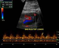
DISCLOSURE TEST YOUR WAVEFORM IQ. Partial volume artifact. 86 yo female with right arm swelling, picc line. AVF on left? Dx?
|
|
|
- Evelyn Stevens
- 5 years ago
- Views:
Transcription


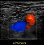
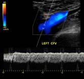
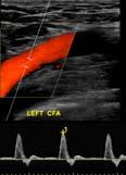
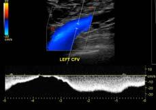

1 Deborah Rubens University of Rochester Rochester, NY DISCLOSURE Neither I nor my immediate family have a financial relationship with a commercial organization that may have a direct or indirect interest in the content of this presentation. Imaging Sciences 86 yo female with right arm swelling, picc line. Partial volume artifact PSA incidentally discovered Thrombin x 3 AVF on left? Dx? 51 yo diabetic with syncope DX? 1


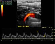
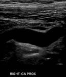
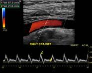
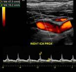
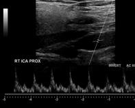
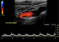
2 44 yo male with right monocular vision loss Gray scale image shows extensive plaque in the ICA Spectral tracing of the CCA has a normal waveform with a PSV of 51cm/sec Prox ICA normal waveform shape, velocity is 58cm/sec ICA/CCA ratio is 1.1 Distal ICA waveform is tardus parvus, velocity 14cm/sec ICA/CCA ratio (14/51) =0.3 How do you report this? Severe Stenosis Velocity increases as diameter reduction increases from 50 to 90% If stenosis is near complete, velocity will drop Diagnosis: Severe Stenosis 72 year old male with abnormal mental status Abn grayscale Low velocity flow Tardus parvus waveform 2


3 Q: What to do next? Left ICA: Small, blunt percussive waveforms Low PSV No diastolic flow Right ICA: Normal waveforms and PSV No Significant Stenosis: exam is complete Severe Stenosis of the LT CCA with low velocities: do a CTA Thorax and Neck More distal stenosis or occlusion: evaluate intracranial circulation Dx: Intracranial ICA occlusion 45 year old male with a bruit A knocking waveform is characterized by diminished peak systolic velocity and absent or even reversed diastolic flow Knocking waveforms occur proximal to an occlusion or severe stenosis Normal flow void indicating patent artery This patient had an occluded left internal carotid artery, resulting in an acute stroke Abnormal signal indicating occlusion of the artery Carotid dissection Highly variable, irregular waveform Abnormally low peak systolic velocity Bidirectional flow throughout the cardiac cycle This patient suffered a traumatic aortic dissection with extension into both common carotid arteries The waveform tends to be bizarre, highly irregular, and dampened Waveforms will vary according to the extent of the dissection and relative sizes of true and false lumen 3


4 63 year old male with abnormal physical exam Low velocity waveforms in the left and right carotid systems, involving CCA, ICA and vertebral arteries Waveform has 2 peaks, one in systole and the second in early diastole Diastolic flow reversal at end diastole (arrow) Intra-aortic balloon pump Mid systolic retraction due to pressure drop Inflation of balloon causes 2nd peak of forward flow during early diastole Flow reversal at end of diastole corresponds to deflation of balloon 23 year old male with chest pain and bruit c/o L Scoutt Sharp systolic upstroke and rapid deceleration Reversed early and enddiastolic flow, indicating a widened pulse pressure Markedly elevated peak systolic velocity Aortic regurgitation The water hammer pulse may be seen with severe aortic regurgitation This waveform is characterized by: sharp systolic upstroke with steep drop in late systole reversal of flow in diastole markedly elevated peak systolic velocity 4


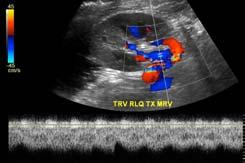
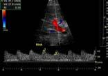
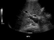
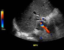
5 49 year old male undergoing heart transplant evaluation Marked tardus parvus waveforms in all vessels Low peak systolic velocity No flow below the baseline Findings should be reproducible in the femoral arteries Left ventricular assist device Blood is diverted from the left ventricular apex and propelled by a pump through a graft into the aorta Most devices in current use provide continuous, forward flow throughout the cardiac cycle Abnormality? Left portal vein antegrade, right portal vein retrograde. Portal Hypertension with Collateral 48 y.o. male, GI bleed Transverse midline 5


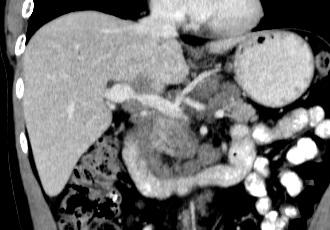
 Tumor")


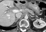
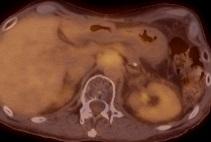
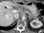
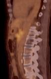
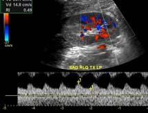
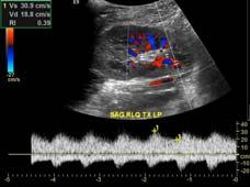
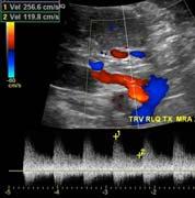
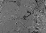
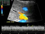
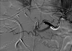
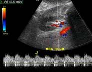
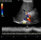
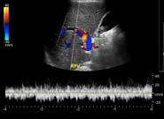
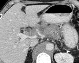
6 Diagnosis: Tumor Thrombus Tumor Thrombus Hepatic artery Portal vein with thrombus Tumor artery Often has visible flow within thrombus Don t mistake residual flow in nonocclusive clot (portal vein wave form) with true vascularized clot (hepatic artery wave form) Tumor within portal vein CT shows occlusion of the portal vein with tumor. Histology demonstrated tumor throughout the liver and in the portal vein CHF 45 yo M with weight loss and diarrhea, r/o mes. ischemia Increased RA pressure reflected in TIPs, IVC, PV PV pulsatility of greater than 50% Severe cases have systolic flow reversal 45 yo M with weight loss and diarrhea, r/o mes. ischemia Dx Criteria: PSV Celiac >200cm/sec SMA > 275 cm/sec IMA > 200cm/sec Ratio > 2.5/3:1 ACR guidelines 2012, Pellerito JUM 2009 Diagnosis? Low resistance in CA Prominent IMA Color bleed over stenosis Dx pancreatic CA invading mesentery 6


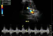
7 Low RI s- HAS? False Positive: Intraparenchymal Shunting Reperfusion Injury First 48 hours post op High velocity diastolic flow May also have tardus parvus waveform Reverses on its own Post Operative Day 0 Post Operative Day 1 Assymptomatic Patient Low RI s Dx: HA-PV Fistula HA- PV Fistula Common Complication Most Asymptomatic Seen in up to 50% of patients within 1 week post biopsy Less than 10% persist beyond a week Most close spontaneously Saad WEA, Lin E, Ormanoski M, Darcy MD, Rubens DJ. Noninvasive Imaging of Liver Transplant Complications.Tech Vasc Interventional Rad 10: , PORTAL VEIN THROMBOSIS? HEMATOMA COMPRESSES MPV No detectable flow in the MPV with reversed flow in R and LPVs? Doesn t make sense. What to do? Get a CT. 7


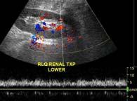


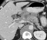
8 FOLLOWING DECOMPRESSION S/P liver Txp, poor fxn Post Coiling Immediate 24 hours later Arterial Steal Syndromes? Consequence of excess PV flow Dx by arteriography-low flow into allograft US shows elevated RI s with low velocity or loss of HA flow signal, loss of diastolic flow most specific Angiography demonstrates increased flow to the splenic a. or gastroduodenal a. Rx includes splenic artery embolization Garcia-Criado AJR 2009;193(1): Sanyal and Shah JUM 2009:28: Day 0 45 yo M, Rising LFT s p/bmt Very poor flow in the parenchyma with tardus parvus waveforms 3:1 ratio Dx? Arterial compression syndrome Following revision returns to normal VOD? Portal Hypertension? PV Stenosis? Thrombosis? 8


9 Bilateral Leg Swelling CHF Waveform extremely pulsatile Large extent above baseline (towards feet) Conclusions THANK YOU Waveform shape and velocity both contribute to diagnoses Technique important to insure proper waveform Waveforms reflect local disease as well as proximal, distal or systemic abnormalities Waveform expertise makes you a Doppler star!! 9
Ultrasound Imaging of The Posterior Circulation
 Ultrasound Imaging of The Posterior Circulation Michigan Sonographers Society 2 Nd Annual Fall Vascular Conference Larry N. Raber RDMS-RVT Clinical Manager General Ultrasound/Neurovascular Laboratory Cleveland
What effects will proximal or distal disease have on an waveform?
 Spectral Doppler Interpretation Director Director of of Ultrasound Ultrasound Education Education & & Quality Quality Assurance Assurance Baylor Baylor College College of of Medicine Medicine Division
Deborah J. Rubens, MD Ultrasound of Liver Transplants 7/8/2015. Indications DISCLOSURES. Ultrasound of Liver Transplants OBJECTIVES
 Ultrasound of Liver Transplants None DISCLOSURES Deborah J. Rubens, M.D. Professor of Imaging Sciences, Oncology and Biomedical Engineering University of Rochester Medical Center Associate Director, Center
Carotid Artery Doppler
 Carotid Artery Doppler Patient Position supine or semisupine head slightly hyper extended rotated 45 away from the side being examined. Higher frequency linear transducers (7 MHz) Vessels should be imaged
Visceral Vascular Ultrasound. Joel Thompson, MD, MPH Borg & Ide Imaging
 Visceral Vascular Ultrasound Joel Thompson, MD, MPH Borg & Ide Imaging Objectives: Review major abdominal vascular structures Identify normal peak systolic velocity (PSV) for major abdominal arteries.
What effects will proximal or distal disease have on a waveform?
 Spectral Doppler Interpretation Director of Ultrasound Education & Quality Assurance Baylor College of Medicine Division of Maternal-Fetal Medicine Maternal Fetal Center Imaging Manager Texas Children
NON-ATHEROSCLEROTIC PATHOLOGY OF THE CAROTID ARTERIES
 NON-ATHEROSCLEROTIC PATHOLOGY OF THE CAROTID ARTERIES Leslie M. Scoutt, MD, FACR Professor of Diagnostic Radiology & Surgery Vice Chair, Dept of Radiology & Biomedical Imaging Chief, Ultrasound Section
Carotid US: More than just a chart on the wall
 Carotid US: More than just a chart on the wall Leslie M. Scoutt, MD, FACR Professor of Diagnostic Radiology & Surgery Vice Chair, Dept of Radiology & Biomedical Imaging Chief, Ultrasound Section Medical
Carotid Abnormalities Coils, Kinks and Tortuosity David Lorelli M.D., RVT, FACS Michigan Vascular Association Conference Saturday, October 20, 2012
 Carotid Abnormalities Coils, Kinks and Tortuosity David Lorelli M.D., RVT, FACS Michigan Vascular Association Conference Saturday, October 20, 2012 Page 1 Table of Contents Carotid Anatomy Carotid Duplex
Vascular Imaging in the Pediatric Abdomen. Jonathan Swanson, MD
 Vascular Imaging in the Pediatric Abdomen Jonathan Swanson, MD Goals and Objectives To understand the imaging approach, appearance, and clinical manifestations of the common pediatric abdominal vascular
HD Scanning: Velocities and Volume Flow
 HD Scanning: Velocities and Volume Flow Non-Invasive Lab Symposium West Orange, NJ April 27, 2018 Volume Flow Cindy Sturt, MD, FACS, RVT 500,000 Americans on dialysis 20-25% annual mortality 65% 5 year
GUNDERSEN/LUTHERAN ULTRASOUND DEPARTMENT POLICY AND PROCEDURE MANUAL
 GUNDERSEN/LUTHERAN ULTRASOUND DEPARTMENT POLICY AND PROCEDURE MANUAL SUBJECT: Carotid Duplex Ultrasound SECTION: Vascular Ultrasound ORIGINATOR: Deborah L. Richert, BSVT, RDMS, RVT DATE: October 15, 2015
No financial or commercial relationships to disclose
 Deanna New, RVT No financial or commercial relationships to disclose IAC REQUIREMENTS: The main duty of a sonographer is to make the physician or radiologists job easier by capturing images and doing
Case Report 1. CTA head. (c) Tele3D Advantage, LLC
 Tele3D Advantage, LLC") Case Report 1 CTA head 1 History 82 YEAR OLD woman with signs and symptoms of increased intra cranial pressure in setting of SAH. CT Brain was performed followed by CT Angiography of head. 2 CT brain Extensive
Case Report 1 CTA head 1 History 82 YEAR OLD woman with signs and symptoms of increased intra cranial pressure in setting of SAH. CT Brain was performed followed by CT Angiography of head. 2 CT brain Extensive
US of Renovascular Hypertension. Jonathan R. Dillman, MD, MSc Associate Professor Director, Thoracoabdominal Imaging
 US of Renovascular Hypertension Jonathan R. Dillman, MD, MSc Associate Professor Director, Thoracoabdominal Imaging Disclosures Nothing Relevant Unrelated grant funding Siemens US Toshiba US Objectives
Abdominal Doppler Mastering the next level of vascular anatomy in the belly. Cindy A. Owen, RDMS, RVT
 Abdominal Doppler Mastering the next level of vascular anatomy in the belly Cindy A. Owen, RDMS, RVT Introduction Abdominal Doppler is a tough exam Success is dependent on: Patient body habitus Patient
Carotid Duplex: Beyond Stenosis Ido Weinberg, MD Vascular Medicine Massachusetts General Hospital Assistant Professor of Medicine Harvard Medical
 Carotid Duplex: Beyond Stenosis Ido Weinberg, MD Vascular Medicine Massachusetts General Hospital Assistant Professor of Medicine Harvard Medical School Boston, Massachusetts Disclosures I do not have
Beyond Stenosis Severity: Top 5 Important Duplex Characteristics to Identify in a Patient with Carotid Disease
 Beyond Stenosis Severity: Top 5 Important Duplex Characteristics to Identify in a Patient with Carotid Disease Jan M. Sloves RVT, RCS, FASE Technical Director New York Cardiovascular Associates Disclosures
ASDIN 7th Annual Scientific Meeting DISCLOSURES TECHNICAL CONSIDERATIONS TECHNICAL CONSIDERATIONS UTILITY OF ULTRASOUND IN EVALUATING ACCESS
 DISCLOSURES UTILITY OF ULTRASOUND IN EVALUATING ACCESS DYSFUNCTION None Vandana Dua Niyyar, MD Assistant Professor of Medicine, Division of Nephrology, Emory University UTILITY OF ULTRASOUND IN ACCESS
Vascular Surgery Cases: Detours. Brian F. Stull, RDMS, RVT UNC REX Healthcare Vascular Specialists
 Vascular Surgery Cases: Detours Brian F. Stull, RDMS, RVT UNC REX Healthcare Vascular Specialists Brian.Stull@Unchealth.unc.edu Objectives Anatomy of a bypass graft Where does it connect, where does it
Carotid Ultrasound: Improving Ultrasound
 Carotid Ultrasound: Improving Ultrasound Edward I. Bluth, M.D., F.A.C.R. Chairman Emeritus, Department of Radiology, Ochsner Clinic Foundation, New Orleans, Louisiana Professor, Ochsner Clinical School,
Doppler Ultrasonography of the Liver: What Every General Radiologist Should Know
 Doppler Ultrasonography of the Liver: What Every General Radiologist Should Know Poster No.: C-1658 Congress: ECR 2014 Type: Authors: Keywords: DOI: Educational Exhibit T. González de la Huebra Labrador,
Copy Here VASCULAR ACCESS COMPLICATIONS TYPICAL ARTERIAL COMPLICATION PROTOCOL ARTERIAL EXAMINATION: NORMAL FINDINGS X X
 VASCULAR ACCESS COMPLICATIONS X X Copy Here Natalia Fendrikova Mahlay, MD, RPVI ARTERIAL EXAMINATION: NORMAL FINDINGS TYPICAL ARTERIAL COMPLICATION PROTOCOL Indications Prior vascular access Pulsatile
Pre-and Post Procedure Non-Invasive Evaluation of the Patient with Carotid Disease
 Pre-and Post Procedure Non-Invasive Evaluation of the Patient with Carotid Disease Michael R. Jaff, D.O., F.A.C.P., F.A.C.C. Assistant Professor of Medicine Harvard Medical School Director, Vascular Medicine
What Do We Know? Disclosure Statement: 3/11/2015. Deep abdominal imaging
 Marsha M. Neumyer, BS, RVT, FSVU, FSDMS, FAIUM International Director Vascular Diagnostic Educational Services Vascular Resource Associates Harrisburg, PA Disclosure Statement: CME Calendar QR Code Marsha
Carotid Imaging IT S ABOUT MORE THAN JUST OBTAINING THE IMAGES
 Carotid Imaging IT S ABOUT MORE THAN JUST OBTAINING THE IMAGES No financial or commercial relationships to disclose Carotid artery disease: Stroke is one of the most serious causes of mortality and morbidity
11 TH ANNUAL VASCULAR NONINVASIVE TESTING SYMPOSIUM NOVEMBER 10, 2018
 11 TH ANNUAL VASCULAR NONINVASIVE TESTING SYMPOSIUM NOVEMBER 10, 2018 RENAL ARTERY DISEASE AND RENOVASCULAR HYPERTENSION 1 WHAT IS RENOVASCULAR HYPERTENSION? https://my.clevelandclinic.org/health/diseases/16459-renovascular-hypertension
Vascular Sonography Examination
 Vascular Sonography Examination The purpose of The American Registry of Radiologic Technologists (ARRT ) Vascular Sonography Examination is to assess the knowledge and cognitive skills underlying the intelligent
1. Long images of aorta (prox, mid, and dist) with AP measurements. 2. Trans images of aorta (prox, mid, and dist) with R/L measurements.
 with AP measurements. 2. Trans images of aorta (prox, mid, and dist) with R/L measurements.") Aorta 1. Long images of aorta (prox, mid, and dist) with AP measurements. 2. Trans images of aorta (prox, mid, and dist) with R/L measurements. 3. Long images of R/L common iliac arteries with AP measurements.
Aorta 1. Long images of aorta (prox, mid, and dist) with AP measurements. 2. Trans images of aorta (prox, mid, and dist) with R/L measurements. 3. Long images of R/L common iliac arteries with AP measurements.
CT Imaging of Blunt and Penetrating Vascular Trauma DENNIS FOLEY MEDICAL COLLEGE WISCONSIN
 CT Imaging of Blunt and Penetrating Vascular Trauma DENNIS FOLEY MEDICAL COLLEGE WISCONSIN THORACO ABDOMINAL TRAUMA 0 10 20 30 40 50 60 5 cc/sec 30 secs 1.25 mm/ 55 mm Z1.375 2.5 mm/ 55 mm Z 1.375 Grade
SCAI Fall Fellows Course Subclavian/Innominate Case Presentation
 SCAI Fall Fellows Course 2012 Subclavian/Innominate Case Presentation Daniel J. McCormick DO, FACC, FSCAI Director, Cardiovascular Interventional Therapy Pennsylvania Hospital University of Pennsylvania
Abdominal Doppler. Cases of Where, Why, and How
 Abdominal Doppler Cases of Where, Why, and How Jill D. Trotter, BS, RT(R), RDMS, RVT Director, Diagnostic Medical Sonography Program Vanderbilt University/Vanderbilt Medical Center Nashville, Tennessee
Visceral aneurysm. Diagnosis and Interventions M.NEDEVSKA
 Visceral aneurysm Diagnosis and Interventions M.NEDEVSKA History 1953 De Bakeyand Cooley Visceral aneurysm VAAs rare, reported incidence of 0.01 to 0.2% on routine autopsies. Clinically important Potentially
The Role of US in Chronic Mesenteric Ischemia. Sagar S. Gandhi, MD Vascular Health Alliance Greenville Health System
 The Role of US in Chronic Mesenteric Ischemia Sagar S. Gandhi, MD Vascular Health Alliance Greenville Health System No Disclosures Mesenteric Ischemia Anatomy Presentation Diagnostic tools Treatment Celiac
Radiologic Evaluation of Peripheral Arterial Disease
 January 2003 Radiologic Evaluation of Peripheral Arterial Disease Grace Tye, Harvard Medical School Year III Patient D.M. CC: 44 y/o male with pain in his buttocks Occurs after walking 2 blocks. Pain is
Non-invasive examination
 Non-invasive examination Segmental pressure and Ankle-Brachial Index (ABI) The segmental blood pressure (SBP) examination is a simple, noninvasive method for diagnosing and localizing arterial disease.
STRUCTURED EDUCATION REQUIREMENTS IMPLEMENTATION DATE: JULY 1, 2016
 STRUCTURED EDUCATION REQUIREMENTS Vascular Sonography The purpose of structured education is to provide the opportunity for individuals to develop mastery of discipline-specific knowledge that, when coupled
Duplex Ultrasound of the Renal Arteries. Duplex Ultrasound. In the Beginning
 Duplex Ultrasound of the Renal Arteries DIMENSIONS IN HEART AND VASCULAR CARE 2013 PENN STATE HEART AND VASCULAR INSTITUTE ROBERT G. ATNIP MD PROFESSOR OF SURGERY AND RADIOLOGY Duplex Ultrasound Developed
Doppler Ultrasound Findings in the Hepatic Artery Shortly After Liver Transplantation
 Gastrointestinal Imaging Pictorial Essay García-riado et al. Hepatic rtery fter Liver Transplantation Gastrointestinal Imaging Pictorial Essay FOUS ON: Ángeles García-riado 1 Rosa Gilabert nnalisa erzigotti
Fibromuscular Dysplasia. Miranda Forrest Baker College
 Fibromuscular Dysplasia Miranda Forrest Baker College Overview Case Study Patient Information Exam Images Findings FMD Types Signs and Symptoms Treatment Case Study Patient Information Female 57 years
How to Determine Tolerance for Branch Vessel Coverage
 How to Determine Tolerance for Branch Vessel Coverage Venita Chandra, MD Clinical Assistant Professor of Surgery Division of Stanford Medical School, Stanford, CA PNEC May 25 th, 2017 DISCLOSURES Venita
Indications: following: embolization. artery that has diseases 5. The evaluation. of suspected. such entities. a cold hand. biopsy
 Peripheral Arterial Ultrasound Protocol Using Color and Spectral Doppler Reviewed by: Mark Yuhasz, MD Last Review Date: January 2015 Contact: (866) 761 4200, Option 1 Indications: The indications for peripheral
Image Formation (10) 2 Evaluation and Selection of Representative Images (10)
 2 Evaluation and Selection of Representative Images (10)") STRUCTURED SELF ASSESSMENT CONTENT SPECIFICATIONS SSA LAUNCH DATE: JANUARY 1, 2018 Vascular Sonography The purpose of continuing qualifications requirements (CQR) is to assist registered technologists
STRUCTURED SELF ASSESSMENT CONTENT SPECIFICATIONS SSA LAUNCH DATE: JANUARY 1, 2018 Vascular Sonography The purpose of continuing qualifications requirements (CQR) is to assist registered technologists
Michigan Vascular Association 2012 Conference Case studies from Massachusetts General Hospital. Our lab
 Michigan Vascular Association 2012 Conference Case studies from Massachusetts General Hospital Kathleen Hannon, MS, RVT, RDMS khannon@partners.org Our lab #1 in the nation! 15 full time RVT s 11 MD s IAC
Introduction History Preceded by Arterial Doppler and ABI Indications
 Elise Brady, RVT, RDMS Introduction History Preceded by Arterial Doppler and ABI Indications 1) Abnormal ABI (within 2weeks of duplex) 2) Abnormal Doppler waveforms 3) Claudication 4) History of PVD 5)
Case 8038 Renal allograft complicated with renal artery stenosis
 Case 8038 Renal allograft complicated with renal artery stenosis Santiago I, Canelas A, Pinto AP Section: Cardiovascular Published: 2009, Nov. 30 Patient: 61 year(s), male Clinical History A 61-year-old
Deb Coghlan AMS (Vascular and General ) Brisbane, Australia
 Brisbane, Australia") Deb Coghlan AMS (Vascular and General ) Brisbane, Australia ANEURYSMAL DIISEASE The infrarenal aorta enlarges with age, and is the commonest site for arterial aneurysms. An aneurysm is a permanent focal
Deb Coghlan AMS (Vascular and General ) Brisbane, Australia ANEURYSMAL DIISEASE The infrarenal aorta enlarges with age, and is the commonest site for arterial aneurysms. An aneurysm is a permanent focal
Protokollanhang zur SPACE-2-Studie Neurology Quality Standards
 Protokollanhang zur SPACE-2-Studie Neurology Quality Standards 1. General remarks In contrast to SPACE-1, the neurological center participating in the SPACE-2 trial will also be involved in the treatment
Disclosure Statement:
 Marsha M. Neumyer, BS, RVT, FSVU, FSDMS, FAIUM International Director Vascular Diagnostic Educational Services Vascular Resource Associates Harrisburg, PA Disclosure Statement: CME Calendar QR Code Marsha
Section 6 Intra Aortic Balloon Pump
 Section 6 Intra Aortic Balloon Pump The Intra Aortic Balloon Pump (IABP) The balloon is synthetic and is made for single use only. It is threaded into the aorta, usually via a femoral approach. The balloon
Neurovascular Ultrasound Course
 Neurovascular Ultrasound Course William M. McKinney (6/6/30-10/24/03) Father of Neurosonology Founder, Neurosonology Course, WFUSM Welcome to Winston-Salem, NC, Wake Forest School of Medicine, and the
Imaging Strategy For Claudication
 Who are the Debators? Imaging Strategy For Claudication Duplex Ultrasound Alone is Adequate to Select Patients for Endovascular Intervention - Pro: Dennis Bandyk MD No Disclosures PRO - Vascular Surgeon
Goals. Access flow and renal artery stenosis evaluation by Doppler ultrasound. Reimbursement. WHY use of Doppler Ultrasound
 Access flow and renal artery stenosis evaluation by Doppler ultrasound Adina Voiculescu, MD Interventional Nephrology Brigham and Women s Hospital Boston Instructor at Harvard Medical School Understand
Challenges. 1. Sizing. 2. Proximal landing zone 3. Distal landing zone 4. Access vessels 5. Spinal cord ischemia 6. Endoleak
 Disclosure I have the following potential conflicts of interest to report: Consulting: Medtronic, Gore Employment in industry Stockholder of a healthcare company Owner of a healthcare company Other(s)
Doppler ultrasound as noninvasive diagnosis of peripheral arterial disease
 Doppler ultrasound as noninvasive diagnosis of peripheral arterial disease Poster No.: C-0246 Congress: ECR 2012 Type: Scientific Exhibit Authors: C. Ballester Valles, F. Aparici-Robles; Valencia/ES Keywords:
Subclavian artery Stenting
 Subclavian artery Stenting Etiology Atherosclerosis Takayasu s arteritis Fibromuscular dysplasia Giant Cell Arteritis Radiation-induced Vascular Injury Thoracic Outlet Syndrome Neurofibromatosis Incidence
Endovascular Treatment of Hepatic Artery Stenosis following Liver Transplantation
 Endovascular Treatment of Hepatic Artery Stenosis following Liver Transplantation W. Charles Sternbergh, III, MD Professor and Chief Vascular and Endovascular Surgery Vice Chair for Research Department
Doppler Waveform Parvus and Tardus A Sign of Proximal Flow Obstruction
 Doppler Waveform Parvus and Tardus A Sign of Proximal Flow Obstruction Pesho S. Kotval, MD, PhD The Doppler linear flow velocity versus time spectrum obtained in an arterial flow system in which there
TRANSCRANIAL DOPPLER ULTRASOUND INTRODUCTION TO TCD INTERPRETATION
 TRANSCRANIAL DOPPLER ULTRASOUND INTRODUCTION TO TCD INTERPRETATION ---Rune Aaslid First TCD Publication 1982 WHAT IS TCD? Uses 2 MHz pulsed Doppler ultrasound Passes through cranial windows Provides information
MINIMALLY INVASIVE MANAGEMENT OF RENOVASCULAR COMPLICATIONS AFTER RENAL GRAFT TRANSPLANTATION
 MINIMALLY INVASIVE MANAGEMENT OF RENOVASCULAR COMPLICATIONS AFTER RENAL GRAFT TRANSPLANTATION Gortes, Francisco Javier B.S; Salsamendi, Jason Thomas M.D LEARNING OBJECTIVES Educate physicians on the prompt
Multidetector CTA for Diagnosing Blunt Cerebrovascular Injuries
 Multidetector CTA for Diagnosing Blunt Cerebrovascular Injuries 4 th Nordic Trauma Course 2006 Stuart E. Mirvis, M.D., FACR Department of Diagnostic Radiology and Nuclear Medicine, University of Maryland
The production of murmurs is due to 3 main factors:
 Heart murmurs The production of murmurs is due to 3 main factors: high blood flow rate through normal or abnormal orifices forward flow through a narrowed or irregular orifice into a dilated vessel or
Antegrade and retrograde flow of carotid
 Antegrade and retrograde flow of carotid The ECA waveform is high resistance and may have retrograde flow in diastole.. They should always demonstrate antegrade flow (toward the brain) and be. external
MESENTERIC ISCHEMIA. Phillip J Bendick, PhD
 MESENTERIC ISCHEMIA Phillip J Bendick, PhD Arterial Celiac - Hepatic - Splenic Superior Mesenteric Artery Inferior Mesenteric Artery Venous Mesenteric system Porto - hepatic system Inferior Vena Cava Acute
PERPHERAL ARTERY ANEURYSM. By Pooja Sharma and Susanna Sebastianpillai
 PERPHERAL ARTERY ANEURYSM By Pooja Sharma and Susanna Sebastianpillai Defintions True Aneurysm Involves all three layers of the vessel. Have two basic shapes; Fusiform = symmetric widening of the vessels
The role for contrast-enhanced ultrasonography outside of focal liver lesions
 The role for contrast-enhanced ultrasonography outside of focal liver lesions Paul S. Sidhu King s College Hospital, London, UK Introduction Contrast-enhanced ultrasonography (US) of focal liver lesions
Recommendations for Follow-up After Vascular Surgery Arterial Procedures SVS Practice Guidelines
 Recommendations for Follow-up After Vascular Surgery Arterial Procedures 2018 SVS Practice Guidelines vsweb.org/svsguidelines About the guidelines Published in the July 2018 issue of Journal of Vascular
Treatment of choice for end stage renal disease Imaging to establish baseline and diagnosis of potential complications Review common surgical
 Treatment of choice for end stage renal disease Imaging to establish baseline and diagnosis of potential complications Review common surgical techniques Review normal appearance Discuss US diagnosis of
Radiologic Importance of a High- Resistive Vertebral Artery Doppler Waveform on Carotid Duplex Ultrasonography
 CME Article Radiologic Importance of a High- Resistive Vertebral Artery Doppler Waveform on Carotid Duplex Ultrasonography Esther S. H. Kim, MD, MPH, Megan Thompson, Kristine M. Nacion, BA, Carmel Celestin,
Postoperative AV Fistula Evaluation. Postoperative examination protocol. Postoperative AVF Protocol. Hemodialysis Access Surveillance
 Hemodialysis Access Surveillance Postoperative AV Fistula Evaluation Failure of maturation Stenosis Perigraft mass/fluid collection Joseph L. Mills, Sr., M.D. Professor of Surgery Chief, Division of Vascular
TCD IN THE NICU, PICU AND OTHER APPLICATIONS. Dorothy Bulas M.D. Professor of Pediatrics & Radiology Children s National Washington D.C.
 TCD IN THE NICU, PICU AND OTHER APPLICATIONS Dorothy Bulas M.D. Professor of Pediatrics & Radiology Children s National Washington D.C. Objectives Recognize normal and abnormal cranial blood flow patterns
Pitfalls in the evaluation of carotid artery stenosis. Serge Kownator «Centre Cardiologique et Vasculaire» Thionville, Fr
 Pitfalls in the evaluation of carotid artery stenosis Serge Kownator «Centre Cardiologique et Vasculaire» Thionville, Fr Disclosure Statement of Financial Interest I currently have, or have had over the
Mesenteric/Splanchnic Artery Duplex Imaging
 VASCULAR TECHNOLOGY PROFESSIONAL PERFORMANCE GUIDELINES Mesenteric/Splanchnic Artery Duplex Imaging This Guideline was prepared by members of the Society for Vascular Ultrasound (SVU) as a template to
An Overview of Post-EVAR Endoleaks: Imaging Findings and Management. Ravi Shergill BSc Sean A. Kennedy MD Mark O. Baerlocher MD FRCPC
 An Overview of Post-EVAR Endoleaks: Imaging Findings and Management Ravi Shergill BSc Sean A. Kennedy MD Mark O. Baerlocher MD FRCPC Disclosure Slide Mark O. Baerlocher: Current: Consultant for Boston
Carotid Artery Stenting
 Carotid Artery Stenting JESSICA MITCHELL, ACNP CENTRAL ILLINOIS RADIOLOGICAL ASSOCIATES External Carotid Artery (ECA) can easily be identified from Internal Carotid Artery (ICA) by noticing the branches.
Measure #195 (NQF 0507): Radiology: Stenosis Measurement in Carotid Imaging Reports National Quality Strategy Domain: Effective Clinical Care
: Radiology: Stenosis Measurement in Carotid Imaging Reports National Quality Strategy Domain: Effective Clinical Care") Measure #195 (NQF 0507): Radiology: Stenosis Measurement in Carotid Imaging Reports National Quality Strategy Domain: Effective Clinical Care 2017 OPTIONS FOR INDIVIDUAL MEASURES: CLAIMS ONLY MEASURE TYPE:
Measure #195 (NQF 0507): Radiology: Stenosis Measurement in Carotid Imaging Reports National Quality Strategy Domain: Effective Clinical Care 2017 OPTIONS FOR INDIVIDUAL MEASURES: CLAIMS ONLY MEASURE TYPE:
Carotid Stenosis (carotid artery disease)
") 1 Carotid Stenosis (carotid artery disease) Overview Carotid stenosis is a narrowing of the carotid arteries, the two major arteries that carry oxygenrich blood from the heart to the brain. Also called
1 Carotid Stenosis (carotid artery disease) Overview Carotid stenosis is a narrowing of the carotid arteries, the two major arteries that carry oxygenrich blood from the heart to the brain. Also called
Endovascular treatment of acquired arteriovenous fistula with severe hemodynamic effects: a case report
 Endovascular treatment of acquired arteriovenous fistula with severe hemodynamic effects: a case report The Leipzig Interventional Course, January 24 27, 2017 El Samman K., Šedivý P., Šnajdrová A., Přindišová
Quality ID #195 (NQF 0507): Radiology: Stenosis Measurement in Carotid Imaging Reports National Quality Strategy Domain: Effective Clinical Care
: Radiology: Stenosis Measurement in Carotid Imaging Reports National Quality Strategy Domain: Effective Clinical Care") Quality ID #195 (NQF 0507): Radiology: Stenosis Measurement in Carotid Imaging Reports National Quality Strategy Domain: Effective Clinical Care 2018 OPTIONS FOR INDIVIDUAL MEASURES: REGISTRY ONLY MEASURE
Quality ID #195 (NQF 0507): Radiology: Stenosis Measurement in Carotid Imaging Reports National Quality Strategy Domain: Effective Clinical Care 2018 OPTIONS FOR INDIVIDUAL MEASURES: REGISTRY ONLY MEASURE
US of Neurovascular Occlusive Disease: Interpretive Pearls and Pitfalls 1
 EDUCATION EXHIBIT 1165 US of Neurovascular Occlusive Disease: Interpretive Pearls and Pitfalls 1 CME FEATURE See accompanying test at http:// www.rsna.org /education /rg_cme.html LEARNING OBJECTIVES FOR
Vascular Technology Examination Content Outline
 Vascular Technology Examination Content Outline (Outline Summary) # Domain Subdomain Percentage 1 Normal Anatomy, Perfusion, and Function Evaluate normal anatomy, perfusion, function 2 Pathology, Perfusion,
Transjugular Intrahepatic Portosystemic Shunt Reduction for Management of Recurrent Hepatic Encephalopathy
 CLINICAL IMAGES Ochsner Journal 17:311 316, 2017 Ó Academic Division of Ochsner Clinic Foundation Transjugular Intrahepatic Portosystemic Shunt Reduction for Management of Recurrent Hepatic Encephalopathy
Vertebral Artery Doppler Waveform Changes Indicating Subclavian Steal Physiology
 Downloaded from www.ajronline.org by 7.44.00.5 on 0/09/8 from IP address 7.44.00.5. Copyright RRS. For personal use only; all rights reserved Mark. Kliewer arbara S. Hertzberg David H. Kim James D. owie
General Imaging. Imaging modalities. Incremental CT. Multislice CT Multislice CT [ MDCT ]
![General Imaging. Imaging modalities. Incremental CT. Multislice CT Multislice CT [ MDCT ]](/thumbs/76/74079340.jpg "General Imaging. Imaging modalities. Incremental CT. Multislice CT Multislice CT [ MDCT ]") General Imaging Imaging modalities Conventional X-rays Ultrasonography [ US ] Computed tomography [ CT ] Radionuclide imaging Magnetic resonance imaging [ MRI ] Angiography conventional, CT,MRI Interventional
General Imaging Imaging modalities Conventional X-rays Ultrasonography [ US ] Computed tomography [ CT ] Radionuclide imaging Magnetic resonance imaging [ MRI ] Angiography conventional, CT,MRI Interventional
Introduction to the Native Arteriovenous Fistula: A primer for medical students and radiology residents
 Introduction to the Native Arteriovenous Fistula: A primer for medical students and radiology residents Jesus Contreras, D.O. PGY-4 John Yasmer, D.O. Department of Radiology No Disclosures Objectives Introduce
Internal Carotid Artery Dissection
 May 2011 Internal Carotid Artery Dissection Carolyn April, HMS IV Agenda Presentation of a clinical case Discussion of the clinical features of ICA dissection Discussion of the imaging modalities used
 CLINICAL FEATURES THAT SUPPORT ATHEROSCLEROTIC STROKE 1. cerebral cortical impairment (aphasia, neglect, restricted motor involvement, etc.) or brain stem or cerebellar dysfunction 2. lacunar clinical
Adult Echocardiography Examination Content Outline
 Adult Echocardiography Examination Content Outline (Outline Summary) # Domain Subdomain Percentage 1 2 3 4 5 Anatomy and Physiology Pathology Clinical Care and Safety Measurement Techniques, Maneuvers,
Job Task Analysis for ARDMS Abdomen Data Collected: June 30, 2011
 Job Task Analysis for ARDMS Abdomen Data Collected: June 30, 2011 Reported: Analysis Summary for: Abdomen Examination Survey Dates 06/13/2011-06/26/2011 Invited Respondents 6,000 Surveys with Demographics
How to manage the left subclavian and left vertebral artery during TEVAR
 How to manage the left subclavian and left vertebral artery during TEVAR Jürg Schmidli Chief of Vascular Surgery Inselspital Hamburg 2017 Dept Cardiovascular Surgery, Bern, Switzerland Disclosure No Disclosures
RENAL AND MESENTERIC ARTERY STENTS Are There Standard Velocity Criteria for Restenosis?
 RENAL AND MESENTERIC ARTERY STENTS Are There Standard Velocity Criteria for Restenosis? R. Eugene Zierler, M.D. The D. E. Strandness, Jr. Vascular Laboratory University of Washington Medical Center Division
Echo Emergencies. Outline. Michael H. Picard, MD Massachusetts General Hospital Harvard Medical School No disclosures
 Echo Emergencies Michael H. Picard, MD Massachusetts General Hospital Harvard Medical School No disclosures Outline Common emergency / on call scenarios Tamponade Pulmonary embolism/rv strain Cardiogenic
Lower Extremity Arterial Doppler
 Lower Extremity Arterial Doppler 1. Spectral Doppler waveform should be taken in distal aorta and common iliac arteries. 2. R/L common femoral artery (CFA) color Doppler with velocity and B-mode. 3. R/L
Physician s Vascular Interpretation Examination Content Outline
 Physician s Vascular Interpretation Examination Content Outline (Outline Summary) # Domain Subdomain Percentage 1 2 3 4 5 6 Cerebrovascular Abdominal Peripheral Arterial - Duplex Imaging Peripheral Arterial
TO CATCH A THIEF: IMAGING OF SUBCLAVIAN STEAL
 October 2013 TO CATCH A THIEF: IMAGING OF SUBCLAVIAN STEAL Sumir Pandit, Harvard Medical School, Year III 1 AGENDA Introduction to our patient A.B. Anatomy review of aorta and branches CT imaging of our
Competency Title: Caring for a patient with an arterial line
 Appendix 2 Competency Title: Caring for a patient with an arterial line Aims and objectives Following completion of this competency document the practitioner will be able to: Discuss the indications for
Hemodynamically significant subclavian artery stenosis
 REVIEW ARTICLE Duplex Ultrasonography of Vertebral and Subclavian Arteries Vijay G. Kalaria, MD, FACC, FSCAI, Sony Jacob, MD, William Irwin, RVT, and Robert M. Schainfeld, DO, Indianapolis, Indiana, and
(Department of Radiology, Beylikdüzü State Hospital, İstanbul, Turkey) Corresponding Author: Dr. Mete Özdikici
 Corresponding Author: Dr. Mete Özdikici") Quest Journals Journal of Medical and Dental Science Research Volume 5~ Issue 6 (2018) pp: 61-65 ISSN(Online) : 2394-076X ISSN (Print):2394-0751 www.questjournals.org Research Paper Quantitative Measurements
Quest Journals Journal of Medical and Dental Science Research Volume 5~ Issue 6 (2018) pp: 61-65 ISSN(Online) : 2394-076X ISSN (Print):2394-0751 www.questjournals.org Research Paper Quantitative Measurements
NCVH. Ultrasongraphy: State of the Art Vein Forum 2015 A Multidisciplinary Approach to Otptimizing Venous Circulation From Wounds to WOW
 Ultrasongraphy: State of the Art 2015 NCVH New Cardiovascular Horizons Vein Forum 2015 A Multidisciplinary Approach to Otptimizing Venous Circulation From Wounds to WOW Anil K. Chagarlamudi, M.D. Cardiovascular
Interventional Radiology in Trauma. Vikash Prasad, MD, FRCPC Vascular and Interventional Radiology The Moncton Hospital
 Interventional Radiology in Trauma Vikash Prasad, MD, FRCPC Vascular and Interventional Radiology The Moncton Hospital Disclosures None relevant to this presentation Shareholder Johnson and Johnson Goal