Lezione 3 Tronchi Sovraortici
|
|
|
- Julianna Richard
- 6 years ago
- Views:
Transcription

1 CORSO DI CERTIFICAZIONE DI COMPETENZA in ECOGRAFIA VASCOLARE GENERALE Lezione 3 Tronchi Sovraortici Settore formazione : Direttore: Paolo G. Pino Marco Campana, Antonella Moreo, Fausto Rigo, Ketty Savino
2 Overview Epidemiology Clinical trials Anatomy Hemodynamic flow patterns Assessment of stenosis Classification of disease Video case presentation Dipartimento Cardio-Toracico - Università di Pisa
3 Causes of Stroke: Cerebrovascular Atherosclerosis 85% Infarction 15% Hemorrhage - Intracerebral - Subarachnoid 60% Cerebrovascular atherosclerosis - Stenosis (flow reduction) - Ulcerated plaque (artery-to-artery emboli) 20% Penetrating artery disease (lacunes) 15% Cardiogenic embolism Cardiac and Thoracic Department - University of Pisa 5% Other, unusual causes


4 ARIC Atherosclerosis Risk in Communities patients - Age: range years - Carotid plaque prevalence: 34% - Gender: male > female R. Li; Stroke patients (Northern Italy) - Age: range years -Carotid plaque prevalence: 25,4% M - 26,4% F Carotid plaque prevalence: 30,8% M - 21,9% F Patients: years P. Prati; Stroke 1992
5 ASYMPTOMATIC CAROTID LESIONS IN 755 SUBJECTS IN RELATION WITH A SINGLE NORMALS FINDINGS (%) IMT (%) A.C.P. (%) RF R.F R.F * 14.7 * HYPERTENS * * NIDDM * 16.7 * HYPER-C * FAMILY H. CVD HYPER-TG SMOKING * 0 OBESITY IMT+ACP (%) * 65.5 * 41.7 * 26.6 * * 4.7 Novo S.: Adv. Vasc. Pathol. 1997; Excerpta Medica, Amsterdam 1997: 36-46
6 Prevalence and outcome of asymptomatic carotid stenosis: a population-based ultrasonographic study 500 volunteers 200 men and 300 female Without signs and symptoms of cerebrovascular disease aged years The prevalence of asymptomatic carotid stenosis of 50% or greater was 6.4%. Severe carotid stenosis was detected in only 0.4% of the subjects examined. European Journal of Neurology
7
8 Intrinsic risk of embologenic carotid stenosis 13.1% major ipsilateral stroke in 2 years in NASCET (about 6.5%/yr) - 13%/yr for ANY ipsilateral stroke NASCET 16.2% ANY ipsilateral stroke in 3 years in ECST (about 5.4%/yr) MOST strokes are within first year of signal event. Cardiac and Thoracic Department - University of Pisa
 Lower")
 3) High mortality in patients with")

9 EPIDEMIOLOGICAL DATA: Conclusion 1) High prevalence of Carotid Artery plaques 2) Lower prevalence of carotid Stenosis 50% - (6.4%) 3) High mortality in patients with symptomatic carotid stenosis 4) High risk for embologenic carotid stenosis Cardiac and Thoracic Department - University of Pisa
10 THERAPEUTIC OPTIONS IN PATIENTS WITH CAROTID ARTERY DISEASE 1. Medical Therapy 2. Surgical Therapy 3. Interventional Therapy Cardiac and Thoracic Department - University of Pisa
11 THERAPEUTIC OPTIONS IN PATIENTS WITH CAROTID ARTERY DISEASE NASCET (North American Symptomatic Carotid Endarterectomy Trial) - Carotid Stenosis > 70% - TEA stroke risk of 17% at 2 years ECST (European Carotid Surgery Trials) - Symptomatic patients with stenosis > 70% - Stroke incidence 14,9% (Surgery) (Medical Treatment) ACAS (Asymptomatic Carotid Atherosclerosis Study) - Carotid Stenosis > 60% - TEA stroke risk of 5.8% at 5 years Cardiac and Thoracic Department - University of Pisa
12 Intrinsic risk of embologenic carotid stenosis 13.1% major ipsilateral stroke in 2 years in NASCET (about 6.5%/yr) - 13%/yr for ANY ipsilateral stroke NASCET 16.2% ANY ipsilateral stroke in 3 years in ECST (about 5.4%/yr) MOST strokes are within first year of signal event Risk is directly proportional to degree of stenosis!! - UNSTABLE PLAQUE IS VULNERABLE TO FLOW! Cardiac and Thoracic Department - University of Pisa
13 Overview Epidemiology Clinical trials Anatomy Hemodynamic flow patterns Assessment of stenosis Classification of disease Video case presentation Dipartimento Cardio-Toracico - Università di Pisa
14 Anatomy Common Carotid Artery Right CCA originates from brachiocephalic Artery Left CCA originates from Aorta Difficult to image origin of left CCA
 into internal and external carotid arteries")

15 CAROTID BULB CCA bifurcates at upper level of thyroid cartilage (C4) into internal and external carotid arteries Fusiform dilatation Unique flow patterns Exact location varies among patients Usually symmetric within a patient Anatomy
 Bifurcates into middle and anterior cerebral arteries in the circle of")
16 Anatomy INTERNAL CAROTID ARTERY Supplies the brain No branches in the neck Inside the skull makes S shaped curve called the carotid siphon First major branch is the ophthalmic artery (eye) Bifurcates into middle and anterior cerebral arteries in the circle of Willis
17 Anatomy EXTERNAL CAROTID ARTERY Supplies the muscle and skin of the face and scalp Lies anterior and medial to ICA Numerous branches in the neck May provide collateral supply to brain with ICA stenosis/occlusion


18 Anatomy
19 Overview Epidemiology Clinical trials Anatomy Hemodynamic flow patterns Assessment of stenosis Classification of disease Video case presentation Dipartimento Cardio-Toracico - Università di Pisa
20 Flow Patterns Types of flow Laminar Disturbed Turbulent Major determinants Geometry of the arterial system Resistance to flow offered by the tissue/organ
21 Flow Patterns : Geometry Helical flow and boundary flow separation occur at bifurcations

 End diastolic flow")
22 Flow Patterns : Resistance LOW RESISTANCE TO FLOW Brain, liver, kidney End diastolic flow always above zero HIGH RESISTANCE TO FLOW Muscles, intestine Resistance dependant on metabolic activity Triphasic response (forward-reverseforward flow) End diastolic flow near zero
23 Flow Patterns : Normal Carotid Artery ICA : brain is low resistance organ ECA : facial muscles show variable resistance CCA : supplies brain (70-80%) and facial muscles


24 Normal Flow Patterns


25 Normal Flow Pattern : CCA Mid CCA 2cm from bifurcation EDV above zero baseline Distal CCA Just proximal to bifurcation CCA begins to dilate Peak systolic velocity decreases EDV increases Normal flow disturbances


26 Normal Flow Pattern : ECA Supplies face and scalp, not a source of cerebral emboli Characteristics of high resistance vessel Peak systolic velocity higher than ICA Rapid systolic acceleration Prominent dichrotic wave in late systole or early diastole EDV near zero baseline


27 Normal Flow Pattern : Carotid Bulb and Proximal ICA Complex flow patterns due to dilatation and bifurcation Anatomic changes create boundary layer separation and helical flows Unidirectional flow near flow divider Transient flow reversal near outer wall When present, these confirm normal carotid bulb


28 Normal Flow Pattern : Mid ICA Low resistance flow pattern EDV above zero baseline Peak systolic velocity may be higher than the proximal ICA due to smaller diameter Normal flow disturbances from carotid bulb may travel into the mid ICA
29 Vessel Identification : CCA Proximal CCA Courses toward skin surface then curves parallel Curve creates normal flow disturbances Origins often difficult to image (esp left CCA) Mid CCA Straightens out and runs parallel to skin Lies approximately 1-2 cm deep Atherosclerosis rarely involves this segment Distal CCA Continuation of vessel to the bifurcation Dilates into carotid bulb Disease frequently occurs here


30 Vessel Identification : ECA vs ICA Anatomic location ICA : lateral and posterior ECA : medial and anterior Size of Vessel ICA usually larger diameter at the origin


31 Vessel Identification : ECA vs ICA Location of the bulb and associated flow pattern Bulb most commonly involves distal CCA and proximal ICA Flow reversal due to boundary layer separation usually extends into ICA Not useful if bulb includes only distal CCA or both ICA and ECA

32 Vessel Identification : ECA vs ICA Presence of branches ECA has eight branches ICA has no branches in ICA has no branches in the neck
33 Vessel Identification : ECA vs ICA Hemodynamic flow patterns ICA : supplies low resistance bed EDV above zero Lower systolic velocity Rounded systolic peak ECA: supplies high resistance bed EDV at or near zero Higher systolic velocity Flow reversal in late systole Direct comparison of waveforms is essential
34 Vessel Identification : ECA vs ICA Color Doppler Hemodynamic differences created by differing vascular resistance can be seen in color Doppler ICA : continuous color throughout cardiac cycle ECA : color decrease significantly in diastole Superficial temporal tap Superficial temporal artery is a branch off ECA than can be palpated in front of the ear Gentle compressions result in oscillations in spectral waveforms of ECA Spectral waveforms of ICA not affected


35 B-mode Imaging :Normal Vessel Wall Double line First line represents interface blood and intimal surface Second line represents interface between intima-media complex and the adventitia As intima thickens, lines become more widely separated As plaque develops, lines become indistinguishable due to deposition of echogenic material


36 EXAMINATION PROTOCOL anteriore laterale Asse corto Postero-laterale posteriore
37 Overview Epidemiology Clinical trials Anatomy Hemodynamic flow patterns Assessment of stenosis Classification of disease Video case presentation Dipartimento Cardio-Toracico - Università di Pisa


38
39 Atherosclerosis of the Carotid Bulb Disease of branch points and bifurcations Some arterial segments spared Distal to subclavian origin Internal mammary arteries Carotid bifurcation Carotid bulb is only prominent dilatation in an arterial segment Disease starts in the posterolateral aspect of bulb Progresses circumferentially to involve entire bulb Rarely extends beyond the distal margin of the bulb
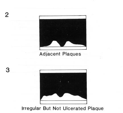
40 STUDIO ECOGRAFICO DELLA MALATTIA CAROTIDEA STUDIO DELLA PLACCA SEDE ED ESTENSIONE (asse longitudinale e trasversale) A. MODULO ECOCOLOR MORFOLOGIA CARATTERIZZAZIONE PLACCA BASSA-MEDIA ALTA ECOGENICITA OMOGENEA/ETEROGENEA SUPERFICIE (REGOLARE, IRREGOLARE, ULCERATA MODULO DOPPLER SEVERITA STENOSI ( GRADING ) Dip. di Cardio-Toracico - Università di Pisa


41


42


43

44


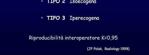
45 PLACCA CAROTIDEA MORFOLOGIA ECOGENICITA ASPETTO TIPO SUPERFICIE


46


47


48

49


50


51


52 PLACCA CAROTIDEA
53


54


55


56


57


58


59


60

61


62


63

64


65


66


67


68

69


70


71


72 VALUTAZIONE CORRETTA DEL GRADO DI STENOSI CAROTIDEA Asse lungo Asse corto Percentuale di stenosi lineare Analisi del Segnale Doppler % stenosi planimetrica

73
74 Assessment of Severity Minimal to moderate disease (<50%) B-mode imaging is highly sensitive and reliable for identifying stenoses of less than 50% Plaques, wall, and lumens usually clearly defined Severe disease (>50%) Doppler spectral waveform most accurate method for defining severity of disease Typical complex plaques seen in high grade lesions are usually not well visualized


75 Nature of flow patterns distal to stenosis
76 VALUTAZIONE DOPPLER DELLA STENOSI CAROTIDEA 90% Dip. di Cardio-Toracico - Università di Pisa


77 Over the years, many hybrid and different stenosis measurement algorithms arose!!! Each with their own set cut points of validity, most appropriately based on ROC curve analysis. The problem was, there were just so many different velocity criteria being used that there was substantial difficulty getting comparisons and standardization in readings between facilities.


78 Cardiac and Thoracic Department - University of Pisa


79 DOPPLER EVALUATION OF CAROTID STENOSIS developed recommendations for diagnosis and stratification of ICA stenosis derived from analysis of numerous studies. Cardiac and Thoracic Department - University of Pisa
80 DOPPLER EVALUATION OF CAROTID STENOSIS internal validation is encouraged when possible. Important points from the Consensus Panel each laboratory should have a single set of diagnostic criteria that is applied uniformly.


81 DOPPLER EVALUATION OF CAROTID STENOSIS: The consensus recommended that the NASCET method of carotid stenosis measurement should be employed...when angiography is used to correlate the US findings.
82 Additional Doppler parameters other than ICA PSV The ICA/CCA ratio becomes important to use in situations where the ICA PSV may not be by itself, representative of the extent of ICA disease due to: Tandem ICA stenosis Elevated CCA velocities or stenosis Contralateral high-grade ICA stenosis Discrepancy between visual assessment of plaque and ICAPSV Significantly altered Cardiac output states (high or low) Cardiac and Thoracic Department - University of Pisa
83 Additional Doppler parameters other than ICA PSV End diastolic velocity Really starts to increase at higher levels of stenosis, usually at or above 75% diameter luminal narrowing. Cardiac and Thoracic Department - University of Pisa
84 Accuracy predicated upon Proper angle of insonation = always less than or equal to 60 degrees. Proper placement of the Doppler cursor in the center of the flow stream with the cursor in the direction of flow. Cardiac and Thoracic Department - University of Pisa


85

86


87


88


89


90


91


92


93

94


95


96


97


98 Normal carotid bifurcation Cardiac and Thoracic Department - University of Pisa
99 Criteria for Less Than 50% Diameter Reduction Normal Peak systolic velocity less than 125 cm/s No visible plaque on B-mode Boundary layer separation in carotid bulb 1% to 15% diameter reduction Peak systolic velocity less than 125 cm/s No boundary separation in carotid bulb Minimal spectral broadening 16% to 49% diameter reduction Peak systolic velocity less than 125 cm/s Marked spectral broadening with no systolic window


100 50-69 % Stenosis ө=48 Cardiac and Thoracic Department - University of Pisa


101 Velocities of > 70 % stenosis (subset > 80 % ) Cardiac and Thoracic Department - University of Pisa


102 Detection of Carotid Artery Stenosis by in vivo Duplex Ultrasound Correlation with Planimetric Measurements of the Corresponding Post-mortem Specimens Stroke 2002 Steno-occlusive occlusive range from 8.5% to 100% Cardiac and Thoracic Department - University of Pisa
103 Cardiac and Thoracic Department - University of Pisa
104 Why do we report stenosis in a Range? Significant overlap of velocities at the same level of stenosis exists this is the reason that stenoses are reported in ranges and not specific %. The higher the velocity, the tighter the stenosis up to about 97 %, then the velocities start to decrease (string sign) trickle of flow. Cardiac and Thoracic Department - University of Pisa
105 ABILITY TO USE DUPLEX US TO QUANTIFY ICA STENOSES: FACT OR FINCTION? Radiology 2000 PSV AND ICA/CCA ratio INCREASED WITH STENOSIS LEVEL p<.01 that the average Doppler velocity rises in direct proportion to the degree of stenosis as determined with angiography. there are very wide ranges of Doppler values around those means, which makes it impossible to classify lesions into gradations as narrow as 10%.


106 ABILITY TO USE DUPLEX US TO QUANTIFY ICA STENOSES: FACT OR FINCTION? Radiology 2000 US is most accurate when lesions are classified as being above or below a single level, such as 60% stenosis or 70% stenosis
107
108
109
110


111


112


113


114


115


116


117


118

119


120


121


122


123


124


125
126 CORSO DI CERTIFICAZIONE DI COMPETENZA in ECOGRAFIA VASCOLARE GENERALE Lezione 3 Tronchi Sovraortici Settore formazione : Direttore: Paolo G. Pino Marco Campana, Antonella Moreo, Fausto Rigo, Ketty Savino
Carotid Abnormalities Coils, Kinks and Tortuosity David Lorelli M.D., RVT, FACS Michigan Vascular Association Conference Saturday, October 20, 2012
 Carotid Abnormalities Coils, Kinks and Tortuosity David Lorelli M.D., RVT, FACS Michigan Vascular Association Conference Saturday, October 20, 2012 Page 1 Table of Contents Carotid Anatomy Carotid Duplex
Disclosure Statement:
 Marsha M. Neumyer, BS, RVT, FSVU, FSDMS, FAIUM International Director Vascular Diagnostic Educational Services Vascular Resource Associates Harrisburg, PA Disclosure Statement: CME Calendar QR Code Marsha
GUNDERSEN/LUTHERAN ULTRASOUND DEPARTMENT POLICY AND PROCEDURE MANUAL
 GUNDERSEN/LUTHERAN ULTRASOUND DEPARTMENT POLICY AND PROCEDURE MANUAL SUBJECT: Carotid Duplex Ultrasound SECTION: Vascular Ultrasound ORIGINATOR: Deborah L. Richert, BSVT, RDMS, RVT DATE: October 15, 2015
Carotid Artery Doppler
 Carotid Artery Doppler Patient Position supine or semisupine head slightly hyper extended rotated 45 away from the side being examined. Higher frequency linear transducers (7 MHz) Vessels should be imaged
Carotid Ultrasound: Improving Ultrasound
 Carotid Ultrasound: Improving Ultrasound Edward I. Bluth, M.D., F.A.C.R. Chairman Emeritus, Department of Radiology, Ochsner Clinic Foundation, New Orleans, Louisiana Professor, Ochsner Clinical School,
Protokollanhang zur SPACE-2-Studie Neurology Quality Standards
 Protokollanhang zur SPACE-2-Studie Neurology Quality Standards 1. General remarks In contrast to SPACE-1, the neurological center participating in the SPACE-2 trial will also be involved in the treatment
Pre-and Post Procedure Non-Invasive Evaluation of the Patient with Carotid Disease
 Pre-and Post Procedure Non-Invasive Evaluation of the Patient with Carotid Disease Michael R. Jaff, D.O., F.A.C.P., F.A.C.C. Assistant Professor of Medicine Harvard Medical School Director, Vascular Medicine
Beyond Stenosis Severity: Top 5 Important Duplex Characteristics to Identify in a Patient with Carotid Disease
 Beyond Stenosis Severity: Top 5 Important Duplex Characteristics to Identify in a Patient with Carotid Disease Jan M. Sloves RVT, RCS, FASE Technical Director New York Cardiovascular Associates Disclosures
(Department of Radiology, Beylikdüzü State Hospital, İstanbul, Turkey) Corresponding Author: Dr. Mete Özdikici
 Corresponding Author: Dr. Mete Özdikici") Quest Journals Journal of Medical and Dental Science Research Volume 5~ Issue 6 (2018) pp: 61-65 ISSN(Online) : 2394-076X ISSN (Print):2394-0751 www.questjournals.org Research Paper Quantitative Measurements
Quest Journals Journal of Medical and Dental Science Research Volume 5~ Issue 6 (2018) pp: 61-65 ISSN(Online) : 2394-076X ISSN (Print):2394-0751 www.questjournals.org Research Paper Quantitative Measurements
Measure #195 (NQF 0507): Radiology: Stenosis Measurement in Carotid Imaging Reports National Quality Strategy Domain: Effective Clinical Care
: Radiology: Stenosis Measurement in Carotid Imaging Reports National Quality Strategy Domain: Effective Clinical Care") Measure #195 (NQF 0507): Radiology: Stenosis Measurement in Carotid Imaging Reports National Quality Strategy Domain: Effective Clinical Care 2017 OPTIONS FOR INDIVIDUAL MEASURES: CLAIMS ONLY MEASURE TYPE:
Measure #195 (NQF 0507): Radiology: Stenosis Measurement in Carotid Imaging Reports National Quality Strategy Domain: Effective Clinical Care 2017 OPTIONS FOR INDIVIDUAL MEASURES: CLAIMS ONLY MEASURE TYPE:
TRANSCRANIAL DOPPLER ULTRASOUND INTRODUCTION TO TCD INTERPRETATION
 TRANSCRANIAL DOPPLER ULTRASOUND INTRODUCTION TO TCD INTERPRETATION ---Rune Aaslid First TCD Publication 1982 WHAT IS TCD? Uses 2 MHz pulsed Doppler ultrasound Passes through cranial windows Provides information
Quality ID #195 (NQF 0507): Radiology: Stenosis Measurement in Carotid Imaging Reports National Quality Strategy Domain: Effective Clinical Care
: Radiology: Stenosis Measurement in Carotid Imaging Reports National Quality Strategy Domain: Effective Clinical Care") Quality ID #195 (NQF 0507): Radiology: Stenosis Measurement in Carotid Imaging Reports National Quality Strategy Domain: Effective Clinical Care 2018 OPTIONS FOR INDIVIDUAL MEASURES: REGISTRY ONLY MEASURE
Quality ID #195 (NQF 0507): Radiology: Stenosis Measurement in Carotid Imaging Reports National Quality Strategy Domain: Effective Clinical Care 2018 OPTIONS FOR INDIVIDUAL MEASURES: REGISTRY ONLY MEASURE
8/20/18. The Doppler Effect. Objectives. What is the Doppler Effect. Doppler principles. Spectral Waveform. Image recognition. Vascular Ultrasound
 Vascular Ultrasound: Physics and Haemodynamics Objectives Doppler principles Spectral Waveform Key factors Haemodynamics: Stenosis Waveforms Image recognition Vascular Ultrasound: A flawed paradigm What
Neurovascular Ultrasound Course
 Neurovascular Ultrasound Course William M. McKinney (6/6/30-10/24/03) Father of Neurosonology Founder, Neurosonology Course, WFUSM Welcome to Winston-Salem, NC, Wake Forest School of Medicine, and the
Fibromuscular Dysplasia. Miranda Forrest Baker College
 Fibromuscular Dysplasia Miranda Forrest Baker College Overview Case Study Patient Information Exam Images Findings FMD Types Signs and Symptoms Treatment Case Study Patient Information Female 57 years
NON-ATHEROSCLEROTIC PATHOLOGY OF THE CAROTID ARTERIES
 NON-ATHEROSCLEROTIC PATHOLOGY OF THE CAROTID ARTERIES Leslie M. Scoutt, MD, FACR Professor of Diagnostic Radiology & Surgery Vice Chair, Dept of Radiology & Biomedical Imaging Chief, Ultrasound Section
Policies and Statements D16. Intracranial Cerebrovascular Ultrasound
 Policies and Statements D16 Intracranial Cerebrovascular Ultrasound SECTION 1: INSTRUMENTATION Policies and Statements D16 Intracranial Cerebrovascular Ultrasound May 2006 (Reaffirmed July 2007) Essential
What Do We Know? Disclosure Statement: 3/11/2015. Deep abdominal imaging
 Marsha M. Neumyer, BS, RVT, FSVU, FSDMS, FAIUM International Director Vascular Diagnostic Educational Services Vascular Resource Associates Harrisburg, PA Disclosure Statement: CME Calendar QR Code Marsha
TO CATCH A THIEF: IMAGING OF SUBCLAVIAN STEAL
 October 2013 TO CATCH A THIEF: IMAGING OF SUBCLAVIAN STEAL Sumir Pandit, Harvard Medical School, Year III 1 AGENDA Introduction to our patient A.B. Anatomy review of aorta and branches CT imaging of our
Treatment Considerations for Carotid Artery Stenosis. Danielle Zielinski, RN, MSN, ACNP Rush University Neurosurgery
 Treatment Considerations for Carotid Artery Stenosis Danielle Zielinski, RN, MSN, ACNP Rush University Neurosurgery 4.29.2016 There is no actual or potential conflict of interest in regards to this presentation
Neuro Quiz 29 Transcranial Doppler Monitoring
 Verghese Cherian, MD, FFARCSI Penn State Hershey Medical Center, Hershey Quiz Team Shobana Rajan, M.D Suneeta Gollapudy, M.D Angele Marie Theard, M.D Neuro Quiz 29 Transcranial Doppler Monitoring This
Transcranial Doppler (Basic Step) Dae-il Chang, M.D., Sung Sang Yoon, M.D. Department of Neurology, College of Medicine, Kyunghee university
 Dae-il Chang, M.D., Sung Sang Yoon, M.D. Department of Neurology, College of Medicine, Kyunghee university") Transcranial Doppler (Basic Step) Dae-il Chang, M.D., Sung Sang Yoon, M.D. Department of Neurology, College of Medicine, Kyunghee university Principles of Doppler Ultrasonography Major target Speed & direction
Transcranial Doppler (Basic Step) Dae-il Chang, M.D., Sung Sang Yoon, M.D. Department of Neurology, College of Medicine, Kyunghee university Principles of Doppler Ultrasonography Major target Speed & direction
Recommendations for documentation of neurosonographic examinations
 Recommendations for documentation of neurosonographic examinations The documentation of ultrasound examinations is subject to a dynamic development particularly as regards newer applications. The present
Carotid Imaging IT S ABOUT MORE THAN JUST OBTAINING THE IMAGES
 Carotid Imaging IT S ABOUT MORE THAN JUST OBTAINING THE IMAGES No financial or commercial relationships to disclose Carotid artery disease: Stroke is one of the most serious causes of mortality and morbidity
Carotid Imaging. Dr Andrew Farrall. Consultant Neuroradiologist
 20121123 SSCA http://www.neuroimage.co.uk/network Andrew Farrall Carotid Imaging Dr Andrew Farrall Consultant Neuroradiologist SFC Brain Imaging Research Centre (www.sbirc.ed.ac.uk), SINAPSE Collaboration
Screening for asymptomatic internal artery stenosis: Duplex criteria for discriminating 60% to 99% stenosis
 Screening for asymptomatic internal artery stenosis: Duplex criteria for discriminating 60% to 99% stenosis carotid Gregory L. Moneta, MD, James M. Edwards, MD, George Papanicolaou, MD, Thomas Hatsukami,
Essentials of Clinical MR, 2 nd edition. 99. MRA Principles and Carotid MRA
 99. MRA Principles and Carotid MRA As described in Chapter 12, time of flight (TOF) magnetic resonance angiography (MRA) is commonly utilized in the evaluation of the circle of Willis. TOF MRA allows depiction
Duplex Criteria for Determination of 50% or Greater Carotid Stenosis
 Article Duplex Criteria for Determination of 50% or Greater Carotid Stenosis David G. Neschis, MD, Frank J. Lexa, MD, Julia T. Davis, RN, RVT, Jeffrey P. Carpenter, MD, RVT Recently the North American
Vascular Modeling & Hemodynamics Research OUTLINE. CLINICAL MOTIVATION: Stroke. BACKGROUND: Diagnosis
 Biomedical Ultrasonics Research Lab Vascular Modeling & Hemodynamics Research Tamie Poepping Biomedical Ultrasonics Research Laboratory Dept. of Physics & Astronomy The University of Western Ontario UWO:
Pitfalls in the evaluation of carotid artery stenosis. Serge Kownator «Centre Cardiologique et Vasculaire» Thionville, Fr
 Pitfalls in the evaluation of carotid artery stenosis Serge Kownator «Centre Cardiologique et Vasculaire» Thionville, Fr Disclosure Statement of Financial Interest I currently have, or have had over the
HD Scanning: Velocities and Volume Flow
 HD Scanning: Velocities and Volume Flow Non-Invasive Lab Symposium West Orange, NJ April 27, 2018 Volume Flow Cindy Sturt, MD, FACS, RVT 500,000 Americans on dialysis 20-25% annual mortality 65% 5 year
MORTALITY AND MORBIDITY RISK FROM CAROTID ARTERY ATHEROSCLEROSIS. 73 year old NS right-handed male applicant for $1 Million life insurance
 MORTALITY AND MORBIDITY RISK FROM CAROTID ARTERY ATHEROSCLEROSIS October 17, 2012 AAIM Triennial Conference, San Diego Robert Lund, MD What Is The Risk? 73 year old NS right-handed male applicant for $1
MORTALITY AND MORBIDITY RISK FROM CAROTID ARTERY ATHEROSCLEROSIS. 73 year old NS right-handed male applicant for $1 Million Life Insurance
 MORTALITY AND MORBIDITY RISK FROM CAROTID ARTERY ATHEROSCLEROSIS October 17, 2012 AAIM Triennial Conference, San Diego Robert Lund, MD What Is The Risk? 73 year old NS right-handed male applicant for $1
What effects will proximal or distal disease have on an waveform?
 Spectral Doppler Interpretation Director Director of of Ultrasound Ultrasound Education Education & & Quality Quality Assurance Assurance Baylor Baylor College College of of Medicine Medicine Division
Ultrasound Imaging of The Posterior Circulation
 Ultrasound Imaging of The Posterior Circulation Michigan Sonographers Society 2 Nd Annual Fall Vascular Conference Larry N. Raber RDMS-RVT Clinical Manager General Ultrasound/Neurovascular Laboratory Cleveland
Carotid Artery Stenting
 Carotid Artery Stenting JESSICA MITCHELL, ACNP CENTRAL ILLINOIS RADIOLOGICAL ASSOCIATES External Carotid Artery (ECA) can easily be identified from Internal Carotid Artery (ICA) by noticing the branches.
Non-invasive examination
 Non-invasive examination Segmental pressure and Ankle-Brachial Index (ABI) The segmental blood pressure (SBP) examination is a simple, noninvasive method for diagnosing and localizing arterial disease.
Diagnosis of Middle Cerebral Artery Occlusion with Transcranial Color-Coded Real-Time Sonography
 Diagnosis of Middle Cerebral Artery Occlusion with Transcranial Color-Coded Real-Time Sonography Kazumi Kimura, Yoichiro Hashimoto, Teruyuki Hirano, Makoto Uchino, and Masayuki Ando PURPOSE: To determine
Contemporary Carotid Imaging and Approach to Treatment: Course Notes Thursday, June 22, 2017 David M. Pelz, MD, FRCPC
 CNSF Meeting, Victoria, BC. June 2017 Contemporary Carotid Imaging and Approach to Treatment: Course Notes Thursday, June 22, 2017 David M. Pelz, MD, FRCPC A. Objectives 1. To understand the current imaging
Carotid Artery Disease and What s Pertinent JOSEPH A PAULISIN DO
 Carotid Artery Disease and What s Pertinent JOSEPH A PAULISIN DO Goal of treatment of carotid disease Identify those at risk of developing symptoms Prevent patients at risk from developing symptoms Prevent
Carotid intima media thickness as an usefull tool in predicting cerebrovaskular events
 Carotid intima media thickness as an usefull tool in predicting cerebrovaskular events Poster No.: C-0005 Congress: ECR 2015 Type: Authors: Keywords: DOI: Scientific Exhibit A. Rahimic - Catic; Sarajevo/BA
Carotid US: More than just a chart on the wall
 Carotid US: More than just a chart on the wall Leslie M. Scoutt, MD, FACR Professor of Diagnostic Radiology & Surgery Vice Chair, Dept of Radiology & Biomedical Imaging Chief, Ultrasound Section Medical
Carotid Duplex: Beyond Stenosis Ido Weinberg, MD Vascular Medicine Massachusetts General Hospital Assistant Professor of Medicine Harvard Medical
 Carotid Duplex: Beyond Stenosis Ido Weinberg, MD Vascular Medicine Massachusetts General Hospital Assistant Professor of Medicine Harvard Medical School Boston, Massachusetts Disclosures I do not have
Management of cervicocephalic arterial dissection. Ciro G. Randazzo, MD, MPH Thomas Jefferson University Hospital, Department of Neurosurgery
 Management of cervicocephalic arterial dissection Ciro G. Randazzo, MD, MPH Thomas Jefferson University Hospital, Department of Neurosurgery Definition Disruption of arterial wall, either at level of intima-media
Transducer Selection. Renal Artery Duplex Exam. Renal Scan. Renal Scan Echogenicity. How to Perform an Optimal Renal Artery Doppler Examination
 How to Perform an Optimal Renal Artery Doppler Examination Director of Ultrasound Education & Quality Assurance Baylor College of Medicine Division of Maternal-Fetal Medicine Maternal Fetal Center Imaging
Vascular Portfolio: Carotid Reflection. Paige Fabre
 Vascular Portfolio: Carotid Reflection Paige Fabre 13654584 14 Carotid Reflection For this portfolio I produced three pieces of work; a case study, a PowerPoint of study protocol and a poster of stenosis
Vascular Sonography Examination
 Vascular Sonography Examination The purpose of The American Registry of Radiologic Technologists (ARRT ) Vascular Sonography Examination is to assess the knowledge and cognitive skills underlying the intelligent
Intracranial Cerebrovascular Evaluation Transcranial Doppler (Non-Imaging) and Transcranial Duplex Imaging (TCD-I)
 and Transcranial Duplex Imaging (TCD-I)") VASCULAR TECHNOLOGY PROFESSIONAL PERFORMANCE GUIDELINES Intracranial Cerebrovascular Evaluation Transcranial Doppler (Non-Imaging) and Transcranial Duplex Imaging (TCD-I) This Guideline was prepared by
VASCULAR TECHNOLOGY PROFESSIONAL PERFORMANCE GUIDELINES Intracranial Cerebrovascular Evaluation Transcranial Doppler (Non-Imaging) and Transcranial Duplex Imaging (TCD-I) This Guideline was prepared by
Categorical Course: Update of Doppler US 8 : 00 8 : 20
 159 Categorical Course: Update of Doppler US 8 : 00 8 : 20 160 161 Table 1.Comparison of Recommended Values from Data in the Published Literature* S t u d y Lesion PSV E D V VICA/VCCA S e v e r i t y (
Carotid Artery Revascularization: Current Strategies. Shonda Banegas, D.O. Vascular Surgery Carondelet Heart and Vascular Institute September 6, 2014
 Carotid Artery Revascularization: Current Strategies Shonda Banegas, D.O. Vascular Surgery Carondelet Heart and Vascular Institute September 6, 2014 Disclosures None 1 Stroke in 2014 Stroke kills almost
Indications: following: embolization. artery that has diseases 5. The evaluation. of suspected. such entities. a cold hand. biopsy
 Peripheral Arterial Ultrasound Protocol Using Color and Spectral Doppler Reviewed by: Mark Yuhasz, MD Last Review Date: January 2015 Contact: (866) 761 4200, Option 1 Indications: The indications for peripheral
No financial or commercial relationships to disclose
 Deanna New, RVT No financial or commercial relationships to disclose IAC REQUIREMENTS: The main duty of a sonographer is to make the physician or radiologists job easier by capturing images and doing
STRUCTURED EDUCATION REQUIREMENTS IMPLEMENTATION DATE: JULY 1, 2016
 STRUCTURED EDUCATION REQUIREMENTS Vascular Sonography The purpose of structured education is to provide the opportunity for individuals to develop mastery of discipline-specific knowledge that, when coupled
Carotid Endarterectomy for Symptomatic Complete Occlusion of the Internal Carotid Artery
 2011 65 4 239 245 Carotid Endarterectomy for Symptomatic Complete Occlusion of the Internal Carotid Artery a* a b a a a b 240 65 4 2011 241 9 1 60 10 2 62 17 3 67 2 4 64 7 5 69 5 6 71 1 7 55 13 8 73 1
Carotid Artery Stenting (CAS) Pathophysiology. Technical Considerations. Plaque characteristics: relevant concepts. CAS and CEA
 Pathophysiology. Technical Considerations. Plaque characteristics: relevant concepts. CAS and CEA") Carotid Artery Stenting (CAS) Carotid Artery Stenting for Stroke Risk Reduction Matthew A. Corriere MD, MS, RPVI Assistant Professor of Surgery Department of Vascular and Endovascular Surgery Rationale:
Carotid Artery Stenting (CAS) Carotid Artery Stenting for Stroke Risk Reduction Matthew A. Corriere MD, MS, RPVI Assistant Professor of Surgery Department of Vascular and Endovascular Surgery Rationale:
Intima-Media Thickness
 European Society of Cardiology Stockholm, 30th August 2010 Intima-Media Thickness Integration of arterial assessment into clinical practice Prof Arno Schmidt-Trucksäss, MD Institute of Exercise and Health
The CARENET all-comer trial using the CGuard micronet covered carotid embolic prevention stent
 The CARENET all-comer trial using the CGuard micronet covered carotid embolic prevention stent 6 month data Piotr Musialek, MD DPhil FESC Jagiellonian University Dept. of Cardiac & Vascular Diseases John
Antegrade and retrograde flow of carotid
 Antegrade and retrograde flow of carotid The ECA waveform is high resistance and may have retrograde flow in diastole.. They should always demonstrate antegrade flow (toward the brain) and be. external
Image Formation (10) 2 Evaluation and Selection of Representative Images (10)
 2 Evaluation and Selection of Representative Images (10)") STRUCTURED SELF ASSESSMENT CONTENT SPECIFICATIONS SSA LAUNCH DATE: JANUARY 1, 2018 Vascular Sonography The purpose of continuing qualifications requirements (CQR) is to assist registered technologists
STRUCTURED SELF ASSESSMENT CONTENT SPECIFICATIONS SSA LAUNCH DATE: JANUARY 1, 2018 Vascular Sonography The purpose of continuing qualifications requirements (CQR) is to assist registered technologists
Noninvasive transcranial Doppler ultrasound recording of flow velocity in basal cerebral arteries
 J Neurosurg 57:769-774, 1982 Noninvasive transcranial Doppler ultrasound recording of flow velocity in basal cerebral arteries RUNE AASLID, PH.D., THOMAS-MARC MARKWALDER, M.D., AND HEt,CE NORNES, M.D.
a physician-initiated study investigating the RoadSaver stent in carotid lesions Dr. Michel Bosiers
 The study a physician-initiated study investigating the RoadSaver stent in carotid lesions Dr. Michel Bosiers Conflict of interest have the following potential conflicts of interest to report: Consulting
Physician s Vascular Interpretation Examination Content Outline
 Physician s Vascular Interpretation Examination Content Outline (Outline Summary) # Domain Subdomain Percentage 1 2 3 4 5 6 Cerebrovascular Abdominal Peripheral Arterial - Duplex Imaging Peripheral Arterial
Aortic arch pathology. Cerebral ischemia following carotid artery stenosis.
 Important: -Subclavian Steal Syndrome -Cerebral ischemia Aortic arch pathology. Cerebral ischemia following carotid artery stenosis. Mina Aubeed & Alba Hernández Pinilla Aortic arch pathology Common arch
Carotid arterial ultrasound scan imaging: A direct approach to stenosis measurement
 Carotid arterial ultrasound scan imaging: A direct approach to stenosis measurement Hugh G. Beebe, MD, Sergio X. Salles-Cunha, PhD, Robert P. Scissons, RVT, Steven M. Dosick, MD, Ralph C. Whalen, MD, Steven
DISCLOSURE TEST YOUR WAVEFORM IQ. Partial volume artifact. 86 yo female with right arm swelling, picc line. AVF on left? Dx?
 Deborah Rubens University of Rochester Rochester, NY DISCLOSURE Neither I nor my immediate family have a financial relationship with a commercial organization that may have a direct or indirect interest
Duplex Carotid Sonography Peak Systolic Velocity in Quantifying Internal Carotid Artery Stenosis
 Duplex Carotid Sonography Peak Systolic Velocity in Quantifying Internal Carotid Artery Stenosis Cynthia E Withers, MD", Barbara B Gosink, MD", Alison M Keightley, MD", Giovanna Casola, MD", Arthur A Lee,
Radiologic Importance of a High- Resistive Vertebral Artery Doppler Waveform on Carotid Duplex Ultrasonography
 CME Article Radiologic Importance of a High- Resistive Vertebral Artery Doppler Waveform on Carotid Duplex Ultrasonography Esther S. H. Kim, MD, MPH, Megan Thompson, Kristine M. Nacion, BA, Carmel Celestin,
Color Doppler Imaging Evaluation of Proximal Vertebral Artery Stenosis
 Vascular and Interventional Radiology Original Research Hua et al. Color Doppler Imaging of Proximal Vertebral Artery Stenosis Vascular and Interventional Radiology Original Research Yang Hua 1 Xiu-Feng
Index. interventional.theclinics.com. Note: Page numbers of article titles are in boldface type.
 Index Note: Page numbers of article titles are in boldface type. A ACAS (Asymptomatic Carotid Atherosclerosis Study), 65 66 ACST (Asymptomatic Carotid Surgery Trial), 6 7, 65, 75 Age factors, in carotid
US of Neurovascular Occlusive Disease: Interpretive Pearls and Pitfalls 1
 EDUCATION EXHIBIT 1165 US of Neurovascular Occlusive Disease: Interpretive Pearls and Pitfalls 1 CME FEATURE See accompanying test at http:// www.rsna.org /education /rg_cme.html LEARNING OBJECTIVES FOR
Report For Center Created Gender D.O.B Page 1 Sean Breen HeartSmart IMT plus 3/29/2012 Male 11/26/1973 B C D E
 Report For Center Created Gender D.O.B Page 1 Carotid Assessment A B C D E Good Satisfactory Concern Serious Highest Risk Intima-Media Thickness Additional Findings Plaque Character Percent Stenosis Comments:
Author Query: 1. Please provide yellow arrow in figure as you mentioned in the legend (caption). Peripheral Vascular Ultrasound
. Peripheral Vascular Ultrasound") uthor Query: 1. Please provide yellow arrow in figure 33.16 as you mentioned in the legend (caption). CHPTER 33 Peripheral Vascular Ultrasound Ricardo enenstein, Muhamed Saric Snapshot ¾Ultrasound Diagnosis
uthor Query: 1. Please provide yellow arrow in figure 33.16 as you mentioned in the legend (caption). CHPTER 33 Peripheral Vascular Ultrasound Ricardo enenstein, Muhamed Saric Snapshot ¾Ultrasound Diagnosis
Identification, Prognosis, and Management of Patients with Carotid Artery Near Occlusion
 AJNR Am J Neuroradiol 26:2086 2094, September 2005 Identification, Prognosis, and Management of Patients with Carotid Artery Near Occlusion Allan J. Fox, Michael Eliasziw, Peter M. Rothwell, Matthias H.
Proposed duplex velocity criteria for carotid restenosis following carotid endarterectomy with patch closure
 From the Southern Association for Vascular Surgery Proposed duplex velocity criteria for carotid restenosis following carotid endarterectomy with patch closure Ali F. AbuRahma, MD, a Patrick Stone, MD,
Deb Coghlan AMS (Vascular and General ) Brisbane, Australia
 Brisbane, Australia") Deb Coghlan AMS (Vascular and General ) Brisbane, Australia ANEURYSMAL DIISEASE The infrarenal aorta enlarges with age, and is the commonest site for arterial aneurysms. An aneurysm is a permanent focal
Deb Coghlan AMS (Vascular and General ) Brisbane, Australia ANEURYSMAL DIISEASE The infrarenal aorta enlarges with age, and is the commonest site for arterial aneurysms. An aneurysm is a permanent focal
Vascular disease. Structural evaluation of vascular disease. Goo-Yeong Cho, MD, PhD Seoul National University Bundang Hospital
 Vascular disease. Structural evaluation of vascular disease Goo-Yeong Cho, MD, PhD Seoul National University Bundang Hospital resistance vessels : arteries
Duplex US of the External Carotid Artery
 Acta Radiologica ISSN: 0284-1851 (Print) 1600-0455 (Online) Journal homepage: https://www.tandfonline.com/loi/iard20 Duplex US of the External Carotid Artery M. J. Päivänsalo, T. M. J. Siniluoto, T. A.
What effects will proximal or distal disease have on a waveform?
 Spectral Doppler Interpretation Director of Ultrasound Education & Quality Assurance Baylor College of Medicine Division of Maternal-Fetal Medicine Maternal Fetal Center Imaging Manager Texas Children
EXPLORING ULTRASOUND IMAGES OF THE CAROTID ARTERIES USING NEURAL NETWORK TOOLS
 Proceedings of the 6th International Conference on Mechanics and Materials in Design, Editors: J.F. Silva Gomes & S.A. Meguid, P.Delgada/Azores, 26-30 July 2015 PAPER REF: 5430 EXPLORING ULTRASOUND IMAGES
Duplex Ultrasound of the Renal Arteries. Duplex Ultrasound. In the Beginning
 Duplex Ultrasound of the Renal Arteries DIMENSIONS IN HEART AND VASCULAR CARE 2013 PENN STATE HEART AND VASCULAR INSTITUTE ROBERT G. ATNIP MD PROFESSOR OF SURGERY AND RADIOLOGY Duplex Ultrasound Developed
DICOM Correction Item
 Correction Number DICOM Correction Item CP-449 Log Summary: Type of Modification Correct Value Name of Standard PS 3.16-2004 Rationale for Correction: Clinical applications use arterial and venous branches
Vasculature and Aneurysm Design
 Vasculature and Aneurysm Design Amy Swartz November 18, 2016 Team 23: In-vitro Tube Model System ME450 Section 3 Dr. David Trevas and Dr. Sarah Oman Introduction The primary goal of this capstone project
[(PHY-3a) Initials of MD reviewing films] [(PHY-3b) Initials of 2 nd opinion MD]
![[(PHY-3a) Initials of MD reviewing films] [(PHY-3b) Initials of 2 nd opinion MD]](/thumbs/89/98619893.jpg "[(PHY-3a) Initials of MD reviewing films] [(PHY-3b) Initials of 2 nd opinion MD]") 2015 PHYSICIAN SIGN-OFF (1) STUDY NO (PHY-1) CASE, PER PHYSICIAN REVIEW 1=yes 2=no [strictly meets case definition] (PHY-1a) CASE, IN PHYSICIAN S OPINION 1=yes 2=no (PHY-2) (PHY-3) [based on all available
2015 PHYSICIAN SIGN-OFF (1) STUDY NO (PHY-1) CASE, PER PHYSICIAN REVIEW 1=yes 2=no [strictly meets case definition] (PHY-1a) CASE, IN PHYSICIAN S OPINION 1=yes 2=no (PHY-2) (PHY-3) [based on all available
New Trials in Progress: ACT 1. Jon Matsumura, MD Cannes, France June 28, 2008
 New Trials in Progress: ACT 1 Jon Matsumura, MD Cannes, France June 28, 2008 Faculty Disclosure I disclose the following financial relationships: Consultant, CAS training director, and/or research grants
Carotid artery occlusion: Positive predictive value of duplex sonography compared with arteriography
 Carotid artery occlusion: Positive predictive value of duplex sonography compared with arteriography Jonathan D. Kirsch, MD, Louis R. Wagner, MD, E. Meredith James, MD, J. William Charboneau, MD, Douglas
4/19/2018. St. Cloud Hospital Perinatology Kristin Olson, RDMS, RVT
 St. Cloud Hospital Perinatology Kristin Olson, RDMS, RVT Review Fetal Circulation Provide Indications for Umbilical Artery, Middle Cerebral Artery, and Ductus Venosus Doppler studies. Demonstrate normal
History of revascularization
 History of revascularization Author (year) Kredel, 1942 Woringer& Kunlin, 1963 Donaghy& Yasargil, 1968 Loughheed 1971 Kikuchini & Karasawa1973 Karasawa, 1977 Story, 1978 Sundt, 1982 EC/IC bypass study
ULTRASOUND BLOOD FLOW IMAGING IN CAROTID ARTERIES BEFORE AND AFTER ENDARTERECTOMY
 ULTRASOUND BLOOD FLOW IMAGING IN CAROTID ARTERIES BEFORE AND AFTER ENDARTERECTOMY G. BAMBI 1, F. GUIDI 1, S. RICCI 1, P. TORTOLI 1 M.R. CIRELLI 2, L. PEDRINI 2 1 Electronics & Telecommunications Department,University
Carotid Stenosis 1/24/2019. Review of Primary Studies. NASCET- Moderate stenosis. ACAS (Asymptomatic Carotid Atherosclerosis Study) NASCET
 NASCET") Review of Primary Studies Carotid Stenosis NINDS National Institute of Neurological Disorders and Stroke 2 large studies to determine who would benefit from surgery NASCET North American Symptomatic Carotid
Review of Primary Studies Carotid Stenosis NINDS National Institute of Neurological Disorders and Stroke 2 large studies to determine who would benefit from surgery NASCET North American Symptomatic Carotid
COPYRIGHTED MATERIAL. I How to Perform Ultrasound Tests
 I How to Perform Ultrasound Tests COPYRIGHTED MATERIAL 1 2 1 Principles of Extracranial Ultrasound Examination Andrei V. Alexandrov 1, Alice Robinson-Vaughn 1, Clotilde Balucani 2 & Marsha M. Neumyer 3
Enhancement of Cranial US: Utility of Supplementary Acoustic Windows and Doppler Harriet J. Paltiel, MD
 Enhancement of Cranial US: Utility of Supplementary Acoustic Windows and Doppler Harriet J. Paltiel, MD Boston Children s Hospital Harvard Medical School None Disclosures Conventional US Anterior fontanelle
PTA 106 Unit 1 Lecture 3
 PTA 106 Unit 1 Lecture 3 The Basics Arteries: Carry blood away from the heart toward tissues. They typically have thicker vessels walls to handle increased pressure. Contain internal and external elastic
Doppler ultrasound as noninvasive diagnosis of peripheral arterial disease
 Doppler ultrasound as noninvasive diagnosis of peripheral arterial disease Poster No.: C-0246 Congress: ECR 2012 Type: Scientific Exhibit Authors: C. Ballester Valles, F. Aparici-Robles; Valencia/ES Keywords:
Pulsed Doppler techniques are commonly used
 336 Accurate Noninvasive Method to Diagnose Minor Atherosclerotic Lesions in Carotid Artery Bulb Tiny van Merode, MD, Jan Lodder, MD, Frans A.M. Smeets, Arnold P.G. Hoeks, PhD, and Robert S. Reneman, MD,
Coronary artery disease (CAD) risk factors
 risk factors") Background Coronary artery disease (CAD) risk factors CAD Risk factors Hypertension Insulin resistance /diabetes Dyslipidemia Smoking /Obesity Male gender/ Old age Atherosclerosis Arterial stiffness precedes
Background Coronary artery disease (CAD) risk factors CAD Risk factors Hypertension Insulin resistance /diabetes Dyslipidemia Smoking /Obesity Male gender/ Old age Atherosclerosis Arterial stiffness precedes
Carotid Artery Stenting
 Carotid Artery Stenting Woong Chol Kang M.D. Gil Medical Center, Gachon University of Medicine and Science, Incheon, Korea Carotid Stenosis and Stroke ~25% of stroke is due to carotid disease, the reminder
Evaluation of Carotid Vessels and Vertebral Artery in Stroke Patients with Color Doppler Ultrasound and MR Angiography
 Evaluation of Carotid Vessels and Vertebral Artery in Stroke Patients with Color Doppler Ultrasound and MR Angiography Dr. Pramod Shaha 1, Dr. Vinay Raj R 2, Dr. (Brig) K. Sahoo 3 Abstract: Aim & Objectives:
Reappraisal of Flow Velocity Ratio in Common Carotid Artery to Predict Hemodynamic Change in Carotid Stenosis
 AJNR Am J Neuroradiol 26:957 962, April 2005 Reappraisal of Flow Velocity Ratio in Common Carotid Artery to Predict Hemodynamic Change in Carotid Stenosis Masahiro Kamouchi, Kazuhiro Kishikawa, Yasushi
COLOUR DOPPLER EVALUATION OF DEGREE OF STENOSIS AND PLAQUE MORPHOLOGY IN EXTRACRANIAL CAROTID ARTERIES IN PATIENTS OF STROKE
 wjpmr, 2019, 5(1), 122-128 SJIF Impact Factor: 4.639 Hassan et al. Research Article WORLD JOURNAL OF PHARMACEUTICAL AND MEDICAL RESEARCH ISSN 2455-3301 www.wjpmr.com WJPMR COLOUR DOPPLER EVALUATION OF
Carotid Stenosis (carotid artery disease)
") 1 Carotid Stenosis (carotid artery disease) Overview Carotid stenosis is a narrowing of the carotid arteries, the two major arteries that carry oxygenrich blood from the heart to the brain. Also called
1 Carotid Stenosis (carotid artery disease) Overview Carotid stenosis is a narrowing of the carotid arteries, the two major arteries that carry oxygenrich blood from the heart to the brain. Also called