Non-invasive Imaging of Carotid Artery Atherosclerosis
|
|
|
- Jeffery Ford
- 5 years ago
- Views:
Transcription
1 Non-invasive Imaging of Carotid Artery Atherosclerosis 최연현 성균관의대삼성서울병원영상의학과
2 Noninvasive Techniques US with Doppler CT MRI


3 Ultrasonography
4 Techniques of Carotid US US Anatomy (ICA vs ECA) Gray scale and color Doppler exam Transducer selection (4-7 vs MHz) Artifacts New techniques
5 경동맥협착의 Doppler 진단 Criteria: Parameters ICA 의 peak systolic velocity ICA 의 end diastolic velocity ICA/CCA 의 peak systolic velocity ratio Proximal ICA(stenotic lesion)/distal ICA peak systolic velocity ratio

6 Duplex Criteria: Comparison of Recommended Values Study Severity PSV EDV VICA/VCCA Ratio Moneta > 70% (to 4.2) Carpenter > 70% Neale > 70% NA Hood > 70% NA Browman > 70% NA NA Moneta > 60% Carpenter > 60% Derdeyn > 60% NA NA
7 Duplex Criteria: for 50% and 70% Stenosis. (Robinson et al. AJR,1988) Dm PSV EDV VICA/VCCA Stenosis 50% > 150 >50 >2 70% > 230 >75 >3 Ratio
8 경동맥협착의 Doppler 진단 Criteria CCA 의 peak velocity 는근위부와원위부에서측정할때차이 (23 cm/sec 범위 ) Stenosis > 90-95%: PSV< 25 cm/sec


9
10 Pitfalls Hypertension or hypotension Severe stenosis of distal ICA Stenosis of contralateral ICA Tandem lesions Ulceration of plaque Vessel tortuosity


11 Intimomedial Thickness Measurement
12 US quanfication of carotid atheroma Manual or semi-automated method Thickness, area, and volume measurement of plaques IMT measurement Objectiveness? (operator-dependent)


13
14 Carotid IMT and Brain Infarction Touboul et al. Circulation 2000;102: pts with brain infarction and 463 controls Mean IMT was higher in pts with brain infarction (0.797 mm vs mm, p <0.0001) Odds ratio per SD increase (0.150) = 1.82 (95% conf. Level = 1.54~2.15)


15 Enhanced CT


16 Maximum Intensity Projection


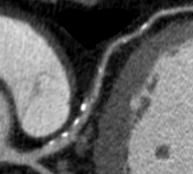
17 Noncalcified Coronary Artery Plaque
18 Plaque Characterization: ICUS vs CT Schroeder Leber Soft HU HU Intermed HU Calcified HU HU Schroeder et al. J Am Coll Cardiol 2001;37:1430 Leber et al. J Am Coll Cardiol. 2004;43:1241

19 Plaque Quantification HanWonhee

20
21 MR Angiography: Techniques Noncontrast: time-of-flight, phase contrast, true FISP (b-ffe, FIESTA) Longer time, motion artifact, flow sensitive Contrast-enhanced MRA High SI, quick, less artifacts, depiction of vessel origins from the aortic arch


22 CE-MRA vs TOF MRA
23 Sonography in Imaging Atheroma Fibrofatty, fibrocalcific, calcific type Inhomogeneous atheroma: calcifications, cholesterol crystals, hemorrhage Plaque cap rupture: irregular surface, ulcer

24 US Assessment of Plaque Composition


25 Ulcerated Plaques

26 Inhomogeneous Plaque
27 MRI for Carotid Atheroma Excellent tissue contrast (fat, fibrous tissue, collagen, thrombus) Noninvasive More accurate and objective estimation of stenosis degree than US
28 MRI Techniques T1-weighed spin echo imaging (precontrast axial, again with fat-saturation, postcontrast axial) Double inversion-recovery SE imaging (axial) T2-weighted spin echo imaging (axial) Proton-WI (axial) 3D Time-of-flight MRA (source and 3D images) Two 3-inch phased-array coils, bilateral application Field of view: 8x8 cm or cm Slice thickness = 2-3 mm, matrix, NEX = 1 or 2

29 Atheromatous Core Mostly composed of cholesterol and cholesterol esters in solid (crystal) and smectic (liquid-crystaline) state Short T2 of core due to Micellar structure of lipoproteins Denaturation by oxidation Exchange btw CEs and water molecules T2WI
30 Regions with Fibers Collagen, elastin, proteoglycan Moderately high SI areas on T2WI
31 MRI Appearance of Plaque Components on Various Imaging Sequences Plaque Components T2WI T1WI T1WI, Proton WI TOF CE on Fat- Post- Suppr. Contrast Recent Hemorrhage Variable H-to-M H Variable H No Lipid-rich Necrotic Core Variable (L) H M-to-H H M No Fibrous Tissue Variable M M H M-to-L Yes Calcification L L L L L No Tissue contrast is relative to SI of muscle


32 Stable Plaque T2WI Pathology


33 Lipid-rich Plaque FISSURE T1WI Proton-WI T2WI TOF


34 Fibrous Cap on MRI LeeSG


35 Intact plaque cap with intraplaque hemorrhage (fibrin) TOF Cap T1FS Fibrin


36 Contrast-enhancement in fibrous component Pre Post
37 Increased neovasculature within plaque may contribute to plaque instability. Contrast-enhancement represents neovasculature in the histopathology (sensitivity 76%, specificity 79% by Yuan). Contrast-enhancement improves differentiation of tissue types (fibrous tissue, necrotic core, calcifications).


38 Plaque Characterization by Contrast-enhancement Pre Post Chinyongil
39 MRI Accuracy Ex vivo T2WI: Accurate for fibrocalcific plaques (90% sensitivity, 100% specificity) -Serfaty et al In vivo multispectral MRI for lipid-rich cores and intraplaque hemorrahge: overall accuracy 87%, sensitivity 85%, specificity 92% -Yuan et al
40 Fibrous Cap Rupture at MRI and Recent TIA or Stroke (Yuan, Circulation 2002) Intact thick FC (n = 11) % Symptomatic Likelihood to have TIA/stroke 9 1X Odds Ratio Intact thin FC (n = 12) Ruptured FC (n = 30) 50 10X (95% CI = 1.0, 104) 70 23X (95% CI = 3, 210) 6 18
41 Lipid lowering drugs Direct effects on Endothelial cell function Inflamm. cell activity VSMC proliferation Platelet aggregation Thrombus formation
42 Effects of Lipid-Lowering Therapy on Carotid Plaques at MRI Significant reduction in vessel wall thickness and area at 12 m and after (Corti, 2001) Pts with 10-y intensive therapy had a smaller core lipid area and lipid composition than control group (Zhao 2001)
43 Changes in Carotid Plaques after Statin Therapy: MRI Evaluation 45 pts with 30-70% stenosis PreTx (Simbastatin 20 mg) and F-up MRI (19 ± 7.5 m) Decreased mean total chol., LDL, & CRP compared with control group: Total Chol = 188 ± 26 vs 158 ± 50, p<.001 LDL = 125 ± 23 vs 110± 42, p <.001 CRP = 0.41 ± 0.59 vs 0.10 ± 1.3, p<0.01
44 Results Stabilization of plaque components on MRI in 16% (7/45) Decreased plaque thickness: 4.2 mm± 1.0 vs 3.4 mm ± 1.3, p < 0.001


45 Increased Fibrotic Component on T2WI at 20 m f-up YoonJY


46 Improved Wall Thickness, at 1 y (0.5 mm) YoonYCh
47 Contrast Agents for MR Imaging of Atherosclerosis Ultrasmall superparamagnetic particles of iron oxide (USPIO) Fibrin-specific polypeptide (EPIX) Plaque detection by Gadofluorine (Schering)
 Watanabe Control Watanabe USPIO USPIO No")
48 USPIO in Watanabe Hyperlipidemic Rabbits (5 ds) Watanabe Control Watanabe USPIO USPIO No USPIO


49 Gadofluorine M Courtesy of Dr Whal Lee, SNUH
 Botnar, Circulation")
50 Fibrin-specific polypeptide (20 h post EP-1873, EPIX) Botnar, Circulation 2004
51 Summary Noninvasive imaging studies may eliminate the need of DSA in assessment of stenosis degree. CT angiography and high-resolution MRI/MRA can be useful in further evaluation of intracranial/extracranial carotid arteries and coronary arteries in selected patients High-resolution MRI can identify vulnerable carotid plaques and allows early intervention on unstable plaques.
MR Imaging of Atherosclerotic Plaques
 MR Imaging of Atherosclerotic Plaques Yeon Hyeon Choe, MD Department of Radiology, Samsung Medical Center, Sungkyunkwan University, Seoul MRI for Carotid Atheroma Excellent tissue contrast (fat, fibrous
Carotid Abnormalities Coils, Kinks and Tortuosity David Lorelli M.D., RVT, FACS Michigan Vascular Association Conference Saturday, October 20, 2012
 Carotid Abnormalities Coils, Kinks and Tortuosity David Lorelli M.D., RVT, FACS Michigan Vascular Association Conference Saturday, October 20, 2012 Page 1 Table of Contents Carotid Anatomy Carotid Duplex
Contemporary Carotid Imaging and Approach to Treatment: Course Notes Thursday, June 22, 2017 David M. Pelz, MD, FRCPC
 CNSF Meeting, Victoria, BC. June 2017 Contemporary Carotid Imaging and Approach to Treatment: Course Notes Thursday, June 22, 2017 David M. Pelz, MD, FRCPC A. Objectives 1. To understand the current imaging
Vascular disease. Structural evaluation of vascular disease. Goo-Yeong Cho, MD, PhD Seoul National University Bundang Hospital
 Vascular disease. Structural evaluation of vascular disease Goo-Yeong Cho, MD, PhD Seoul National University Bundang Hospital resistance vessels : arteries
Pre-and Post Procedure Non-Invasive Evaluation of the Patient with Carotid Disease
 Pre-and Post Procedure Non-Invasive Evaluation of the Patient with Carotid Disease Michael R. Jaff, D.O., F.A.C.P., F.A.C.C. Assistant Professor of Medicine Harvard Medical School Director, Vascular Medicine
Carotid Ultrasound: Improving Ultrasound
 Carotid Ultrasound: Improving Ultrasound Edward I. Bluth, M.D., F.A.C.R. Chairman Emeritus, Department of Radiology, Ochsner Clinic Foundation, New Orleans, Louisiana Professor, Ochsner Clinical School,
Categorical Course: Update of Doppler US 8 : 00 8 : 20
 159 Categorical Course: Update of Doppler US 8 : 00 8 : 20 160 161 Table 1.Comparison of Recommended Values from Data in the Published Literature* S t u d y Lesion PSV E D V VICA/VCCA S e v e r i t y (
Invasive Coronary Imaging Modalities for Vulnerable Plaque Detection
 Invasive Coronary Imaging Modalities for Vulnerable Plaque Detection Gary S. Mintz, MD Cardiovascular Research Foundation New York, NY Greyscale IVUS studies have shown Plaque ruptures do not occur randomly
GUNDERSEN/LUTHERAN ULTRASOUND DEPARTMENT POLICY AND PROCEDURE MANUAL
 GUNDERSEN/LUTHERAN ULTRASOUND DEPARTMENT POLICY AND PROCEDURE MANUAL SUBJECT: Carotid Duplex Ultrasound SECTION: Vascular Ultrasound ORIGINATOR: Deborah L. Richert, BSVT, RDMS, RVT DATE: October 15, 2015
Pathology of Coronary Artery Disease
 Pathology of Coronary Artery Disease Seth J. Kligerman, MD Pathology of Coronary Artery Disease Seth Kligerman, MD Assistant Professor Medical Director of MRI University of Maryland Department of Radiology
Imaging Atheroma The quest for the Vulnerable Plaque
 Imaging Atheroma The quest for the Vulnerable Plaque P.J. de Feijter 1. Department of Cardiology 2. Department of Radiology Coronary Heart Disease Remains the Leading Cause of Death in the U.S, Causing
Plaque Imaging: What It Can Tell Us. Kenneth Snyder, MD, PhD L Nelson Hopkins MD FACS Elad Levy MD MBA FAHA FACS Adnan Siddiqui MD PhD
 Plaque Imaging: What It Can Tell Us Kenneth Snyder, MD, PhD L Nelson Hopkins MD FACS Elad Levy MD MBA FAHA FACS Adnan Siddiqui MD PhD Buffalo Disclosure Information FINANCIAL DISCLOSURE: Research and consultant
Noninvasive Coronary Imaging: Plaque Imaging by MDCT
 Coronary Physiology & Imaging Summit 2007 Noninvasive Coronary Imaging: Plaque Imaging by MDCT Byoung Wook Choi Department of Radiology Yonsei University, Seoul, Korea Stary, H. C. et al. Circulation
Disclosure Statement:
 Marsha M. Neumyer, BS, RVT, FSVU, FSDMS, FAIUM International Director Vascular Diagnostic Educational Services Vascular Resource Associates Harrisburg, PA Disclosure Statement: CME Calendar QR Code Marsha
Atherosclerosis is a chronic disease that affects medium
 Classification of Human Carotid Atherosclerotic Lesions With In Vivo Multicontrast Magnetic Resonance Imaging Jian-Ming Cai, MD, PhD; Thomas S. Hatsukami, MD; Marina S. Ferguson, BS; Randy Small, BS; Nayak
OCT. molecular imaging J Jpn Coll Angiol, 2008, 48: molecular imaging MRI positron-emission tomography PET IMT
 48 6 CT MRI PET OCT molecular imaging J Jpn Coll Angiol, 2008, 48: 456 461 atherosclerosis, imaging gold standard computed tomography CT magnetic resonance imaging MRI CT B intima media thickness IMT B
Arteriosclerosis & Atherosclerosis
 Arteriosclerosis & Atherosclerosis Arteriosclerosis = hardening of arteries = arterial wall thickening + loss of elasticity 3 types: -Arteriolosclerosis -Monckeberg medial sclerosis -Atherosclerosis Arteriosclerosis,
Imaging Overview for Vulnerable Plaque: Data from IVUS Trial and An Introduction to VH-IVUS Imgaging
 Imaging Overview for Vulnerable Plaque: Data from IVUS Trial and An Introduction to VH-IVUS Imgaging Gary S. Mintz,, MD Cardiovascular Research Foundation New York, NY Today, in reality, almost everything
Added Value of Invasive Coronary Imaging for Plaque Rupture and Erosion
 Assessment of Coronary Plaque Rupture and Erosion Added Value of Invasive Coronary Imaging for Plaque Rupture and Erosion Yukio Ozaki, MD, PhD, FACC, FESC Cardiology Dept., Fujita Health Univ. Toyoake,
Department of Radiology University of California San Diego. MR Angiography. Techniques & Applications. John R. Hesselink, M.D.
 Department of Radiology University of California San Diego MR Angiography Techniques & Applications John R. Hesselink, M.D. Vascular Imaging Arterial flow void Flow enhancement Gadolinium enhancement Vascular
Non-Contrast MRA. How and When 1996! Why Non-Contrast MRA? Angiography: What are our goals? Inflow Techniques Differences in excitation hx
 A major teaching hospital of Harvard Medical School Angiography: What are our goals? Non-Contrast MRA: How and When Neil M. Rofsky, M.D. Professor of Radiology, Harvard Medical School Director of MRI &
Coronary Artery Imaging. Suvipaporn Siripornpitak, MD Inter-hospital Conference : Rajavithi Hospital
 Coronary Artery Imaging Suvipaporn Siripornpitak, MD Inter-hospital Conference : Rajavithi Hospital Larger array : cover scan area Detector size : spatial resolution Rotation speed : scan time Retrospective
OCT; Comparative Imaging Results with IVUS, VH and Angioscopy
 OCT; Comparative Imaging Results with IVUS, VH and Angioscopy Takashi Akasaka, M.D. Department of Cardiovascular Medicine Wakayama, Japan Comparison among coronary imaging techniques OCT IVUS MRI CAG Angioscopy
Evaluation of Carotid Vessels and Vertebral Artery in Stroke Patients with Color Doppler Ultrasound and MR Angiography
 Evaluation of Carotid Vessels and Vertebral Artery in Stroke Patients with Color Doppler Ultrasound and MR Angiography Dr. Pramod Shaha 1, Dr. Vinay Raj R 2, Dr. (Brig) K. Sahoo 3 Abstract: Aim & Objectives:
Carotid Imaging. Dr Andrew Farrall. Consultant Neuroradiologist
 20121123 SSCA http://www.neuroimage.co.uk/network Andrew Farrall Carotid Imaging Dr Andrew Farrall Consultant Neuroradiologist SFC Brain Imaging Research Centre (www.sbirc.ed.ac.uk), SINAPSE Collaboration
Detection of carotid plaque neovascularization with Superb Micro-Vascular Imaging
 Detection of carotid plaque neovascularization with Superb Micro-Vascular Imaging Yong Qiang Professor Ultrasonic Department, Beijing An-Zhen Hospital, Capital Medical University, China 1. Background Conventional
CT Imaging of Atherosclerotic Plaque. William Stanford MD Professor-Emeritus Radiology University of Iowa College of Medicine Iowa City, IA
 CT Imaging of Atherosclerotic Plaque William Stanford MD Professor-Emeritus Radiology University of Iowa College of Medicine Iowa City, IA PREVALENCE OF CARDIOVASCULAR DISEASE In 2006 there were 80 million
Duplex Ultrasound of the Renal Arteries. Duplex Ultrasound. In the Beginning
 Duplex Ultrasound of the Renal Arteries DIMENSIONS IN HEART AND VASCULAR CARE 2013 PENN STATE HEART AND VASCULAR INSTITUTE ROBERT G. ATNIP MD PROFESSOR OF SURGERY AND RADIOLOGY Duplex Ultrasound Developed
Carotid Artery Stenting
 Carotid Artery Stenting JESSICA MITCHELL, ACNP CENTRAL ILLINOIS RADIOLOGICAL ASSOCIATES External Carotid Artery (ECA) can easily be identified from Internal Carotid Artery (ICA) by noticing the branches.
Identification of Fibrous Cap Rupture With Magnetic Resonance Imaging Is Highly Associated With Recent Transient Ischemic Attack or Stroke
 Identification of Fibrous Cap Rupture With Magnetic Resonance Imaging Is Highly Associated With Recent Transient Ischemic Attack or Stroke Chun Yuan, PhD; Shao-xiong Zhang, MD, PhD; Nayak L. Polissar,
Contemporary carotid imaging: from degree of stenosis to plaque vulnerability
 literature review J Neurosurg 124:27 42, 2016 Contemporary carotid imaging: from degree of stenosis to plaque vulnerability Waleed Brinjikji, MD, 1 John Huston III, MD, 1 Alejandro A. Rabinstein, MD, 2
Carotid Artery Stenting (CAS) Pathophysiology. Technical Considerations. Plaque characteristics: relevant concepts. CAS and CEA
 Pathophysiology. Technical Considerations. Plaque characteristics: relevant concepts. CAS and CEA") Carotid Artery Stenting (CAS) Carotid Artery Stenting for Stroke Risk Reduction Matthew A. Corriere MD, MS, RPVI Assistant Professor of Surgery Department of Vascular and Endovascular Surgery Rationale:
Carotid Artery Stenting (CAS) Carotid Artery Stenting for Stroke Risk Reduction Matthew A. Corriere MD, MS, RPVI Assistant Professor of Surgery Department of Vascular and Endovascular Surgery Rationale:
MR Advance Techniques. Vascular Imaging. Class II
 MR Advance Techniques Vascular Imaging Class II 1 Vascular Imaging There are several methods that can be used to evaluate the cardiovascular systems with the use of MRI. MRI will aloud to evaluate morphology
Impact of Body Mass Index and Metabolic Syndrome on the Characteristics of Coronary Plaques Using Computed Tomography Angiography
 Impact of Body Mass Index and Metabolic Syndrome on the Characteristics of Coronary Plaques Using Computed Tomography Angiography Cardiovascular Division, Faculty of Medicine, University of Tsukuba Akira
Journal of the American College of Cardiology Vol. 51, No. 10, by the American College of Cardiology Foundation ISSN /08/$34.
 Journal of the American College of Cardiology Vol. 51, No. 10, 2008 2008 by the American College of Cardiology Foundation ISSN 0735-1097/08/$34.00 Published by Elsevier Inc. doi:10.1016/j.jacc.2007.10.054
High-risk vulnerable plaques. Kostis Raisakis G.Gennimatas General Hospital of Athens
 High-risk vulnerable plaques. Kostis Raisakis G.Gennimatas General Hospital of Athens Overview: 1 Definition-Pathology 2 3 Diagnostic Strategies Invasive Non Invasive Prognostic Value of Detection 4 Treatment
Gary S. Mintz,, MD. IVUS Observations in Acute (vs Chronic) Coronary Artery Disease: Structure vs Function
 Coronary Artery Disease: Structure vs Function") Gary S. Mintz,, MD IVUS Observations in Acute (vs Chronic) Coronary Artery Disease: Structure vs Function Important IVUS Observations: Remodeling Originally used (first by Glagov) ) to explain atherosclerosis
Gary S. Mintz,, MD IVUS Observations in Acute (vs Chronic) Coronary Artery Disease: Structure vs Function Important IVUS Observations: Remodeling Originally used (first by Glagov) ) to explain atherosclerosis
Pathology of Vulnerable Plaque Angioplasty Summit 2005 TCT Asia Pacific, Seoul, April 28-30, 2005
 Pathology of Vulnerable Plaque Angioplasty Summit 25 TCT Asia Pacific, Seoul, April 28-3, 25 Renu Virmani, MD CVPath, A Research Service of the International Registry of Pathology Gaithersburg, MD Plaque
Cardiac CT Angiography
 Cardiac CT Angiography Dr James Chafey, Radiologist Why do we need a better test for C.A.D? 1. CAD is the leading cause of death in the US CAD 31% Cancer 23% Stroke 7% 2. The prevalence of atherosclerosis
Cardiovascular Research Foundation and Columbia University Medical Center, New York.
 Virtual Histology Intravascular Ultrasound Analysis of Non-culprit Attenuated Plaques Detected by Grayscale Intravascular Ultrasound in Patients with Acute Coronary Syndromes Xiaofan Wu, Akiko Maehara,
Cottrell Memorial Lecture. Has Reversing Atherosclerosis Become the New Gold Standard in the Treatment of Cardiovascular Disease?
 Cottrell Memorial Lecture Has Reversing Atherosclerosis Become the New Gold Standard in the Treatment of Cardiovascular Disease? Stephen Nicholls MBBS PhD @SAHMRI_Heart Disclosures Research support: AstraZeneca,
頸動脈の MRI (A) はじめに 狭窄の評価 特集 : 脈管疾患診断における非侵襲的画像診断 : 進歩と現状. MRA MRI MRI J Jpn Coll Angiol :
 はじめに 狭窄の評価 特集 : 脈管疾患診断における非侵襲的画像診断 : 進歩と現状. MRA MRI MRI J Jpn Coll Angiol :") Online publication January 22, 2010 総 説 特集 : 脈管疾患診断における非侵襲的画像診断 : 進歩と現状 頸動脈の MRI (A) 1 2 3 要旨 : MRA MRI MRI J Jpn Coll Angiol 2009 49: 459 464 Key words: carotid artery, magnetic resonance (MR), atherosclerosis,
Online publication January 22, 2010 総 説 特集 : 脈管疾患診断における非侵襲的画像診断 : 進歩と現状 頸動脈の MRI (A) 1 2 3 要旨 : MRA MRI MRI J Jpn Coll Angiol 2009 49: 459 464 Key words: carotid artery, magnetic resonance (MR), atherosclerosis,
Beyond Stenosis Severity: Top 5 Important Duplex Characteristics to Identify in a Patient with Carotid Disease
 Beyond Stenosis Severity: Top 5 Important Duplex Characteristics to Identify in a Patient with Carotid Disease Jan M. Sloves RVT, RCS, FASE Technical Director New York Cardiovascular Associates Disclosures
Can We Identify Vulnerable Patients & Vulnerable Plaque?
 Can We Identify Vulnerable Patients & Vulnerable Plaque? We Know Enough to Treat High-Risk Lesions? Takashi Akasaka, MD, PhD Department of Cardiovascular Medicine, Japan Disclosure Statement of Financial
Essentials of Clinical MR, 2 nd edition. 99. MRA Principles and Carotid MRA
 99. MRA Principles and Carotid MRA As described in Chapter 12, time of flight (TOF) magnetic resonance angiography (MRA) is commonly utilized in the evaluation of the circle of Willis. TOF MRA allows depiction
Pitfalls in the evaluation of carotid artery stenosis. Serge Kownator «Centre Cardiologique et Vasculaire» Thionville, Fr
 Pitfalls in the evaluation of carotid artery stenosis Serge Kownator «Centre Cardiologique et Vasculaire» Thionville, Fr Disclosure Statement of Financial Interest I currently have, or have had over the
Neuroradiology MR Protocols
 Neuroradiology MR Protocols Brain protocols N 1: Brain MRI without contrast N 2: Pre- and post-contrast brain MRI N 3 is deleted N 4: Brain MRI without or pre-/post-contrast (seizure protocol) N 5: Pre-
Carotid Artery Disease and What s Pertinent JOSEPH A PAULISIN DO
 Carotid Artery Disease and What s Pertinent JOSEPH A PAULISIN DO Goal of treatment of carotid disease Identify those at risk of developing symptoms Prevent patients at risk from developing symptoms Prevent
Blood Vessels. Dr. Nabila Hamdi MD, PhD
 Blood Vessels Dr. Nabila Hamdi MD, PhD ILOs Understand the structure and function of blood vessels. Discuss the different mechanisms of blood pressure regulation. Compare and contrast the following types
Chapter 43 Noninvasive Coronary Plaque Imaging
 hapter 43 Noninvasive oronary Plaque Imaging NIRUDH KOHLI The goal of coronary imaging is to define the extent of luminal narrowing as well as composition of an atherosclerotic plaque to facilitate appropriate
MRI of carotid atherosclerotid plaques
 MRI of carotid atherosclerotid plaques Poster No.: C-0208 Congress: ECR 2015 Type: Scientific Exhibit Authors: N. Ananyeva, T. Rostovtseva, R. Ezhova, O. Zheleznyakova, K. Laptev; Saint-Petersburg/RU Keywords:
Carotid intima media thickness as an usefull tool in predicting cerebrovaskular events
 Carotid intima media thickness as an usefull tool in predicting cerebrovaskular events Poster No.: C-0005 Congress: ECR 2015 Type: Authors: Keywords: DOI: Scientific Exhibit A. Rahimic - Catic; Sarajevo/BA
Optical Coherence Tomography (OCT): A New Imaging Tool During Carotid Artery Stenting
: A New Imaging Tool During Carotid Artery Stenting") Chapter 6 Optical Coherence Tomography (OCT): A New Imaging Tool During Carotid Artery Stenting Shinichi Yoshimura, Masanori Kawasaki, Kiyofumi Yamada, Arihiro Hattori, Kazuhiko Nishigaki, Shinya Minatoguchi
Chapter 6 Optical Coherence Tomography (OCT): A New Imaging Tool During Carotid Artery Stenting Shinichi Yoshimura, Masanori Kawasaki, Kiyofumi Yamada, Arihiro Hattori, Kazuhiko Nishigaki, Shinya Minatoguchi
Can IVUS Define Plaque Features that Impact Patient Care?
 Can IVUS Define Plaque Features that Impact Patient Care? A Pichard L Satler, K Kent, R Waksman, W Suddath, N Bernardo, N Weissman, M Angelo, D Harrington, J Lindsay, J Panza. Washington Hospital Center
1st Department of Cardiology, University of Athens, Hippokration Hospital, Athens, Greece
 Konstantinos Toutouzas, Maria Riga, Antonios Karanasos, Eleftherios Tsiamis, Andreas Synetos, Maria Drakopoulou, Chrysoula Patsa, Georgia Triantafyllou, Aris Androulakis, Christodoulos Stefanadis 1st Department
Noninvasive Imaging of Atherosclerotic Plaques Using MRI and CT
 Original Review rticle Korean Circulation J 2005;35:1-14 ISSN 1738-5520 c 2005, The Korean Society of Circulation Noninvasive Imaging of therosclerotic Plaques Using MRI and CT Yeon Hyeon Choe, MD Department
NON-ATHEROSCLEROTIC PATHOLOGY OF THE CAROTID ARTERIES
 NON-ATHEROSCLEROTIC PATHOLOGY OF THE CAROTID ARTERIES Leslie M. Scoutt, MD, FACR Professor of Diagnostic Radiology & Surgery Vice Chair, Dept of Radiology & Biomedical Imaging Chief, Ultrasound Section
Assessment of vulnerable plaque by OCT
 Assessment of vulnerable plaque by OCT Comparison with histology and possible clinical applications Takashi Akasaka, M.D. Department of Cardiovascular Medicine Wakayama, Japan Identification of vulnerable
Carotid Ddisease, Carotid IMT and Risk of Stroke
 Carotid Ddisease, Carotid IMT and Risk of Stroke TATJANA RUNDEK, MD PhD Professor of Neurology, Epidemiology and Public Health Director, Clinical translational Division Department of Neurology, Miller
Defining Plaque Composition by CTA: The Latest Tool to Monitor Therapy?
 Defining Plaque Composition by CTA: The Latest Tool to Monitor Therapy? John McB. Hodgson, M.D., FSCAI Chairman, Department of Cardiology Geisinger Health System Wilkes Barre,, Pa Disclosure Information
Assessment of plaque morphology by OCT in patients with ACS
 Assessment of plaque morphology by OCT in patients with ACS Takashi Akasaka, M.D. Department of Cardiovascular Medicine Wakayama, Japan Unstable plaque Intima Lipid core Plaque rupture and coronary events
Protokollanhang zur SPACE-2-Studie Neurology Quality Standards
 Protokollanhang zur SPACE-2-Studie Neurology Quality Standards 1. General remarks In contrast to SPACE-1, the neurological center participating in the SPACE-2 trial will also be involved in the treatment
(Department of Radiology, Beylikdüzü State Hospital, İstanbul, Turkey) Corresponding Author: Dr. Mete Özdikici
 Corresponding Author: Dr. Mete Özdikici") Quest Journals Journal of Medical and Dental Science Research Volume 5~ Issue 6 (2018) pp: 61-65 ISSN(Online) : 2394-076X ISSN (Print):2394-0751 www.questjournals.org Research Paper Quantitative Measurements
Quest Journals Journal of Medical and Dental Science Research Volume 5~ Issue 6 (2018) pp: 61-65 ISSN(Online) : 2394-076X ISSN (Print):2394-0751 www.questjournals.org Research Paper Quantitative Measurements
Morphological duplex ultrasound criteria how to assess and report echolucency, inhomogeneity and ulceration
 Morphological duplex ultrasound criteria how to assess and report echolucency, inhomogeneity and ulceration Prof. Daniel Staub, Angiology, University Hospital Basel, Switzerland daniel.staub@usb.ch Disclosure
Culprit Lesion Remodeling and Long-term (> 5years) Prognosis in Patients with Acute Coronary Syndrome
 Prognosis in Patients with Acute Coronary Syndrome") Culprit Lesion Remodeling and Long-term (> 5years) Prognosis in Patients with Acute Coronary Syndrome Hiroyuki Okura*, MD; Nobuya Matsushita**,MD Kenji Shimeno**, MD; Hiroyuki Yamaghishi**, MD Iku Toda**,
Culprit Lesion Remodeling and Long-term (> 5years) Prognosis in Patients with Acute Coronary Syndrome Hiroyuki Okura*, MD; Nobuya Matsushita**,MD Kenji Shimeno**, MD; Hiroyuki Yamaghishi**, MD Iku Toda**,
DISCLOSURE TEST YOUR WAVEFORM IQ. Partial volume artifact. 86 yo female with right arm swelling, picc line. AVF on left? Dx?
 Deborah Rubens University of Rochester Rochester, NY DISCLOSURE Neither I nor my immediate family have a financial relationship with a commercial organization that may have a direct or indirect interest
Carotid Artery Doppler
 Carotid Artery Doppler Patient Position supine or semisupine head slightly hyper extended rotated 45 away from the side being examined. Higher frequency linear transducers (7 MHz) Vessels should be imaged
Ultrasound Imaging of The Posterior Circulation
 Ultrasound Imaging of The Posterior Circulation Michigan Sonographers Society 2 Nd Annual Fall Vascular Conference Larry N. Raber RDMS-RVT Clinical Manager General Ultrasound/Neurovascular Laboratory Cleveland
State of the Art. Advances in Cardiovascular Imaging. ESC Congres Stockholm September 1, 2010 Frank E. Rademakers, MD, PhD, FESC
 State of the Art Advances in Cardiovascular Imaging ESC Congres Stockholm September 1, 2010 Frank E. Rademakers, MD, PhD, FESC Coronary Artery Disease Content Patho Physiology Imaging requirements Economical
Coronary Artery Calcification
 Coronary Artery Calcification Julianna M. Czum, MD OBJECTIVES CORONARY ARTERY CALCIFICATION Julianna M. Czum, MD Dartmouth-Hitchcock Medical Center 1. To review the clinical significance of coronary heart
Report For Center Created Gender D.O.B Page 1 Sean Breen HeartSmart IMT plus 3/29/2012 Male 11/26/1973 B C D E
 Report For Center Created Gender D.O.B Page 1 Carotid Assessment A B C D E Good Satisfactory Concern Serious Highest Risk Intima-Media Thickness Additional Findings Plaque Character Percent Stenosis Comments:
The aorta is an integral part of the cardiovascular system and should not be considered as just a conduit for blood supply from the heart to the
 The aorta is an integral part of the cardiovascular system and should not be considered as just a conduit for blood supply from the heart to the limbs and major organs. A range of important pathologies
Dr Rodney Itaki Lecturer Anatomical Pathology Discipline. University of Papua New Guinea School of Medicine & Health Sciences Division of Pathology
 Arterial Diseases Dr Rodney Itaki Lecturer Anatomical Pathology Discipline University of Papua New Guinea School of Medicine & Health Sciences Division of Pathology Disease Spectrum Arteriosclerosis Atherosclerosis
Asymptomatic Carotid Stenosis To Do or Not To Do
 Asymptomatic Carotid Stenosis To Do or Not To Do October 22, 2016 Neurosciences: Updates and Controversies Andrew C. MacDougall, MD Advocate Medical Group Advocate Lutheran General Hospital Principle
Carotid Artery Wall Imaging: Perspective and. Guidelines from the ASNR Vessel Wall. Imaging Study Group and Expert Consensus
 Carotid Artery Wall Imaging: Perspective and Guidelines from the ASNR Vessel Wall Imaging Study Group and Expert Consensus Recommendations of the American Society of Neuroradiology Type of manuscript:
Coronary artery disease (CAD) risk factors
 risk factors") Background Coronary artery disease (CAD) risk factors CAD Risk factors Hypertension Insulin resistance /diabetes Dyslipidemia Smoking /Obesity Male gender/ Old age Atherosclerosis Arterial stiffness precedes
Background Coronary artery disease (CAD) risk factors CAD Risk factors Hypertension Insulin resistance /diabetes Dyslipidemia Smoking /Obesity Male gender/ Old age Atherosclerosis Arterial stiffness precedes
Coronary Artery Thermography
 Coronary Artery Thermography The 10th Anniversary, Interventional Vascular Therapeutics Angioplasty Summit 2005 TCT Asia Pacific Christodoulos Stefanadis Professor of Cardiology Athens Medical School In
The 10 th International & 15 th National Congress on Quality Improvement in Clinical Laboratories
 The 10 th International & 15 th National Congress on Quality Improvement in Clinical Laboratories Cardiac biomarkers in atherosclerosis Najma Asadi MD-APCP Ross and Colleagues in 1973: Response to Injury
Vulnerable Plaque Pathophysiology, Detection, and Intervention. VP: A Local Problem or Systemic Disease. Erling Falk, Denmark
 Vulnerable Plaque Pathophysiology, Detection, and Intervention VP: A Local Problem or Systemic Disease Erling Falk, Denmark Vulnerable Plaque Pathophysiology, Detection, and Intervention VP: A Local Problem
Screening for asymptomatic internal artery stenosis: Duplex criteria for discriminating 60% to 99% stenosis
 Screening for asymptomatic internal artery stenosis: Duplex criteria for discriminating 60% to 99% stenosis carotid Gregory L. Moneta, MD, James M. Edwards, MD, George Papanicolaou, MD, Thomas Hatsukami,
IVUS Analysis. Myeong-Ki. Hong, MD, PhD. Cardiac Center, Asan Medical Center University of Ulsan College of Medicine, Seoul, Korea
 IVUS Analysis Myeong-Ki Hong, MD, PhD Cardiac Center, Asan Medical Center University of Ulsan College of Medicine, Seoul, Korea Intimal disease (plaque) is dense and will appear white Media is made of
Complications of Diabetes mellitus. Dr Bill Young 16 March 2015
 Complications of Diabetes mellitus Dr Bill Young 16 March 2015 Complications of diabetes Multi-organ involvement 2 The extent of diabetes complications At diagnosis as many as 50% of patients may have
Magnetic Resonance Imaging. Basics of MRI in practice. Generation of MR signal. Generation of MR signal. Spin echo imaging. Generation of MR signal
 Magnetic Resonance Imaging Protons aligned with B0 magnetic filed Longitudinal magnetization - T1 relaxation Transverse magnetization - T2 relaxation Signal measured in the transverse plane Basics of MRI
Fielder XT: Initial and. Department of Cardiology, Asan Medical Center, Ulsan University of college of medicine
 Fielder XT: Initial and Professional Use for CTO Seung-Whan Lee, MD, PhD D t t f C di l A M di l C t Department of Cardiology, Asan Medical Center, Ulsan University of college of medicine Plastic-Jacket
Left main coronary artery (LMCA): The proximal segment
: The proximal segment") Anatomy and Pathology of Left main coronary artery G Nakazawa Tokai Univ. Kanagawa, Japan 1 Anatomy Difinition Left main coronary artery (LMCA): The proximal segment RCA AV LAD LM LCX of the left coronary
Anatomy and Pathology of Left main coronary artery G Nakazawa Tokai Univ. Kanagawa, Japan 1 Anatomy Difinition Left main coronary artery (LMCA): The proximal segment RCA AV LAD LM LCX of the left coronary
PATIENTS AND METHODS:
 BACKGROUND: Rheumatoid arthritis (RA) is a chronic systemic inflammatory disease characterized by erosive synovitis that involves peripheral joints and implicates an important influence in the quality
Neurovascular Ultrasound Course
 Neurovascular Ultrasound Course William M. McKinney (6/6/30-10/24/03) Father of Neurosonology Founder, Neurosonology Course, WFUSM Welcome to Winston-Salem, NC, Wake Forest School of Medicine, and the
ATHEROSCLEROSIS زيد ثامر جابر. Zaid. Th. Jaber
 ATHEROSCLEROSIS زيد ثامر جابر Zaid. Th. Jaber Objectives 1- Review the normal histological features of blood vessels walls. 2-define the atherosclerosis. 3- display the risk factors of atherosclerosis.
Normal blood vessels A= artery V= vein
 Normal blood vessels A= artery V= vein Artery (A) versus vein (V) ARTERIOSCLEROSIS Arteriosclerosis ="hardening of the arteries" arterial wall thickening and loss of elasticity. Three patterns are recognized,
Plaque protrusion during carotid artery stenting: risk factors determined by MR plaque imaging
 Plaque protrusion during carotid artery stenting: risk factors determined by MR plaque imaging Katsutoshi Takayama, M.D., Ph.D. Department of Radiology and Interventional neuroradiology, Ishinkai Yao General
Carotid Revascularization
 Options for Carotid Disease Carotid Revascularization Wayne Causey, MD 2 nd Year Vascular Surgery Fellow Best medical therapy, Carotid Endarterectomy, and Carotid Stenting Who benefits from best medical
ATHEROSCLEROSIS. Secondary changes are found in other coats of the vessel wall.
 ATHEROSCLEROSIS Atherosclerosis Atherosclerosis is a disease process affecting the intima of the aorta and large and medium arteries, taking the form of focal thickening or plaques of fibrous tissue and
Carotid Artery Revascularization: Current Strategies. Shonda Banegas, D.O. Vascular Surgery Carondelet Heart and Vascular Institute September 6, 2014
 Carotid Artery Revascularization: Current Strategies Shonda Banegas, D.O. Vascular Surgery Carondelet Heart and Vascular Institute September 6, 2014 Disclosures None 1 Stroke in 2014 Stroke kills almost
CLINICAL APPLICATIONS OF OPTICAL COHERENCE TOMOGRAPHY. Konstantina P. Bouki, FESC 2 nd Department of Cardiology General Hospital Of Nikea, Pireaus
 CLINICAL APPLICATIONS OF OPTICAL COHERENCE TOMOGRAPHY Konstantina P. Bouki, FESC 2 nd Department of Cardiology General Hospital Of Nikea, Pireaus OPTICAL COHERENCE TOMOGRAPHY (OCT) IVUS and OCT IVUS OCT
CAROTID INTIMA-MEDIA THICKNESS. Dimitrios N. Nikas, MD, PhD, FESC Interventional Cardiologist Ioannina University Hospital
 CAROTID INTIMA-MEDIA THICKNESS Dimitrios N. Nikas, MD, PhD, FESC Interventional Cardiologist Ioannina University Hospital I, DIMITRIOS N. NIKAS, MD, PHD, FESC, HAVE NO CONFLICT OF INTEREST TO DECLARE RELATED
The PROSPECT Trial. A Natural History Study of Atherosclerosis Using Multimodality Intracoronary Imaging to Prospectively Identify Vulnerable Plaque
 The PROSPECT Trial Providing Regional Observations to Study Predictors of Events in the Coronary Tree A Natural History Study of Atherosclerosis Using Multimodality Intracoronary Imaging to Prospectively
Imaging in the Evaluation of Coronary Artery Disease and Abdominal Aortic Aneurysm
 Imaging in the Evaluation of Coronary Artery Disease and Abdominal Aortic Aneurysm Mark J. Sands, MD Vice Chairman, Imaging Institute Clinical Operations and Quality Objectives Review of available radiologic
CARDIAC IMAGING FOR SUBCLINICAL CAD
 CARDIAC IMAGING FOR SUBCLINICAL CAD WHY DON'T YOU ADOPT MORE SMART TECHNIQUE? Whal Lee, M.D. Seoul National University Hospital Department of Radiology We are talking about Coronary artery Calcium scoring,