CTS NEWS. President s Message. Dear members and friends of the California Thoracic Society,
|
|
|
- Shanna Conley
- 5 years ago
- Views:
Transcription


1 January 30, 2019 CTS NEWS President s Message Dear members and friends of the California Thoracic Society, I am incredibly honored to be serving as the President of CTS for the next year during such an exciting time of growth for our society. Our membership and our educational conferences are now thriving, and we remain one of the strongest chapters of the American Thoracic Society. I would like to personally thank Dr. Philippe Montgrain for his strong leadership and guidance of the society over the past year. Having just returned from another remarkable CTS conference in Monterey with very useful handson sessions and world-renowned speakers, I was reflecting back to that same conference 16 years ago when I presented a case at this conference as a fellow. These conferences with their great educational programs and opportunities for networking are one of the highlights of my year. Thank you to Drs. Michelle Cao, George Su, and William Stringer for volunteering so many hours over the past year to assemble this year s amazing conference! Congratulations to Ana Carolina Costa Monteiro, Janelle Pugashetti and Michelle Quan, the winners of our 5 th annual CTS Fellow / Multidisciplinary Poster Competition. CTS has impressed me with the collegiality of its members, the amazing educational programs that are now offered twice a year, and the advocacy for advancing the respiratory health of our patients and our communities. Over the next year, we will be expanding our efforts to support our members who are early in their careers with our newly formed Career Development Committee to be chaired by Dr. Nicholas Kolaitis. I would like to invite all of you to become more involved in CTS this year. One way to do that is to help CTS as we advocate for our patients with lung disease. We are currently trying to remedy the under-funding for COPD research by the NIH, and we are collaborating with the American Lung Association of California to raise awareness about the effects of air pollution and climate on health. I hope that you will join with me in inviting other members of our community to join the California Thoracic Society and moving forward issues that are of most interest to our patients and their health. Please feel free to contact me if you would like to become more involved in CTS or if you have ideas for advancing our mission! Respectfully, Lorriana Leard, MD President, California Thoracic Society

2 CTS Inspirations Page 2 January 30, 2019 Editor s Note: This month, we feature the first in a short series of clinical articles on Restless Legs Syndrome by Dr. Buchfuhrer at Stanford University. In addition, Dr. Sunwoo from UCSD contributes a clinical article on Cystic Lung Diseases. CTS Inspirations features succinct scholarly overviews of topics in pulmonary, critical care and sleep medicine. If you are interested in contributing a piece, or have an announcement regarding educational or advocacy events that would be of interest to California s respiratory health community, please contact me at wang.angela@scrippshealth.org. Diagnosing and Treating Restless Legs Syndrome (RLS) Mark J. Buchfuhrer, M.D. FRCP(C), FCCP, FASSM Stanford School of Medicine, Sleep Disorders Center Key Points: 1) Every patient with insomnia should be questioned about possible RLS symptoms as it is a common cause of insomnia and patients usually do not mention their symptoms since they have difficulty describing them. 2) RLS patients do not need a sleep study (unless superimposed sleep apnea is suspected) to make a diagnosis but RLS patients should have serum ferritin and iron levels and if below mcg/l, iron supplementation should be considered. 3) When choosing a dopamine agonist for first line therapy, consider using a long acting drug such as the rotigotine patch and do not exceed the recommended FDA approved maximum doses (.5 mg for pramipexole, 4 mg for ropinirole and 3 mg for the rotigotine patch). Even better would be to keep the short acting dopamine drugs at much lower than approved doses. Lower doses of the dopamine agonists also decrease the risk of ICDs (Impulse Control Disorders). 4) Consider augmentation (worsening of RLS symptoms after starting a medication) to treat RLS whenever a patient who has been on stable dopamine agonist treatment for at least 6 months requests more medication. Try to avoid increasing the dopamine agonist dose to treat worsening symptoms. 5) Initiate treatment for new RLS patients with an alpha-2-delta ligand (gabapentin, gabapentin enacarbil or pregabalin) unless there are concerns with the side effects of these drugs (sedation, weight gain, depression/suicidal ideation). Despite being frequently underdiagnosed and misdiagnosed, RLS is a very common disorder affecting about 10% of the adult Caucasian population and about 2% of the population with RLS severe enough to require treatment. The diagnosis is established solely by identifying and fulfilling all the 5 clinical criteria (no tests including sleep studies are required) which can be remembered easily by the acronym URGES: 1. Urge to move the legs associated with unpleasant leg sensations. 2. Rest induces symptoms 3. Gets better with activity 4. Evening and nighttime worsening. 5. Solely not accounted by another medical or behavioral condition
3 CTS Inspirations Page 3 January 30, 2019 When treatment is required for patients with more severe disease that occurs for 2 or more days per week, therapy choices may be based on the algorithm detailed in the Mayo Clinic Proceedings in The drugs of choice include dopamine agonists (pramipexole, ropinirole and the rotigotine patch) or alpha-2-delta ligands (gabapentin, gabapentin enacarbil or pregabalin). However, there are more recent concerns with using the dopamine agonists which include ICDs and augmentation. ICDs (compulsive gambling, shopping, eating, internet use or hypersexuality) may occur in about 5-10% of RLS patients on dopamine agonists and often result in disastrous consequences such as the loss of hundreds of thousands of dollars. Augmentation is a worsening of RLS symptoms due to taking a dopamine agonist drug and occurs at a rate of 7-8% per year with the short acting dopamine agonists (pramipexole and ropinirole). Since these drugs have been available for over 20 years and prescribed for over 90% of RLS patients for the past 10 years, augmentation with marked worsening of RLS symptoms requiring very high doses of the dopamine agonists has become a very common treatment emergent issue requiring significant expertise to resolve. Since the occurrence of augmentation is dose related, patients needing a dopamine agonist should be kept on the lowest dose possible and a better choice may be the longer acting rotigotine patch which is associated with less augmentation problems. Because of the above treatment emergent problems with dopamine agonists, many experts suggest starting patients on an alpha-2-delta ligand. However, CNS depressive side effects such as sedation, confusion and dizziness may limit their use. Therefore, combining lower doses of a dopamine agonist and an alpha-2-delta may relieve RLS symptoms while limiting side effects. In more severe cases, opioids may be very helpful (especially in cases of augmentation) as add on or solo therapy for RLS. All RLS patients should have serum iron and ferritin levels checked as many RLS patients are iron deficient and respond to iron supplementation. Patients with ferritin levels below mcg/l may improve with iron supplementation which often is best achieved with intravenous administration. REFERENCES: Silber MH, Becker PM, Earley C, et al. Medical Advisory Board of the Willis-Ekbom Disease Foundation. Willis-Ekbom Disease Foundation revised consensus statement on the management of restless legs syndrome. Mayo Clin Proc Sep;88(9): Garcia-Borreguero D, Silber M, Winkelman J, et.al. Guidelines for the first-line treatment of restless legs syndrome/willis Ekbom disease, prevention and treatment of dopaminergic augmentation: a combined task force of the IRLSSG, EURLSSG, and the RLS-foundation. Sleep Med. 2016; 21:1 11. Allen RP, Picchietti DL, Auerbach M, et al. International Restless Legs Syndrome Study Group (IRLSSG). Evidence-based and consensus clinical practice guidelines for the iron treatment of restless legs syndrome/ Willis-Ekbom disease in adults and children: an IRLSSG task force report. Sleep Med Jan;41: Silber MH, Becker PM, Buchfuhrer MJ, et al. Scientific and Medical Advisory Board, Restless Legs Syndrome Foundation. The Appropriate Use of Opioids in the Treatment of Refractory Restless Legs Syndrome. Mayo Clin Proc Jan;93(1):59-67.
.")
4 CTS Inspirations Page 4 January 30, 2019 Cystic Lung Diseases Bernie Y. Sunwoo, MBBS Associate Professor of Clinical Medicine Medical Director, Pulmonary and Sleep Medicine University of California San Diego Cysts are commonly seen on chest computed tomography (CT). Pathologically, a cyst is any round circumscribed space surrounded by an epithelial or fibrous outer wall. Radiographically, a cyst is a round parenchymal lucency or low attenuation area with a well-defined, typically thin-walled interface with normal lung, usually containing air [1]. As the use of CT increases, cystic lung disease is being more frequently encountered in clinical practice, necessitating an awareness of the range of pulmonary and systemic disorders that can manifest with pulmonary cysts. When approaching cystic lung disease the first step is to discriminate pulmonary cysts from potential radiographic mimickers including cavities, emphysema, bronchiectasis and honeycombing [2]. Cavities typically have a wall thickness >4 mm within pulmonary consolidation, a mass or a nodule. Emphysema, unlike cysts, lack distinct walls. Cystic bronchiectasis or irreversible dilation of the bronchi is differentiated from cysts based on continuity with the airways on continuous CT sections. Honeycombing is characterized by clustered, thicker walled airspaces, typically 3 to 10 mm in diameter stacked on each other, and is seen in pulmonary fibrosis. The differential diagnosis for cystic lung disease is broad and can be narrowed depending on whether cysts are solitary or diffuse. Solitary cysts are often congenital or sequelae of prior infections or chest trauma. Diffuse cystic lung diseases (DCLDs) can be caused by low-grade or high-grade metastasizing neoplasms, polyclonal or monoclonal lymphoproliferative disorders, infections, interstitial lung diseases, smoking, and congenital or developmental defects, but more common causes include lymphangioleiomyomatosis, pulmonary Langerhans cell histiocytosis, Birt-Hogg-Dube syndrome and cystic diseases associated with lymphoproliferative disorders including lymphocytic interstitial pneumonia and light-chain deposition disease [3, 4]. Critical review of chest high-resolution CT by an expert radiologist is essential in accurately diagnosing DCLDs. Expert radiologists were able to accurately diagnose DCLDs in approximately 80% of cases based on review of HRCT features alone [5]. The number, distribution and morphology of the cysts and associated CT findings in the right clinical context can help narrow the differential diagnosis. Lymphangioleieomyomatosis (LAM) is a cause of diffuse cystic lung disease characterized by proliferation of abnormal smooth-muscle like cells. LAM can be associated with tuberous sclerosis or occur sporadically. Sporadic LAM is almost exclusively seen in women of childbearing age. It is characterized by pulmonary cysts, lymphatic abnormalities including chylothorax and renal angiomyolipomas. The cysts in LAM are typically multiple, diffusely distributed, thin walled, 0.2 to 2 cm and round. Clinically pulmonary cysts can manifest as progressive dyspnea and hypoxemia due to loss of lung parenchyma or as a pneumothorax, often recurrent. Pulmonary Langerhans Cell Histiocytosis (PLCH) is an inflammatory bronchiolitis with loosely formed nodules of dendritic cells aggregating around small airways, resulting in varying degrees of interstitial inflammation, alveolar macrophage infiltration and proliferative vasculopathy [6]. In children LCH is part of a multisystem disorder while in adults it is generally isolated to the lungs and almost universally seen in young adult smokers. Chest CT reveals nodules and cysts that can be irregular, bizarre-shaped and thick walled with relative sparing of the lung bases and costophrenic angles. In the appropriate clinical context, a typical chest CT can be highly suggestive of the diagnosis. Birt-Hogg-Dube (BHD) syndrome is a rare, autosomal dominant disorder caused by mutations in the Folliculin gene, and is characterized by the formation of fibrofolliculomas (small, dome-shaped, papules), renal cancers and pulmonary cysts. There is great phenotypic variability in the clinical features of BHD. Multiple and bilateral pulmonary cysts are seen in greater than 80% of BHD patient. Cysts associated with BHD are typically basilar predominant, round-lentiform in shape and usually encompass less than 30% of the total lung field. The surrounding pulmonary parenchyma is largely normal and patients are typically asymptomatic from a pulmonary perspective until the development of a pneumothorax.
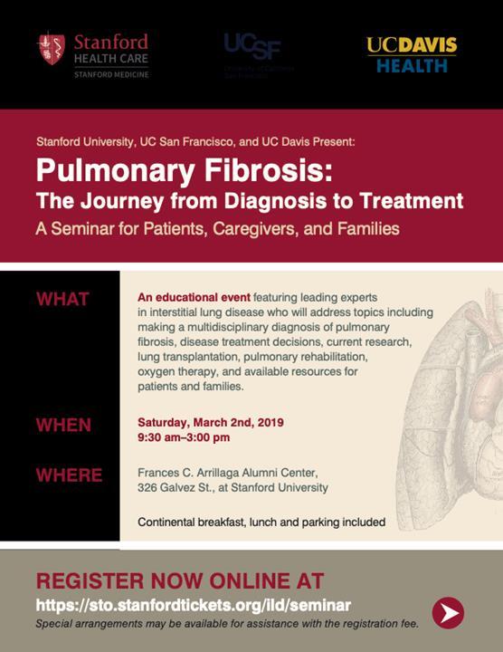
5 CTS Inspirations Page 5 January 30, 2019 The benign end of a spectrum of pulmonary lymphoproliferative disorders including follicular bronchiolitis (FB) and lymphocytic interstitial pneumonia (LIP) can be associated with pulmonary cysts. FB is characterized by peribronchial and peribronchiolar lymphoid follicles with reactive germinal centers while in LIP dense interstitial lymphocyte infiltrates are seen. The cysts in FB/LIP are typically thin-walled, usually affecting <10% of lung field and may be bordered by an eccentric vessel. It can be associated with areas of groundglass opacities, centrilobular and subpleural nodules, thickened bronchovascular bundles, interlobular septal thickening and lymphadenopathy. LIP can be idiopathic but it is more commonly associated with various systemic and infectious disorders that are associated with dysgammaglobulinemia and lymphocytic infiltration including Sjogren syndrome and HIV [7]. REFERENCES 1. Hansell, D.M., et al., Fleischner Society: glossary of terms for thoracic imaging. Radiology, (3): p Jawad, H., et al., Cystic interstitial lung diseases: recognizing the common and uncommon entities. Curr Probl Diagn Radiol, (3): p Gupta, N., et al., Diffuse Cystic Lung Disease. Part II. Am J Respir Crit Care Med, (1): p Gupta, N., et al., Diffuse Cystic Lung Disease. Part I. Am J Respir Crit Care Med, (12): p Gupta, N., et al., Accuracy of chest high-resolution computed tomography in diagnosing diffuse cystic lung diseases. Eur Respir J, (4): p DeMartino, E., R.S. Go, and R. Vassallo, Langerhans Cell Histiocytosis and Other Histiocytic Diseases of the Lung. Clin Chest Med, (3): p Panchabhai, T.S., C. Farver, and K.B. Highland, Lymphocytic Interstitial Pneumonia. Clin Chest Med, (3): p ANNOUNCEMENTS: Stanford University, in collaboration with UC San Francisco and UC Davis, presents an Interstitial Lung Disease educational event for patients, families, and caregivers featuring leading experts who will address topics including making a multidisciplinary diagnosis of pulmonary fibrosis, disease treatment decisions, current research, lung transplantation, pulmonary rehabilitation, oxygen therapy, and available resources. The event will be held on the beautiful Stanford campus at the Frances C. Arrilaga Alumni Center on March 2, For the full program, directions, and online registration information please go to:


6 CTS Inspirations Page 6 January 30, 2019 ************************************************************************************ To view Volume 17, Issue 6 of the SWJPCC Journal, click on the following link:
Cystic Lung Disease. Cristopher A. Meyer, MD
 Cystic Lung Disease Cristopher A. Meyer, MD Air filled structure with definable wall typically less than 1 mm thick Cris A. Meyer, M.D. Professor of Radiology University of Wisconsin School of Medicine
HRCT in Diffuse Interstitial Lung Disease Steps in High Resolution CT Diagnosis. Where are the lymphatics? Anatomic distribution
 Steps in High Resolution CT Diagnosis Pattern of abnormality Distribution of disease Associated findings Clinical history Tomás Franquet MD What is the diagnosis? Hospital de Sant Pau. Barcelona Secondary
The Prevention & Treatment of Augmentation
 The Prevention & Treatment of Augmentation MARK J BUCHFUHRER, MD S T A N F O R D U N I VERSITY S L E E P M E D I C IN E C E N T ER, R E D W O O D C I T Y, C A P R I VATE P R ACTICE, D O W N E Y, C A 2017
Spectrum of Cystic Lung Disease and its Mimics. Kathleen Jacobs MD and Elizabeth Weihe MD UC San Diego Medical Center, Department of Radiology
 Spectrum of Cystic Lung Disease and its Mimics Kathleen Jacobs MD and Elizabeth Weihe MD UC San Diego Medical Center, Department of Radiology No Financial Disclosures Learning Objectives 1. Review the
Basics of Restless Legs Syndrome (Willis-Ekbom Disease)
") Basics of Restless Legs Syndrome (Willis-Ekbom Disease) Michael H. Silber, M.B.Ch.B. Professor of Neurology Mayo Clinic College of Medicine Objectives Understand how RLS is diagnosed Understand what we
Basics of Restless Legs Syndrome (Willis-Ekbom Disease) Michael H. Silber, M.B.Ch.B. Professor of Neurology Mayo Clinic College of Medicine Objectives Understand how RLS is diagnosed Understand what we
Workshop Cyst & Lucency. How to Approach
 Workshop Cyst & Lucency How to Approach To Approach Cystic Lung Disease True cysts? Cavitary disease Cystic bronchiectasis Mosaic attenuation Subpleural cysts Bullae Paraseptal emphysema Honeycombing Birt
Financial disclosure COMMON DIAGNOSES IN HRCT. High Res Chest HRCT. HRCT Pre test. I have no financial relationships to disclose. Anatomy Nomenclature
 Financial disclosure I have no financial relationships to disclose. Douglas Johnson D.O. Cardiothoracic Imaging Gaston Radiology COMMON DIAGNOSES IN HRCT High Res Chest Anatomy Nomenclature HRCT Sampling
Cystic lung diseases 7/21/2017. Cystic Lung Diseases. CT definition of a lung cyst. Important clinical clues
 Cystic Lung Diseases Cystic lung diseases Aurelie Fabre Increased Awareness Spontaneous Pneumothorax High resolution imaging (HRCT) Multidisciplinary approach like interstitial lung disease CT definition
NONE OVERVIEW FINANCIAL DISCLOSURES UPDATE ON IDIOPATHIC PULMONARY FIBROSIS/IPF (UIP) FOR PATHOLOGISTS. IPF = Idiopathic UIP Radiologic UIP Path UIP
 FOR PATHOLOGISTS. IPF = Idiopathic UIP Radiologic UIP Path UIP") UPDATE ON IDIOPATHIC PULMONARY FIBROSIS/IPF () FOR PATHOLOGISTS Thomas V. Colby, M.D. Professor of Pathology (Emeritus) Mayo Clinic Arizona FINANCIAL DISCLOSURES NONE OVERVIEW IPF Radiologic Dx Pathologic
UPDATE ON IDIOPATHIC PULMONARY FIBROSIS/IPF () FOR PATHOLOGISTS Thomas V. Colby, M.D. Professor of Pathology (Emeritus) Mayo Clinic Arizona FINANCIAL DISCLOSURES NONE OVERVIEW IPF Radiologic Dx Pathologic
Diffuse Cystic Lung Disease at High-Resolution CT
 Cardiopulmonary Imaging Pictorial Essay Seaman et al. HRCT of Diffuse Cystic Lung Disease Cardiopulmonary Imaging Pictorial Essay Downloaded from www.ajronline.org by 37.44.193.85 on 01/05/18 from IP address
An Image Repository for Chest CT
 An Image Repository for Chest CT Francesco Frajoli for the Chest CT in Antibody Deficiency Group An Image Repository for Chest CT he Chest CT in Antibody Deficiency Group is an international and interdisciplinary
Acute and Chronic Lung Disease
 KATHOLIEKE UNIVERSITEIT LEUVEN Faculty of Medicine Acute and Chronic Lung Disease W De Wever, JA Verschakelen Department of Radiology, University Hospitals Leuven, Belgium Clinical utility of HRCT To detect
Usual Interstitial pneumonia and Nonspecific Interstitial Pneumonia. Nitra and the Gangs.
 Usual Interstitial pneumonia and Nonspecific Interstitial Pneumonia Nitra and the Gangs. บทน ำและบทท ๓, ๑๐, ๑๒, ๑๓, ๑๔, ๑๕, ๑๗ Usual Interstitial Pneumonia (UIP) Most common & basic pathologic pattern
Outline Definition of Terms: Lexicon. Traction Bronchiectasis
 HRCT OF IDIOPATHIC INTERSTITIAL PNEUMONIAS Disclosures Genentech, Inc. Speakers Bureau Tadashi Allen, MD University of Minnesota Assistant Professor Diagnostic Radiology 10/29/2016 Outline Definition of
11/10/2014. Multi-disciplinary Approach to Diffuse Lung Disease: The Imager s Perspective. Radiology
 Multi-disciplinary Approach to Diffuse Lung Disease: The Imager s Perspective Radiology Pathology Clinical 1 Role of HRCT Diagnosis Fibrosis vs. inflammation Next step in management Response to treatment
Manish Powari Regional Training Day 10/12/2014
 Manish Powari Regional Training Day 10/12/2014 Large number of different types of Interstitial Lung Disease (ILD). Most are very rare Most patients present with one of a smaller number of commoner diseases
Radiologic Approach to Smoking Related Interstitial Lung Disease
 Radiologic Approach to Smoking Related Interstitial Lung Disease Poster No.: C-1854 Congress: ECR 2013 Type: Educational Exhibit Authors: K.-N. Lee, J.-Y. Han, E.-J. Kang, J. Kang; Busan/KR Keywords: Toxicity,
A Rare Case of Lymphangioleiomyomatosis in Sri Lanka
 A Rare Case of Lymphangioleiomyomatosis in Sri Lanka Author s Details: (1) Dushantha Madegedara (2) Asela Rasika Bandara (3) Sachini Seneviratne (4) Samadara Nakandala (5) Rathnayake R.M.D.H.M - (1) (2)
Liebow and Carrington's original classification of IIP
 Liebow and Carrington's original classification of IIP-- 1969 Eric J. Stern MD University of Washington UIP Usual interstitial pneumonia DIP Desquamative interstitial pneumonia BIP Bronchiolitis obliterans
Pediatric High-Resolution Chest CT
 Pediatric High-Resolution Chest CT Alan S. Brody, MD Professor of Radiology and Pediatrics Chief, Thoracic Imaging Cincinnati Children s s Hospital Cincinnati, Ohio, USA Pediatric High-Resolution CT Short
Monday 10 September Interstitial lung disease 15:10 15:35. The uncommon interstitial lung diseases (ILD)
") Interstitial lung disease 15:10 15:35 The uncommon interstitial lung diseases (ILD) Dr Grant Griffiths, Cwm Taf University Health Board, Cardiff Be familiar with the Diagnostic criteria for idiopathic
Interstitial lung disease 15:10 15:35 The uncommon interstitial lung diseases (ILD) Dr Grant Griffiths, Cwm Taf University Health Board, Cardiff Be familiar with the Diagnostic criteria for idiopathic
Systemic lupus erythematosus (SLE): Pleuropulmonary Manifestations
: Pleuropulmonary Manifestations") 08/30/10 09/26/10 Systemic lupus erythematosus (SLE): Pleuropulmonary Manifestations Camila Downey S. Universidad de Chile, School of Medicine, Year VII Harvard University, School of Medicine Sept 17,
08/30/10 09/26/10 Systemic lupus erythematosus (SLE): Pleuropulmonary Manifestations Camila Downey S. Universidad de Chile, School of Medicine, Year VII Harvard University, School of Medicine Sept 17,
HYPERSENSITIVITY PNEUMONITIS
 HYPERSENSITIVITY PNEUMONITIS A preventable fibrosis MOSAVIR ANSARIE MB., FCCP INTERSTITIAL LUNG DISEASES A heterogeneous group of non infectious, non malignant diffuse parenchymal disorders of the lower
AUGMENTATION SUFFERING AND WHAT CAN BE DONE ABOUT IT. John W. Winkelman MD PhD Massachusetts General Hospital Harvard Medical School Boston, MA
 AUGMENTATION SUFFERING AND WHAT CAN BE DONE ABOUT IT John W. Winkelman MD PhD Massachusetts General Hospital Harvard Medical School Boston, MA Disclosure Information Type of Affiliation Commercial Entity
Cryptogenic Organizing Pneumonia Diagnosis Approach Based on a Clinical-Radiologic-Pathologic Consensus
 Cryptogenic Organizing Pneumonia Diagnosis Approach Based on a Clinical-Radiologic-Pathologic Consensus Poster No.: C-1622 Congress: ECR 2012 Type: Scientific Exhibit Authors: C. Cordero Lares, E. Zorita
Diffuse Interstitial Lung Diseases: Is There Really Anything New?
 : Is There Really Anything New? Sujal R. Desai, MBBS, MD ESTI SPEAKER SUNDAY Society of Thoracic Radiology San Antonio, Texas March 2014 Diffuse Interstitial Lung Disease The State of Play DILDs Is There
5/9/2015. Multi-disciplinary Approach to Diffuse Lung Disease: The Imager s Perspective. No, I am not a pulmonologist! Radiology
 Multi-disciplinary Approach to Diffuse Lung Disease: The Imager s Perspective No, I am not a pulmonologist! Radiology Pathology Clinical 1 Everyone needs a CT Confidence in diagnosis Definitive HRCT +
10/17/2016. Nuts and Bolts of Thoracic Radiology. Objectives. Techniques
 Nuts and Bolts of Thoracic Radiology October 20, 2016 Carleen Risaliti Objectives Understand the basics of chest radiograph Develop a system for interpreting chest radiographs Correctly identify thoracic
Bubbles in the lung. Page 1 of 29
 Bubbles in the lung. Poster No.: C-1048 Congress: ECR 2015 Type: Educational Exhibit Authors: A. Arango, A. Martínez de Alegría, R. García Figueiras, S. Baleato González, M. C. Ageitos Casais, M. V. Trujillo
DIAGNOSTIC NOTE TEMPLATE
 DIAGNOSTIC NOTE TEMPLATE SOAP NOTE TEMPLATE WHEN CONSIDERING A DIAGNOSIS OF IDIOPATHIC PULMONARY FIBROSIS (IPF) CHIEF COMPLAINT HISTORY OF PRESENT ILLNESS Consider IPF as possible diagnosis if any of the
Thoracic lung involvement in rheumatoid arthritis: Findings on HRCT
 Thoracic lung involvement in rheumatoid arthritis: Findings on HRCT Poster No.: C-2488 Congress: ECR 2015 Type: Educational Exhibit Authors: R. E. Correa Soto, M. J. Martín Sánchez, J. M. Fernandez 1 1
Differential diagnosis
 Differential diagnosis Idiopathic pulmonary fibrosis (IPF) is part of a large family of idiopathic interstitial pneumonias (IIP), one of four subgroups of interstitial lung disease (ILD). Differential
ARDS - a must know. Page 1 of 14
 ARDS - a must know Poster No.: C-1683 Congress: ECR 2016 Type: Authors: Keywords: DOI: Educational Exhibit M. Cristian; Turda/RO Education and training, Edema, Acute, Localisation, Education, Digital radiography,
Bronchiectasis: An Imaging Approach
 Bronchiectasis: An Imaging Approach Travis S Henry, MD Associate Professor of Clinical Radiology Cardiac and Pulmonary Imaging Section University of California, San Francisco Large Middle Small 1 Bronchiectasis
Multiple Cystlike Lung Lesions
 Residents Section Pattern of the Month Cantin et al. Multiple Cystlike Lung Lesions in the dult Residents Section Pattern of the Month Residents inradiology Luce Cantin 1 lexander. ankier Ronald L. Eisenberg
Coexistence of Lymphangioleiomyomatosis and Angiomyolipomas in a Patient of Tuberous Sclerosis Complex: a case report
 Chin J Radiol 2003; 28: 329-333 329 Coexistence of Lymphangioleiomyomatosis and Angiomyolipomas in a Patient of Tuberous Sclerosis Complex: a case report FENG-CHI HSIEH 1 KAO-LANG LIU 1 YIH-LEONG CHANG
Connective Tissue Disorder- Associated Interstitial Lung Disease (CTD-ILD) and Updates
 and Updates") Connective Tissue Disorder- Associated Interstitial Lung Disease (CTD-ILD) and Updates Maria Elena Vega, M.D Assistant Professor of Medicine Lewis Katz School of Medicine at Temple University Nothing to
Connective Tissue Disorder- Associated Interstitial Lung Disease (CTD-ILD) and Updates Maria Elena Vega, M.D Assistant Professor of Medicine Lewis Katz School of Medicine at Temple University Nothing to
Smoking-related Interstitial Lung Diseases: High-Resolution CT Findings
 Smoking-related Interstitial Lung Diseases: High-Resolution CT Findings Poster No.: C-2358 Congress: ECR 2013 Type: Educational Exhibit Authors: V. Cuartero Revilla, M. Nogueras Carrasco, P. Olmedilla
Daria Manos RSNA 2016 RC 401. https://medicine.dal.ca/departments/depar tment-sites/radiology/contact/faculty/dariamanos.html
 Daria Manos RSNA 2016 RC 401 https://medicine.dal.ca/departments/depar tment-sites/radiology/contact/faculty/dariamanos.html STEP1: Is this fibrotic lung disease? STEP 2: Is this a UIP pattern? If yes:
4/17/2010 C ini n ca c l a Ev E a v l a ua u t a ion o n of o ILD U dat a e t e i n I LDs
 Update in ILDs Diagnosis 101: Clinical Evaluation April 17, 2010 Jay H. Ryu, MD Mayo Clinic, Rochester MN Clinical Evaluation of ILD Outline General aspects of ILDs Classification of ILDs Clinical evaluation
Imaging Small Airways Diseases: Not Just Air trapping. Eric J. Stern MD University of Washington
 Imaging Small Airways Diseases: Not Just Air trapping Eric J. Stern MD University of Washington What we are discussing SAD classification SAD imaging with MDCT emphasis What is a small airway? Airway with
Epidemiology and classification of smoking related interstitial lung diseases
 Epidemiology and classification of smoking related interstitial lung diseases Šterclová M. Department of Respiratory Diseases, Thomayer Hospital, Prague, Czech Republic Supported by an IGA Grant No G 1207
August 2018 Imaging Case of the Month: Dyspnea in a 55-Year-Old Smoker. Michael B. Gotway, MD
 August 2018 Imaging Case of the Month: Dyspnea in a 55-Year-Old Smoker Michael B. Gotway, MD Department of Radiology Mayo Clinic Arizona Scottsdale, AZ USA Clinical History: A 55 year old woman presented
ARTICLE IN PRESS. Ahuva Grubstein a, Daniele Bendayan b, Ithak Schactman c, Maya Cohen a, David Shitrit b, Mordechai R. Kramer b,
 Respiratory Medicine (2005) 99, 948 954 Concomitant upper-lobe bullous emphysema, lower-lobe interstitial fibrosis and pulmonary hypertension in heavy smokers: report of eight cases and review of the literature
Pulmonary Manifestations Of Skeletal Disorders
 Pulmonary Manifestations Of Skeletal Disorders U. A. Saeed, MBBS FCPS, J. Nair, MBBS MD, R. Khosla, MD FRCR, K. Sayegh, MD FRCPC, J. Kosiuk, MD FRCPC, J. Taylor, MD FRCPC; Department of Radiology, McGill
Radiologic-pathologic correlation of pulmonary diseases
 The 1578 th Chest Conference/ 3 rd Biennial Clinical- Radiologic-Pathologic Correlation Radiologic-pathologic correlation of pulmonary diseases Harumi Itoh, M.D. University of Fukui, Japan Centriacinar
September 2014 Imaging Case of the Month. Michael B. Gotway, MD. Department of Radiology Mayo Clinic Arizona Scottsdale, AZ
 September 2014 Imaging Case of the Month Michael B. Gotway, MD Department of Radiology Mayo Clinic Arizona Scottsdale, AZ Clinical History: A 57-year-old non-smoking woman presented to her physician as
Case 1: Question. 1.1 What is the main pattern of this HRCT? 1. Intralobular line 2. Groundglass opacity 3. Perilymphatic nodule
 HRCT WORK SHOP Case 1 Case 1: Question 1.1 What is the main pattern of this HRCT? 1. Intralobular line 2. Groundglass opacity 3. Perilymphatic nodule Case 1: Question 1.2 What is the diagnosis? 1. Hypersensitivity
Case Presentations in ILD. Harold R. Collard, MD Department of Medicine University of California San Francisco
 Case Presentations in ILD Harold R. Collard, MD Department of Medicine University of California San Francisco Outline Overview of diagnosis in ILD Definition/Classification High-resolution CT scan Multidisciplinary
Progress in Idiopathic Pulmonary Fibrosis
 Progress in Idiopathic Pulmonary Fibrosis David A. Lynch, MB Disclosures Progress in Idiopathic Pulmonary Fibrosis David A Lynch, MB Consultant: t Research support: Perceptive Imaging Boehringer Ingelheim
Restrictive lung diseases
 Restrictive lung diseases Restrictive lung diseases are diseases that affect the interstitium of the lung. Interstitium of the lung is the very thin walls surrounding the alveoli, it s formed of epithelium
CTD-related Lung Disease
 13 th Cambridge Chest Meeting King s College, Cambridge April 2015 Imaging of CTD-related Lung Disease Dr Sujal R Desai King s College Hospital, London Disclosure Statement No Disclosures / Conflicts of
Lung Allograft Dysfunction
 Lung Allograft Dysfunction Carlos S. Restrepo M.D. Ameya Baxi M.D. Department of Radiology University of Texas Health San Antonio Disclaimer: We do not have any conflict of interest or financial gain to
PULMONARY TUBERCULOSIS RADIOLOGY
 PULMONARY TUBERCULOSIS RADIOLOGY RADIOLOGICAL MODALITIES Medical radiophotography Radiography Fluoroscopy Linear (conventional) tomography Computed tomography Pulmonary angiography, bronchography Ultrasonography,
Case of the Day Chest
 Case of the Day Chest Darin White MDCM FRCPC Department of Radiology, Mayo Clinic 76 th Annual Scientific Meeting Canadian Association of Radiologists Montreal, QC April 26, 2013 2013 MFMER slide-1 Disclosures
TB Radiology for Nurses Garold O. Minns, MD
 TB Nurse Case Management Salina, Kansas March 31-April 1, 2010 TB Radiology for Nurses Garold O. Minns, MD April 1, 2010 TB Radiology for Nurses Highway Patrol Training Center Salina, KS April 1, 2010
An Introduction to Radiology for TB Nurses
 An Introduction to Radiology for TB Nurses Garold O. Minns, MD September 14, 2017 TB Nurse Case Management September 12 14, 2017 EXCELLENCE EXPERTISE INNOVATION Garold O. Minns, MD has the following disclosures
Bronkhorst colloquium Interstitiële longziekten. Katrien Grünberg, klinisch patholoog
 Bronkhorst colloquium 2013-2014 Interstitiële longziekten De pathologie achter de CT Katrien Grünberg, klinisch patholoog K.grunberg@vumc.nl Preparing: introduction and 3 cases The introduction on microscopic
INTERSTITIAL LUNG DISEASE. Radhika Reddy MD Pulmonary/Critical Care Long Beach VA Medical Center January 5, 2018
 INTERSTITIAL LUNG DISEASE Radhika Reddy MD Pulmonary/Critical Care Long Beach VA Medical Center January 5, 2018 Interstitial Lung Disease Interstitial Lung Disease Prevalence by Diagnosis: Idiopathic Interstitial
MANAGEMENT RECOMMENDATIONS
 1 MANAGEMENT RECOMMENDATIONS 1. Adrenal masses!!!!!!! page 2 2. Liver Masses!!!!!!! page 3 3. Obstetric US Soft Markers for Aneuploidy!! pages 4-6 4. Ovarian and Adnexal Cysts!!!!! pages 7-10 5. Pancreatic
Diagnosing Idiopathic Pulmonary Fibrosis on Evidence-Based Guidelines
 Diagnosing Idiopathic Pulmonary Fibrosis on Evidence-Based Guidelines Rebecca Keith, MD Assistant Professor, Division of Pulmonary and Critical Care Medicine National Jewish Health, Denver, CO Objectives
Imaging: how to recognise idiopathic pulmonary fibrosis
 REVIEW IDIOPATHIC PULMONARY FIBROSIS Imaging: how to recognise idiopathic pulmonary fibrosis Anand Devaraj Affiliations: Dept of Radiology, St George s Hospital, London, UK. Correspondence: Anand Devaraj,
Clinical History. 29 yo woman with polyhydramnios Cardiac mass at fetal ultrasound At 35 weeks, newborn died 30 minutes after delivery
 CASE 1 a Clinical History 29 yo woman with polyhydramnios Cardiac mass at fetal ultrasound At 35 weeks, newborn died 30 minutes after delivery Interface between tumor and normal myocardium Smaller well-demarcated
Excavated pulmonary nodule: steps to diagnosis?
 Excavated pulmonary nodule: steps to diagnosis? Poster No.: C-1044 Congress: ECR 2014 Type: Authors: Keywords: DOI: Educational Exhibit W. Mnari, M. MAATOUK, A. Zrig, B. Hmida, M. GOLLI; Monastir/ TN Metastases,
Case 1 : Question. 1.1 What is the intralobular distribution? 1. Centrilobular 2. Perilymphatic 3. Random
 Interesting case Case 1 Case 1 : Question 1.1 What is the intralobular distribution? 1. Centrilobular 2. Perilymphatic 3. Random Case 1: Answer 1.1 What is the intralobular distribution? 1. Centrilobular
Follicular bronchiolitis in surgical lung biopsies: Clinical implications in 12 patients
 Respiratory Medicine (2008) 102, 307 312 Follicular bronchiolitis in surgical lung biopsies: Clinical implications in 12 patients Michelle R. Aerni a, Robert Vassallo a,, Jeffrey L. Myers b, Rebecca M.
Histopathologic Approach to Interstitial Lung Disease
 Histopathologic Approach to Interstitial Lung Disease Kirk D. Jones, MD UCSF Dept of Pathology kirk.jones@ucsf.edu Disclosures I have nothing to disclose 1 Why? Much of interstitial lung disease biopsies
IPF: Epidemiologia e stato dell arte
 IPF: Epidemiologia e stato dell arte Clinical Classification Diffuse parenchimal lung diseases Exposure-related: - occupational - environmental - medication Desquamative interstitial pneumonia Idiopathic
Smoking-related interstitial lung disease
 Smoking-related interstitial lung disease Sergio Harari U.O. di Pneumologia UTIR Servizio di Fisiopatologia Respiratoria e Emodinamica Polmonare Ospedale S. Giuseppe MultiMedica Milano Milano, 7 Ottobre
Cystic Lung Disease: a Comparison of Cystic Size, as Seen on Expiratory and Inspiratory HRCT Scans
 Cystic Lung Disease: a Comparison of Cystic Size, as Seen on Expiratory and Inspiratory HRCT Scans Ki-Nam Lee, MD 1 Seong-Kuk Yoon, MD 1 Seok Jin Choi, MD 2 Jin Mo Goo, MD 3 Kyung-Jin Nam, MD 1 Index words:
Idiopathic interstitial pneumonias (IIPs) are a group of
 are a group of") SYMPOSIA C. Isabela S. Silva, MD, PhD and Nestor L. Müller, MD, PhD Abstract: The idiopathic interstitial pneumonias (IIPs) are a group of diffuse parenchymal lung diseases of unknown etiology characterized
SYMPOSIA C. Isabela S. Silva, MD, PhD and Nestor L. Müller, MD, PhD Abstract: The idiopathic interstitial pneumonias (IIPs) are a group of diffuse parenchymal lung diseases of unknown etiology characterized
Eun-Young Kang, M.D., Jae Wook Lee, M.D., Ji Yung Choo, M.D., Hwan Seok Yong, M.D., Ki Yeol Lee, M.D., Yu-Whan Oh, M.D.
 Eun-Young Kang, M.D., Jae Wook Lee, M.D., Ji Yung Choo, M.D., Hwan Seok Yong, M.D., Ki Yeol Lee, M.D., Yu-Whan Oh, M.D. Department of Radiology, Korea University Guro Hospital, College of Medicine, Korea
From the Archives of the AFIP
 AFIP ARCHIVES 821 CME FEATURE See accompanying test at http:// www.rsna.org /education /rg_cme.html LEARNING OBJECTIVES FOR TEST 6 After reading this article and taking the test, the reader will be able
Patient with IPF and no honeycombing on HRCT. Case 1 Demosthenes Bouros, Vasilios Tzilas University of Athens
 Patient with IPF and no honeycombing on HRCT Case 1 Demosthenes Bouros, Vasilios Tzilas University of Athens CASE OVERVIEW A 76-year-old male patient presented with progressive exertional dyspnoea refractory
Long-Term Treatment of Restless Legs Syndrome (RLS): An Approach to Management of Worsening Symptoms, Loss of Efficacy, and Augmentation
: An Approach to Management of Worsening Symptoms, Loss of Efficacy, and Augmentation") CNS Drugs (2015) 29:351 357 DOI 10.1007/s40263-015-0250-2 CURRENT OPINION Long-Term Treatment of Restless Legs Syndrome (RLS): An Approach to Management of Worsening Symptoms, Loss of Efficacy, and Augmentation
CNS Drugs (2015) 29:351 357 DOI 10.1007/s40263-015-0250-2 CURRENT OPINION Long-Term Treatment of Restless Legs Syndrome (RLS): An Approach to Management of Worsening Symptoms, Loss of Efficacy, and Augmentation
CT Findings in the Elderly Lung
 CT Findings in the Elderly Lung Poster No.: C-2498 Congress: ECR 2015 Type: Educational Exhibit Authors: P. Ananias, R. Coelho, H. M. R. Marques, O. Fernandes, M. Simões, L. Figueiredo; Lisbon/PT Keywords:
Pulmonary Sarcoidosis - Radiological Evaluation
 Original Research Article Pulmonary Sarcoidosis - Radiological Evaluation Jayesh Shah 1, Darshan Shah 2*, C. Raychaudhuri 3 1 Associate Professor, 2 1 st Year Resident, 3 Professor and HOD Radiology Department,
Case 1. A. Insomnia B. Restless leg syndrome C. Peripheral neuropathy D. Osteoarthritis of the hip. Disclosures. Diagnosis for trouble falling asleep
 Disclosures I have no disclosures Case 1 Liza Ashbrook, MD Assistant Clinical Professor UCSF Department of Neurology History of Present Illness Diagnosis for trouble falling asleep 70-year-man with obstructive
Micronodular lung pattern - Differential diagnosis
 Micronodular lung pattern - Differential diagnosis Poster No.: P-0074 Congress: ESTI 2015 Type: Educational Poster Authors: P. Ninitas, F. Marinho, P. Campos, I. Távora ; Lisbon/PT, 1 2 2 3 1 1 3 Funchal/PT,
The radiological differential diagnosis of the UIP pattern
 5th International Conference on Idiopathic Pulmonary Fibrosis, Modena, 2015, June 12th The radiological differential diagnosis of the UIP pattern Simon Walsh King s College Hospital Foundation Trust London,
T he diagnostic evaluation of a patient with
 546 REVIEW SERIES Challenges in pulmonary fibrosis? 1: Use of high resolution CT scanning of the lung for the evaluation of patients with idiopathic interstitial pneumonias Michael B Gotway, Michelle M
R E S T L E S S L E G S S Y N D R O M E. Causes, diagnosis and treatment. For the patient living with restless legs syndrome (RLS)
") R E S T L E S S L E G S S Y N D R O M E Causes, diagnosis and treatment For the patient living with restless legs syndrome (RLS) www.rls.org RESTLESS LEGS SYNDROME Restless legs syndrome (RLS), also called
R E S T L E S S L E G S S Y N D R O M E Causes, diagnosis and treatment For the patient living with restless legs syndrome (RLS) www.rls.org RESTLESS LEGS SYNDROME Restless legs syndrome (RLS), also called
I don t need you. Disclosure Statement. Pathology Approach to ILD 11/5/2016. Kirk D. Jones, MD UCSF Dept of Pathology
 Pathology Approach to ILD Disclosure Statement Relevant financial relationships with a commercial interest: Boeringer Ingleheim, speaker Kirk D. Jones, MD UCSF Dept of Pathology kirk.jones@ucsf.edu I don
A Review of Interstitial Lung Diseases. Paul J. Wolters, MD Associate Professor Department of Medicine University of California San Francisco
 A Review of Interstitial Lung Diseases Paul J. Wolters, MD Associate Professor Department of Medicine University of California San Francisco Outline Overview of diagnosis in ILD Why it is important Definition/Classification
Thoracic CT pattern in lung cancer: correlation of CT and pathologic diagnosis
 19 th Congress of APSR PG of Lung Cancer (ESAP): Update of Lung Cancer Thoracic CT pattern in lung cancer: correlation of CT and pathologic diagnosis Kazuma Kishi, M.D. Department of Respiratory Medicine,
Medical Policy An independent licensee of the Blue Cross Blue Shield Association
 Idiopathic Pulmonary Fibrosis Page 1 of 10 Medical Policy An independent licensee of the Blue Cross Blue Shield Association Title: Idiopathic Pulmonary Fibrosis (Esbriet /pirfenidone, Ofev /nintedanib)
TBLB is not recommended as the initial biopsy option in cases of suspected IPF and is unreliable in the diagnosis of rare lung disease (other than
 TBLB is not recommended as the initial biopsy option in cases of suspected IPF and is unreliable in the diagnosis of rare lung disease (other than PAP) BAL is not required as a diagnostic tool in patients
I have no relevant conflicts of interest to disclose
 I have no relevant conflicts of interest to disclose Diffuse parenchymal lung disease (DPLD) and its associations Secondary lobular anatomy DPLD History, clinical findings, temporal evolution, and exposures
Is it really honeycombing? Limitations and pitfalls in radiological diagnosis of honeycombing.
 Is it really honeycombing? Limitations and pitfalls in radiological diagnosis of honeycombing. J. Arenas-Jiménez, E. García-Garrigós, M. Sirera-Matilla; M.C. Planells-Alduvin, F.I. Aranda* Hospital General
Thoracic sarcoidosis: Pictoral review of typical and atypical findings
 Thoracic sarcoidosis: Pictoral review of typical and atypical findings Poster No.: C-0804 Congress: ECR 2010 Type: Educational Exhibit Topic: Chest Authors: A. Ferreira, J. Calha; Lisbon/PT Keywords: Sarcoidosis,
SCLERODERMA LUNG DISEASE: WHAT THE PATIENT SHOULD KNOW
 SCLERODERMA LUNG DISEASE: WHAT THE PATIENT SHOULD KNOW Lung disease can be a serious complication of scleroderma. The two most common types of lung disease in patients with scleroderma are interstitial
Nestor L. Müller, MD, PhD Professor Emeritus of Radiology University of British Columbia Vancouver, BC, Canada
 Atlas of Interstitial Lung Disease Pathology Pathology with High Resolution CT Correlations First Edition Andrew Churg, MD Professor of Pathology University of British Columbia Pathologist, Vancouver General
LAM 101. Lymph-angio-leiomyo-matosis
 LAM 101 Lymph-angio-leiomyo-matosis Charlie Strange, MD Division of Pulmonary and Critical Care Medicine Medical University of South Carolina Dr. Strange has been a grant recipient in LAM from the NIH,
Lymphangioleiomyomatosis (LAM)
") Lymphangioleiomyomatosis (LAM) is a rare lung condition that mainly affects women of childbearing age. Although it has been reported in men, it is extremely rare. It is estimated that three to five in
Lymphangioleiomyomatosis (LAM) is a rare lung condition that mainly affects women of childbearing age. Although it has been reported in men, it is extremely rare. It is estimated that three to five in
TB Intensive Houston, Texas
 TB Intensive Houston, Texas October 15-17, 17 2013 Diagnosis of TB: Radiology Rosa M Estrada-Y-Martin, MD MSc FCCP October 16, 2013 Rosa M Estrada-Y-Martin, MD MSc FCCP, has the following disclosures to
The Management of Restless Legs Syndrome in Adults in Primary Care Version 1.1 February 2019
 The Management of Restless Legs Syndrome in Adults in Primary Care Version 1.1 February 2019 VERSION CONTROL Version Number Date Amendments made 1.0 December 2015 1.1 February 2019 Version 1. Approved
Idiopathic Pulmonary of Care
 Chapter 6.1 Living Medical etextbook A Digital Tool at the Point of Care From Projects In Knowledge Pulmonology Idiopathic Pulmonary Fibrosis @Point of Care IPF Case Study: Typical Presentation, Role of
A Review of Interstitial Lung Diseases
 Outline A Review of Interstitial Lung Diseases Paul J. Wolters, MD Associate Professor Department of Medicine University of California San Francisco Overview of diagnosis in ILD Why it is important Definition/Classification