8/3/2016. Consultant for / research support from: Astellas Bayer Bracco GE Healthcare Guerbet Medrad Siemens Healthcare. Single Energy.
|
|
|
- Brent Woods
- 5 years ago
- Views:
Transcription


1 U. Joseph Schoepf, MD Prof. (h.c.), FAHA, FSCBT-MR, FNASCI, FSCCT Professor of Radiology, Medicine, and Pediatrics Director, Division of Cardiovascular Imaging Consultant for / research support from: Astellas Bayer Bracco GE Healthcare Guerbet Medrad Siemens Healthcare Single Energy Dual Energy Pre-Contrast Arterial Portal Late 1
2 INFORMATION Single Energy Dual Energy Qualitative & Quantitative Information Main applications in oncologic imaging include: Virtual non-contrast (VNC) images to lower radiation dose Improving contrast using virtual monoenergetic images (Mono) Quantifying iodine uptake with iodine perfusion maps Material Differentiation Iodine, calcium, barium, uric acid Material Detection and Quantification 1) Tumor detection 2) Tumor characterization 3) Evaluation of response to therapy 4) Detection of oncologic-related disorders (e.g. pulmonary embolism) 2


3 Melanoma Metastasis Renal Cell Carcinoma 1. Lesion Detection 2. Iodine Quantification Simons D et al. Eur Rad 2014 MONOENERGETIC PLUS 40keV 50keV 60keV 70keV 80keV 90keV 110 kev 120keV 150keV 190keV Lesion detection and contrast medium reduction (higher attenuation at lower kev levels Metallic artifacts reduction (high kev levels) Iodine K-edge 33.2 kev 120 kvp 40 kev Stolzmann P Insight Radiol


4 DECT may be useful in detecting underlying tumors in patients with intracerebral hemorrhage of unknown origin Sens 94% Spec 97% Kim SJ AJNR 2012 Differentiating metastatic lymph nodes Improved head and neck tumor staging and assessment Differentiating metastatic lymph nodes Metastatic Normal Inflammatory Tawfik AM Eur Radiol


5 DECT can increase diagnostic performance and reproducibility for evaluation of thyroid cartilage invasion Kuno H Radiology 2012 Kun H Eur J Radiol 2013 The ability of DECT to visualize and quantify the iodine uptake of nodules may help in differentiating benign from malignant pulmonary nodules VNC data-set can help in reducing radiation dose 5


6 Iodine Map Degree Enhancement Sensitivity 92% 72% Specificity 70% 70% Accuracy 82.2% 71.1% Chae EJ Radiology 2008 In patients with NSCLC, iodine uptake quantified by DECT has been shown to correlate with metabolic activity on FDG-PET Schmid-Bindert G Eur Radiol 2012 Arterial Enhancement Fraction (AEF) Ratio of early and late post-contrast iodine uptake Baxa J Eur Radiol

 Evaluation of")


")
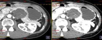
7 Radiation dose reduction using the VNC data-set Nodule detection and characterization (Iodine Map & Monoenergetic Imaging 50-55/70-75 kev) Evaluation of response to therapy Real Virtual Arterial Virtual Portal Which One is the Real, the Virtual Arterial and the Virtual Portal Unenhanced? 120 kvp 80 kvp Improved lesion detection and conspicuity (HCC, hypervascular metastases) Robinson E Invest Radiol 2010, Altenberd J Eur Rad 2010, Marin D Radiology


8 Mixed 120 kvp MonoPlus 40 kev MRI HCC Virtual Monoenergetic Images at low kev may improve lesion detection and conspicuity Mixed 120 kvp MonoBasic 70 kev MonoPlus 50 kev HCC Nodule MRI 8
")


9 DECT can increase the visualization of small (< 2 cm) or isoattenuating pancreatic adeno-ca Cystic lesion characterization (Serous vs Mucinous) Neuroendocrine tumor detection Hypoattenuating solid mass in 90-95% Hypoattenuation due to desmoplastic fibrotic component CT Sensitivity: > 2 cm: up to 98% < 2 cm: 68-77% MonoPlus at low energies (kev) Mono 40 kev Mono+ 40 kev Improved image quality and pancreasto-lesion contrast in the diagnosis of pancreatic adenocarcinoma Mono 55 kev Mono+ 55 kev Substantial noise reduction compared to the prior algorithm Mixed M_0.6 Frellesen et al, EJR


")


10 120 kvp 50 kev Iodine Map Mc Namara MM Abdom Imaging 2015 MACROCYSTIC SCN (10%) Difficult DD with pseudocyst and MCN SCN MCN MCN DD: Calcifications / intramural nodules / enhancing septa 70 kev 52 kev Morgan DE Abdom Imaging


11 Pancreatic insulinomas are usually small tumors that are difficult to localize with conventional CT Lin XZ Eur J Radiol 2012 Agrawal MD Radiographics


12 The differential diagnosis between benign cyst and solid tumor can be a challenge Iodine uptake attenuation increase of at least HU DECT significantly increases reader confidence in differentiating cystic from solid renal masses Lesion Iodine Threshold: 0.5 mg/ml Chandarana H AJR 2011 Sens Spec PPV NPV Iodine-related attenuation: 15 HU 91.4% 93.3% 91.4% 93.3% Iodine concentration: 0.5 mg/ml 100% 97.7% 97.2% 100% Mileto A AJR kvp 80 kev Mileto A Radiology
13 Incidental adrenal masses occur in up to 5% of CT examinations Most of these lesions are benign even in oncologic patients (adrenal adenomas) Characterization: Unenhanced + Enhanced CT Unenhanced + Venous + Delayed 1. Unenhanced CT: Threshold 10 HU Lipid Rich Adenoma: Sens 71%, Spec 98% 2. Absolute Washout: (E D)/(E U)] 100 Threshold 60% Lipid Rich/Lipid Poor: Sens 88%, Spec 96% 3. Relative Washout: (E D)/E 100) Threshold 50% at 9 min Lipid Rich/Lipid Poor: Sens 98%, Spec 100% Boland GW AJR 1998, Caoili EM AJR 2000, Caoili Radiology 2002 DECT can improve characterization of adrenal nodules: VNC replacing True Unenhanced data-set Material Density Analysis Low kv imaging Monoenergetic Imaging 13


14 Ho LM AJR 2012 Gnnant R AJR 2012 Glazer D AJR 2013 Helck A Eur Rad 2014 Material Density Analysis: Sens 96%, Spec 100% Non Enhanced CT: Sens 67%, Spec 100% Mileto A Radiology 2014 Lipid Rich Adenoma Lipid Poor Adenoma Mileto A Radiology


15 pnet Metastasis Mileto A Radiology 2014 Mileto A Radiology 2014 Material decomposition based on the difference in absorption characteristics for different elements. Fat, Iodine, Water (mg/ml) Morgan DE JCAT


16 Iodine-related attenuation (IRA) of DECT correlates with the Choi criteria DECT showed significantly higher IRA in progressive ( HU) lesions compared with stable or regressive ( HU) lesions Apfaltrer P Invest Radiol 2012 Morgan DE Abdom Imaging 2013 Pulmonary Embolism Meinel FG Invest Radiol


 Assessment of response to therapy and quantification")
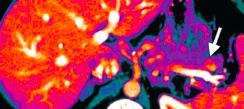
17 Small bowel ischemia in Leriche syndrome Iodine map Optimum Contrast Combined Approach 1. Mixed 120 kvp 2. Virtual Unenhanced Image (VNC) 3. Virtual Monoenergetic Images (50-55 kev Mono+ or kev Standard Mono) 4. Iodine Map Use Iterative Reconstruction DECT can significantly improve lesion detection and characterization (MonoPlus and Iodine Map) Assessment of response to therapy and quantification of enhancement Radiation dose reduction with VNC imaging Oncologic related disease and metallic artifacts reduction 17


18 Thank you! 18
Dual Energy CT of Pulmonary Embolism
 Dual Energy CT of Pulmonary Embolism U. Joseph Schoepf, MD, FAHA, FSCBT MR, FSCCT Professor of Radiology, Medicine, and Pediatrics Director of Cardiovascular Imaging Disclosures Consultant for / research
Dual-Energy CT: The Technological Approaches
 Dual-Energy CT: The Technological Approaches Dushyant Sahani, M.D Director of CT Associate Professor of Radiology Massachusetts General Hospital Harvard Medical School Email-dsahani@partners.org Disclosure
Role of DE-CT in Oncology
 Role of DE-CT in Oncology Dushyant Sahani, M.D Director of CT Associate Professor of Radiology Massachusetts General Hospital Harvard Medical School Email-dsahani@partners.org Disclosure Research Grant
ESUR 2018, Sept. 13 th.-16 th., 2018 Barcelona, Spain
 ESUR 2018, Sept. 13 th.-16 th., 2018 Barcelona, Spain OUR APPROACH Incidental adrenal nodule/mass Isaac R Francis, M.B;B.S University of Michigan, Ann Arbor, Michigan Disclosures None (in memory) M Korobkin,
Dual-Energy CT Applications in Radiation Therapy
 THE UNIVERSITY OF WISCONSIN MADISON Dual-Energy CT Applications in Radiation Therapy - Jessica Miller 1 Disclosures Funding provided by Siemens Medical 2 Learning objectives General principles of dual
CT Perfusion. U. Joseph Schoepf, MD, FAHA, FSCBT MR, FSCCT Professor of Radiology, Medicine, and Pediatrics Director of Cardiovascular Imaging
 CT Perfusion U. Joseph Schoepf, MD, FAHA, FSCBT MR, FSCCT Professor of Radiology, Medicine, and Pediatrics Director of Cardiovascular Imaging Disclosures Consultant for / research support from Bayer Bracco
MEDICAL UNIVERSITY of SOUTH CAROLINA
 U. Joseph Schoepf, MD Prof. (h.c.), FAHA, FSCBT-MR, FNASCI, FSCCT Professor of Radiology, Medicine, and Pediatrics Director, Division of Cardiovascular Imaging MEDICAL UNIVERSITY of SOUTH CAROLINA Disclosures
CT Myocardial Perfusion: Is there Added Value to Coronary CT?
 CT Myocardial Perfusion: Is there Added Value to Coronary CT? U. Joseph Schoepf, MD, FAHA, FSCBT MR, FSCCT Professor of Radiology, Medicine, and Pediatrics Director of Cardiovascular Imaging Disclosures
Dual Energy CT of the Heart: Perfusion and Beyond
 Dual Energy CT of the Heart: Perfusion and Beyond U. Joseph Schoepf, MD, FAHA, FSCBT MR, FSCCT Professor of Radiology, Medicine, and Pediatrics Director of Cardiovascular Imaging Disclosures Consultant
Liver, Liver Fat, Fibrosis and HCC Screening with DECT
 Liver, Liver Fat, Fibrosis and HCC Screening with DECT Dushyant Sahani, M.D Director of CT Associate Professor of Radiology Massachusetts General Hospital Harvard Medical School Email-dsahani@partners.org
Dual Energy CT Aortography: Can We Reduce Iodine Dose??
 Dual Energy CT Aortography: Can We Reduce Iodine Dose?? William P. Shuman MD, FACR FSCBTMR Department of Radiology University of Washington SCBTMR Annual Course Boston, October 10, 2012 Conflict of Interest
ADRENAL LESIONS 10/09/2012. Adrenal + lesion. Introduction. Common causes. Anatomy. Financial disclosure. Dr. Boraiah Sreeharsha. Nothing to declare
 ADRENAL LESIONS Financial disclosure Nothing to declare Dr. Boraiah Sreeharsha MBBS;FRCR;FRCPSC Introduction Adrenal + lesion Adrenal lesions are common 9% of the population Increase in the detection rate
Dual Energy CT of the Liver
 34th Annual Course October 2011 Washington, DC Dual Energy CT of the Liver Vassilios Raptopoulos, MD Beth Israel Deaconess Medical Center Harvard Medical School Dual Energy CT (DECT) Different materials
Cardiac CT Lowering the Dose Dramatically
 Cardiac CT Lowering the Dose Dramatically U. Joseph Schoepf, MD, FAHA, FSCBT MR, FSCCT Professor of Radiology, Medicine, and Pediatrics Director of Cardiovascular Imaging Disclosures Consultant for / research
Dual Energy Spectral CT of Focal Liver Lesions in Advanced Cirrhosis: Early Experience
 Dual Energy Spectral CT of Focal Liver Lesions in Advanced Cirrhosis: Early Experience William P. Shuman MD, FACR University of Washington SCBTMR Annual Course Washington DC, October 23-26, 2011 Conflict
STANDARDIZED MANAGEMENT RECOMMENDATIONS FOR ADRENAL NODULES: EVIDENCE-BASED CONSENSUS POWERSCRIBE MACROS FROM AN ACADEMIC/PRIVATE PRACTICE
 STANDARDIZED MANAGEMENT RECOMMENDATIONS FOR ADRENAL NODULES: EVIDENCE-BASED CONSENSUS POWERSCRIBE MACROS FROM AN ACADEMIC/PRIVATE PRACTICE COLLABORATIVE Pamela Johnson 1, Darcy Wolfman 2, Upma Rawal 3,
Imaging Pancreatic Neuroendocrine Tumors (PNETs): CT, MRI, EUS, Nuclear
: CT, MRI, EUS, Nuclear") Imaging Pancreatic Neuroendocrine Tumors (PNETs): CT, MRI, EUS, Nuclear Eric Tamm, M.D. Department of Diagnostic Radiology Division of Diagnostic Imaging MD Anderson Cancer Center Houston, TX Disclosure
Imaging Pancreatic Neuroendocrine Tumors (PNETs): CT, MRI, EUS, Nuclear Eric Tamm, M.D. Department of Diagnostic Radiology Division of Diagnostic Imaging MD Anderson Cancer Center Houston, TX Disclosure
Computer Aided Detection and Diagnosis: Cardiac Imaging Applications
 Computer Aided Detection and Diagnosis: Cardiac Imaging Applications U. Joseph Schoepf, MD, FAHA, FSCBT MR, FSCCT Professor of Radiology, Medicine, and Pediatrics Director of Cardiovascular Imaging Disclosures
Imaging Pancreatic Neuroendocrine Tumors (PNETs): CT, MRI, EUS, Nuclear
: CT, MRI, EUS, Nuclear") Imaging Pancreatic Neuroendocrine Tumors (PNETs): CT, MRI, EUS, Nuclear Eric Tamm, M.D. Department of Diagnostic Radiology Division of Diagnostic Imaging MD Anderson Cancer Center Houston, TX Disclosure
Imaging Pancreatic Neuroendocrine Tumors (PNETs): CT, MRI, EUS, Nuclear Eric Tamm, M.D. Department of Diagnostic Radiology Division of Diagnostic Imaging MD Anderson Cancer Center Houston, TX Disclosure
REVIEW. Distinguishing benign from malignant adrenal masses
 Cancer Imaging (2003) 3, 102 110 DOI: 10.1102/1470-7330.2003.0006 CI REVIEW Distinguishing benign from malignant adrenal masses Isaac R Francis Professor of Radiology, Department of Radiology, University
True Dual Energy. Dr. Stefan Ulzheimer, Siemens Healthcare GmbH. DEfinitely Siemens
 DEfinitely Siemens True Dual Energy Dr. Stefan Ulzheimer, Siemens Healthcare GmbH International version. Not for distribution in the US. Unrestricted Siemens AG 2015 All rights reserved. The products/features
Neuro-endocrine and pancreatic non-adenocarcinomas. Marc Engelbrecht, AMC, Amsterdam
 Neuro-endocrine and pancreatic non-adenocarcinomas Marc Engelbrecht, AMC, Amsterdam Pancreatic Tumors q Epithelial Exocrine q Mesenchymal Ductal Adenocarcinoma (85-95%) Metastasis Lymfoma Acinar Cell Carcinoma
Gemstone Spectral Imaging quantifies lesion characteristics for a confident diagnosis
 GE Healthcare Gemstone Spectral Imaging quantifies lesion characteristics for a confident diagnosis CT clinical case study lesion characterization Desiree Morgan, MD Vice Chair of Clinical Research Professor
Functional CT imaging techniques for the assessment of angiogenesis in lung cancer
 Perspective Functional CT imaging techniques for the assessment of angiogenesis in lung cancer Thomas Henzler 1, Jingyun Shi 2, Hashim Jafarov 1, Stefan O. Schoenberg 1, Christian Manegold 3, Christian
SA CME Information SA CME INFORMATION. Target Audience
 SA CME INFORMATION SA CME Information Description Adrenal Imaging: A Three-category Approach To Managing The Adrenal "Incidentaloma" Imaging plays a critical role in the work-up and clinical management
Dual-Energy CT Applications in the Abdomen
 Dual-Energy CT Review Dual-Energy CT Review Tobias Heye 1 Rendon C. Nelson Lisa M. Ho Daniele Marin Daniel T. Boll Heye T, Nelson RC, Ho LM, Marin D, Boll DT Keywords: abdomen, dual-energy CT, iodine extraction,
ABDOMINAL DIFFUSION WEIGHTED MR
 ABDOMINAL DIFFUSION WEIGHTED MR Frank Miller, M.D. FACR Professor of Radiology Chief, Body Imaging Section Medical Director, MR Imaging Northwestern University Feinberg School of Medicine fmiller@northwestern.edu
Positron Emission Tomography in Lung Cancer
 May 19, 2003 Positron Emission Tomography in Lung Cancer Andrew Wang, HMS III Patient DD 53 y/o gentleman presented with worsening dyspnea on exertion for the past two months 30 pack-year smoking Hx and
Imaging of Neuroendocrine Metastases
 Imaging of Neuroendocrine Metastases Aoife Kilcoyne, Shaunagh McDermott, Colin McCarthy,Manuel Patino, Dushyant Sahani, Michael Blake Abdominal Imaging Division Massachusetts General Hospital Disclosure
PET IMAGING (POSITRON EMISSION TOMOGRAPY) FACT SHEET
 FACT SHEET") Positron Emission Tomography (PET) When calling Anthem (1-800-533-1120) or using the Point of Care authorization system for a Health Service Review, the following clinical information may be needed to
Positron Emission Tomography (PET) When calling Anthem (1-800-533-1120) or using the Point of Care authorization system for a Health Service Review, the following clinical information may be needed to
CT Contrast Protocols for Different Organ Imaging
 CT Contrast Protocols for Different Organ Imaging g Paul Shreve, M.D. Advanced Radiology Services, P.C. & Spectrum Health Grand Rapids, MI, USA Correlative Imaging Council Society of Nuclear Medicine 56
ADRENAL MR: PEARLS AND PITFALLS
 ADRENAL MR: PEARLS AND PITFALLS Frank Miller, M.D. Lee F. Rogers MD Professor of Medical Education Chief, Body Imaging Section and Fellowship Medical Director, MR Imaging Professor of Radiology Northwestern
SCBT-MR 2015 Incidentaloma on Chest CT
 SCBT-MR 2015 Incidentaloma on Chest CT Reginald F. Munden MD, DMD, MBA I have no conflicts of interest to report Incidentaloma Pulmonary Nodule Mediastinal Lymph Node Coronary Artery Calcium Incidental
Dr Claire Smith, Consultant Radiologist St James University Hospital Leeds
 Dr Claire Smith, Consultant Radiologist St James University Hospital Leeds Imaging in jaundice and 2ww pathway Image protocol Staging Limitations Pancreatic cancer 1.2.4 Refer people using a suspected
New Visions in PET: Surgical Decision Making and PET/CT
 New Visions in PET: Surgical Decision Making and PET/CT Stanley J. Goldsmith, MD Director, Nuclear Medicine Professor, Radiology & Medicine New York Presbyterian Hospital- Weill Cornell Medical Center
CT 101 :Pancreas and Spleen
 CT 101 :Pancreas and Spleen Shikha Khullar,, MD, MPH Division of Radiology University of South Alabama The Pancreas Normal Pancreas 3 Phase Pancreatic CT Non contrast Arterial phase : 30-35 35 second
Role of imaging in RCC. Ultrasonography. Solid lesion. Cystic RCC. Solid RCC 31/08/60. From Diagnosis to Treatment: the Radiologist Perspective
 Role of imaging in RCC From Diagnosis to Treatment: the Radiologist Perspective Diagnosis Staging Follow up Imaging modalities Limitations and pitfalls Duangkamon Prapruttam, MD Department of Therapeutic
performed to help sway the clinician in what the appropriate diagnosis is, which can substantially alter the treatment of management.
 Hello, I am Maura Polansky at the University of Texas MD Anderson Cancer Center. I am a Physician Assistant in the Department of Gastrointestinal Medical Oncology and the Program Director for Physician
PET/CT in lung cancer
 PET/CT in lung cancer Andrei Šamarin North Estonia Medical Centre 3 rd Baltic Congress of Radiology 08.10.2010 Imaging in lung cancer Why do we need PET/CT? CT is routine imaging modality for staging of
Contrast Enhanced Ultrasound of Parenchymal Masses in Children
 Contrast Enhanced Ultrasound of Parenchymal Masses in Children Sue C Kaste, DO On behalf of Beth McCarville, MD St. Jude Children s Research Hospital Memphis, TN Overview Share St. Jude experience with
INTERDISCIPLINARY DISCUSSIONS IN LOCALISED RCC DIAGNOSIS AND SURGICAL STRATEGIES FOR ATYPICAL RENAL CYSTIC LESIONS. Maria Cova
 INTERDISCIPLINARY DISCUSSIONS IN LOCALISED RCC DIAGNOSIS AND SURGICAL STRATEGIES FOR ATYPICAL RENAL CYSTIC LESIONS Maria Cova Radiology Department University of Trieste (IT) Eleventh European International
Disclosures. Diffusion and Perfusion Imaging in the Head and Neck. Learning objectives ???
 Disclosures No relevant financial disclosures Diffusion and Perfusion Imaging in the Head and Neck Ashok Srinivasan, MD Associate Professor Director of Neuroradiology University of Michigan Health System
Radiology Update 2017
 Radiology Update 2017 John K. Phillips, MD Affiliated Assistant Professor of Radiology University of Tennessee Health Sciences Center Chief, Radiology and Nuclear Medicine VA Memphis Disclosures Financial:
Index. radiologic.theclinics.com. Note: Page numbers of article titles are in boldface type.
 Index Note: Page numbers of article titles are in boldface type. A ACC. See Adrenal cortical carcinoma. Acromegaly and the pituitary gland, 551 Acute suppurative thyroiditis, 405, 406 Addison, Thomas and
11/1/2014. Radiologic incidentalomas Ordering pitfalls Newer technology and applications
 Bilal Tahir, MD Gitasree Borthakur, MD Indiana University School of Medicine Department of Radiology & Imaging Sciences October 31, 2014 ACP 2014 Dr. V. Aaron Nuclear (vaaron@iupui.edu) Dr. S. Westphal
PET/CT Frequently Asked Questions
 PET/CT Frequently Asked Questions General Q: Is FDG PET specific for cancer? A: No, it is a marker of metabolism. In general, any disease that causes increased metabolism can result in increased FDG uptake
Dual-Energy 101: Principles, Methods and Dose
 Dual-Energy 101: Principles, Methods and Dose Juan Carlos Ramirez-Giraldo, Ph.D Staff Scien2st, Collabora2ons Manager SE Region ISCT San Francisco, 2017 Siemens Medical Solu2ons USA, Inc., 2017 Page 1
Jeffrey C. Weinreb, MD, FACR Yale School of Medicine Yale-New Haven Hospital
 Jeffrey C. Weinreb, MD, FACR Yale School of Medicine Yale-New Haven Hospital jeffrey.weinreb@yale.edu 1991 1997 Whole body MRI: multistation approach x z Isocenter: Table Move: Multiple Steps Whole body
Radiological staging of lung cancer. Shukri Loutfi,MD,FRCR Consultant Thoracic Radiologist KAMC-Riyadh
 Radiological staging of lung cancer Shukri Loutfi,MD,FRCR Consultant Thoracic Radiologist KAMC-Riyadh Bronchogenic Carcinoma Accounts for 14% of new cancer diagnoses in 2012. Estimated to kill ~150,000
Evaluation and Management of Thyroid Nodules. Nick Vernetti, MD, FACE Palm Medical Group Las Vegas, Nevada
 Evaluation and Management of Thyroid Nodules Nick Vernetti, MD, FACE Palm Medical Group Las Vegas, Nevada Disclosure Consulting Amgen Speaking Amgen Objectives Understand the significance of incidental
Breast Cancer PET/CT Imaging Protocol
 Breast Cancer PET/CT Imaging Protocol Scanning Protocol: Patients are scanned from the top of the neck through the pelvis. Arms-up position is used to avoid beam-hardening artifact in the chest and abdomen.
Wednesday 12 September Advanced imaging science to practice 14:30 14:55. Recent advances in computed tomography (CT) technology
 technology") 14:30 14:55 Recent advances in computed tomography (CT) technology Dr Gareth Iball, Leeds Teaching Hospitals NHS Trust All modern CT systems incorporate iterative reconstruction (IR) techniques. Their
14:30 14:55 Recent advances in computed tomography (CT) technology Dr Gareth Iball, Leeds Teaching Hospitals NHS Trust All modern CT systems incorporate iterative reconstruction (IR) techniques. Their
Recommendations for cross-sectional imaging in cancer management, Second edition
 www.rcr.ac.uk Recommendations for cross-sectional imaging in cancer management, Second edition Renal and adrenal tumours Faculty of Clinical Radiology www.rcr.ac.uk Contents Renal cell carcinoma 3 Clinical
Innovations in HCC Imaging: MDCT/MRI
 Innovations in HCC Imaging: MDCT/MRI Anthony E. Cheng, M.D. Cardinal MRI Center Cardinal Santos Medical Center, Wilson Street, San Juan Innovations in HCC Imaging: Goals/Objectives MDCT/MRI Learn the diagnostic
Diffusion Weighted Imaging in Prostate Cancer
 Diffusion Weighted Imaging in Prostate Cancer Disclosure Information Vikas Kundra, M.D, Ph.D. No financial relationships to disclose. Education Goals and Objectives To describe the utility of diffusion-weighted
X-ray Corner. Imaging of The Pancreas. Pantongrag-Brown L
 X-ray Corner 125 Imaging of The Pancreas Modern imaging modalities commonly used in pancreas include ultrasound (US), CT, and MRI. Pancreas is a retroperitoneal organ which makes it difficult to visualize
Imaging Work-Up of a Neck Mass - Adults & Children
 Disclosures Imaging Work-Up of a Neck Mass - Adults & Children I have nothing to disclose Christine M Glastonbury MBBS Professor of Radiology & Biomedical Imaging Otolaryngology-Head & Neck Surgery and
Evaluation of Thyroid Nodules
 Evaluation of Thyroid Nodules Stephan Kowalyk, MD January 25 28, 2018 1 Primary goal Exclude malignancy Incidental thyroid nodules If found on CT, MRI, PET scan, carotid Doppler ULTRASOUND!! January 25
An Introduction to Dual Energy Computed Tomography
 An Introduction to Dual Energy Computed Tomography Michael Riedel University of Texas Health Science Center at San Antonio Introduction The idea of computed tomography (CT) was first introduced in the
Traumatic and Non Traumatic Adrenal Emergencies
 Traumatic and Non Traumatic Adrenal Emergencies Michael N. Patlas, MD, FRCPC (1), Christine O. Menias, MD (2), Douglas S. Katz, MD, FACR (3), Ania Z. Kielar, MD, FRCPC (4), Alla M. Rozenblit, MD (5), Jorge
objectives Pitfalls and Pearls in PET/CT imaging Kevin Robinson, DO Assistant Professor Department of Radiology Michigan State University
 objectives Pitfalls and Pearls in PET/CT imaging Kevin Robinson, DO Assistant Professor Department of Radiology Michigan State University To determine the regions of physiologic activity To understand
Financial Disclosure
 Benign Liver Masses Adil Abdalla, MBBS Creighton University-CHI Health August 25, 2018 Financial Disclosure Nothing to disclose Financial Disclosure 1 Objectives To assess patients with benign liver tumors
Contrast-Enhanced Ultrasonograpic Findings in Pancreatic Tumors
 Int. J. Med. Sci. 2008, 5 203 Short Research Communication International Journal of Medical Sciences ISSN 1449-1907 www.medsci.org 2008 5(4):203-208 Ivyspring International Publisher. All rights reserved
Papillary Thyroid Cancer: Dual-Energy Spectral CT Quantitative Parameters for Preoperative Diagnosis of Metastasis to the Cervical Lymph Nodes 1
 Note: This copy is for your personal non-commercial use only. To order presentation-ready copies for distribution to your colleagues or clients, contact us at www.rsna.org/rsnarights. Xuewen Liu, MD, PhD
Imaging Decisions Start Here SM
 Owing to its high resolution and wide anatomic coverage, dynamic first-pass perfusion 320-detector-row CT outperforms PET/CT for distinguishing benign from malignant lung nodules, researchers from Japan
PARATHYROID IMAGING. James Lee, MD Chief, Endocrine Surgery Co-Director NY Thyroid-Parathyroid Center Columbia University Medical Center
 PARATHYROID IMAGING James Lee, MD Chief, Endocrine Surgery Co-Director NY Thyroid-Parathyroid Center Columbia University Medical Center NO DISCLOSURES Overview The hallmarks of the ideal test Benefits
(2/3 PRCC!) (2/3 PRCC!)
 (2/3 PRCC!)") Approach to the Incidental Solid Renal Mass Stuart G. Silverman, MD, FACR Professor of Radiology Harvard ard Medical School Director, Abdominal Imaging and Intervention Brigham and Women s Hospital Boston,
Approach to the Incidental Solid Renal Mass Stuart G. Silverman, MD, FACR Professor of Radiology Harvard ard Medical School Director, Abdominal Imaging and Intervention Brigham and Women s Hospital Boston,
Updated Imaging for Novel Pancreatic Cancer Therapy. Desiree E. Morgan, MD FSCBTMR Professor and Vice Chair Education
 Updated Imaging for Novel Pancreatic Cancer Therapy Desiree E. Morgan, MD FSCBTMR Professor and Vice Chair Education Disclosures I have no financial disclosures relevant to the educational content of this
Acknowledgements. Update of Focal Liver Lesions Goals. Focal Liver Lesions. Imaging Choices For Liver Lesions. Focal Liver Lesions
 Acknowledgements Update of Focal Liver Lesions 2012 Giles Boland Massachusetts General Hospital Harvard Medical School No disclosures Dushyant Sahani Mukesh Harisinghani Goals Focal liver lesions Imaging
Anatomical and Functional MRI of the Pancreas
 Anatomical and Functional MRI of the Pancreas MA Bali, MD, T Metens, PhD Erasme Hospital Free University of Brussels Belgium mbali@ulb.ac.be Introduction The use of MRI to investigate the pancreas has
FDG PET/CT STAGING OF LUNG CANCER. Dr Shakher Ramdave
 FDG PET/CT STAGING OF LUNG CANCER Dr Shakher Ramdave FDG PET/CT STAGING OF LUNG CANCER FDG PET/CT is used in all patients with lung cancer who are considered for curative treatment to exclude occult disease.
Evidence based imaging of the pancreas
 Evidence based imaging of the pancreas D.Vanbeckevoort, D.Bielen, K.Op de beeck, R.Vanslembrouck Department of Radiology Chairman Prof. Dr. R.Oyen Non-invasive imaging tests available for the diagnosis
Los Angeles Radiological Society 62 nd Annual Midwinter Radiology Conference January 31, 2010
 Los Angeles Radiological Society 62 nd Annual Midwinter Radiology Conference January 31, 2010 Self Assessment Module on Nuclear Medicine and PET/CT Case Review FDG PET/CT IN LYMPHOMA AND MELANOMA Submitted
THYROID NODULES: THE ROLE OF ULTRASOUND
 THYROID NODULES: THE ROLE OF ULTRASOUND NOVEMBER 2017 DR. DEAN DURANT DEFINITION Thyroid nodule: Focal area within the thyroid gland with echogenicity different from surrounding parenchyma. THYROID NODULES
Hepatobiliary and Pancreatic Malignancies
 Hepatobiliary and Pancreatic Malignancies Gareth Eeson MD MSc FRCSC Surgical Oncologist and General Surgeon Kelowna General Hospital Interior Health Consultant, Surgical Oncology BC Cancer Agency Centre
Low-Dose CT: Clinical Studies & the Radiologist Perspective
 Low-Dose CT: Clinical Studies & the Radiologist Perspective RD-ASiR RD-MBIR SD-FBP RD=0.35 msv (80% dose reduction) Perry J. Pickhardt, MD UW School of Medicine & Public Health Low-Dose CT: Clinical Overview
MDCT combined parameters in characterization of adrenal masses in cancer patients; A prospective study in South Egypt Cancer Institute
 walaa et al. SECI Oncology 2018 MDCT combined parameters in characterization of adrenal masses in cancer patients; A prospective study in South Egypt Cancer Institute Walaa Talaat 1, Haisam A.Samy 2, Hosam
CT Urography. Ureter. Stuart G. Silverman, M.D.
 CT Urography Stuart G. Silverman, M.D. Professor of Radiology Harvard Medical School Director, Abdominal Imaging and Intervention Brigham and Women s Hospital Ureter Boston, MA CT Urography Stuart G. Silverman,
Ultralow Dose Chest CT with MBIR
 Ultralow Dose Chest CT with MBIR Ella A. Kazerooni, M.D. Professor & Director Cardiothoracic Radiology Associate Chair for Clinical Affairs University of Michigan Disclosures Consultant: GE Healthcare
Clinical indications for positron emission tomography
 Clinical indications for positron emission tomography Oncology applications Brain and spinal cord Parotid Suspected tumour recurrence when anatomical imaging is difficult or equivocal and management will
Liver Tumors. Prof. Dr. Ahmed El - Samongy
 Liver Tumors Prof. Dr. Ahmed El - Samongy Objective 1. Identify the most important features of common benign liver tumors 2. Know the risk factors, diagnosis, and management of hepatocellular carcinoma
MEASUREMENT OF EFFECT SOLID TUMOR EXAMPLES
 MEASUREMENT OF EFFECT SOLID TUMOR EXAMPLES Although response is not the primary endpoint of this trial, subjects with measurable disease will be assessed by standard criteria. For the purposes of this
Nuclear Sciences and Medicine
 Nuclear Sciences and Medicine Rethy Chhem, MD, PhD (Edu), PhD (His), FRCPC Division of Human Health Guest Professor, Medical University of Vienna International Atomic Energy Agency Medical Imaging X-rays
Newcastle HPB MDM updated radiology imaging protocol recommendations. Author Dr John Scott. Consultant Radiologist Freeman Hospital
 Newcastle HPB MDM updated radiology imaging protocol recommendations Author Dr John Scott. Consultant Radiologist Freeman Hospital This document is intended as a guide to aid radiologists and clinicians
Computed Diffusion-Weighted Image in the Abdomen
 Computed Diffusion-Weighted Image in the Abdomen Poster No.: C-0234 Congress: ECR 2014 Type: Scientific Exhibit Authors: T. Yoshikawa 1, N. Aoyama 1, Y. Ohno 1, K. Kyotani 1, Y. Kassai 2, K. Sofue 1, M.
Radiology- Pathology Conference 4/29/2012. Lymph Nodes. John McGrath
 Radiology- Pathology Conference 4/29/2012 Lymph Nodes John McGrath 1 Presentation material is for education purposes only. All rights reserved. 2012 URMC Radiology Page 1 of 24 Case 1: 51 year-old male
Liver MRI in 30 minutes
 X Liver MRI in 30 minutes SCBT/MR Annual Meeting Salt Lake City September 18, 2016 Scott B. Reeder, MD, PhD Department of Radiology University of Wisconsin Madison, WI Disclosures University of Wisconsin-Madison
Emerging Referral Patterns for Whole-Body Diffusion Weighted Imaging (WB-DWI) in an Oncology Center
 in an Oncology Center") Emerging Referral Patterns for Whole-Body Diffusion Weighted Imaging (WB-DWI) in an Oncology Center Poster No.: C-1296 Congress: ECR 2014 Type: Scientific Exhibit Authors: G. Petralia 1, G. Conte 1, S.
Emerging Referral Patterns for Whole-Body Diffusion Weighted Imaging (WB-DWI) in an Oncology Center Poster No.: C-1296 Congress: ECR 2014 Type: Scientific Exhibit Authors: G. Petralia 1, G. Conte 1, S.
Update on RECIST and Staging of Common Pediatric Tumors Ethan A. Smith, MD
 Update on RECIST and Staging of Common Pediatric Tumors Ethan A. Smith, MD Section of Pediatric Radiology C.S. Mott Children s Hospital University of Michigan ethans@med.umich.edu Disclosures No relevant
Imaging of liver and pancreas
 Imaging of liver and pancreas.. Disease of the liver Focal liver disease Diffusion liver disease Focal liver disease Benign Cyst Abscess Hemangioma FNH Hepatic adenoma HCC Malignant Fibrolamellar carcinoma
Disclosure. Acknowledgement. What is the Best Workup for Rectal Cancer Staging: US/MRI/PET? Rectal cancer imaging. None
 What is the Best Workup for Rectal Cancer Staging: US/MRI/PET? Zhen Jane Wang, MD Assistant Professor in Residence UC SF Department of Radiology Disclosure None Acknowledgement Hueylan Chern, MD, Department
What Radiologists do?
 Multimodality Imaging in Oncology 2018 March 5 th 9th Diagnostic Imaging in Oncology What Radiologists do? Chikako Suzuki, MD, PhD Department of Diagnostic Radiology, KS Solna Department of Molecular Medicine
The Diagnosis of Hypovascular Hepatic Lesions Showing Hypo-intensity in the Hepatobiliary Phase of Gd-EOB- DTPA-enhanced MR Imaging in High-risk
 2013 67 4 239 244 The Diagnosis of Hypovascular Hepatic Lesions Showing Hypo-intensity in the Hepatobiliary Phase of Gd-EOB- DTPA-enhanced MR Imaging in High-risk Patients for Hepatocellular Carcinoma
Index. Surg Oncol Clin N Am 16 (2007) Note: Page numbers of article titles are in boldface type.
 Note: Page numbers of article titles are in boldface type.") Surg Oncol Clin N Am 16 (2007) 465 469 Index Note: Page numbers of article titles are in boldface type. A Adjuvant therapy, preoperative for gastric cancer, staging and, 339 B Breast cancer, metabolic
Surg Oncol Clin N Am 16 (2007) 465 469 Index Note: Page numbers of article titles are in boldface type. A Adjuvant therapy, preoperative for gastric cancer, staging and, 339 B Breast cancer, metabolic
ADRENAL MEDULLARY DISORDERS: PHAEOCHROMOCYTOMAS AND MORE
 ADRENAL MEDULLARY DISORDERS: PHAEOCHROMOCYTOMAS AND MORE DR ANJU SAHDEV READER AND CONSULTANT RADIOLOGIST QUEEN MARY UNIVERSITY AND ST BARTHOLOMEW S HOSPITAL BARTS HEALTH, LONDON, UK DISCLOSURE OF CONFLICT
42 yr old male with h/o Graves disease and prior I 131 treatment presents with hyperthyroidism and undetectable TSH. 2 hr uptake 20%, 24 hr uptake 50%
 Pinhole images of the neck are acquired in multiple projections, 24hrs after the oral administration of approximately 200 µci of I123. Usually, 24hr uptake value if also calculated (normal 24 hr uptake
DETECTING EARLY LIVER METASTASIS: THE POWER OF MRI WITH LIVER SPECIFIC CONTRAST
 DETECTING EARLY LIVER METASTASIS: THE POWER OF MRI WITH LIVER SPECIFIC CONTRAST LINDA PANTONGRARG-BROWN, MD ADVANCED DIAGNOSTIC IMAGING, RAMATHIBODI HOSPITAL, BANGKOK, THAILAND OUTLINE Choice of imaging
Common and unusual CT and MRI manifestations of pancreatic adenocarcinoma: a pictorial review
 Review Article Common and unusual CT and MRI manifestations of pancreatic adenocarcinoma: a pictorial review Min-Jie Yang, Su Li, Yong-Guang Liu, Na Jiao, Jing-Shan Gong Department of Radiology, Shenzhen
An Approach to Pancreatic Cysts. Introduction
 An Approach to Pancreatic Cysts Nalini M. Guda, MD Aurora St. Luke s Medical Center, Milwaukee Clinical Adjunct Professor of Medicine, University of Wisconsin School of Medicine and Public Health Introduction
CASE 1 11/1/2016 HEPATOBILIARY IMAGING CASE PRESENTATIONS DECLARATION. Dr. Chirag Patel ORGAN IMAGING yr old lady
 HEPATOBILIARY IMAGING CASE PRESENTATIONS DECLARATION No financial disclosures or affiliations with commercial organisations No discussion of investigational or off-label use of medical devices, products