JMSCR Vol 04 Issue 01 Page January 2016
|
|
|
- William Reynolds
- 6 years ago
- Views:
Transcription
1 Impact Factor 3.79 Index Copernicus Value: 5.88 ISSN (e) x ISSN (p) DOI: A Spectrum of Morphological Variations in the human liver lobes and its Clinical importance; a Cadaveric Study Authors Dr S.Saritha 1, Dr Usha rani 2, Dr Gayatri 3 & Dr Peter 3, Mrs Asra Anjum 4 Mrs Himabindu.N 4 1 Professor & HOD, 2 Associate Professor, 3 Assistant Professor & 4 Lecture Department of Anatomy, KAMSRC (Medical College) L.B Nagar, Hyderabad INDIA Correspondence Author Professor & HOD, Dept of Anatomy, KAMSRC (Medical College) L.B Nagar, Hyderabad INDIA Dr S. Saritha kmr.saritha@gmail.com Abstract Most common morphological variations of liver are irregularities in the form, shape, and presence of number of accessory lobes, accessory fissures or abnormal ligaments. Knowledge and awareness of these anomalies is useful to the clinician to rule out diseases, surgeons during segmental resection of liver and radiologist when interpreting liver radiologic findings. The exact origin of accessory lobes of liver in man is unknown and may stimulate tumours. Accessory fissure may mimic internal trauma at the post-mortem study. Rare abnormalities are atrophy or complete absence of one of the lobes. The developmental anomalies of liver may cause confusion to clinician during procedures like biopsy, transplantation & other important surgical or radiological procedures. Aim of the present study comprises a systematic analysis of the anatomical variations exhibited by 50 formalinised and glycerinated adult human livers collected from department of Anatomy, KAMSRC(Hyderabad) &KIMS (Narketpally). Results & Conclusion: We found accessory liver lobes in 8 cadavers (16%), accessory fissures in 15 cases (30%), abnormal connection between left lobe and quadrate lobe in 2 cases (4%) and other morphological variations were also present. Detailed knowledge of anatomical variations in the human liver could be valuable in improving diagnostic procedures.in conditions associated with some liver diseases. Hence we undertook this comprehensive study to identify important accessory lobes and fissures. Key words: quadrate lobe, caudate lobe, liver, accessory lobes & accessory fissures.. Introduction The liver is responsible for a wide range of vital functions including blood detoxification and purification, synthesis of plasma proteins, production of bile, metabolism of carbohydrates, fats & proteins. In man the Liver is essential for survival, no artificial organ or equipment has capacity to compensate for absence of liver function. Gross anatomy of the liver divided into four lobes and eight segments. The Falciform ligament on the anterior surface divides the organ into the right Dr S.Saritha et al Volume 04 Issue 01 January Page 8818
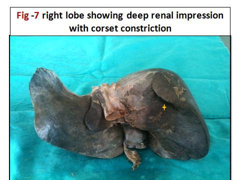
2 &left anatomical lobes. Two additional lobes are seen on visceral surface, superiorly Caudate lobe & inferiorly Quadrate lobe divided by ligamentum Venosum & ligamentum teres. The accessory lobes are found in infra hepatic position. Riedel s lobe is the best known example of a sessile accessory lobe. 1,2 Abnormalities of liver are rare inspite of its complex development. Common abnormalities are irregularities in form, one or more accessory lobes or fissures. The variations have been observed in human liver and have classified as congenital or acquired. A sound knowledge of normal & variant liver is a prerequisite for safe surgical approaches& diagnostic imaging. A presence of abnormal liver has to be kept in mind when an unexplained abdominal mass is encountered. The study was undertaken to investigate the type & frequency of morphological variations in cadaveric Livers available from the department of anatomy, KAMSRC and KIMS. The result of liver, morphological variations have been identified, described and photographed in detail. Materials& methods The formalin fixed adult 50 liver specimens available in the Anatomy department of KAMSRC&KIMS constituted the study material. The livers were dissected during human cadaveric dissection classes for IMBBS medical students over period of 5 years. The embalmed liver lobes that is right lobe, left lobe, caudate lobe & quadrate lobe were studied in detail for presence of accessory lobes and accessory fissures. Specimens were photographed, the findings were appropriately documented Exclusion criteria: age below 20years, specimens with cirrhotic liver.. Inclusion criteria: age between years, weight between 1.2-2kg. Intact liver specimens with normal anatomical features. Observation & Results: Total number of livers studied was 50 Table 1: Showing the incidence of liver morphology types and variations from organ collection at KAMSRC & KIMS S.NO Characteristic features No.of examples Percentage 1. Normal liver 33/50 66% 2. Hypotrophy of left lobe with deep impressions on costal surface 2 4% 3. Transverse liver with large left lobe 2 4% 4. Liver with lingual process of left lobe 2 4% 5. Liver with lingular process of right lobe(rediel s lobe) 1 2% 6. Liver with deep renal impressions& corset constriction 3 6% 7. Liver with diaphragmatic impressions 2 4% 8. Liver with accessory lobes 8 16% 9. Liver with Accessory fissures 15 30% 10. Pons Hepatis- Left lobe connected to quadrate lobe 2 4% 11. Narrow (ill-defined) quadrate lobe 1 2% 12 Total Number of Variation in livers 17/50 34% Dr S.Saritha et al Volume 04 Issue 01 January Page 8819
3 Table 2: Showing the incidence of accessory lobes & accessory fissures in various lobes of Liver S.NO Type of variations Accessory lobes & % Accessory fissures & % 1. Right lobe 1 specimen (2%) 8specimen (16%) 2. Left lobes - 1specimen (2%) 3. Caudate lobe 2 specimens (4%) 3specimen (6%) 4. Quadrate lobe 5specimens (10%) 5specimens (10%) 5. Superior And Inferior Quadrate Lobe 1specimens (2%) Table 3: Classification of liver according to Netter s 17 Netter type No of specimens Type 1 very small left lobe, deep costal impressions. 1 (2%) Type 2-complete atrophy of left lobe Nil Type 3-transverse saddle like liver with relatively large left lobe 2(4%) Type 4- tongue like process of right lobe(reidel s lobe) 1(2%) Type 5- very deep renal impression & corset constriction 3(6%) Type 6-diaphragmatic grooves 2(4%) Observation & Results continued It was noteworthy that 17 cadaveric livers out of 50 were exhibited a range of morphological variations &, while 33 livers. considered to be anatomically normal. Normal surfaces, fissures and borders were observed in 33 livers (66%) and were without accessory lobes & fissures. 17 Liver specimens showed the following morphological variations 1.Accessory fissures in different lobes in 15 livers (30%)-Table 1&2; fig-6,8,9,10,14,16,17,18,19,20, 21 & Accessory lobes were seen in 8 livers specimens (16%).Table 1& 2, fig 8,16,19,20,24&26) 3. Pons hepatis joining left lobe with quadrate lobe was seen in 2 specimens 4%, table- 1, fig-11 & A complete transverse fissure dividing quadrate lobe into superior& inferior lobes(fig 20;table-2) was seen in 1specimen(2%), while mini accessory quadrate lobe was seen in 1specimen 2%.(fig--20) 5. Elongation of left lobe(lingular process)was observed in 2 cases.(table-1; fig-12 & One specimen showed the presence of Reidel s lobe, where right lobe extended downward to right of cystic notch.(table-1 &3; fig-3 ; 7.Abnormal leftlobe L-shapeseen in one specimen with shift of quadrate lobe& fissure for ligamentum teres to the right (fig-27) such a case was not reported in the literature. Two livers presented deep renal impression with corset constriction (table1 &3; fig 6&7, 8.Two liver specimens(4%) had deep costal impressions on anterior surface of right lobe(table1&3; fig 1&2. The liver specimens were classified according to 6 types of liver variations as described by Netter 17 Table 3 These data suggest that there is high incidence of anatomical variation in human liver Discussion In this world of the modern imaging period, it becomes very important to, surgeon,radiologist and clinician to have a thorough knowledge of anatomy and commonly occurring variations in liver. The liver is known to show lobe and fissure anomalies The congenital abnormalities of human liver are rareinspite of its complex development and they are rarer than any other organ of the body. The anomalies may be high in society but we do not notice them, because they are usually asymptomatic. 3 Dr S.Saritha et al Volume 04 Issue 01 January Page 8820
4 They may present in any age group as an accidental findings. Congenital malformations of liver are irregularities in form or occurrence of one or more lobes. Other includes agenesis of lobes and atrophy of lobes. 4 The variations in human liver have been classified as congenital or acquired The Congenital anomalies of liver can be categorized into two; due to defective development or due to excessive development. The anomalies are sometimes associated with malformations of other organs like diaphragm. 5 The embryological basis of anomalies of liver morphology occurs in the course of organogenesis. The defective development of left lobe of liver can lead to gastric volvulus whereas defective development of right lobe may progress to portal hypertension. The excessive development of liver results in formation of accessory lobe which is very rare. The accessory lobes carry risk of torsion or may remain silent in many subjects. The accessory lobes arise most common from the right lobe & may project in any direction. Most common among them is the Riedel s lobe which descends inferiorly along the right lateral surface as tongue like projection. Riedel s lobe is the best example for excessive development of liver. 6 It was described that the hepatic malformations are common in perinatal age group and liver undergoes reformation postnatal. Accordingly all fissures and lobes of liver should disappear during postnatal. 7. In the present case Riedel s lobe may be due to defective development. Multiple hepatic lobes and fissures were common on the under surface of liver opposite to quadrate lobe or left lobeor in the region of gall bladder. 8, The present study had 5accessory lobes in quadrate lobe (10%) & one specimen had accessory lobe near right border of fossa for gall bladder Two accessory lobes in caudate lobe, i.e. One liver specimen had with complete deep vertical fissure dividing the caudate lobe into duplicated caudate lobes. The accessory fissures are potential sources of diagnostic errors in sonography or CT 9 The Multiple accessory fissures may mimic pathologic macro nodular liver on CT The Fissure may be associated with diaphragmatic scalloping or eventration on chest film. Fluid collection in these fissures may mistake for liver cyst, liver abscess or implantation of disseminated tumours cells. The fissures are formed by the invagination of the muscular diaphragm into the liver on the costal surface. Hussein Muktyazet et al 10 found accessory liver lobes in 6 cadavers(14.6%), atrophy of left lobe in 2 cadavers, accessory fissures in 5 cases (12.1%) and evidence of ectopic liver tissue. Sato et al 11 found incidence ectopic liver lobe and accessory liver lobe in 0.7%, according to him accessory lobes are most commonly found on the undersurface of liver, but also seen on gall bladder. According to him ectopic livers are seen in Hepatogastric ligament, near the umbilicus, adrenal gland, pancreas and thoracic cavity. Intra thoracic liver lobe was reported by Hansborough & Lipin in Joshi et alreported 13 notching along inferior border of caudate lobe in 18% of livers, vertical fissure in 30% and prominent papillary process in 32% of livers in their extensive study on lobes and fissures. Our present study had notching along the inferior border of caudate lobe dividing caudate process and papillary process in 3 specimens (6%), a vertical fissure in one specimen dividing the caudate lobe into two or Duplicate caudate lobes and prominent papillary process in 3 specimens. Pujari& Deodhare 14 reported presence of a symptomatic accessory lobe may herniated into thorax through diaphragm and cause serious problems. He also reported a case of bifid liver presenting with anomalous quadrate and caudate lobes with transverse gall bladder. Very recently Anjamrooz and Azari reported 15 a case of coexistences of multiple anomalies of hepatobiliary system. Reports on presence of Dr S.Saritha et al Volume 04 Issue 01 January Page 8821


.")

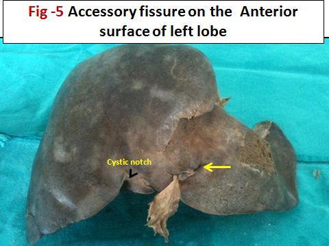
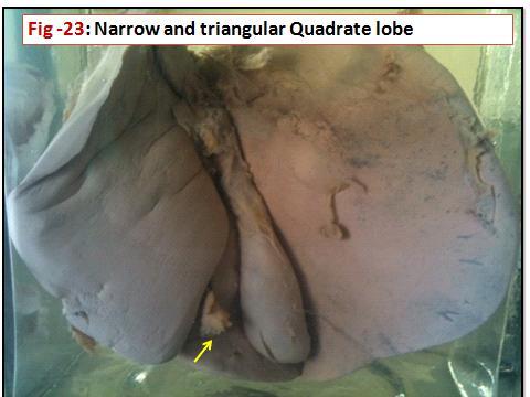
5 accessory liver sulci, absence of quadrate lobe and fissure for ligamentum teres, gall bladder fossa was broad shifted to left. Lobar atrophy of the liver due to causes other than liver tumor or liver cirrhosis is a relatively rare pathological condition, and there are only a few reports in the literature. We report two case of hypotrophy of left lobe of liver (2%) but no case of lobar atrophy except there was one case of small narrow triangular quadrate lobe Acquired morphology in liver are represented as linguiform lobes, small left lobe, deep renal impression with corset type constriction. The present study had linguiform lobe of left lobe in two specimens(4%), hypotrophy left lobe in one specimen (2%), hypertrophy of left lobe with transverse saddle shape liver in two specimens(4%)& renal impression with corset type of constriction in three specimens.(6%). Riedel in 1888described the occasional tonguelike projection of the right lobe of the liver, extending to or below theumbilicus. It has been observed almost exclusively seen in females. Reidel s lobe may extend into the iliac fossa or may extend to below the anterior superior iliac spine. The causes to this condition may be pushing down of right lobe of liver by an enlarging gall bladder, 16 Our study study includes accessory lobes in right lobe one on inferior surface, two in caudate lobe, 5 in quadrate lobe& one liver specimen with Rediel s lobe of right lobe of liver & there was no evidence of ectopic liver tissue. Total number of accessory lobesin our study was in 8 liver specimens. The current study also showed deep diaphragmatic grooves in two liver specimens and 15 accessory fissures in different parts of liver surfaces including caudate lobe & quadrate lobe. Our study also alighted multiple anomalies in one cadaver (fig-27) which was first of its kind in literature. The liver showed L shaped large left lobe, with the shift of quadrate lobe(shape of foot)& fissure for ligamentum teres to right, inferior border of quadrate lobe had small accessory lobes, with Riedel s Lobe with accessory fissure and deep renal impression with corset constriction. I.Variation in the anterior & superior surface of liver Dr S.Saritha et al Volume 04 Issue 01 January Page 8822


6 II. Variarions In The Right Lobe Of Liver Dr S.Saritha et al Volume 04 Issue 01 January Page 8823


7 III.Variations In Left Lobe Of Liver IV.Variation In Caudate Lobe Dr S.Saritha et al Volume 04 Issue 01 January Page 8824


8 V. Variation In Quadrate Lobe Dr S.Saritha et al Volume 04 Issue 01 January Page 8825


9 Conclusion This work was taken up to enlighten the anatomists, morphologists, clinicians and embryologists to update the knowledge of morphological variations of liver. Knowledge of liver variations like atrophy, agenesis, and presence of accessory fissures or lobes, absence of normal fissure or lobe can cause diagnostic error in interpretation. and avoid fatal or serious complications. Acknowledgements All authors are thankful to Department of Anatomy, KAMSRC & KIMS; Hyderabad. Authors of this study also acknowledged to authors, editors, and publishers of all those articles, journals and books from where literature for this article has been reviewed and discussed. Conflict of interest: Nil References 1. Standring.S. Gray s Anatomy: The Anatomical Basis of Clinical Practice 40th ed. New York: Churchill Livingstone 2008; GUYTON, CA. and HALL, JH. Tratado de FisiologiaMédica. Rio de Janeiro: Elsevier, AKTAN, ZA., SAVAS, R., PINAR, Y. and ARSLAN, O. Lobe and segment anomalies of the liver. Journal of Anatomical Society ofindia., 2001, vol. 50, n. 1, p O. C. Bradley, A contribution to the morphology and development of the mammalian liver, Journal of Anatomy and Physiology, vol. 43, pp. 1 42, View at Google Scholar 5. Daver GB, Bakhshi GD, Patil A, Ellur S, Jain M, DaverNG. Bifid liver in a patient Dr S.Saritha et al Volume 04 Issue 01 January Page 8826
10 with diaphragmatic hernia. Indian J Gastroenterol. 2005;24(1): Champetier J, Yver R, Létoublon C, Vigneau B. Ageneral review of anomalies of hepatic morphologyand their clinical implications. AnatomiaClinica,1985;7(4): ). 7. PARKE, WW., SETTLES, HE., BUNGER, PC. and VAN DEMARK, RE. Malformations of the liver: some prenatal and postnatal developmental aspects. Clinical Anatomy, 1996, vol. 9,n. 5, p T. S. Cullen, Accessory lobes of the liver, Archives of Surgery, vol. 11, pp , View at Google Scholar 9. Y. H. Auh, W. A. Rubenstein, and K. Zirinsky, Accessory fissures of the liver: CT and sonographic appearance, American Journal of Roentgenology, vol. 143, no. 3, pp , View at Google Scholar View at Scopus 10. Hussein M, Usman N, Gupta R, Sharma Kr.Morphological variations of liver lobes and itsclinical significance in north Indian populationg.j M.M.S. 2013; 1(1): Sato S, Watanabe M, Nagasawa S, Niigaki M, Sakai S, Akagi S (1998) Laproscopic observations of congenital anomalies of the liver. GastrointestEndosc.; 47: Hansborough ET, Lipin RJ.Intrathoracic accessory lobe of the liver. Ann Surg.1975; 145: Joshi SD, Joshi SS, Athavale SA. Some interesting observations on the surface features of the liver and their clinical implications. Singapore Med J 2009;50: Pujari BD, Deodhare SG. Symptomatic accessory lobe of liver with a review of the literature. Postgrad Med J. 1976; 52: Anjamrooz and Azari Coexistence of multiple anomalies in the hepatobiliary system Anat Cell Biol Mar;45(1):62-5. doi: /acb Epub 2012 Mar Riedel BM. Uber den zungenformigen Fortsatzdesrechten Leberlappens und seine pathognostische Bedeutung fur die Erkrankungder Gallenblasenebst Bemerkungenuber Gallenstein operationen. Berlin Klin Wschr. 1888;25: NETTER, FH. Atlas de Anatomia Humana. Porto Alegre: Artmed, Dr S.Saritha et al Volume 04 Issue 01 January Page 8827
MORPHOLOGICAL STUDY OF ANOMALIES OF LOBES AND SEGMENTS OF HUMAN LIVER
 Original Research Article Anatomy International Journal of Pharma and Bio Sciences ISSN 0975-6299 MORPHOLOGICAL STUDY OF ANOMALIES OF LOBES AND SEGMENTS OF HUMAN LIVER 1* SHARMILA ARISTOTLE 1* Professor,
Access this Article online. Original Article
 Original Article MORPHOLOGICAL STUDY OF HUMAN LIVER AND ITS SURGICAL IMPORTANCE Sachin Patil * 1, Madhu Sethi 2, Smita Kakar 3. * 1, 2 Senior Resident, 3 Director, Professor. Department Of Anatomy, Maulana
MORPHOLOGICAL STUDY OF ADULT HUMAN CADAVERIC LIVER
 Original Research Article MORPHOLOGICAL STUDY OF ADULT HUMAN CADAVERIC LIVER Mohini M.Joshi * 1, Sushama K. Chavan 2. ABSTRACT Background: The liver is the largest of the abdominal viscera, occupying a
ACCESSORY LIVER A RARE FINDING: A CADAVERIC STUDY
 Volume-5, Issue-3, July-Sept-2015 Coden: IJPAJX-CAS-USA, Copyrights@2015 ISSN-2231-4490 Received: 19 th June-2015 Revised: 6 th July -2015 Accepted: 6 th July-2015 Research Article ACCESSORY LIVER A RARE
Lingular Extension of Left Lobe of Liver: A Case Report
 CASE REPORT Lingular Extension of Left Lobe of Liver: A Case Report Nilesh Bhosale 1 and Anjali Gosavi 2 Assistant Professor, Department Of Anatomy, Ashwini Rural Medical College & Hospital, Kumbhari,
Morphometric Study of Caudate Lobe of Liver.
 Original Article ISSN (O):2395-2822; ISSN (P):2395-2814 Morphometric Study of Caudate Lobe of Liver. Neel Kamal Arora 1, Stuti Srivastava 2, Mahboobul Haque 3, Abeer Zubair Khan 2, Karamvir Singh 2 1 Professor
Some interesting observations on the surface features of the liver and their clinical implications
 715 Original Article Some interesting observations on the surface features of the liver and their clinical implications Joshi S D, Joshi S S, Athavale S A Department of Anatomy, Rural Medical College,
Lecture 02 Anatomy of the LIVER
 Lecture 02 Anatomy of the LIVER BY Dr Farooq Khan Aurakzai Dated: 02.01.2018 Introduction to Liver Largest gland in the body. 2 nd largest organ of the body. Weight approximately 1500 gm, and is roughly
Tongue-like elongation of the left lobe of liver
 Page 1 of 5 Anatomy Tongue-like elongation of the left lobe of liver P Baruah, PR Choudhury* Abstract Introduction The liver is the largest gland in our body. Liver normally has a larger right lobe and
Duodenum retroperitoneal
 Duodenum retroperitoneal C shaped Initial region out of stomach into small intestine RETROperitoneal viscus Superior 1 st part duodenal cap ; moves upwards and backwards to lie on the R crura medial to
CONGENITAL ANOMALY OF THE LIVER AND GALL BLADDER
 Abstract CONGENITAL ANOMALY OF THE LIVER AND GALL BLADDER Pages with reference to book, From 202 To 204 Shahnaz Taqi ( Department of Paediatrics, Liaquat National Hospital, Karachi. ) Congenital anomaly
Accessory Glands of Digestive System
 Accessory Glands of Digestive System The liver The liver is soft and pliable and occupies the upper part of the abdominal cavity just beneath the diaphragm. The greater part of the liver is situated under
To describe the liver. To list main structures in porta hepatis.
 GI anatomy Lecture: 6 د. عصام طارق Objectives: To describe the liver. To list main structures in porta hepatis. To define portal system & portosystemic anastomosis. To list parts of biliary system. To
Variations In Branching Pattern Of Coeliac Trunk
 IOSR Journal of Dental and Medical Sciences (IOSR-JDMS) e-issn: 2279-0853, p-issn: 2279-0861.Volume 14, Issue 11 Ver. IV (Nov. 2015), PP 54-58 www.iosrjournals.org Variations In Branching Pattern Of Coeliac
A STUDY OF MORPHOLOGICAL VARIATIONS OF FISSURES AND LOBES IN HUMAN CADAVERIC LUNGS CORRELATING WITH SUR- GICAL IMPLICATIONS IN THE TELANGANA ZONE
 Original Research Article A STUDY OF MORPHOLOGICAL VARIATIONS OF FISSURES AND LOBES IN HUMAN CADAVERIC LUNGS CORRELATING WITH SUR- GICAL IMPLICATIONS IN THE TELANGANA ZONE Gayathri. P *1, S.Saritha 2,
VARIATION OF FISSURE AND LOBAR PATTERN OF LUNG: A CASE REPORT
 Original Research Article DOI - 10.26479/2016.0203.10 VARIATION OF FISSURE AND LOBAR PATTERN OF LUNG: A CASE REPORT Adhanom Gebreslassie Berhe, Dr. Peter Ekanem, Hafte Assefa Beyene Department of Anatomy
Surface Anatomy. Location Shape Weight Role of Five Surfaces Borders Fissures Lobes Peritoneal Lig
 The Liver Functions Bile production and secretion Detoxification Storage of glycogen Protein synthesis Production of heparin and bile pigments Erythropoiesis (in fetus) Surface Anatomy Location Shape Weight
NEW VARIANT LIVER SURFACE MORPHOLOGY ACCORDING TO PORTAL VEIN SEGMENTATION
 NEW VARIANT LIVER SURFACE MORPHOLOGY ACCORDING TO PORTAL VEIN SEGMENTATION Dragica Jurkovikj Institute of Anatomy, Medical Faculty, University Ss. Cyril and Methodius, Skopje 1000, R. Macedonia Abstract
A STUDY OF MORPHOLOGY AND VARIATIONS OF LUNGS IN ADULTS AND FOETUS
 International Journal of Advancements in Research & Technology, Volume 3, Issue 4, April-2014 150 A STUDY OF MORPHOLOGY AND VARIATIONS OF LUNGS IN ADULTS AND FOETUS ZAREENA.SK (assistant professor of anatomy)
Normal Sonographic Anatomy
 hapter 2:The Liver DUNSTAN ABRAHAM Normal Sonographic Anatomy Homogeneous, echogenic texture (Figure 2-1) Measures approximately 15 cm in length and 10 12.5 cm anterior to posterior; measurement taken
Pancreas & Biliary System. Dr. Vohra & Dr. Jamila
 Pancreas & Biliary System Dr. Vohra & Dr. Jamila 1 Objectives At the end of the lecture, the student should be able to describe the: Location, surface anatomy, parts, relations & peritoneal reflection
Block 3: DISSECTION 2 CELIAC TRUNK, JEJUNUM/ILEUM, LARGE INTESTINE, DUODENUM, PANCREAS, PORTAL VEIN; MOBILIZATION OF THE LIVER
 1 Block 3: DISSECTION 2 CELIAC TRUNK, JEJUNUM/ILEUM, LARGE INTESTINE, DUODENUM, PANCREAS, PORTAL VEIN; MOBILIZATION OF THE LIVER Attempt to complete as much as you can of the dissection explained in the
LECTURE 11 & 12: ABDOMINAL VISCERA ABDOMINAL CONTENTS DIVISION. The location of abdominal viscera is divided into 4 quadrants:
 LECTURE 11 & 12: ABDOMINAL VISCERA ABDOMINAL CONTENTS DIVISION The location of abdominal viscera is divided into 4 quadrants: - horizontal line across the umbilicus divides the upper quadrants from the
Intrahepatic ramifications of the portal vein in the horse
 Intrahepatic ramifications of the portal vein in the horse Tadjalli, M. 1* and Moslemy, H. R. 2 1 Department of Anatomical Sciences, School of Veterinary Medicine, University of Shiraz, Shiraz, Iran; 2
Variations of Lung Fissures: A Cadaveric Study
 JKIMSU, Vol. 3, No. 1, JanJune 2014 ISSN 22314261 ORIGINAL ARTICLE Variations of Lung Fissures: A Cadaveric Study Ambali Manoj P 1*, Jadhav Surekha D 2, Doshi Medha 1, Patil Raosaheb 1 Roy Priya 1, Desai
Surgical anatomy of the biliary tract
 HPB, 2008; 10: 7276 REVIEW ARTICLE Surgical anatomy of the biliary tract DENIS CASTAING Centre hépato-biliaire, Hôpital Paul Brousse, Assistance Publique- Hôpitaux de Paris, Université Paris XI, Paris,
Accessory Renal Arteries: A Cadaveric Study
 Accessory Renal Arteries: A Cadaveric Study Bina.K.Katariya 1*, Priyank Bhabhor 2, H.R.Shah 3. 1, 2 Third year resident, 3 Additional Professor, Department of anatomy, B.J.Medical College, Ahmedabad, Gujarat
The External Anatomy of the Lungs. Prof Oluwadiya KS
 The External Anatomy of the Lungs Prof Oluwadiya KS www.oluwadiya.com Introduction The lungs are the vital organs of respiration Their main function is to oxygenate the blood by bringing inspired air into
STUDY OF VASCULAR SEGMENTS OF LIVER IN DISSECTED CADAVERIC LIVER SPECIMEN
 Research Article International Journal of Bioassays ISSN: 2278-778X www.ijbio.com STUDY OF VASCULAR SEGMENTS OF LIVER IN DISSECTED CADAVERIC LIVER SPECIMEN Anju Balaji More Department of Anatomy, Sree
Lecture 01 Internal surface of anterolateral abdominal wall. BY Dr Farooq Khan Aurakzai
 Lecture 01 Internal surface of anterolateral abdominal wall BY Dr Farooq Khan Aurakzai Dated: 21.12.2017 Internal surface of the anterolateral abdominal wall The internal ( posterior ) surface of the anterolateral
1 Right & left Hepatic ducts Gastric Impression of spleen
 Pancreatic Model 1 Right & left Hepatic ducts 14 Gastric Impression of spleen 2 Common hepatic duct 15 Renal Impression of spleen 3 Cystic Duct 16 Colic Impression of spleen 4 Common Bile Duct 17 Splenic
Synostosis of First and Second Ribs: A Case Report
 Synostosis of First and Second Ribs: A Case Report VIDYA K. SHIVAKUMAR 1 & PRIYA RANGANATH 2 Department of Anatomy, Bangalore Medical College & Research Institute, Bangalore 560002, Karnataka E-mail: priya_ranganath@rediffmail.com
Morphometric characteristic of thyroid cartilage in Gujarat region: A cadaveric study
 Original Article Morphometric characteristic of thyroid cartilage in Gujarat region: A cadaveric study Shital Patel, Rashmi Bhardwaj, Priyanka Parmar, Vasant H Vaniya Department of Anatomy, Government
Basic Abdominal Sonography
 24S Basic Abdominal Sonography Procedural Overview JOHN FATCHETT II, RDMS is provided. Patient preparation (i.e., fasting) scanning techniques, spleen, transducer. evaluation of abdominal anatomy in the
Appendix 5. EFSUMB Newsletter. Gastroenterological Ultrasound
 EFSUMB Newsletter 87 Examinations should encompass the full range of pathological conditions listed below A log book listing the types of examinations undertaken should be kept Training should usually
Liver Ultrasound - Beyond the Basics. Pamela Parker Lead Sonographer
 Liver Ultrasound - Beyond the Basics Pamela Parker Lead Sonographer Aims Review what we know about the liver Reasons for imaging Focal lesions Diffuse disease Can we do more? The Liver The Liver The Liver
STUDY OF AZYGOS SYSTEM AND ITS VARIATIONS B. Vijaya Nirmala 1, Teresa Rani S 2
 STUDY OF AZYGOS SYSTEM AND ITS VARIATIONS B. Vijaya Nirmala 1, Teresa Rani S 2 HOW TO CITE THIS ARTICLE: B. Vijaya Nirmala, Teresa Rani S. Study of Azygos System and its Variations. Journal of Evolution
Liver lacerations in abdominal trauma management based on anatomical knowledge: A Case report
 American Journal of Advances in Medical Science www.arnaca.com eissn: 2347-2766 Case Report Liver lacerations in abdominal trauma management based on anatomical Ashfaq ul Hassan 1*, Rohul 1, Shifan 2,
East and Central African Journal of Surgery Volume 12 Number 1 - April 2007
 Gross Anatomical Variations and Congenital Anomalies of Surgical Importance in Hepatobiliary Surgery in Uganda. 93 C.B.R. Ibingira Senior Lecturer and Head of Department of Anatomy Faculty of Medicine
The peritoneum. Prof. Oluwadiya KS, MBBS, FMCS(Orthop) Website:
 Website:") The peritoneum Prof. Oluwadiya KS, MBBS, FMCS(Orthop) Website: http://oluwadiya.com The peritoneum Serous membrane that lines the abdominopelvic cavity and invests the viscera The largest serous membrane
The peritoneum Prof. Oluwadiya KS, MBBS, FMCS(Orthop) Website: http://oluwadiya.com The peritoneum Serous membrane that lines the abdominopelvic cavity and invests the viscera The largest serous membrane
Lecturer: Ms DS Pillay ROOM 2P24 25 February 2013
 Lecturer: Ms DS Pillay ROOM 2P24 25 February 2013 Thoracic Wall Consists of thoracic cage Muscle Fascia Thoracic Cavity 3 Compartments of the Thorax (Great Vessels) (Heart) Superior thoracic aperture
Prevalence of anatomical variations of cystic artery in South Indian cadavers
 International Journal of Research in Medical Sciences Tejaswi HL et al. Int J Res Med Sci. 2013 Nov;1(4):424-428 www.msjonline.org pissn 2320-6071 eissn 2320-6012 Research Article DOI: 10.5455/2320-6012.ijrms20131122
Jhia Anjela D. Rivera 1 1. BS Biology, Department of Biology, College of Science, Polytechnic University of the Philippines
 DIGESTIVE SYSTEM Jhia Anjela D. Rivera 1 1 BS Biology, Department of Biology, College of Science, Polytechnic University of the Philippines DIGESTIVE SYSTEM Consists of the digestive tract (gastrointestinal
Variations of Fissures and Lobes of the Lungs in Human Cadavers in Selected Universities of Ethiopia
 Variations of Fissures and Lobes of the Lungs in Human Cadavers in Selected Universities of Ethiopia 1.Azmera Gebregziabher Lecturer of Anatomy College of Health Sciences Aksum University, Ethiopia 2.Tesfamichael
Cruveilhier-Baumgarten syndrome: anatomical and pathologic imaging of periumbilical venous network
 Cruveilhier-Baumgarten syndrome: anatomical and pathologic imaging of periumbilical venous network Poster No.: C-0442 Congress: ECR 2014 Type: Educational Exhibit Authors: J. Isogai, H. Sakamoto ; Asahi/JP,
PRESENCE OF LOWER ACCESSORY LOBES IN THE LUNGS
 Int. J. Pharm. Med. & Bio. Sc. 2013 Hemanth Kommuru et al., 2013 Research Paper ISSN 2278 5221 www.ijpmbs.com Vol. 2, No. 3, July 2013 2013 IJPMBS. All Rights Reserved PRESENCE OF LOWER ACCESSORY LOBES
Lung sequestration and Scimitar syndrome
 Lung sequestration and Scimitar syndrome Imaging approaches M. Mearadji International Foundation for Pediatric Imaging Aid Rotterdam, The Netherlands Pulmonary sequestration Pulmonary sequestration (PS)
Liver o The liver is the largest gland in the body and has a wide variety of functions. - It s an accessory organ of GIT
 بسم رلاهللا You don t need to refer to the slides, we included everything here In this lecture we will talk about Liver & Gallbladder Liver o The liver is the largest gland in the body and has a wide variety
A rare bilateral varaiation in renal vascular pedicle
 Case Report: A rare bilateral varaiation in renal vascular pedicle *Anshu Mishra, *Parmatma Prasad Mishra, **Gyan Prakash Mishra *Department of Anatomy, Integral Institute of Medical Sciences and Research,
UNIVERSITY DEVELOPMENT CENTER. Course Specification 2015/2016 For the Anatomy (first year) Medicine Anatomy and Embryology Department 29/12/2015
 Medicine Anatomy and Embryology Department 29/12/2015") Course Specification 2015/2016 For the Anatomy (first year) Faculty : Department : Medicine Anatomy and Embryology Department Course Specification: Programme (s) on which the course is given : M.B.B.Ch
Course Specification 2015/2016 For the Anatomy (first year) Faculty : Department : Medicine Anatomy and Embryology Department Course Specification: Programme (s) on which the course is given : M.B.B.Ch
Anatomical Study of Pectoral Nerves and its Implications in Surgery
 DOI: 10.7860/JCDR/2014/8631.4545 Anatomy Section Original Article Anatomical Study of Pectoral Nerves and its Implications in Surgery Prakash KG 1, Saniya K 2 ABSTRACT Introduction: This anatomical study
INTRA HEPATIC PATTERN OF PORTAL VEIN IN DOG S LIVER: A CORROSION CAST STUDY
 Original Research Article INTRA HEPATIC PATTERN OF PORTAL VEIN IN DOG S LIVER: A CORROSION CAST STUDY Shikha Sharma *1, Tejendra Singh, Ekramuddin, Bhawani Shankar Modi. ABSTRACT Introduction: The variations
MORPHOMETRIC STUDY OF GALL BLADDER IN SOUTH INDIAN POPULATION (CADAVERIC STUDY)
") ORIGINAL PAPER ABSTRACT MORPHOMETRIC STUDY OF GALL BLADDER IN SOUTH INDIAN POPULATION (CADAVERIC STUDY) Rajendra R 1,*, Makandar UK. 2, Tejaswi H L 3, Patil. B. G. 4 1Professor, 2 Associate Professor,
ORIGINAL PAPER ABSTRACT MORPHOMETRIC STUDY OF GALL BLADDER IN SOUTH INDIAN POPULATION (CADAVERIC STUDY) Rajendra R 1,*, Makandar UK. 2, Tejaswi H L 3, Patil. B. G. 4 1Professor, 2 Associate Professor,
Pancreas and Biliary System
 Pancreas and Biliary System Please view our Editing File before studying this lecture to check for any changes. Color Code Important Doctors Notes Notes/Extra explanation Objectives At the end of the lecture,
EFSUMB Course Book, 2 nd Edition
 Ultrasound of the liver. 11.04.2018 10:01 1 EFSUMB Course Book, 2 nd Edition Editor: Christoph F. Dietrich Ultrasound of the liver Christoph F. Dietrich, Carla Serra 2, Maciej Jedrzejczyk 3 1 Caritas-Krankenhaus
Title: Post traumatic Diaphragmatic hernia in children: Diagnostic Dilemmas and lessons learned. Type: Original article
 Title: Post traumatic Diaphragmatic hernia in children: Diagnostic Dilemmas and lessons learned. Type: Original article Authors: Dr Vaibhav Pandey 1*, Dr. Pranay Panigrahi 2 Srivastav 4 & Dr Rakesh Kumar
Accessory Inferior Sulci of the Liver in an Afro-Caribbean Population
 International journal of Biomedical science ORIGINAL ARTICLE Accessory Inferior Sulci of the Liver in an Afro-Caribbean Population Shamir O. Cawich 2, Michael T. Gardner 1, Ramnanand Shetty 1, Neil W.
PLEURAE and PLEURAL RECESSES
 PLEURAE and PLEURAL RECESSES By Dr Farooq Aman Ullah Khan PMC 26 th April 2018 Introduction When sectioned transversely, it is apparent that the thoracic cavity is kidney shaped: a transversely ovoid space
International Journal of Pharma and Bio Sciences MORPHOMETRIC STUDY OF MITRAL VALVE IN HUMAN HEARTS A COMPARATIVE ANATOMICAL STUDY ABSTRACT
 Research Article Anatomy International Journal of Pharma and Bio Sciences ISSN 0975-6299 MORPHOMETRIC STUDY OF MITRAL VALVE IN HUMAN HEARTS A COMPARATIVE ANATOMICAL STUDY DR. B. SENTHIL KUMAR 1* DR. A.
د. عصام طارق. Objectives:
 GI anatomy Lecture: 5 د. عصام طارق Objectives: To describe anatomy of stomach, duodenum & pancreas. To list their main relations. To define their blood & nerve supply. To list their lymph drainage. To
بسم هللا الرحمن الرحيم
 بسم هللا الرحمن الرحيم **As we remember from the last lecture: The arterial supply which comes from the single branches of the aorta drains in the portal vein (venous drainage of the gut = portal vein).
Early View Article: Online published version of an accepted article before publication in the final form.
 Early View Article: Online published version of an accepted article before publication in the final form. Journal Name: Edorium Journal of Anatomy and Embryology Type of Article: Case Report Title: Pulmonary
ANALYSIS ANATOMY Medical Science, Volume 5, Number 18, February 26, 2014
 ANALYSIS ANATOMY Medical Science, Volume 5, Number 18, February 26, 2014 ISSN 2321 7359 EISSN 2321 7367 Medical Science The International Weekly Journal for Medicine A Study of Pulmonary Vein Variations
Anatomical Considerations for Lab Practical II
 Anatomical Considerations for Lab Practical II For each of the following please be prepared to provide: Identification System Organ(s) or ducts to Function(s) location which it is attached Use your lecture
Research Article The Prevalence and Classification of the Cystoduodenal Ligament
 Anatomy Volume 2015, Article ID 742621, 4 pages http://dx.doi.org/10.1155/2015/742621 Research Article The Prevalence and Classification of the Cystoduodenal Ligament J. O. Ashaolu, 1 J. Olayinka, 1 and
MORPHOLOGICAL VARIATIONS OF VERMIFORM APPENDIX AND CAECUM: A CADAVERIC STUDY
 Original Research Article MORPHOLOGICAL VARIATIONS OF VERMIFORM APPENDIX AND CAECUM: A CADAVERIC STUDY Sheela D. Kadam 1, Priya P Roy * 2, Doshi Megha A 3, Kadam Ankur D 4, Mohite Hema 5. 1,5 Department
A Rare Case of Bilateral Jugular Venous Malformation
 JOURNAL OF CASE REPORTS 2013;3(2):326-330 A Rare Case of Bilateral Jugular Venous Malformation Prasanna LC, Alva R, D Souza AS, Bhat KMR Department of Anatomy, Kasturba Medical College, Manipal University,
GASTRO-PANCREATIC AND GASTRO-DUODENO- PANCREATIC LIGAMENTS: A CASE OF TWO UNUSUAL INHABITANTS OF THE OMENTAL BURSA AND THEIR CLINICAL IMPLICATIONS
 CASE REPORT Anatomy Journal of Africa. 2015. Vol 4 (1): 440-443 GASTRO-PANCREATIC AND GASTRO-DUODENO- PANCREATIC LIGAMENTS: A CASE OF TWO UNUSUAL INHABITANTS OF THE OMENTAL BURSA AND THEIR CLINICAL IMPLICATIONS
Calvin 9 year old NM DLH. Dr. Norman Ackerman Memorial Radiography Case Challenge
 September 2014 Dr. Norman Ackerman served the University of Florida, College of Veterinary Medicine with distinction as Professor of Radiology from 1979 to 1994. A concerned teacher of veterinary students
Clinical Anatomy of the Biliary Apparatus: Relations & Variations
 Clinical Anatomy of the Biliary Apparatus: Relations & Variations Handout download: http://www.oucom.ohiou.edu/dbms-witmer/gs-rpac.htm 27 March 2007 Lawrence M. Witmer, PhD Professor of Anatomy Department
Mousa Salah. Dr. Mohammad Al. Mohtasib. 1 P a g e
 8 Mousa Salah Dr. Mohammad Al. Mohtasib 1 P a g e In the previous lecture we talked about the peritoneum, and we said that the peritonium is a serous sac, and it consists of two layers, visceral and parietal.
The Spleen. Dr Fahad Ullah
 The Spleen BY Dr Fahad Ullah Spleen The spleen is an largest lymphoid organ shaped like a shoe that lies relative to the 9th and 11th ribs and is located in the left hypochondrium. Thus, the spleen is
Job Task Analysis for ARDMS Abdomen Data Collected: June 30, 2011
 Job Task Analysis for ARDMS Abdomen Data Collected: June 30, 2011 Reported: Analysis Summary for: Abdomen Examination Survey Dates 06/13/2011-06/26/2011 Invited Respondents 6,000 Surveys with Demographics
CT abdomen and pelvis
 CT abdomen and pelvis General indications: Assessment of vague abdominal symptoms (pain, colics,distenstion,...) Varifecation of a lesion discovered by other diagnostic modalities as US, barium,ivp, Staging
Guidelines, Policies and Statements D5 Statement on Abdominal Scanning
 Guidelines, Policies and Statements D5 Statement on Abdominal Scanning Disclaimer and Copyright The ASUM Standards of Practice Board have made every effort to ensure that this Guideline/Policy/Statement
Development of the Liver and Pancreas
 Development of the Liver and Pancreas Professor Alfred Cuschieri Department of Anatomy University of Malta Three glandular buds arise from the distal end of the foregut during the fourth week Day 22 -The
Any of the vertebra in the cervical (neck) region of the spinal column. The cervical vertebra are the smallest vertebra in the spine, reflective of th
 region of the spinal column. The cervical vertebra are the smallest vertebra in the spine, reflective of th") Any of the vertebra in the cervical (neck) region of the spinal column. The cervical vertebra are the smallest vertebra in the spine, reflective of the fact that they support the least load. In humans,
Any of the vertebra in the cervical (neck) region of the spinal column. The cervical vertebra are the smallest vertebra in the spine, reflective of the fact that they support the least load. In humans,
Thoracolumbar Anatomy Eric Shamus Catherine Patla Objectives
 1 2 Thoracolumbar Anatomy Eric Shamus Catherine Patla Objectives List the muscular and ligamentous attachments of the thoracic and lumbar spine Describe how the muscles affect the spine and upper extremity
Situs inversus. Dr praveena pulmonology- final year post graduate
 Situs inversus Dr praveena pulmonology- final year post graduate Definiton History Types Cause Clinical features Diagnosis Treatment Definition The term situs inversus is a short form of the latin phrase
Anatomy: Know Your Abdomen
 Anatomy: Know Your Abdomen Glossary Abdomen - part of the body below the thorax (chest cavity); separated by the diaphragm. Anterior - towards the front of the body. For example, the umbilicus is anterior
Two Cases of Incidentally Picked Up Adult Unilateral Pulmonary Artery Atresia with Variable Imaging Features
 IOSR Journal of Dental and Medical Sciences (IOSR-JDMS) e-issn: 2279-0853, p-issn: 2279-0861.Volume 16, Issue 12 Ver. III (Dec. 2017), PP 45-49 www.iosrjournals.org Two Cases of Incidentally Picked Up
Gastro system. Examination
 Gastro system Examination 1. INSPECTION: Skin lesions- scars Blood vessels: ABDOMEN Nine regions Inf vena cava Obstruction shows veins in flanks and emptying from distal to proximal SVC Portal vein Obstruction
Peritoneum: Def. : It is a thin serous membrane that lines the walls of the abdominal and pelvic cavities and clothes the viscera.
 Peritoneum: Def. : It is a thin serous membrane that lines the walls of the abdominal and pelvic cavities and clothes the viscera. Layers of the peritoneum: 1. Outer Layer ( Parietal Peritoneum) : lines
Development of the Digestive System. W.S. O School of Biomedical Sciences, University of Hong Kong.
 Development of the Digestive System W.S. O School of Biomedical Sciences, University of Hong Kong. Organization of the GI tract: Foregut (abdominal part) supplied by coeliac trunk; derivatives include
Lab Monitor Images Dissection of the Abdominal Vasculature + Lower Digestive System
 Lab Monitor Images Dissection of the Abdominal Vasculature + Lower Digestive System Stomach & Duodenum Frontal (AP) View Nasogastric tube 2 1 3 4 Stomach Pylorus Duodenum 1 Duodenum 2 Duodenum 3 Duodenum
Original article Journal of International Medicine and Dentistry 2014; 1 (1): 10-18
: 10-18") Original article JOURNAL OF INTERNATIONAL MEDICINE AND DENTISTRY To search..to know...to share ISSN 2350-045X Study of variations in medial sural cutaneous nerve, lateral sural cutaneous nerve and peroneal
Original article JOURNAL OF INTERNATIONAL MEDICINE AND DENTISTRY To search..to know...to share ISSN 2350-045X Study of variations in medial sural cutaneous nerve, lateral sural cutaneous nerve and peroneal
International Journal of Medical and Health Sciences
 International Journal of Medical and Health Sciences Journal Home Page: http://www.ijmhs.net ISSN: 2277-4505 Case Report An Unusual Branching Pattern of the Axillary Artery and Brachial Artery- A Case
Indian Journal of Basic & Applied Medical Research; June 2013: Issue-7, Vol.-2, P
 Case Report: An Unusual Formation of the Femoral Nerve - A Case Report 1Dr. Indrajit Gupta, 2 Dr. Sudeshna Majumdar*, 3 Dr. Santanu Bhattacharya, 4 Dr. Seikh Ali Amam, 5Dr. Susmita Ghosh, 6 Dr. Lopamudra
Anatomy Jessica Ferguson Ashley Dobos May 31, 2006 LIVER
 Anatomy Jessica Ferguson Ashley Dobos May 31, 2006 LIVER 1) Other Names: Reidel s Lobe normal anatomic variant; projection of the right lobe that can extend as far as the iliac crest (Tempkin, p.54, Anatomy).
International Journal of Health Sciences and Research ISSN:
 International Journal of Health Sciences and Research www.ijhsr.org ISSN: 2249-9571 Original Research Article Morphometry of the Posterior Border of the Hip Bone Lakshmi TA 1, Jose A 2, Nisha T 2, Pallavi
The sinus venosus represent the venous end of the heart It receives 3 veins: 1- Common cardinal vein body wall 2- Umbilical vein from placenta 3-
 1 2 The sinus venosus represent the venous end of the heart It receives 3 veins: 1- Common cardinal vein body wall 2- Umbilical vein from placenta 3- Vitelline vein from yolk sac 3 However!!!!! The left
ANATOMY OF THE DIGESTIVE SYSTEM PART II
 ANATOMY OF THE DIGESTIVE SYSTEM PART II 9.12.2014 Kaan Yücel M.D., Ph.D. http://fhs121.org Dr.Kaan Yücel http://fhs121.org Digestive system Part II 1. LIVER The liver is the largest gland in the body and,
THE THORACIC WALL. Boundaries Posteriorly by the thoracic part of the vertebral column. Anteriorly by the sternum and costal cartilages
 THE THORACIC WALL Boundaries Posteriorly by the thoracic part of the vertebral column Anteriorly by the sternum and costal cartilages Laterally by the ribs and intercostal spaces Superiorly by the suprapleural
Lab 9 Abdomen MUSCLES
 Lab 9 Abdomen MUSCLES External abdominal oblique continuous with the external intercostal muscle; its fibers point in a caudal direction as it moves anteriorly until it inserts on the linea alba via its
Study of Coeliac Trunk Length and Its Branching Pattern
 IOSR Journal of Dental and Medical Sciences (IOSR-JDMS) e-issn: 2279-0853, p-issn: 2279-0861. Volume 8, Issue 6 (Jul.- Aug. 2013), PP 60-65 Study of Coeliac Trunk Length and Its Branching Pattern Suman
BLOCK IV: OFFICIAL BODY PARTS LIST FOR ANTERIOR ABDOMINAL WALL AND ABDOMINAL CONTENTS
 BLOCK IV: OFFICIAL BODY PARTS LIST FOR ANTERIOR ABDOMINAL WALL AND ABDOMINAL CONTENTS External oblique muscle Muscular portion Aponeurotic portion Superficial inguinal ring Lateral (inferior) crus Medial
VARIANT ORIGIN OF RENAL ARTERIES AND ITS CLINICAL IMPLICATION
 VARIANT ORIGIN OF RENAL ARTERIES AND ITS CLINICAL IMPLICATION Krunal Chauhan,*Shweta J. Patel, Rashvaita K. Patel, Mehta C.D. and Maunil Desai Department of Anatomy, Government Medical College, Surat,
Case Report Coexistent Congenital Diaphragmatic Hernia with Extrapulmonary Sequestration
 Canadian Respiratory Journal Volume 2016, Article ID 1460480, 4 pages http://dx.doi.org/10.1155/2016/1460480 Case Report Coexistent Congenital Diaphragmatic Hernia with Extrapulmonary Sequestration Nao
GASTROINTESTINAL SYSTEM
 GASTROINTESTINAL SYSTEM Topographic Anatomy of the Abdomen Surface Landmarks Xiphoid process T9/T10 Inferior costal margin L2/L3 Iliac Crest L4 level ASIS L5/S1 level Pubic symphysis level of greater trochanter