The sinus venosus represent the venous end of the heart It receives 3 veins: 1- Common cardinal vein body wall 2- Umbilical vein from placenta 3-
|
|
|
- Vivien Briggs
- 5 years ago
- Views:
Transcription
1 1
2 2

3 The sinus venosus represent the venous end of the heart It receives 3 veins: 1- Common cardinal vein body wall 2- Umbilical vein from placenta 3- Vitelline vein from yolk sac 3
4 However!!!!! The left sinus horn of the sinus venosus is loosing its importance and blood from the left side is rechanneled toward the right 4
5 Due to left-to-right shunts of blood 1-The first left-to-right shunt is a result of the transformation of both vitelline and umbilical veins 2-The second left-to-right shunt occurs when the left anterior cardinal vein becomes connected To the right anterior cardinal vein by an oblique anastomosis???????? 5
6 6
 veins 2-The growing liver cords interrupt the course of the vitelline veins, and form an extensive vascular")
7 1-The vitelline veins form a plexus around the developing duodenum then it enters the sinus venosus Vitelline Veins (omphalomesenteric) veins 2-The growing liver cords interrupt the course of the vitelline veins, and form an extensive vascular network Called 7
8 THE HEPATIC SINUSOIDS What is the origin of the HEPATIC SINUSOIDS? 8
 Notice how the left vitelline vein Is redirected to the right vitelline vein which is in its turn getting bigger")
9 It should be noted that at this time the left sinus horn of the sinus venosus is loosing its importance and blood from the left side of the liver is rechanneled toward the right, resulting in an enlargement of the right vitelline vein Also called (right hepatocardiac channel) Notice how the left vitelline vein Is redirected to the right vitelline vein which is in its turn getting bigger 3-The right hepatocardiac channel forms the hepatocardiac portion of The inferior vena cava 4-The proximal part of the left vitelline vein disappears 9

10 5- The anastomotic network around the duodenum develops into a single vessel, The portal vein 6- The superior mesenteric vein, which drains the primary intestinal loop, derives from the right vitelline vein The distal portion of the left vitelline vein also disappear
11 Right vitelline vein You should know by now; 1-the origin of all of the following: THE HEPATIC SINUSOIDS The hepatocardiac portion of the inferior vena cava The portal vein The superior mesenteric vein 2- what is the fate of the left vitelline vein The proximal part of the left vitelline vein disappear The distal portion of the left vitelline vein also disappear 11
12 1-Initially the umbilical veins pass on each side of the liver Transformation of the umbilical veins B-Umbilical Veins 3-The proximal part of both umbilical veins disappear No connection between the sinus venosus and the umbilical veins 2-Some connect to the hepatic sinusoids 12 A Compare between umbilical veins in A and B B 4-The remainder of the right umbilical vein then disappear, so that the left vein is the only one to carry blood from the placenta to the liver

13 5-With the increase of the placental circulation, a direct communication forms between the left umbilical vein and the right hepatocardiac channel To Form The ductus venosus This vessel bypasses the sinusoidal plexus of the liver and directly connects the left umbilical vein to HEPATIC PORTION OF THE INFERIOR VENA CAVA After birth the left umbilical vein and ductus venosus are obliterated left umbilical vein forms.. the ligamentum teres hepatis ductus venosus forms the ligamentum venosum

14 you should have known the answer to the question asked previously Umbilical Veins 14

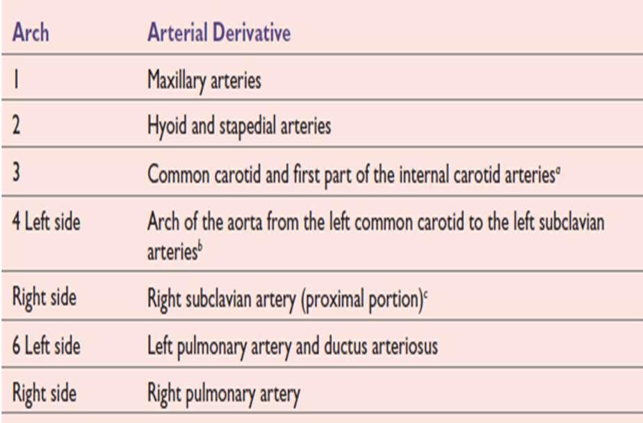
15 C- Cardinal Veins 1-This system consists of: Right and left anterior cardinal veins Right and left posterior cardinal which drain the cephalic part of the embryo veins which drain the rest of the join before entering the embryo sinus horn and form the short right and left common cardinal veins 15
16 The anterior cardinal veins common cardinal veins The posterior cardinal veins 16
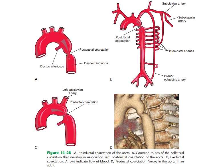
17 additional veins are formed: 2- During the fourth week, the cardinal veins form a symmetrical system During the fifth to the seventh week a number of additional veins are formed: (a)the subcardinal veins which mainly drain the kidneys (b) The sacrocardinal veins which drain the lower extremities (c) The supracardinal veins which drain the body wall by way of the intercostal veins, taking over the functions of the posterior cardinal veins 17
18 3-The anastomosis between the anterior cardinal veins develops into the left brachiocephalic vein. 4- Most of the blood from the left side of the head and the left upper extremity is then channeled to the right Now, you should have understood 18 The second left-to-right shunt occurs when the left anterior cardinal vein becomes connected To the right anterior cardinal vein by an oblique anastomosis

19 5- The terminal portion of the left posterior cardinal vein entering into the left brachiocephalic vein is retained as a small vessel, the left superior intercostal vein This vessel receives blood from the second and third intercostal spaces You should understand, by now, why the left superior intercostal vein Drains into the left brachiocephalic vein 19

20 6- The superior vena cava is formed by A-The right common cardinal vein B-The proximal portion of the right anterior cardinal vein A B 20


21 Clinical correlates Failure of the right brachiocephalic vein to form Persistence of the left anterior cardinal vein Left superior vena cava Double superior vena cava
22 AORTIC ARCH SYSTEM The major arteries in an early embryo are represented by a pair of vessels THE DORSAL AORTAE, which run with the long axis of the embryo and form the continuation of the endocardial heart tubes. The cranial portion of each dorsal aorta forms an arc on both sides of the foregut, thus establishing the first pair of aortic arch arteries, termed aortic arches 22

23 Aortic Arches Arterial System they run within branchial (pharyngeal) arches These arteries, the aortic arches, arise from the aortic sac, the most distal part of the truncus arteriosus. The aortic sac, giving rise to a total of five pairs of arteries. The pharyngeal arches and their vessels appear in a cranial-to-caudal sequence, so that they are not all present simultaneously. Consequently, the five arches are numbered I, II, III, IV, and VI. During further development, this arterial pattern becomes modified, and some vessels regress completely.

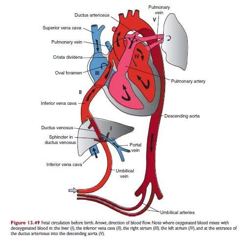
24 The fifth aortic arch either never forms or forms incompletely and then regresses. The sixth aortic arch also known as the pulmonary arch, gives off an important branch that grows toward the developing lung bud. On the right side, the proximal part becomes the proximal segment of the right pulmonary artery. The distal portion of this arch loses its connection with the dorsal aorta and disappears. On the left, the distal part persists during intrauterine life as THE DUCTUS ARTERIOSUS The proximal part of the artery persists as the proximal 24 part of the left pulmonary artery
25
 occurs in approximately 10% of children with CHDs.")
26 Coarctation of the aorta is a congenital narrowing of the aorta just proximal, opposite, or distal to the site of attachment of the ligamentum arteriosum However, most constrictions occur distal to the origin of the left subclavian artery, at the entrance of the DA (juxtaductal coarctation) occurs in approximately 10% of children with CHDs. A classification system of preductal and postductal coarctations is commonly used; however, in 90% of cases, the coarctation is directly opposite the DA. Coarctation occurs two times as often in males as in females, 26
27 Cause: this condition is believed to result from an unusual quantity of ductus arteriosus muscle tissue in the wall of the aorta. When the ductus arteriosus contracts, the ductal muscle in the aortic wall also contracts, and the aortic lumen becomes narrowed. Later, when fibrosis takes place, the aortic wall also is involved, and permanent narrowing occurs 27

28 Clinically, the cardinal sign of aortic coarctation is absent or diminished pulses in the femoral arteries of both lower limbs. To compensate for the diminished volume of blood reaching the lower part of the body, an enormous collateral circulation develops, with dilatation of the internal thoracic, subclavian, and posterior intercostal arteries. The dilated intercostal arteries erode the lower borders of the ribs, producing characteristic notching, which is seen on radiographic examination. The condition should be treated surgically 28
29 29
30 Ductus Arteriosus and Ligamentum Arteriosum Functional closure of the DA is usually completed 10 to 15 hours after birth. Anatomical closure of the DA and formation of the ligamentum arteriosum usually occurs by the 12th postnatal week 30
, aortic blood is shunted into the pulmonary artery PDA is the most common birth defect associated with maternal rubella infection during early pregnancy.")
31 Patent ductus arteriosus (PDA) a common birth defect, occurs two to three times more frequently in females than in males Functional closure of the PDA usually occurs soon after birth; however, if it remains patent (open), aortic blood is shunted into the pulmonary artery PDA is the most common birth defect associated with maternal rubella infection during early pregnancy. Preterm neonates and those born at high altitude may have PDA; this patency is the result of hypoxia (decrease of oxygen) and immaturity. The embryologic basis of PDA is failure of the DA to involute after birth and form the ligamentum arteriosum 31
32 32
33 Arterial systems associated with the fetal heart During fetal circulation, oxygenated blood flood from the placenta to the fetus passes through the umbilical vein. Three vascular shunts develop in the fetal circulation to bypass blood flow around the liver and lungs The ductus venosus allows oxygenated blood in the umbilical vein to bypass the sinusoids of the liver into the inferior vena cava and to the right atrium. From the right atrium, oxygenated blood flows mostly through the foramen ovale into the left atrium then left ventricle and into the systemic circulation. The foramen ovale develops during atrial septation to allow oxygenated blood to bypass the pulmonary circulation. Note that this is a right-toleft shunting of blood during fetal life. During fetal circulation, the superior vena cava drains deoxygenated blood from the upper limbs and head into the right atrium. Most of this blood flow is directed into the right ventricle and into the pulmonary trunk. The ductus arteriosus opens into the underside of the aorta just distal to the origin of the left subclavian artery and shunts this deoxygenated blood from the pulmonary trunk to the aorta to bypass the pulmonary circulation 33
34 fetal circulation
35
36 Circulatory Changes at Birth During prenatal life, the placental circulation provides the fetus with its oxygen, but after birth, the lungs take on gas exchange. In the circulatory system, the following changes take place at birth and in the first postnatal months: (1) the ductus arteriosus closes (2) the oval foramen closes (3) the umbilical vein and ductus venosus close and remain as the ligamentum teres hepatis and ligamentum venosum (4) the umbilical arteries form the medial umbilical ligaments.

37 37

38 Ligamentum arteriosum ligamentum venosum ligamentum teres hepatis the umbilical arteries form the medial umbilical ligaments. 38
39 انتهى بحمد هللا 39
The Cardiovascular System (Part II)
") The Cardiovascular System (Part II) 黃敏銓 mchuang@ntu.edu.tw 解剖學暨細胞生物學研究所 1 Development of veins Three paired veins drain into the tubular heart of a 4-week embryo Vitelline veins: poorly oxygenated blood
The Cardiovascular System (Part II) 黃敏銓 mchuang@ntu.edu.tw 解剖學暨細胞生物學研究所 1 Development of veins Three paired veins drain into the tubular heart of a 4-week embryo Vitelline veins: poorly oxygenated blood
DEVELOPMENT OF THE CIRCULATORY SYSTEM L E C T U R E 5
 DEVELOPMENT OF THE CIRCULATORY SYSTEM L E C T U R E 5 REVIEW OF CARDIAC ANATOMY Heart 4 chambers Base and apex Valves Pericardial sac 3 layers: epi, myo, endo cardium Major blood vessels Aorta and its
Large veins of the thorax Brachiocephalic veins
 Large veins of the thorax Brachiocephalic veins Right brachiocephalic vein: formed at the root of the neck by the union of the right subclavian & the right internal jugular veins. Left brachiocephalic
Heart & vascular system I. Dawei Dong
 Heart & vascular system I Dawei Dong Lecture goal Learn the basics of heart and vascular development. Development of Heart, Blood, and Blood Vessels LEARNING GOALS: 1. explain the early development of
Embryology of the Heart
 *Page 1A: Embryology of the Heart Human embryonic disc is divided into three layers: ectoderm, intraembryonic mesoderm, and endoderm. The embryonic disc lies between the amniotic cavity and the primary
Development of the Great Vessels and Conduc6on Tissue
 Development of the Great Vessels and Conduc6on Tissue Development of the heart fields h:p://php.med.unsw.edu.au/embryology/ index.php?6tle=advanced_- _Heart_Fields! 2 Septa6on of the Bulbus Cordis Bulbus
Transcription for Narration of Embryology of the Great Arteries
 Transcription for Narration of Embryology of the Great Arteries Slide 1: In this presentation I am going to describe for you the development of what are known as the great arteries. The great arteries
Blood Vessels. Types of Blood Vessels Arteries carry blood away from the heart Capillaries smallest blood vessels. Veins carry blood toward the heart
 C H A P T E R Blood Vessels 20 Types of Blood Vessels Arteries carry blood away from the heart Capillaries smallest blood vessels The site of exchange of molecules between blood and tissue fluid Veins
CONGENITAL HEART DISEASE (CHD)
") CONGENITAL HEART DISEASE (CHD) DEFINITION It is the result of a structural or functional abnormality of the cardiovascular system at birth GENERAL FEATURES OF CHD Structural defects due to specific disturbance
CONGENITAL HEART DISEASE (CHD) DEFINITION It is the result of a structural or functional abnormality of the cardiovascular system at birth GENERAL FEATURES OF CHD Structural defects due to specific disturbance
Development and teratology of cardiovascular and lymphatic systems. Repetition: Muscle tissue
 Development and teratology of cardiovascular and lymphatic systems Repetition: Muscle tissue Beginning of the cardiovascular system development the 3rd week: Hemangiogenesis (day 15 16) blood islets (insulae
Circumcaval Ureter: Embryology
 european urology supplements 5 (2006) 444 448 available at www.sciencedirect.com journal homepage: www.europeanurology.com Circumcaval Ureter: Embryology Arianna Lesma *, Aldo Bocciardi, Patrizio Rigatti
The Blood Vessels of the Thorax
 5 The Blood Vessels of the Thorax Chapter Outline Large Arteries of the Thorax 78 The Aorta 78 Ascending Aorta and the Pericardium 78 Aneurysm of the Arch of the Aorta 78 Coarctation of the Aorta 78 Trauma
The Physiology of the Fetal Cardiovascular System
 The Physiology of the Fetal Cardiovascular System Jeff Vergales, MD, MS Department of Pediatrics Division of Pediatric Cardiology jvergales@virginia.edu Disclosures I serve as the medical director for
Anatomy & Physiology
 1 Anatomy & Physiology Heart is divided into four chambers, two atrias & two ventricles. Atrioventricular valves (tricuspid & mitral) separate the atria from ventricles. they open & close to control flow
Development of the Liver and Pancreas
 Development of the Liver and Pancreas Professor Alfred Cuschieri Department of Anatomy University of Malta Three glandular buds arise from the distal end of the foregut during the fourth week Day 22 -The
2. capillaries - allow exchange of materials between blood and tissue fluid
 Chapter 19 - Vascular System A. categories and general functions: 1. arteries - carry blood away from heart 2. capillaries - allow exchange of materials between blood and tissue fluid 3. veins - return
Cardiovascular System Blood Vessels
 Cardiovascular System Blood Vessels Structure of Blood Vessels The three layers (tunics) Tunica intima composed of simple squamous epithelium Tunica media sheets of smooth muscle Contraction vasoconstriction
A. Incorrect! Think of a therapy that reduces prostaglandin synthesis. B. Incorrect! Think of a therapy that reduces prostaglandin synthesis.
 USMLE Step 1 - Problem Drill 02: Embryology Question No. 1 of 10 1. A premature infant is born with a patent ductus arteriosis. Which of the following treatments may be used as part of the treatment regimen?
IN THE NAME OF GOD. Development of the Heart and Vasculature
 IN THE NAME OF GOD Development of the Heart and Vasculature Overview vascular system appears (middle of 3 rd week) when the embryo is not able to satisfy its nutrition by diffusion Heart is the first functional
Circulatory system. Lecture #2
 Circulatory system Lecture #2 The essential components of the human cardiovascular system: Heart Blood Blood vessels Arteries - blood vessels that conduct arterial blood from heart ventricle to organs
Spleen. Vertebrate hearts Pericardial cavity division in coelum. Vessel walls. Endocardium = endothelium of blood vessels. Artery elastic tissue
 Spleen White pulp macrophages, monocyte storage Red pulp - (RBC) storage, and prod n (in nonmammals) Vertebrate hearts Pericardial cavity division in coelum Endocardium = endothelium of blood vessels Fig.
Development of Blood Vessels and Fetal Circulation *
 OpenStax-CNX module: m46610 1 Development of Blood Vessels and Fetal Circulation * OpenStax This work is produced by OpenStax-CNX and licensed under the Creative Commons Attribution License 3.0 By the
REVIEW SHEET Anatomy of Blood Vessels
 REVIEW SHEET Anatomy of Blood Vessels Name LabTime/Date Microscopic Structure of the Blood Vessels 1. Cross-sectional views of an aftery of a vein are shown here. ldentify each; on the lines to the sides,
The Cardiovascular System (Part I) 黃敏銓 解剖學暨細胞生物學研究所
 黃敏銓 解剖學暨細胞生物學研究所") The Cardiovascular System (Part I) 黃敏銓 解剖學暨細胞生物學研究所 1 Congenital heart defects (CHDs) 台灣兒童心臟學會 Sinus venarum Membranous septum Conus arteiosus (infundibulum) Aortic vestibule The Cardiovascular System
The Cardiovascular System (Part I) 黃敏銓 解剖學暨細胞生物學研究所 1 Congenital heart defects (CHDs) 台灣兒童心臟學會 Sinus venarum Membranous septum Conus arteiosus (infundibulum) Aortic vestibule The Cardiovascular System
The Cardiovascular System
 PowerPoint Lecture Slide Presentation by Patty Bostwick-Taylor, Florence-Darlington Technical College The Cardiovascular System 11PART B The Heart: Cardiac Output Cardiac output (CO) Amount of blood pumped
6. Development of circulatory system II. Cardiac looping. Septation of atria and ventricles. Common heart malformations.
 6. Development of circulatory system II. Cardiac looping. Septation of atria and ventricles. Common heart malformations. Formation of heart tube paired endothelial-lined heart tube is formed from blood
Large Arteries of Heart
 Cardiovascular System (Part A-2) Module 5 -Chapter 8 Overview Arteries Capillaries Veins Heart Anatomy Conduction System Blood pressure Fetal circulation Susie Turner, M.D. 1/5/13 Large Arteries of Heart
Development of the heart
 Development of the heart Prof. Abdulameer Al-Nuaimi E-mail: a.al-nuaimi@sheffield.ac.uk abdulameerh@yahoo.com Early Development of the Circulatory System Appears in the middle of the third week, when the
BOGOMOLETS NATIONAL MEDICAL UNIVERSITY DEPARTMENT OF HUMAN ANATOMY. Guidelines. Module 2 Topic of the lesson Aorta. Thoracic aorta.
 BOGOMOLETS NATIONAL MEDICAL UNIVERSITY DEPARTMENT OF HUMAN ANATOMY Guidelines Academic discipline HUMAN ANATOMY Module 2 Topic of the lesson Aorta. Thoracic aorta. Course 1 The number of hours 3 1. The
VESSELS: GROSS ANATOMY
 ACTIVITY 10: VESSELS AND CIRCULATION OBJECTIVES: 1) How to get ready: Read Chapter 23, McKinley et al., Human Anatomy, 4e. All text references are for this textbook. 2) Observe and sketch histology slide
Notes: 1)Membranous part contribute in the formation of small portion in the septal cusp.
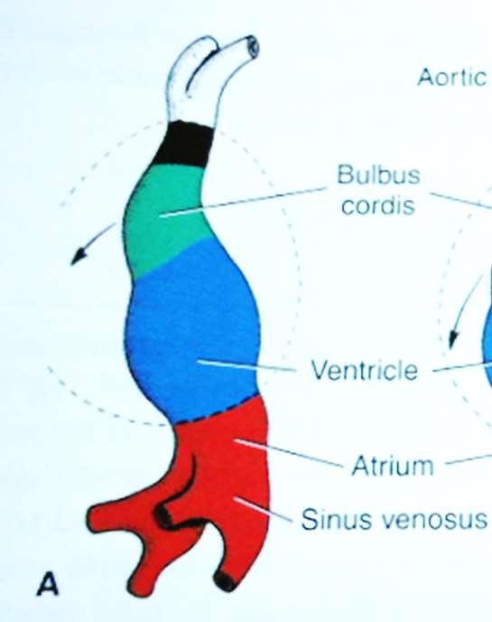
Membranous part contribute in the formation of small portion in the septal cusp.") Embryology 9 : Slide 16 : There is a sulcus between primitive ventricular and bulbis cordis that will disappear gradually and lead to the formation of one chamber which is called bulboventricular chamber.
Embryology 9 : Slide 16 : There is a sulcus between primitive ventricular and bulbis cordis that will disappear gradually and lead to the formation of one chamber which is called bulboventricular chamber.
The Cardiovascular System (Part II)
") The Cardiovascular System (Part II) 1 Aortic arch derivatives Aortic arches 2 Pharyngeal and aortic arches 4 th week 3 (1st, 2nd pairs disappeared) 6 th week (37 days) 4 8th week: transformed into the
The Cardiovascular System (Part II) 1 Aortic arch derivatives Aortic arches 2 Pharyngeal and aortic arches 4 th week 3 (1st, 2nd pairs disappeared) 6 th week (37 days) 4 8th week: transformed into the
When you see this diagram, remember that you are looking at the embryo from above, through the amniotic cavity, where the epiblast appears as an oval
 When you see this diagram, remember that you are looking at the embryo from above, through the amniotic cavity, where the epiblast appears as an oval disc 2 Why the embryo needs the vascular system? When
PIAF study: Placental insufficiency and aortic isthmus flow Jean-Claude Fouron, MD
 Dear colleagues, I would like to thank you very sincerely for agreeing to participate in our multicentre study on the clinical significance of recording fetal aortic isthmus flow during placental circulatory
Heart Development and Congenital Heart Disease
 Heart Development and Congenital Heart Disease Sally Dunwoodie s.dunwoodie@victorchang.edu.au Developmental and Stem Cell Biology Division Victor Chang Cardiac Research Institute for the heart of Australia...
Overview of Anatomy and Physioloy II Second Year Students
 University of Baghdad College of Nursing Department of Basic Medical Sciences Overview of Anatomy and Physioloy II Second Year Students Asaad Ismail Ahmad, Ph.D. Asaad Ismail Ahmad, Ph.D. Electrolyte and
YOU MUST BRING GLOVES FOR THIS ACTIVITY
 ACTIVITY 10: VESSELS AND CIRCULATION OBJECTIVES: 1) How to get ready: Read Chapter 23, McKinley et al., Human Anatomy, 5e. All text references are for this textbook. 2) Observe and sketch histology slide
THE CIRCULATORY SYSTEM
 Biology 30S THE CIRCULATORY SYSTEM Name: This module adapted from bblearn.merlin.mb.ca 1 Introduction to Circulation The first organ to form, and the last organ to die. The heart is the pump of life. The
3 Circulatory Pathways
 40 Chapter 3 Circulatory Pathways Systemic Arteries -Arteries carry blood away from the heart to the various organs of the body. -The aorta is the longest artery in the body; it branches to give rise to
Figure 10.1A Transparency Master 79
 Brain Carotid arteries Jugular vein Right front leg Lungs (inflated) Cranial Right atrium To left front leg Left subclavian Bronchus capillaries Brachiocephalic vein Left atrium Dorsal aorta Right ventricle
Heart and Lungs. LUNG Coronal section demonstrates relationship of pulmonary parenchyma to heart and chest wall.
 Heart and Lungs Normal Sonographic Anatomy THORAX Axial and coronal sections demonstrate integrity of thorax, fetal breathing movements, and overall size and shape. LUNG Coronal section demonstrates relationship
Arteries. Lecture #2
 Arteries Lecture #2 The essential components of the human cardiovascular system: Heart Blood Blood vessels Arteries - blood vessels that conduct arterial blood from heart ventricle to organs and tissues
Surgical Management Of TAPVR. Daniel A. Velez, M.D. Congenital Cardiac Surgeon Phoenix Children s Hospital
 Surgical Management Of TAPVR Daniel A. Velez, M.D. Congenital Cardiac Surgeon Phoenix Children s Hospital No Disclosures Goals Review the embryology and anatomy Review Surgical Strategies for repair Discuss
Dr. Weyrich G07: Superior and Posterior Mediastina. Reading: 1. Gray s Anatomy for Students, chapter 3
 Dr. Weyrich G07: Superior and Posterior Mediastina Reading: 1. Gray s Anatomy for Students, chapter 3 Objectives: 1. Subdivisions of mediastinum 2. Structures in Superior mediastinum 3. Structures in Posterior
Development of the Heart
 Development of the Heart Thomas A. Marino, Ph.D. Temple University School of Medicine Stages of Development of the Heart 1. The horseshoe-shaped pericardial cavity. 2. The formation of the single heart
By Dickens ATURWANAHO & ORIBA DAN LANGOYA MAKchs, MBchB CONGENTAL HEART DISEASE
 By Dickens ATURWANAHO & ORIBA DAN LANGOYA MAKchs, MBchB CONGENTAL HEART DISEASE Introduction CHDs are abnormalities of the heart or great vessels that are present at birth. Common type of heart disease
Congenital Heart Defects
 Normal Heart Congenital Heart Defects 1. Patent Ductus Arteriosus The ductus arteriosus connects the main pulmonary artery to the aorta. In utero, it allows the blood leaving the right ventricle to bypass
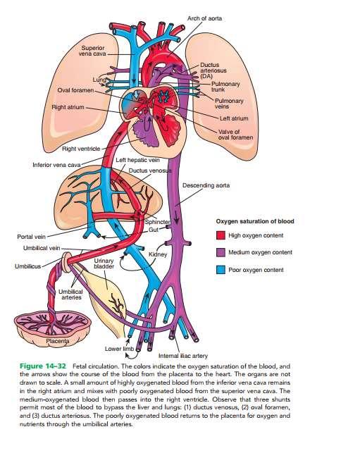
Chapter 14. The Cardiovascular System
 Chapter 14 The Cardiovascular System Introduction Cardiovascular system - heart, blood and blood vessels Cardiac muscle makes up bulk of heart provides force to pump blood Function - transports blood 2
Cardiovascular Anatomy Dr. Gary Mumaugh
 Cardiovascular Anatomy Dr. Gary Mumaugh Location of Heart Approximately the size of your fist Location o Superior surface of diaphragm o Left of the midline in mediastinum o Anterior to the vertebral column,
Unit 11 - The Cardiovascular System 1
 Unit 11 - The Cardiovascular System 1 I. Unit 11: The Cardiovascular System A. The Cardiovascular System 1. A closed system of the heart and blood vessels a) The heart pumps blood b) Blood vessels allow
COPYRIGHTED MATERIAL. The fetal circulation CHAPTER 1. Postnatal circulation
 1 CHAPTER 1 The fetal circulation The circulation in the fetus differs from that in the adult. Knowledge of the course and distribution of the fetal circulation is important to our understanding of the
MODULE 2: CARDIOVASCULAR SYSTEM ANTOMY An Introduction to the Anatomy of the Heart and Blood vessels
 MODULE 2: CARDIOVASCULAR SYSTEM ANTOMY An Introduction to the Anatomy of the Heart and Blood vessels The cardiovascular system includes a pump (the heart) and the vessels that carry blood from the heart
Day 5 Respiratory & Cardiovascular: Respiratory System
 Day 5 Respiratory & Cardiovascular: Respiratory System Be very careful not to damage the heart and lungs while separating the ribs! Analysis Questions-Respiratory & Cardiovascular Log into QUIA using your
AORTIC COARCTATION. Synonyms: - Coarctation of the aorta
 AORTIC COARCTATION Synonyms: - Coarctation of the aorta Definition: Aortic coarctation is a congenital narrowing of the aorta, usually located after the left subclavian artery, near the ductus or the ligamentum
Application of a flow chart to evaluate placement of umbilical venous catheters on frontal radiogram in radiologist resident
 Application of a flow chart to evaluate placement of umbilical venous catheters on frontal radiogram in radiologist resident Poster No.: C-0883 Congress: ECR 2015 Type: Scientific Exhibit Authors: S. Salerno,
Paediatric Cardiology. Acyanotic CHD. Prof F F Takawira
 Paediatric Cardiology Acyanotic CHD Prof F F Takawira Aetiology Chromosomal Down syndrome, T13, T18 Genetic syndromes (gene defects) Velo-Cardio-facial (22 del) Genetic syndromes (undefined aetiology)
Chapter 14. Circulatory System Images. VT-122 Anatomy & Physiology II
 Chapter 14 Circulatory System Images VT-122 Anatomy & Physiology II The mediastinum Dog heart Dog heart Cat heart Dog heart ultrasound Can see pericardium as distinct bright line Pericardial effusion Fluid
The Cardiovascular System
 PowerPoint Lecture Slide Presentation by Patty Bostwick-Taylor, Florence-Darlington Technical College The Cardiovascular System 11PART A The Cardiovascular System A closed system of the heart and blood
THE VESSELS OF BLOOD CIRCULATION
 THE VESSELS OF BLOOD CIRCULATION scientistcindy.com /the-vessels-of-blood-circulation.html NOTE: You should familiarize yourself with the anatomy of the heart and have a good understanding of the flow
W.S. O The University of Hong Kong
 W.S. O The University of Hong Kong Objectives: Describe early angiogenesis. Describe the heart tube formation. Describe the partitioning into a 4- chambered heart. List the formation of heart valves and
Unit 11: The Cardiovascular System
 Unit 11: The Cardiovascular System I. The Cardiovascular System A. A closed system of the heart and blood vessels 1. The heart pumps blood 2. Blood vessels allow blood to circulate to all parts of the
Approximately the size of your fist Location Superior surface of diaphragm Left of the midline in mediastinum Anterior to the vertebral column,
 Dr. Gary Mumaugh Approximately the size of your fist Location Superior surface of diaphragm Left of the midline in mediastinum Anterior to the vertebral column, posterior to the sternum Posteriorly the
1. Which of the following blood vessels has a thin elastic layer? A. Aorta. B. Pulmonary artery. C. Posterior vena cava. D. Mesenteric capillary.
 CIRCULATORY SYSTEM 1. Which of the following blood vessels has a thin elastic layer? A. Aorta. B. Pulmonary artery. C. Posterior vena cava. D. Mesenteric capillary. 2. Capillary beds are equipped with
Infradiaphragmatic Total Anomalous Pulmonary Venous Return
 Infradiaphragmatic Total Anomalous Pulmonary Venous Return Report of a New and Correctable Variant J. Kent Trinkle, M.D., Gordon K. Danielson, M.D., Jacqueline A. Noonan, M.D., and Charles Stephens, M.D.
Human Anatomy Biology 351
 nnnnn 1 Human Anatomy Biology 351 Exam #2 Please place your name on the back of the last page of this exam. You must answer all questions on this exam. Because statistics demonstrate that, on average,
PRACTICAL GUIDE TO FETAL ECHOCARDIOGRAPHY IC Huggon and LD Allan
 PRACTICAL GUIDE TO FETAL ECHOCARDIOGRAPHY IC Huggon and LD Allan Fetal Cardiology Unit, Harris Birthright Research Centre for Fetal Medicine, King's College Hospital, London, UK IMPORTANCE OF PRENATAL
Development of the Heart *
 OpenStax-CNX module: m46673 1 Development of the Heart * OpenStax This work is produced by OpenStax-CNX and licensed under the Creative Commons Attribution License 3.0 By the end of this section, you will
W.S. O. School of Biomedical Sciences, University of Hong Kong
 W.S. O School of Biomedical Sciences, University of Hong Kong Objectives: Describe early angiogenesis. Describe the heart tube formation. Describe the partitioning into a 4- chambered heart. List the formation
Development of the Digestive System. W.S. O The University of Hong Kong
 Development of the Digestive System W.S. O The University of Hong Kong Plan for the GI system Then GI system in the abdomen first develops as a tube suspended by dorsal and ventral mesenteries. Blood
Midgut. Over its entire length the midgut is supplied by the superior mesenteric artery
 Gi Embryology 3 Midgut the midgut is suspended from the dorsal abdominal wall by a short mesentery and communicates with the yolk sac by way of the vitelline duct or yolk stalk Over its entire length the
Biology 340 Comparative Embryology Lecture 10 Dr. Stuart Sumida. Further Development of the Mesoderm (and Endoderm)
") Biology 340 Comparative Embryology Lecture 10 Dr. Stuart Sumida Further Development of the Mesoderm (and Endoderm) Further Development: Digestive System Foregut, Midgut, Hindgut Heart and Aortic Arches
Biology 340 Comparative Embryology Lecture 10 Dr. Stuart Sumida Further Development of the Mesoderm (and Endoderm) Further Development: Digestive System Foregut, Midgut, Hindgut Heart and Aortic Arches
SLIDES 6 AND 10 MM PIG SLIDES; TRANSVERSE SECTIONS AND SAGITAL; FETAL PIGS-1-8INCH; HUMAN SAGITAL DIAGRAMS:DRAWINGS OF THE PIG SECTIONS TO BE
 SLIDES 6 AND 10 MM PIG SLIDES; TRANSVERSE SECTIONS AND SAGITAL; FETAL PIGS-1-8INCH; HUMAN SAGITAL SECTION DIAGRAMS:DRAWINGS OF THE PIG SECTIONS TO BE LABELLED, AND FOUR DRAWINGS TO BE MADE. REFERENCES:PATTEN:
Chapter 2 Cardiac Interpretation of Pediatric Chest X-Ray
 Chapter 2 Cardiac Interpretation of Pediatric Chest X-Ray Ra-id Abdulla and Douglas M. Luxenberg Key Facts The cardiac silhouette occupies 50 55% of the chest width on an anterior posterior chest X-ray
Lab Activity 25. Blood Vessels & Circulation. Portland Community College BI 232
 Lab Activity 25 Blood Vessels & Circulation Portland Community College BI 232 Artery and Vein Histology Walls have 3 layers: Tunica intima Tunica media Tunica externa 2 Tunica Intima Is the innermost layer
1. Distinguish among the types of blood vessels on the basis of their structure and function.
 Blood Vessels and Circulation Objectives This chapter describes the structure and functions of the blood vessels Additional subjects contained in Chapter 13 include cardiovascular physiology, regulation,
C3, 4, 5, 6, & 7 Worksheet. C3 Describe the inter-relationships of the structures of the heart
 Name: Date: C3, 4, 5, 6, & 7 Worksheet C3 Describe the inter-relationships of the structures of the heart 1. Label and give the functions of the following: a. left and right atrium: b. left and right ventricle:
Development of the Digestive System. W.S. O School of Biomedical Sciences, University of Hong Kong.
 Development of the Digestive System W.S. O School of Biomedical Sciences, University of Hong Kong. Organization of the GI tract: Foregut (abdominal part) supplied by coeliac trunk; derivatives include
The Cardiovascular System. The Structure of Blood Vessels. The Structure of Blood Vessels. The Blood Vessels. Blood Vessel Review
 The Cardiovascular System The Blood Vessels The Structure of Blood Vessels Blood Vessel Review Arteries carry blood away from the heart Pulmonary trunk to lungs Aorta to everything else Microcirculation
Figure ) The specific chamber of the heart that is indicated by letter A is called the. Diff: 1 Page Ref: 364
 The specific chamber of the heart that is indicated by letter A is called the. Diff: 1 Page Ref: 364") Essentials of Anatomy and Physiology, 9e (Marieb) Chapter 11 The Cardiovascular System Short Answer Figure 11.1 Using Figure 11.1, identify the following: 1) The Purkinje fibers are indicated by label.
Essentials of Anatomy and Physiology, 9e (Marieb) Chapter 11 The Cardiovascular System Short Answer Figure 11.1 Using Figure 11.1, identify the following: 1) The Purkinje fibers are indicated by label.
HUMAN HEART. Learn the following structures on the heart models.
 HUMAN HEART Learn the following structures on the heart models. The human heart has four chambers that consist of the right atrium, left atrium, right ventricle, and left ventricle. The atria are smaller
Cardiovascular System. Chapter 22
 Cardiovascular System Blood Vessels Chapter 22 Cardiac Contractions Cardiac cycle consists of alternate periods of contraction and relaxation Contraction is the systolic pressure Blood is ejected into
Chapter 5: Other mediastinal structures. The Large Arteries. The Aorta. Ascending aorta
 Chapter 5: Other mediastinal structures The Large Arteries The Aorta The aorta is the main arterial trunk of the systemic circulation and in the healthy state its wall contain a large amount of yellow
This lab activity is aligned with Visible Body s A&P app. Learn more at visiblebody.com/professors
 1 This lab activity is aligned with Visible Body s A&P app. Learn more at visiblebody.com/professors 2 PRE-LAB EXERCISES: A. Watch the video 29.1 Heart Overview and make the following observations: 1.
Chest and cardiovascular
 Module 1 Chest and cardiovascular A. Doss and M. J. Bull 1. Regarding the imaging modalities of the chest: High resolution computed tomography (HRCT) uses a slice thickness of 4 6 mm to identify mass lesions
Lecture 02 Anatomy of the LIVER
 Lecture 02 Anatomy of the LIVER BY Dr Farooq Khan Aurakzai Dated: 02.01.2018 Introduction to Liver Largest gland in the body. 2 nd largest organ of the body. Weight approximately 1500 gm, and is roughly
Heart and Soul Evaluation of the Fetal Heart
 Heart and Soul Evaluation of the Fetal Heart Ivana M. Vettraino, M.D., M.B.A. Clinical Associate Professor, Michigan State University College of Human Medicine Objectives Review the embryology of the formation
Development of pancreas and Small Intestine. ANATOMY DEPARTMENT DR.SANAA AL-AlSHAARAWY DR.ESSAM Eldin Salama
 Development of pancreas and Small Intestine ANATOMY DEPARTMENT DR.SANAA AL-AlSHAARAWY DR.ESSAM Eldin Salama OBJECTIVES At the end of the lecture, the students should be able to : Describe the development
Systematic approach to Fetal Echocardiography. Objectives. Introduction 11/2/2015
 Systematic approach to Fetal Echocardiography. Pediatric Echocardiography Conference, JCMCH November 7, 2015 Rajani Anand Objectives Fetal cardiology pre-test Introduction Embryology and Physiology of
THE AORTA AND IT S MAJOR BRANCHES
 1 THE AORTA AND IT S MAJOR BRANCHES The aorta commences at the aortic valve, above the vestible of the left ventricle and terminates at the level of the fourth lumbar vertebra (L4), where it bifurcates
The fetal circulation
 Peter John Murphy MB ChB DA FRCA The fetal circulation (Fig. 1) is markedly different from the adult circulation. In the fetus, gas exchange does not occur in the lungs but in the placenta. The placenta
The Foregut. At first the esophagus is short. but with descent of the heart and lungs it lengthens rapidly
 GI embryology 2 The Foregut At first the esophagus is short but with descent of the heart and lungs it lengthens rapidly The muscular coat, which is formed by surrounding splanchnic mesenchyme, is striated
CARDIOVASCULAR SYSTEM
 CARDIOVASCULAR SYSTEM CARDIAC SYSTEM TWO TYPES OF CIRCULATION Systemic system delivers blood to ALL body cells and carries away waste. The red blood cells use hemoglobin to carry oxygen to the cells Pulmonary
4/19/2018. St. Cloud Hospital Perinatology Kristin Olson, RDMS, RVT
 St. Cloud Hospital Perinatology Kristin Olson, RDMS, RVT Review Fetal Circulation Provide Indications for Umbilical Artery, Middle Cerebral Artery, and Ductus Venosus Doppler studies. Demonstrate normal
Skeletal muscle. Flow increases and decreases with each muscular contraction - as a result of compression of the blood vessels by contracted muscle
 Regional blood flow Skeletal muscle Extreme increases during exercises Flow increases and decreases with each muscular contraction - as a result of compression of the blood vessels by contracted muscle
Ch.15 Cardiovascular System Pgs {15-12} {15-13}
 Ch.15 Cardiovascular System Pgs {15-12} {15-13} E. Skeleton of the Heart 1. The skeleton of the heart is composed of rings of dense connective tissue and other masses of connective tissue in the interventricular
6. HEART AND CIRCULATORY SYSTEM I
 6. HEART AND CIRCULATORY SYSTEM I Dr. Taube P. Rothman P&S 12-520 Tpr2@columbia.edu 212-305-7930 RECOMMENDED READING: Larsen Human Embryology, 3rd Edition, pp. 195-199; 157-169 top left; 172-174; bottom
Essentials of Anatony and Physiology, 5e (Martini/Nath) Chapter 13 The Cardiovascular System: Blood Vessels and Circulation
 Chapter 13 The Cardiovascular System: Blood Vessels and Circulation") Essentials of Anatony and Physiology, 5e (Martini/Nath) Chapter 13 The Cardiovascular System: Blood Vessels and Circulation Multiple-Choice Questions 1) The muscular layer of blood vessels is the A) tunica
Essentials of Anatony and Physiology, 5e (Martini/Nath) Chapter 13 The Cardiovascular System: Blood Vessels and Circulation Multiple-Choice Questions 1) The muscular layer of blood vessels is the A) tunica
The posterior abdominal wall. Prof. Oluwadiya KS
 The posterior abdominal wall Prof. Oluwadiya KS www.oluwadiya.sitesled.com Posterior Abdominal Wall Lumbar vertebrae and discs. Muscles opsoas, quadratus lumborum, iliacus, transverse, abdominal wall
COMPREHENSIVE EVALUATION OF FETAL HEART R. GOWDAMARAJAN MD
 COMPREHENSIVE EVALUATION OF FETAL HEART R. GOWDAMARAJAN MD Disclosure No Relevant Financial Relationships with Commercial Interests Fetal Echo: How to do it? Timing of Study -optimally between 22-24 weeks
AN ATOMY OF THE CARDIOVASCULAR SYSTEM
 Student Name CHAPTER 18 AN ATOMY OF THE CARDIOVASCULAR SYSTEM T he heart is actually two pumps one moves blood to the lungs, the other pushes it out into the body. These two functions seem rather elementary