Intracranial spontaneous hemorrhage mechanisms, imaging and management
|
|
|
- Phoebe Craig
- 5 years ago
- Views:
Transcription
1 Intracranial spontaneous hemorrhage mechanisms, imaging and management Dora Zlatareva Department of Diagnostic Imaging Medical University, Sofia, Bulgaria
2 Intracranial hemorrhage (ICH) ICH 15% of strokes 25 per 100,000 pt/year Mortality -40% in 1 mo Multiple intracranial compartments By diverse pathology Neuroimaging Identify the cause of hemorrhage location and severity of hemorrhage To guide patient treatment
 Hemorrhagic necrosis (like tumor and infection) Substance abuse")
3 Mechanisms Hypertensive damage to blood vessels Rupture of ananeurysm Rupture of AVM Cerebral amyloid angiopathy Altered hemostasis (thrombolysis & anticoagulation) Hemorrhagic necrosis (like tumor and infection) Substance abuse (cocaine)


4 Location Basal ganglia Lobes of cerebrum Thalamus Pons Cerebellum Other brainstem sites
5 Causes Hypertension Cerebral amyloid angiopathy Hemorrhagic conversion of ischemic infarction Cerebral aneurysms Cerebral AVM Dural AV fistula Vasculitis Venous sinus thrombosis
6 Imaging CT first modality-acute blood-hyperdense CTA- vascular underlying cause (SAH, IPH) CT venography dural venous sinuses MRI r/o tumour Depend on time, sequence, size, location Cerebral DSA suspected vascular abnormality, CTA is either normal or equivocal, or to treat
7 AHA/ASA Guideline(2010) Guidelines for the Management of Spontaneous Intracerebral Hemorrhage Recommendations for Neuroimaging in ICH Rapid neuroimaging with CT or MRI is recommended to distinguish ischemic stroke from ICH CT angiography and contrast-enhanced CT may be considered to help identify patients at risk for hematoma expansion CTA, CT venography, CeCT, CeMRI, MRA and MRV can be useful to evaluate for underlying structural lesions including vascular malformations and tumors when there is clinical or radiologic suspicion Class I, Level of Evidence A (Unchanged from the previous) Class IIb, Level of Evidence B Class IIa, Level of Evidence B(New recommendation).
 Subacute")


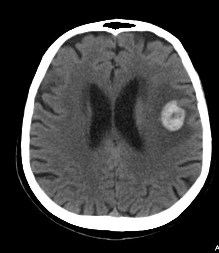
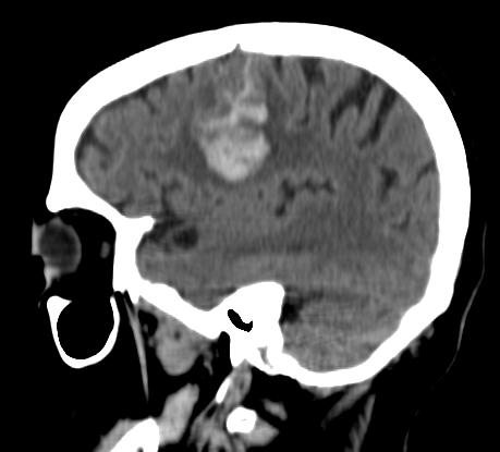
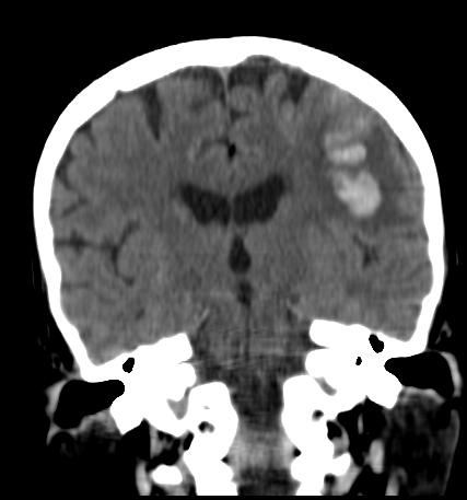
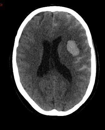
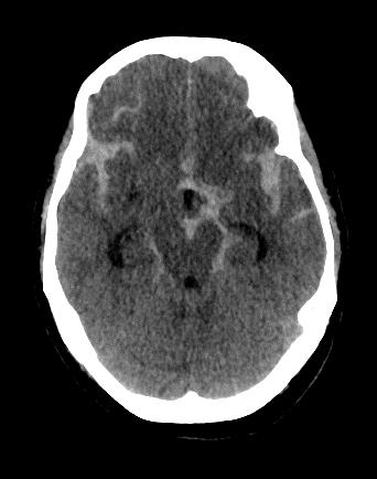
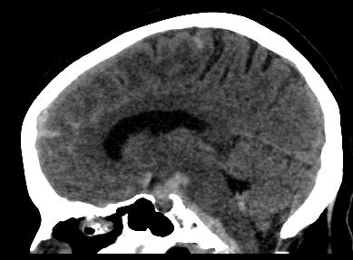
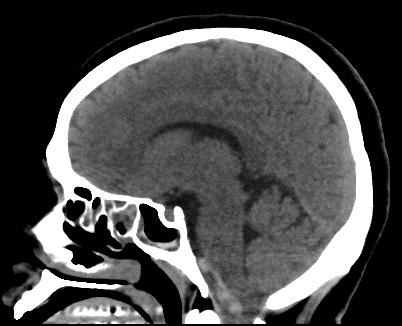
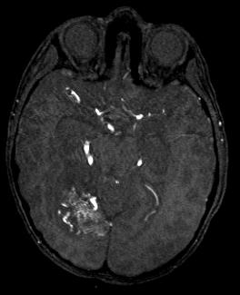
8 ICH: CT appearance Acute phase: Hyperdense mass (50-80 HU) Subacute phase (1-6 weeks) Peripheral edema Attenuation falls1.5 HU / day from the periphery chemical breakdown of globin Hb < 8-10g/dl isodense hematoma


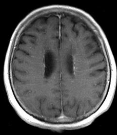
9 ICH: CT appearance Chronic phase Hypodense lesion Sequelae gliosis Hemosiderin!!


10 ICH: CT appearance Still bleeding hematoma Swirl sign Due to semiliquid unretracted clot


11 Parizel, Eur Radiol 2001
12 IPH due to hypertension 60ies -70ies, 30-50% mortality Acute-BG, cerebellum, occipital lobes IPH cerebral cortex -consider other Dg Younger than 50 -?other causes -Tu or vascular malformation


13 IPH due to hypertension Complications Intraventricular hemorrhage Hydrocephalus Herniation Rebleeding Initial hemorrhage different size NCCT -predict outcome Worse prognosis Initial size of the hematoma intraventricular extension of the hemorrhage expansion of the hematoma on serial imaging
14 IPH due to hypertension Management Surgical treatment contoversial Hematoma >3cm Benefit of surgery?? External drainage for hydrocephalus? Treatment of systemic HT


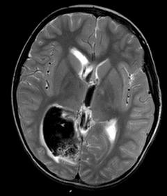
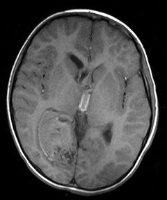
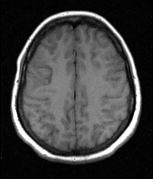
15 IPH-Cerebral amyloid angiopathy Cerebral microhemorrhages Sulcal SAH DD from vasculitis AGE >60 Other areas of ICH Large cerebral IPH -DD hypertensive Subcortical WM Spares BG, posterior fossa, brainstem


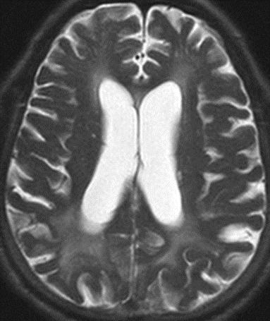
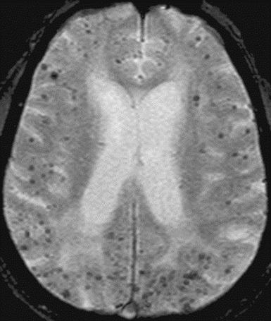

16 Linn, AJNR, 2008

17 Hemorrhagic transformation of ischemic stroke Risk post i.v./i.a. vessel recanaliza on Petechial hemorrhage in margin (HI1) Confluent PH in infarcted tissue (HI2) Parenchymal hematoma <30% infarcted tissue, slight mass effect (PH1) >30% + significant mass effect (PH2) clin. significant
18 Antithrombotic or thrombolytic Th


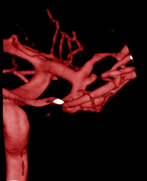
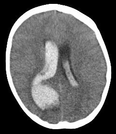
19 Cerebral aneurysms CT -100% sensitivity for acute SAH in the first 6-24H SAH in basal cisterns or diffusely through SA space Ventricles and brain parenchyma (lobar hematoma) Surgical clipping, endovascular coiling/embolization

20 SAH

21 SAH
22

 SAH CTA, MRI,MRA, DSA Heit et al, J")
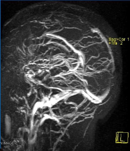
23 Cerebral AVM ICH -most common presentation of cerebral AVM IPH -(young pt or child suspect AVM) Intraventricular hemorrhage (IVH) SAH CTA, MRI,MRA, DSA Heit et al, J stroke 2017
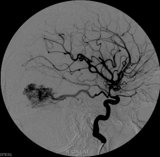
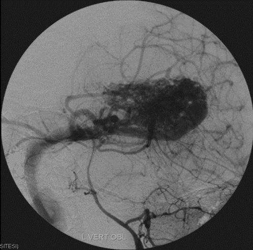

24 Management Embolization, Surgery, Radiosurgery

25 Dural AV fistula
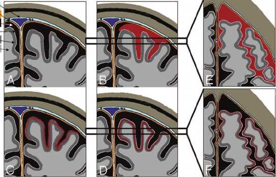
26 Vasculitis Primary, SLE, Behcet ICH or ischemic lesion Sulcal SAH near convexity -most common CT -hyperdensity within cerebral sulci MRI -sulcal hyper FLAIR, hypo GRE or SWI Sulcal SAH +NO trauma -DSAin negative CTA to r/o vasculitis or vasculopathy Immunosuppressive Th


27 CNS vasculıtıs Multiple chronic hemorrhages Perivascular enhancement Irregular vessel walls
28 Venous thrombosis Cortical vein or dural venous sinus Cord sign (hyperdense cortical vein on NCCT), empty delta sign (CT venography) Cerebral edema, parenchymal hemorrhages, ischemic and hemorrhagic infarcts SAH -uncommon, cerebral convexities or Sylvian fissures, sparing the basal cisterns


29 Venous thrombosis Uncommon and often clinically confusing entity Imaging plays an essential role in Dg Secondary to skull base infections, dehydration, hypercoagulable states, compression from meningiomas or other dural tumors


30 Superior sagittal sinusthrombosis


31 Underlying tumors Primary Tu-hemorrhage inside the Tu Metastasis- at the periphery Management-Tutherapy, surgery, Radio, Chemotherapy Choi, ET AL., Glioma Mimicking a Hypertensive Intracerebral Hemorrhage, JKNS 2013

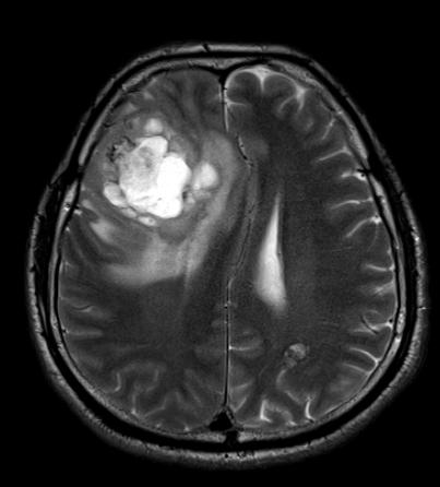
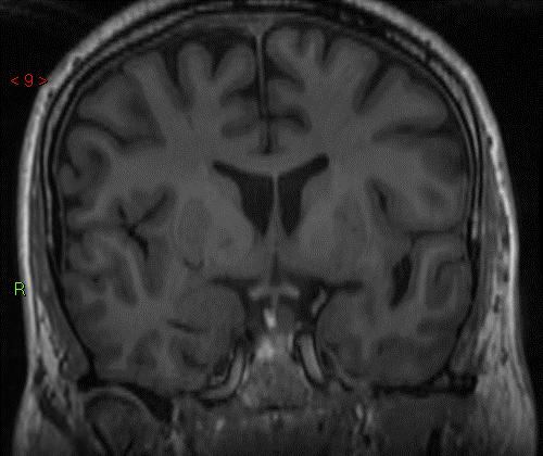
32 Tumors, cavernoma

33 Substance abuse (cocaine) Hemorrhagic or ischemic strokes IPH or SAH -twice as common as ischemic strokes 40% 50% -an underlying AVM or aneurysm Hematoma in BG, thalamus
 Consideration of early surgical intervention Prevention of secondary brain")
34 Management Initial medical stabilization Neuroimaging - establish Dg and elucidate etiology Neurologic exam - determine baseline severity Prevent hematoma expansion (BP management, reversal of coagulopathy) Consideration of early surgical intervention Prevention of secondary brain injury
35 Management Hemostatic agents (factor VIIa) early-reduce hematoma expansion, clinical effectiveness has not been shown Anticoagulation reversal with prothrombin concentrates + Vit K - in VitK antagonist-related ICH Ongoing trials for minimally invasive approaches or hemicraniectomy, role of surgery in ICH to be defined BP control, antithrombotic prevention after ICH - consider the risk of recurrent bleeding and ischemia
36 Take home messages Elderly patient with HT, BG hematoma Hypertensive hemorrhage Do SWI to see other microbleeds Eldery normotensive patient, lobar hematoma Amyloid angiopathy / tumor Do +C MRI, SWI, MRA? DSA? Enhancing lesion Tm Subcortical microbleeds on SWI Amyloid angiopathy
37 Take home messages Young patient with ICH (ALERT!!!) Do +C MRI, MRA / CTA / DSA, SWI Angio abnormal Vasc. Malf, Vasculitis, DVST Enhancing single lesion Tm Enhancing multiple lesions Met, Vasculitis, Septic emboli Multiple black dots on SWI Vasculitis, Cavernomas Patient of any age with associated SAH Aneurysm Patient of any age with ICH, you still cannot decide the etiology Refer to the clinician,short time FUpscan

38 I would like to thank to Cem Calli for ideas and cases


39 Thank you for your kind attention!
ISCHEMIC STROKE IMAGING
 ISCHEMIC STROKE IMAGING ผศ.พญ พญ.จ ร ร ตน ธรรมโรจน ภาคว ชาร งส ว ทยา คณะแพทยศาสตร มหาว ทยาล ยขอนแก น A case of acute hemiplegia Which side is the abnormality, right or left? Early Right MCA infarction
C. Douglas Phillips, MD FACR Director of Head and Neck Imaging Weill Cornell Medical College NewYork-Presbyterian Hospital
 C. Douglas Phillips, MD FACR Director of Head and Neck Imaging Weill Cornell Medical College NewYork-Presbyterian Hospital I have no financial disclosures Understand range of pathology that may present
Cerebro-vascular stroke
 Cerebro-vascular stroke CT Terminology Hypodense lesion = lesion of lower density than the normal brain tissue Hyperdense lesion = lesion of higher density than normal brain tissue Isodense lesion = lesion
NEURO IMAGING 2. Dr. Said Huwaijah Chairman of radiology Dep, Damascus Univercity
 NEURO IMAGING 2 Dr. Said Huwaijah Chairman of radiology Dep, Damascus Univercity I. EPIDURAL HEMATOMA (EDH) LOCATION Seventy to seventy-five percent occur in temporoparietal region. CAUSE Most likely caused
Neuroradiology: Imaging and Stroke
 Neuroradiology: Imaging and Stroke Stroke 2017 William Gallmann January 28, 2017 Stroke Arterial ischemia/infarct accounts for ~85% Cerebral venous occlusions - 0.5-1% Spontaneous intracranial hemorrhage
Vascular Malformations of the Brain: A Review of Imaging Features and Risks
 Vascular Malformations of the Brain: A Review of Imaging Features and Risks Comprehensive Neuroradiology: Best Practices October 27-30, 2016 Sudhakar R. Satti, MD Associate Director Neurointerventional
[(PHY-3a) Initials of MD reviewing films] [(PHY-3b) Initials of 2 nd opinion MD]
![[(PHY-3a) Initials of MD reviewing films] [(PHY-3b) Initials of 2 nd opinion MD]](/thumbs/89/98619893.jpg "[(PHY-3a) Initials of MD reviewing films] [(PHY-3b) Initials of 2 nd opinion MD]") 2015 PHYSICIAN SIGN-OFF (1) STUDY NO (PHY-1) CASE, PER PHYSICIAN REVIEW 1=yes 2=no [strictly meets case definition] (PHY-1a) CASE, IN PHYSICIAN S OPINION 1=yes 2=no (PHY-2) (PHY-3) [based on all available
2015 PHYSICIAN SIGN-OFF (1) STUDY NO (PHY-1) CASE, PER PHYSICIAN REVIEW 1=yes 2=no [strictly meets case definition] (PHY-1a) CASE, IN PHYSICIAN S OPINION 1=yes 2=no (PHY-2) (PHY-3) [based on all available
UPSTATE Comprehensive Stroke Center. Neurosurgical Interventions Satish Krishnamurthy MD, MCh
 UPSTATE Comprehensive Stroke Center Neurosurgical Interventions Satish Krishnamurthy MD, MCh Regional cerebral blood flow is important Some essential facts Neurons are obligatory glucose users Under anerobic
Diagnosis of Subarachnoid Hemorrhage (SAH) and Non- Aneurysmal Causes
 and Non- Aneurysmal Causes") Diagnosis of Subarachnoid Hemorrhage (SAH) and Non- Aneurysmal Causes By Sheila Smith, MD Swedish Medical Center 1 Disclosures I have no disclosures 2 Course Objectives Review significance and differential
Diagnosis of Subarachnoid Hemorrhage (SAH) and Non- Aneurysmal Causes By Sheila Smith, MD Swedish Medical Center 1 Disclosures I have no disclosures 2 Course Objectives Review significance and differential
Acute stroke imaging
 Acute stroke imaging Aims Imaging modalities and differences Why image acute stroke Clinical correlation to imaging appearance What is stroke Classic definition: acute focal injury to the central nervous
An Introduction to Imaging the Brain. Dr Amy Davis
 An Introduction to Imaging the Brain Dr Amy Davis Common reasons for imaging: Clinical scenarios: - Trauma (NICE guidelines) - Stroke - Tumours - Seizure - Neurological degeneration memory, motor dysfunction,
10 May Disclosure. + Outline. Case-based approach to nontraumatic intracranial hemorrhage. Kathleen R. Fink, MD University of Washington
 Kathleen R. Fink, MD University of Washington 5 th Nordic Emergency Radiology Course May 21, 2015 Disclosure My spouse receives research salary support from: Bracco BayerHealthcare Guerbet Outline Case-based
Starting or Resuming Anticoagulation or Antiplatelet Therapy after ICH: A Neurology Perspective
 Starting or Resuming Anticoagulation or Antiplatelet Therapy after ICH: A Neurology Perspective Cathy Sila MD George M Humphrey II Professor and Vice Chair of Neurology Director, Comprehensive Stroke Center
Stroke imaging. Why image stroke patients? Stroke. Treatment of infarct. Methods for infarct diagnosis. Treatment of infarct.
 Stroke imaging Stroke Infarct: -Arterial thrombosis/embolus -Hypoxic/ischemic -Venous thrombosis Non-traumatic hemorrhage: -Intracerebral -Subarachnoid Johan Wikström MD PhD Associate Professor of Radiology
Head CT Scan Interpretation: A Five-Step Approach to Seeing Inside the Head Lawrence B. Stack, MD
 Head CT Scan Interpretation: A Five-Step Approach to Seeing Inside the Head Lawrence B. Stack, MD Five Step Approach 1. Adequate study 2. Bone windows 3. Ventricles 4. Quadrigeminal cistern 5. Parenchyma
For Emergency Doctors. Dr Suzanne Smallbane November 2011
 For Emergency Doctors Dr Suzanne Smallbane November 2011 A: Orbit B: Sphenoid Sinus C: Temporal Lobe D: EAC E: Mastoid air cells F: Cerebellar hemisphere A: Frontal lobe B: Frontal bone C: Dorsum sellae
CLEAR III TRIAL : UPDATE ON SURGICAL MATTERS THAT MATTER
 CLEAR III TRIAL : UPDATE ON SURGICAL MATTERS THAT MATTER CLEAR Surgical Center Team July 2011 Trial Enrollment Status Updates Insert latest enrollment update chart from most recent CLEAR newsletter Imaging
ORIGINAL ARTICLE COMPUTED TOMOGRAPHY EVALUATION OF SPONTANEOUS INTRACRANIAL HAEMORRHAGE
 Available online at www.journalijmrr.com INTERNATIONAL JOURNAL OF MODERN RESEARCH AND REVIEWS IJMRR ISSN: 2347-8314 Int. J. Modn. Res. Revs. Volume 3, Issue 4, pp 647-652, April, 2015 ORIGINAL ARTICLE
Angel J. Lacerda MD PhD, Daisy Abreu MD, Julio A. Díaz MD, Sandro Perez MD, Julio C Martin MD, Daiyan Martin MD.
 Angel J. Lacerda MD PhD, Daisy Abreu MD, Julio A. Díaz MD, Sandro Perez MD, Julio C Martin MD, Daiyan Martin MD. Introduction: Spontaneous intracerebral haemorrhage (SICH) represents one of the most severe
Modern Management of ICH
 Modern Management of ICH Bradley A. Gross, MD Assistant Professor, Dept of Neurosurgery, University of Pittsburgh October 2018 ICH Background Assessment & Diagnosis Medical Management Surgical Management
The central nervous system
 Sectc.qxd 29/06/99 09:42 Page 81 Section C The central nervous system CNS haemorrhage Subarachnoid haemorrhage Cerebral infarction Brain atrophy Ring enhancing lesions MRI of the pituitary Multiple sclerosis
Hypertensive Haemorrhagic Stroke. Dr Philip Lam Thuon Mine
 Hypertensive Haemorrhagic Stroke Dr Philip Lam Thuon Mine Intracerebral Haemorrhage Primary ICH Spontaneous rupture of small vessels damaged by HBP Basal ganglia, thalamus, pons and cerebellum Amyloid
SCCEP 2013 LLSA Course Article 10 AHA/ASA Guidelines for the Management of Spontaneous ICH
 SCCEP 2013 LLSA Course Article 10 AHA/ASA Guidelines for the Management of Spontaneous ICH Morgenstern LB, Hemphill JC. Stroke July 2010;41:2108-2129. Article: This article presents guidelines whose "aim
Stroke - Intracranial hemorrhage. Dr. Amitesh Aggarwal Associate Professor Department of Medicine
 Stroke - Intracranial hemorrhage Dr. Amitesh Aggarwal Associate Professor Department of Medicine Etiology and pathogenesis ICH accounts for ~10% of all strokes 30 day mortality - 35 45% Incidence rates
Vascular Malformations
 Vascular Malformations LTC Robert Shih Chief of Neuroradiology Walter Reed Medical Center Special thanks to LTC Alice Smith (retired) Disclosures: None. This presentation reflects the personal views of
INTRACEREBRAL HAEMORRHAGE:
 INTRACEREBRAL HAEMORRHAGE: WHAT IS THE CAUSE? Prof. Charlotte Cordonnier Head, Department of neurology & stroke centre Director, Lille haemorrhagic stroke research program Lille University Hospital France
41 year old female with headache. Elena G. Violari MD and Leo Wolansky MD
 41 year old female with headache Elena G. Violari MD and Leo Wolansky MD ? Dural Venous Sinus Thrombosis with Hemorrhagic Venous Infarct Acute intraparenchymal hematoma measuring ~3 cm in diameter centered
NEURORADIOLOGY Part I
 NEURORADIOLOGY Part I Vörös Erika University of Szeged Department of Radiology SZEGED BRAIN IMAGING METHODS Plain film radiography Ultrasonography (US) Computer tomography (CT) Magnetic resonance imaging
Pearls and Pitfalls in Neuroradiology of Cerebrovascular Disease The Essentials with MR and CT
 Pearls and Pitfalls in Neuroradiology of Cerebrovascular Disease The Essentials with MR and CT Val M. Runge, MD Wendy R. K. Smoker, MD Anton Valavanis, MD Control # 823 Purpose The focus of this educational
NEURORADIOLOGY DIL part 3
 NEURORADIOLOGY DIL part 3 Bleeds and hemorrhages K. Agyem MD, G. Hall MD, D. Palathinkal MD, Alexandre Menard March/April 2015 OVERVIEW Introduction to Neuroimaging - DIL part 1 Basic Brain Anatomy - DIL
Disclosure. + Outline. What is a stroke? Role of imaging in stroke Ischemic stroke Venous infarct Current topics
 + Kathleen R. Fink, MD University of Washington 5 th Nordic Emergency Radiology Course May 21, 2015 + Disclosure My spouse receives research salary support from: Bracco BayerHealthcare Guerbet Thank you
North Oaks Trauma Symposium Friday, November 3, 2017
 Traumatic Intracranial Hemorrhage Aaron C. Sigler, DO, MS Neurosurgery Tulane Neurosciences None Disclosures Overview Anatomy Epidural hematoma Subdural hematoma Cerebral contusions Outline Traumatic ICH
Cerebrovascular diseases-2
 Cerebrovascular diseases-2 Primary angiitis of CNS - Other causes of infarction i. Hypercoagulable states ii. Drug-abuse such as amphetamine, heroin and cocain Note - The venous side of the circulation
brain MRI for neuropsychiatrists: what do you need to know
 brain MRI for neuropsychiatrists: what do you need to know Christoforos Stoupis, MD, PhD Department of Radiology, Spital Maennedorf, Zurich & Inselspital, University of Bern, Switzerland c.stoupis@spitalmaennedorf.ch
Brain AVM with Accompanying Venous Aneurysm with Intracerebral and Intraventricular Hemorrhage
 Cronicon OPEN ACCESS EC PAEDIATRICS Case Report Brain AVM with Accompanying Venous Aneurysm with Intracerebral and Intraventricular Hemorrhage Dimitrios Panagopoulos* Neurosurgical Department, University
Recombinant Factor VIIa for Intracerebral Hemorrhage
 Recombinant Factor VIIa for Intracerebral Hemorrhage January 24, 2006 Justin Lee Pharmacy Resident University Health Network Outline 1. Introduction to patient case 2. Overview of intracerebral hemorrhage
New Frontiers in Intracerebral Hemorrhage
 New Frontiers in Intracerebral Hemorrhage Ryan Hakimi, DO, MS Director, Neuro ICU Director, Inpatient Neurology Services Greenville Health System Clinical Associate Professor Department of Medicine (Neurology)
Common and uncommon differential diagnosis of cerebral microhemorrhages
 Common and uncommon differential diagnosis of cerebral microhemorrhages Poster No.: C-0261 Congress: ECR 2014 Type: Educational Exhibit Authors: T. C. Rodrigues 1, S. B. Bergamaschi 1, C. F. R. B. Milito
Index. aneurysm, 92 carotid occlusion, 94 ICA stenosis, 95 intracranial, 92 MCA, 94
 A ADC. See Apparent diffusion coefficient (ADC) Aneurysm cerebral artery aneurysm, 93 CT scan, 93 gadolinium, 93 Angiography, 13 Anoxic brain injury, 25 Apparent diffusion coefficient (ADC), 7 Arachnoid
Marc Norman, Ph.D. - Do Not Use without Permission 1. Cerebrovascular Accidents. Marc Norman, Ph.D. Department of Psychiatry
 Cerebrovascular Accidents Marc Norman, Ph.D. Department of Psychiatry Neuropsychiatry and Behavioral Medicine Neuropsychology Clinical Training Seminar 1 5 http://www.nlm.nih.gov/medlineplus/ency/images/ency/fullsize/18009.jpg
Non-Traumatic Neuro Emergencies
 Department of Radiology University of California San Diego Non-Traumatic Neuro Emergencies John R. Hesselink, M.D. Nontraumatic Neuroemergencies 1. Acute focal neurological deficit 2. Worst headache of
A Guide to the Radiologic Evaluation of Extra-Axial Hemorrhage
 July 2013 A Guide to the Radiologic Evaluation of Extra-Axial Hemorrhage John Dickson, Harvard Medical School Year III Agenda 1. Define extra-axial hemorrhage and introduce its subtypes 2. Review coup
Hemorrhagic Stroke. Team Members: Nawaf Aldarwish, Rawan Alqahtani, Talal AlTukhaim, Fatima Altassan.
 Hemorrhagic Stroke Objectives: Introduction Etiology Pathophysiology Clinical presentation Diagnosis and Imaging Treatment Team Members: Nawaf Aldarwish, Rawan Alqahtani, Talal AlTukhaim, Fatima Altassan.
How to Read a Head CT. Andrew D. Perron, MD, FACEP. Head CT. Head CT. Head CT. Head CT. EM Residency Program Director
 Blood Can Be Very Bad How to Read a Head CT EM Residency Program Director (or How I learned to stop worrying and love computed tomography ) Department of Emergency Medicine Maine Medical Center Portland,
CT and MR findings of systemic lupus erythematosus involving the brain: Differential diagnosis based on lesion distribution
 CT and MR findings of systemic lupus erythematosus involving the brain: Differential diagnosis based on lesion distribution Poster No.: C-2723 Congress: ECR 2010 Type: Educational Exhibit Topic: Neuro
Essentials of Clinical MR, 2 nd edition. 14. Ischemia and Infarction II
 14. Ischemia and Infarction II Lacunar infarcts are small deep parenchymal lesions involving the basal ganglia, internal capsule, thalamus, and brainstem. The vascular supply of these areas includes the
Review Article Differentiating between Hemorrhagic Infarct and Parenchymal Intracerebral Hemorrhage
 Hindawi Publishing Corporation Radiology Research and Practice Volume 2012, Article ID 475497, 11 pages doi:10.1155/2012/475497 Review Article Differentiating between Hemorrhagic Infarct and Parenchymal
Neurosurgical decision making in structural lesions causing stroke. Dr Rakesh Ranjan MS, MCh, Dip NB (Neurosurgery)
") Neurosurgical decision making in structural lesions causing stroke Dr Rakesh Ranjan MS, MCh, Dip NB (Neurosurgery) Subarachnoid Hemorrhage Every year, an estimated 30,000 people in the United States experience
Neurosurgical decision making in structural lesions causing stroke Dr Rakesh Ranjan MS, MCh, Dip NB (Neurosurgery) Subarachnoid Hemorrhage Every year, an estimated 30,000 people in the United States experience
Management of Intracerebral Haemorrhage
 Management of Intracerebral Haemorrhage It s the worst type of stroke. Least treatable form of stroke Evidence-base limited most Overall mortality 35-50% Half of these would die within first 24 hours
NEURO IMAGING OF ACUTE STROKE
 1 1 NEURO IMAGING OF ACUTE STROKE ALICIA RICHARDSON, MSN, RN, ACCNS-AG, ANVP-BC WENDY SMITH, MA, RN, MBA, SCRN, FAHA LYNN HUNDLEY, APRN, CNRN, CCNS, ANVP-BC 2 2 1 DISCLOSURES Alicia Richardson: Stryker
Intracerebral Hemorrhage
 Review of Primary Intracerebral Hemorrhage Réza Behrouz, DO Assistant Professor of Neurology University of South Florida College of Medicine STROKE 85% ISCHEMIC 15% HEMORRHAGIC HEMORRHAGIC STROKE 1/3 Subarachnoid
Acute stroke. Ischaemic stroke. Characteristics. Temporal classification. Clinical features. Interpretation of Emergency Head CT
 Ischaemic stroke Characteristics Stroke is the third most common cause of death in the UK, and the leading cause of disability. 80% of strokes are ischaemic Large vessel occlusive atheromatous disease
Background. Recommendations for Imaging of Acute Ischemic Stroke: A Scientific Statement From the American Heart Association
 for Imaging of Acute Ischemic Stroke: A Scientific Statement From the American Heart Association An Scientific Statement from the Stroke Council, American Heart Association and American Stroke Association
Neurosurgical Management of Stroke
 Overview Hemorrhagic Stroke Ischemic Stroke Aneurysmal Subarachnoid hemorrhage Neurosurgical Management of Stroke Jesse Liu, MD Instructor, Neurological Surgery Initial management In hospital management
Surgical Management of Stroke Brandon Evans, MD Department of Neurosurgery
 Surgical Management of Stroke Brandon Evans, MD Department of Neurosurgery 2 Stroke Stroke kills almost 130,000 Americans each year. - Third cause of all deaths in Arkansas. - Death Rate is highest in
Case Report Isolated Central Sulcus Hemorrhage: A Rare Presentation Most Frequently Associated with Cerebral Amyloid Angiopathy
 Case Reports in Radiology Volume 2012, Article ID 574849, 5 pages doi:10.1155/2012/574849 Case Report Isolated Central Sulcus Hemorrhage: A Rare Presentation Most Frequently Associated with Cerebral Amyloid
NEURORADIOLOGY Part I
 NEURORADIOLOGY Part I Vörös Erika University of Szeged Department of Radiology SZEGED DISEASES OF CNS BRAIN Developmental anomalies Cerebrovascular disorders Tumours Inflammatory diseases Trauma DISEASES
M. Edip Gurol, MD, MSc Stroke Service/Neurology, Massachusetts General Hospital, Harvard Medical School
 High Risk of Thromboembolism and ICH: Problems with Medical Management M. Edip Gurol, MD, MSc Stroke Service/Neurology, Massachusetts General Hospital, Harvard Medical School Disclosures Funding from NIH
 www.yassermetwally.com MANAGEMENT OF CEREBRAL HAEMORRHAGE (ICH): A QUICK GUIDE Overview 10% of strokes is caused by ICH. Main Causes: Less than 40 years old: vascular malformations and illicit drug use.
Short Communications. Alcoholic Intracerebral Hemorrhage
 Short Communications 1565 Alcoholic Intracerebral Hemorrhage Leon A. Weisberg, MD Six alcoholic patients developed extensive cerebral hemispheric hemorrhages with both intraventricular and subarachnoid
Benign brain lesions
 Benign brain lesions Diagnostic and Interventional Radiology Hung-Wen Kao Department of Radiology, Tri-Service General Hospital, National Defense Medical Center Computed tomography Hounsfield unit (HU)
Welcome to our MISTIE III Safety Forum September 12, 2016
 Welcome to our MISTIE III Safety Forum September 12, 2016 Agenda: Update from our Surgical Centers: Where we stand surgically Mario Zuccarello, MD, University of Cincinnati Revisiting the Importance of
Symptoms of small vessel strokes. Small Vessel stroke. What is this? Treatment. Large Vessel stroke 6/1/2018
 Stroke Stroke Megan Stevens MD Vascular Neurology SUDDEN Maximal at onset Lateralized weakness, lateralized numbness, vision loss (one eye or portion of field in both eyes), double vision, slurred speech,
CT INTERPRETATION COURSE
 CT INTERPRETATION COURSE Refresher Course ASTRACAT October 2012 Stroke is a Clinical Diagnosis A clinical syndrome characterised by rapidly developing clinical symptoms and/or signs of focal loss of cerebral
Neuroradiological Findings in Non- Accidental Trauma Educational Pictorial Review
 Neuroradiological Findings in Non- Accidental Trauma Educational Pictorial Review M B Moss, MD; L Lanier, MD; R Slater; C L Sistrom, MD; R G Quisling, MD; I M Schmalfuss, MD; and D Rajderkar, MD Contact:
Meninges and Ventricles
 Meninges and Ventricles Irene Yu, class of 2019 LEARNING OBJECTIVES Describe the meningeal layers, the dural infolds, and the spaces they create. Name the contents of the subarachnoid space. Describe the
Attenuation value in HU From -500 To HU From -10 To HU From 60 To 90 HU. From 200 HU and above
 Brain Imaging Common CT attenuation values Structure Air Fat Water Brain tissue Recent hematoma Calcifications Bone Brain edema and infarction Normal liver parenchyma Attenuation value in HU From -500
Nontraumatic Emergent Neuroradiology: Review and Self-Assessment Module
 1.5 CME 1.0 SAM AJR Integrative Imaging LIFELONG LEARNING FOR RADIOLOGY Nontraumatic Emergent Neuroradiology: Review and Self-Assessment Module Yoshimi Anzai 1 and Basavaraj Ghodke 2 Abstract Objective
Intracerebral Hemorrhage
 Update on Intracerebral Hemorrhage November 8, 2013 Anne E. O Duffy, MD Assistant Professor of Neurology Stroke Division Intracerebral Hemorrhage Traumatic Spontaneous Primary (80-88%): due to spontaneous
Controversies in Hemorrhagic Stroke Management. Sarah L. Livesay, DNP, RN, ACNP-BC, ACNS-BC Associate Professor Rush University
 Controversies in Hemorrhagic Stroke Management Sarah L. Livesay, DNP, RN, ACNP-BC, ACNS-BC Associate Professor Rush University Disclosures AHA/ASA Outline Blood pressure VTE Coagulopathy Early mobilization
Classical CNS Disease Patterns
 Classical CNS Disease Patterns Inflammatory Traumatic In response to the trauma of having his head bashed in GM would have experienced some of these features. NOT TWO LITTLE PEENY WEENY I CM LACERATIONS.
Dural Arteriovenous Malformations and Fistulae (DAVM S DAVF S)
") Jorge Guedes Campos NEUROIMAGING DEPARTMENT HOSPITAL SANTA MARIA UNIVERSITY OF LISBON PORTUGAL DEFINITION region of arteriovenous shunting confined to a leaflet of packymeninges often adjacent to a major
Jorge Guedes Campos NEUROIMAGING DEPARTMENT HOSPITAL SANTA MARIA UNIVERSITY OF LISBON PORTUGAL DEFINITION region of arteriovenous shunting confined to a leaflet of packymeninges often adjacent to a major
Giuseppe Micieli Dipartimento di Neurologia d Urgenza IRCCS Fondazione Istituto Neurologico Nazionale C Mondino, Pavia
 Giuseppe Micieli Dipartimento di Neurologia d Urgenza IRCCS Fondazione Istituto Neurologico Nazionale C Mondino, Pavia Charidimou et al, 2012 Pathogenesis of spontaneous and anticoagulationassociated
Identifying Cerebrovascular Disorders. Wengui Yu, MD, PhD Department of Neurology, University of California, Irvine
 Identifying Cerebrovascular Disorders Wengui Yu, MD, PhD Department of Neurology, University of California, Irvine Objectives Review different types of cerebrovascular disorders. Briefly discuss etiology,
William Barr, M.D. January 28, 2017
 William Barr, M.D. January 28, 2017 Types of Stroke Ischemic Stroke Small vessel (20%) Large vessel (31%) Embolic (32%) Hemorrhagic Intracerebral Hemorrhage (10%) Subarachnoid Hemorrhage (7%) The Majority
Diagnostic modalities of the Central Nervous System
 NEURORADIOLOGY Kinga Karlinger, MD, PhD Associate Professor Semmelweis University, Budapest Diagnostic modalities of the Central Nervous System X-ray: screening is not used any more, x-ray images instead
THE ROLE OF IMAGING IN DIAGNOSIS OF SUBDURAL HEMATOMA: REVIEW ARTICLE
 THE ROLE OF IMAGING IN DIAGNOSIS OF SUBDURAL HEMATOMA: REVIEW ARTICLE * Dr. Sumendra Raj Pandey, Prof. Dr. Liu Pei WU, Dr. Sohan Kumar Sah, Dr. Lalu Yadav, Md. Sadam Husen Haque and Rajan KR. Chaurasiya
MD SUBTYPE ADJUDICATION VARIABLE DEFINITIONS MANUAL The following is a list of variables and how to complete each one:
 MD SUBTYPE ADJUDICATION VARIABLE DEFINITIONS MANUAL 2014-15 The following is a list of variables and how to complete each one: (PHY-1) Case, per physician review: The most important task for the physicians
STROKE - IMAGING. Dr RAJASEKHAR REDDY 2nd Yr P.G. RADIODIAGNOSIS KIMS,Narkatpalli.
 STROKE - IMAGING Dr RAJASEKHAR REDDY 2nd Yr P.G. RADIODIAGNOSIS KIMS,Narkatpalli. STROKE Describes a clinical event that consists of sudden onset of neurological symptoms Types Infarction - occlusion of
NEURORADIOLOGY DIL part 5
 NEURORADIOLOGY DIL part 5 Masses and tumors K. Agyem MD, G. Hall MD, D. Palathinkal MD, Alexandre Menard March/April 2015 OVERVIEW Introduction to Neuroimaging - DIL part 1 Basic Brain Anatomy - DIL part
CNS pathology Third year medical students. Dr Heyam Awad 2018 Lecture 7: Non traumatic brain haemorrhage
 CNS pathology Third year medical students Dr Heyam Awad 2018 Lecture 7: Non traumatic brain haemorrhage ILOS To list the causes of intracranial haemorrhage. To understand the pathogenesis of each cause.
Imaging in Stroke. D Nagaraja, N Karthik
 Imaging in Stroke D Nagaraja, N Karthik Cerebro-vascular disease (stroke) is the second leading cause of death. Prior to CT era, diagnosis was essentially clinical supported by angio and lumbar puncture.
IV. Cerebrovascular diseases
 IV. Cerebrovascular diseases - Cerebrovascular disease denotes brain disorders caused by pathologic processes involving the blood vessels. - The three main pathogenic mechanisms are: 1. Thrombotic occlusion
Case 9511 Hypertensive microangiopathy
 Case 9511 Hypertensive microangiopathy Schepers S, Barthels C Section: Neuroradiology Published: 2011, Nov. 3 Patient: 67 year(s), male Authors' Institution Department of Radiology, Jessa ziekenhuis campus
Anoxic brain injury CT and MRI patterns - quick pictoral quide for junior radiologists.
 Anoxic brain injury CT and MRI patterns - quick pictoral quide for junior radiologists. Poster No.: C-1844 Congress: ECR 2017 Type: Educational Exhibit Authors: A. Kecler - Pietrzyk, W. Torreggiani ; Dublin/IE,
Disclosure. + Outline. Case-based approach to neurological emergencies that might present to the ED
 Kathleen R. Fink, MD University of Washington 5 th Nordic Emergency Radiology Course May 21, 2015 Disclosure My spouse receives research salary support from: Bracco BayerHealthcare Guerbet Outline Case-based
Principles Arteries & Veins of the CNS LO14
 Principles Arteries & Veins of the CNS LO14 14. Identify (on cadaver specimens, models and diagrams) and name the principal arteries and veins of the CNS: Why is it important to understand blood supply
CEREBROVASCULAR DISEASES. By: Shifaa AlQa qa
 CEREBROVASCULAR DISEASES By: Shifaa AlQa qa Cerebrovascular diseases Brain disorders caused by pathologic processes involving blood vessels 3 pathogenic mechanisms (1) thrombotic occlusion, (2) embolic
CT INTERPRETATION COURSE
 CT INTERPRETATION COURSE Introductory Lecture on Basic Principles ASTRACAT 2012 Part One Stroke is a Clinical Diagnosis A clinical syndrome characterised by rapidly developing clinical symptoms and/or
Overview of imaging modalities for cerebral aneurysms
 Overview of imaging modalities for cerebral aneurysms Soroush Zaghi BIDMC PCE: Radiology August 2008 (Images from BIDMC, PACS.) Our Patient: Presentation Our patient is a 57 y/o woman who reports blowing
Magnetic resonance imaging (MRI) has the potential to
 has the potential to") Frequency and Location of Microbleeds in Patients With Primary Intracerebral Hemorrhage Gudrun Roob, MD; Anita Lechner, MD; Reinhold Schmidt, MD; Erich Flooh, MSc; Hans-Peter Hartung, MD; Franz Fazekas,
Frequency and Location of Microbleeds in Patients With Primary Intracerebral Hemorrhage Gudrun Roob, MD; Anita Lechner, MD; Reinhold Schmidt, MD; Erich Flooh, MSc; Hans-Peter Hartung, MD; Franz Fazekas,
Intracranial Hemorrhage. Objectives. What Do Need to Know?
 Intracranial Hemorrhage What Do Need to Know? Kerry Brega, MD Associate Professor of Neurosurgery University of Colorado Objectives Know the common types of ICH. Know how they can be differentiated. Know
Magnetic Resonance Imaging for Neurological Conditions. Lawrance Yip Department of Radiology Queen Mary Hospital
 Magnetic Resonance Imaging for Neurological Conditions Lawrance Yip Department of Radiology Queen Mary Hospital Outline Strength and limitations of MRI for neurological conditions MR Imaging techniques
Comparison of Five Major Recent Endovascular Treatment Trials
 Comparison of Five Major Recent Endovascular Treatment Trials Sample size 500 # sites 70 (100 planned) 316 (500 planned) 196 (833 estimated) 206 (690 planned) 16 10 22 39 4 Treatment contrasts Baseline
Interventions in the Management of Acute Stroke. Dr Md Shafiqul Islam Associate Professor Neurosurgery Dhaka Medical College Hospital
 Interventions in the Management of Acute Stroke Dr Md Shafiqul Islam Associate Professor Neurosurgery Dhaka Medical College Hospital Acute stroke intervention Number of stroke patients increasing day by
A pictorial review of neurological complications of systemic lupus erythematosus and antiphospholipid syndrome
 A pictorial review of neurological complications of systemic lupus erythematosus and antiphospholipid syndrome Poster No.: C-2780 Congress: ECR 2010 Type: Educational Exhibit Topic: Neuro Authors: E. Tavernaraki,
Overview of Cerebrovascular Malformations
 Overview of Cerebrovascular Malformations Pursuit of Neurovascular Excellence 8 th annual Barbara Albani, MD Chief, Neurointerventional Surgery Christiana Care Health Systems Newark, DE Financial Disclosures
Intracerebral Hemorrhage
 Intracerebral Hemorrhage J. Claude Hemphill III, MD, MAS Kenneth Rainin Chair in Neurocritical Care Professor of Clinical Neurology and Neurological Surgery University of California, San Francisco Director,
Neuroradiology. J.Lisý
 Neuroradiology J.Lisý X-ray of skull/spine trauma (2 perpendicular projections) congenital developemental errors (scoliosis, spina bifida) Perimyelography (PMG) Lumbar puncture, isoosmolar iodine CM Dural