Development of the Heart
|
|
|
- Arleen Booker
- 6 years ago
- Views:
Transcription
1 Development of the Heart Thomas A. Marino, Ph.D. Temple University School of Medicine
2 Stages of Development of the Heart 1. The horseshoe-shaped pericardial cavity. 2. The formation of the single heart tube. 3. The convolution of the heart tube. 4. The primitive 4-chambered heart. 5. Atrial septation. 6. Ventricular septation 7. Aorticopulmonary septation
3 Development of the Horseshoe- Shaped Pericardial Cavity By day 18 the embryo begins form blood islands that contain hemangioblasts and prospective myoblasts.
4 Development of Horseshoe- Shaped Pericardial Cavity Endocardial Heart tubes These blood islands coalesce in a precephalic area that is in front of the developing brain. The cells form a horseshoe shaped tube called the endocardial heart tube. The ends of the tube are located in the region of the developing septum transversum which will become part of the diaphragm.


5 Development of Horseshoe- Shaped Pericardial Cavity Lateral body folding occurs as well as head folding and brings the two ends of the heart tube together. They approach each other in front of the developing gut tube. Endocardial heart tube


6 This view shows that as lateral body folding occurs the head folding also shifts the heart tubes caudally so that they come to lie in the region of the future neck. Blood islands Heart tube Heart
7 Development of Horseshoe- Shaped Pericardial Cavity If a section is taken in the region of the black line.
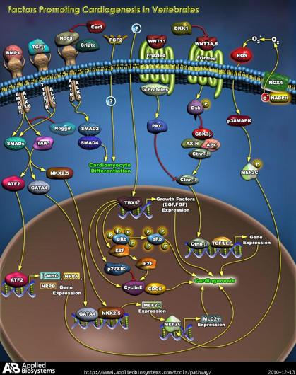
8 BMP expression BMP expression WNT inhibitors (crescent & cerberus) NKX expression GATA4 is important at the beginning of cardiogenesis. BMP 2,4 secreted by endoderm Crescent and cerberus inhibit WNT proteins NKX2.5 is upregulated. FGF8 is also important for heart specific proteins

9 MEF2C - Controls cardiac morphogenesis and myogenesis,
10
11 Development of Horseshoe- Shaped Pericardial Cavity And if you were to look at the embryo from the caudal region.

12 Mesoderm You would see that the heart tubes fuse and that the ventricles fuse first. The single heart tube fuses at day 21 of gestation.
13 Mesoderm As the heart tubes fuse they become surrounded by the myocardial mantle. This group of cells will form one of the heart fields that gives rise to some of the myocardial cells in the heart. It also gives rise to connective tissue cells that will be part of the cardiac jelly.
14 Mesoderm cardiac jelly Cardiac jelly will become the subendocardial connective tissue. It will also give rise to the precursors of the valves. In addition these cells will form the connective tissue of the interatrial and interventricular septa.

15 Mesoderm Foregut Dorsal Aorta The heart has three layers: 1. Endocardial layer 2. Cardiac jelly 3. Myocardial layer.! The heart tube at this point lies in front of the foregut and suspended in the body cavity by the mesocardium. Body Cavity Heart Amniotic Cavity
16 Embryonic Circulation There are three vascular circuits being set up early in development: An embryonic circuit Two extraembryonic circuits Vitelline Umbilical 16

17 Embryonic Circulation Common Cardinal Vein Dorsal Aorta Anterior Cardinal Vein Brain and Spinal Cord Posterior Cardinal Vein Umbilical Artery Aortic Arches Yolk Sac Umbilical Vein Ventricle Atria Vitelline Artery & Vein
18 Endocardial Heart Tube aortic arches II aortic sac III truncus art. conus cordis. I bulbus cordis primitive ventricle atrium r. sinal horn l. sinal horn The heart tube can be subdivided into several different regions. Nkx2.5 is important in the differentiation of different regions of the heart tube.
19 Hand 1 important in conotruncus and left ventricle Hand 2 important in right ventricle. Cells in RV from different source than LV Tbx5 important is specification of atria.
20 Retinoic Acid from mesoderm Atria and sinus venosus then produces RA Commits region Bulbus and ventricles produce lower levels of RA Isotretinoin (vitamin A) Vitamin A embryopathy: small, abnormally shaped ears, mandibular hypoplasia, cleft palate, heart defects RA
21 2 nd heart field Precardiac mesoderm Myogenic progenitor Myocardial progenitor Immature myocyte Differentiated atrial myocyte Myocardial progenitor Endothelial cells Vascular smooth Muscle progenitor Immature myocyte Immature myocyte Differentiated ventricular myocyte LV Differentiated ventricular myocyte RV Blood Cells Conduction tissue Vascular smooth muscle The point of this slide is to note the precursors of the heart cells and origin of the different cell types.
22 Endocardial Heart Tube The atrial end of the heart tube receives new vessels that form in the yolk sac (vitelline) and in the placenta (umbilical). They also are connected to the embryonic circuit (not shown here). Umbilical Vein Vitelline Vein
23 Convolution of the Heart Tube The hear tube then undergoes looping. Looping depends on laterality-inducing genes

24 Convolution of the Heart Tube There are two loops that are formed. 1. a Bulboventricular loop that has the truncus and conus moving ventrally and to the right. 2. An atrioventricular loop that has the atria moving posteriorly and superiorly. Aortic Sac Truncus Ventricle Conus Atria Sinus Venosus
25 Convolution of the Heart Tube This puts the conus and the primitive ventricle on the ventral surface. These chambers extend caudally. The atria are the most cephalic chambers of the heart and also the most posterior. R. Atrium Truncus L. Atrium Conus Primitive Ventricle
26 Primitive Four-Chamber Heart To understand septation of the heart you need to visualize the three dimensional architecture of the heart. So if we take a sagital section of the convoluted heart tube and look from the side we would see the image in the next slide.
27 Primitive 4 Chamber Heart Here the truncus and conus are in the front and the atria are posterior. Note the flow of blood from the atria to the ventricles to the conus and then out the truncus. truncus conus atrium ventricle
28 Primitive 4 Chamber Heart If a coronal section is then take and viewed from the front the next slide shows the view you would see. truncus atrium conus ventricle

29 Primitive Four-Chamber Heart T C AV V on the left the coronal section reveals the ventricle, the conus, the truncus and the black atrioventricular canal.
30 The AV canal has to shift to the right and as it does so thebulboventricular flange regresses This will put the AV canal in continuity with the conus and the ventricle. Bulboventricular flange RA LA RV LV AV canal
31 Primitive Four-Chamber Heart If a sagital section is made again and viewed from the side you see the following section.
32 Primitive 4 Chamber Heart truncus atrium conus ventricle This is the primitive four chambered heart prior to septation.
33 A common way to look at the septating heart is to take a section through the atria and the ventricles like the green line. Primitive 4 Chamber Heart truncus atrium conus ventricle

34 Primitive 4 Chamber Heart truncus atrium conus ventricle Then looking from the front you would see the next image.
35 Primitive 4 Chamber Heart superior R. Atrium L. Atrium Common A-V Canal Right Ventricle Left Ventricle inferior Note the two atria above, the left and right ventricles below. The right ventricle is the structure that develops from the conus. The conus gives rise to the reight ventricle and its outflow tract.
36 Going back to the sagital section, the nest event that occurs is the expansion of the cardic jelly in the region of the AV canal. The develop in the anterior and posterior part of the canal. Endocardial cushions

37 Atrial Septation The first event is atrial septation is the downward growth of a septum that is in between the two atrial chambers. This is called septum primum. Septum primum R. Atrium L. Atrium Foramen Primum Right Ventricle Left Ventricle
38 Atrial Septation Septum primum grows downward toward the endocardial cushions which are in the process of fusing. Before they reach the cushions they leave an opening called the ostium primum. Ostium Primum R. Atrium L. Atrium Septum primum Endocardial Cushion

39 Atrial Septation Here in horizontal (left) and sagital (right) section the endocardial cushion, the ostium primum and the septum primum are seen. Septum Primum R. Atrium L. Atrium Ostium primum Endocardial cushions
40 Atrial Septation As septum primum reaches the endocardial cushions a new ostium forms in the septum. R. Atrium L. Atrium
41 Atrial Septation This is called ostium secumdum. This allows blood to continue to flow from the right atrium to the left atrium (arrow). Ostium Secundum R. Atrium L. Atrium Ostium Primum
42 Atrial Septation As septum primum fuses with the endocardial cushion a second septum (septum secundum) develops to the right of ostium secundum. Septum Secundum R. Atrium L. Atrium
43 Septum spurium L. venous valve Atrial Septation As septum secundum develops the blood now all flows into the right atrium in between the right and left venous valves. R. venous valve
44 Atrial Septation Blood then enters the right atrium and can go either directly to the right ventricle or else flow thru foramen ovale (in septum secundum), thru ostium secundum (in septum primum) to the left ventricle. Foramen Ovale
45 Atrial Septation The sinus venosus tissue gets reabsorbed into the right atrium up to the incorporation of the superior vena cava, the inferior vena cava and the coronary sinus. Coronary sinus SVC IVC foramen ovale Pulmonary veins Valve of the foramen ovale
46 Atrial Development From sinus venosus tissue From pulmonary vein tissue The sinus venosus tissue forms the smooth wall portion of the right atriumfrom the crista termalis up to and including septum secundum of the interatrial septum.
47 Left Atrial Development Here we see the primitive left atrium superior R. Atrium L. Atrium Common A-V Canal Right Ventricle Left Ventricle inferior

48 Left Atrial Development As the pulmonary vein develops it empties into the left atrium. R. Atrium L. Atrium Pulmonary vein
49 Left Atrial Development Pulmonary veins The pulmonary vein gets reabsorbed into the left atrium up to the first bifurcation. It then continues to get incorporated into the left atrium.
50 Left Atrial Development The incorporation of left pulmonary vein tissue continues until the second bifurcation and this accounts for the four pulmonary veins emptying into the left atrium. Pulmonary veins

51 Atrial Development From sinus venosus tissue From pulmonary vein tissue The incorporated pulmonary vein tissue gets reabsorbed and forms the smooth wall portion of the left atrium.

52 Ventricular Septation superior R. Atrium L. Atrium Common A-V Canal Right Ventricle Left Ventricle inferior As atrial septation is taking place the septation of the ventricle is also occurring. Early on the two ventricular chambers are in direct communication with each other.
53 Ventricular Septation Endocardial Cushion Interventricular Foramen Interventricular Septum AS the endocardial cushion grow in the midline dividing the atrioventricular canal into a left and right AV canal, there is a growth of tissue between the two ventricles. This is the muscular interventricular septum.
54 Ventricular Septation E IVS I V E IVS I V The interventricular septum (IV) grows toward the endocardial cushion (E). The space in between the two is the primary IV septum (IVS).
55 Ventricular Septation Secondary Interventricular Foramen RV LV As the muscular interventricular septum reaches the endocardial cushion a small foramen remains and it is called the secondary interventricular foramen.
56 Ventricular Septation The secondary interventricular foramen is closed by the Connective tissue from the muscular interventricular septum. Endocardial cushion tissue. Conal ridges from the septation of the truncus and the conus.
57 Ventricular Septation Endocardial cushion tissue RV I V LV Here in green the contribution from the endocardial cushion is depicted. It will grow toward the IV septum.

58 Ventricular Septation If you take a midsagital section through the interventricular septum and the endocardial cushion you would see the image above. Note the location of the aorta and pulmonary artery as they develop from the truncus and conus. The bulbar septum will septate the conus and truncus and also contribute to the IV septum.

59 Ventricular Septation SVC Aorta Pulmonary Artery Bulbar Septum IV Foramen IVC Foramen Ovale Coronary Sinus Endocarial Cushion Muscular IV Septum Here you can see the formation of the membranous interventricular septum by the muscular IV septum connective tissue, the endocardial cushion and the bulbar septum.
60 Aorta Pulmonary Artery Conal Ridges IV Foramen Membranous Interventricular Septum Endocarial Cushion Muscular IV Septum

61 SeptaIon of the Bulbus Cordis Bulbus Cordis AV Canal Ventricle Looking at a sagital secion of the heart early in development the bulbus cordis is coninuous with the ventricle which is coninuous with the atria. As the AV canal shios to the right the bulbus move to the right as well.

62 SeptaIon of the Bulbus Cordis A P A P The next three slides make the point via cross secions that the aorta and pulmonary arteries rotate around each other. This means the septum between them changes posiion from superior to inferior as well.
63 SeptaIon of the Bulbus Cordis A P A P
64 SeptaIon of the Bulbus Cordis A P P A
65 MigraIon of neural crest cells Neural crest cells migrate from the 3ed, 4th and 6th pharyngeal arches to form some of the populaion of cells forming the aoricopulmonary septum.
66 SeptaIon of the Bulbus Cordis Truncal (Conal) Swellings Bulbus Cordis The cardiac jelly in the region of the truncus and conus adds the neural crest cells and expands as truncal swellings.
67 SeptaIon of the Bulbus Cordis Aorticopulmonary septum These swellings grow toward each other to meet and form the septum between the aorta and pulmonary artery. Aorta Pulmonary Artery

68 SeptaIon of the Bulbus Cordis 1 2 Anterior The aoricopulmonary septum then rotates as it moves inferiorly. However, the exact mechanism for that rotaion remains unclear.
69 However, the aoricopulmonary septum must form properly for the IV septum to be completed. SeptaIon of the Bulbus Cordis Aorta Pulmonary Artery Conal Ridges IV Foramen Membranous Interventricular Septum Endocarial Cushion Muscular IV Septum
70 Embryonic CirculaIon Common Cardinal Vein Dorsal Aorta Anterior Cardinal Vein Brain and Spinal Cord Posterior Cardinal Vein Umbilical Artery AorIc Arches Yolk Sac Umbilical Vein Ventricle Atria Vitelline Artery & Vein Blood leaves the truncus and moves to the aoric arches. There is an aoric arch for each pharyngeal arch.

71 title=file:advanced_heart_development_timeline.jpg 71
When you see this diagram, remember that you are looking at the embryo from above, through the amniotic cavity, where the epiblast appears as an oval
 When you see this diagram, remember that you are looking at the embryo from above, through the amniotic cavity, where the epiblast appears as an oval disc 2 Why the embryo needs the vascular system? When
DEVELOPMENT OF THE CIRCULATORY SYSTEM L E C T U R E 5
 DEVELOPMENT OF THE CIRCULATORY SYSTEM L E C T U R E 5 REVIEW OF CARDIAC ANATOMY Heart 4 chambers Base and apex Valves Pericardial sac 3 layers: epi, myo, endo cardium Major blood vessels Aorta and its
Development of the heart
 Development of the heart Prof. Abdulameer Al-Nuaimi E-mail: a.al-nuaimi@sheffield.ac.uk abdulameerh@yahoo.com Early Development of the Circulatory System Appears in the middle of the third week, when the
Embryology of the Heart
 *Page 1A: Embryology of the Heart Human embryonic disc is divided into three layers: ectoderm, intraembryonic mesoderm, and endoderm. The embryonic disc lies between the amniotic cavity and the primary
The Cardiovascular System (Part I) 黃敏銓 解剖學暨細胞生物學研究所
 黃敏銓 解剖學暨細胞生物學研究所") The Cardiovascular System (Part I) 黃敏銓 解剖學暨細胞生物學研究所 1 Congenital heart defects (CHDs) 台灣兒童心臟學會 Sinus venarum Membranous septum Conus arteiosus (infundibulum) Aortic vestibule The Cardiovascular System
The Cardiovascular System (Part I) 黃敏銓 解剖學暨細胞生物學研究所 1 Congenital heart defects (CHDs) 台灣兒童心臟學會 Sinus venarum Membranous septum Conus arteiosus (infundibulum) Aortic vestibule The Cardiovascular System
W.S. O The University of Hong Kong
 W.S. O The University of Hong Kong Objectives: Describe early angiogenesis. Describe the heart tube formation. Describe the partitioning into a 4- chambered heart. List the formation of heart valves and
Heart & vascular system I. Dawei Dong
 Heart & vascular system I Dawei Dong Lecture goal Learn the basics of heart and vascular development. Development of Heart, Blood, and Blood Vessels LEARNING GOALS: 1. explain the early development of
Development of the Great Vessels and Conduc6on Tissue
 Development of the Great Vessels and Conduc6on Tissue Development of the heart fields h:p://php.med.unsw.edu.au/embryology/ index.php?6tle=advanced_- _Heart_Fields! 2 Septa6on of the Bulbus Cordis Bulbus
6. HEART AND CIRCULATORY SYSTEM I
 6. HEART AND CIRCULATORY SYSTEM I Dr. Taube P. Rothman P&S 12-520 Tpr2@columbia.edu 212-305-7930 RECOMMENDED READING: Larsen Human Embryology, 3rd Edition, pp. 195-199; 157-169 top left; 172-174; bottom
W.S. O. School of Biomedical Sciences, University of Hong Kong
 W.S. O School of Biomedical Sciences, University of Hong Kong Objectives: Describe early angiogenesis. Describe the heart tube formation. Describe the partitioning into a 4- chambered heart. List the formation
6. Development of circulatory system II. Cardiac looping. Septation of atria and ventricles. Common heart malformations.
 6. Development of circulatory system II. Cardiac looping. Septation of atria and ventricles. Common heart malformations. Formation of heart tube paired endothelial-lined heart tube is formed from blood
Development and teratology of cardiovascular and lymphatic systems. Repetition: Muscle tissue
 Development and teratology of cardiovascular and lymphatic systems Repetition: Muscle tissue Beginning of the cardiovascular system development the 3rd week: Hemangiogenesis (day 15 16) blood islets (insulae
Heart Development and Congenital Heart Disease
 Heart Development and Congenital Heart Disease Sally Dunwoodie s.dunwoodie@victorchang.edu.au Developmental and Stem Cell Biology Division Victor Chang Cardiac Research Institute for the heart of Australia...
CARDIAC DEVELOPMENT CARDIAC DEVELOPMENT
 CARDIAC DEVELOPMENT CARDIAC DEVELOPMENT Diane E. Spicer, BS, PA(ASCP) University of Florida Dept. of Pediatric Cardiology Curator Van Mierop Cardiac Archive This lecture is given with special thanks to
Chapter 4: The thoracic cavity and heart. The Heart
 Chapter 4: The thoracic cavity and heart The thoracic cavity is divided into right and left pleural cavities by a central partition, the mediastinum. The mediastinum is bounded behind by the vertebral
Development of the Heart *
 OpenStax-CNX module: m46673 1 Development of the Heart * OpenStax This work is produced by OpenStax-CNX and licensed under the Creative Commons Attribution License 3.0 By the end of this section, you will
IN THE NAME OF GOD. Development of the Heart and Vasculature
 IN THE NAME OF GOD Development of the Heart and Vasculature Overview vascular system appears (middle of 3 rd week) when the embryo is not able to satisfy its nutrition by diffusion Heart is the first functional
Circulatory system. Lecture #2
 Circulatory system Lecture #2 The essential components of the human cardiovascular system: Heart Blood Blood vessels Arteries - blood vessels that conduct arterial blood from heart ventricle to organs
Notes: 1)Membranous part contribute in the formation of small portion in the septal cusp.
Membranous part contribute in the formation of small portion in the septal cusp.") Embryology 9 : Slide 16 : There is a sulcus between primitive ventricular and bulbis cordis that will disappear gradually and lead to the formation of one chamber which is called bulboventricular chamber.
Embryology 9 : Slide 16 : There is a sulcus between primitive ventricular and bulbis cordis that will disappear gradually and lead to the formation of one chamber which is called bulboventricular chamber.
Organogenesis Part 2. V. Lateral Plate Mesoderm VI. Endoderm VII. Development of the Tetrapod Limb VIII. Sex Determination. V. Lateral Plate Mesoderm
 Organogenesis Part 2 V. Lateral Plate Mesoderm VI. Endoderm VII. Development of the Tetrapod Limb VIII. Sex Determination V. Lateral Plate Mesoderm chordamesoderm paraxial mesoderm intermediate mesoderm
human anatomy 2016 lecture thirteen Dr meethak ali ahmed neurosurgeon
 Heart The heart is a hollow muscular organ that is somewhat pyramid shaped and lies within the pericardium in the mediastinum. It is connected at its base to the great blood vessels but otherwise lies
the Cardiovascular System I
 the Cardiovascular System I By: Dr. Nabil A Khouri MD, MsC, Ph.D MEDIASTINUM 1. Superior Mediastinum 2. inferior Mediastinum Anterior mediastinum. Middle mediastinum. Posterior mediastinum Anatomy of
Anatomy of the Heart. Figure 20 2c
 Anatomy of the Heart Figure 20 2c Pericardium & Myocardium Remember, the heart sits in it s own cavity, known as the mediastinum. The heart is surrounded by the Pericardium, a double lining of the pericardial
The sinus venosus represent the venous end of the heart It receives 3 veins: 1- Common cardinal vein body wall 2- Umbilical vein from placenta 3-
 1 2 The sinus venosus represent the venous end of the heart It receives 3 veins: 1- Common cardinal vein body wall 2- Umbilical vein from placenta 3- Vitelline vein from yolk sac 3 However!!!!! The left
Heart and Lungs. LUNG Coronal section demonstrates relationship of pulmonary parenchyma to heart and chest wall.
 Heart and Lungs Normal Sonographic Anatomy THORAX Axial and coronal sections demonstrate integrity of thorax, fetal breathing movements, and overall size and shape. LUNG Coronal section demonstrates relationship
LECTURE 5. Anatomy of the heart
 LECTURE 5. Anatomy of the heart Main components of the CVS: Heart Blood circulatory system arterial compartment haemomicrocirculatory (=microvascular) compartment venous compartment Lymphatic circulatory
CARDIOVASCULAR SYSTEM
 CARDIOVASCULAR SYSTEM Overview Heart and Vessels 2 Major Divisions Pulmonary Circuit Systemic Circuit Closed and Continuous Loop Location Aorta Superior vena cava Right lung Pulmonary trunk Base of heart
Chapter 20 (1) The Heart
 The Heart") Chapter 20 (1) The Heart Learning Objectives Describe the location and structure of the heart Describe the path of a drop of blood from the superior vena cava or inferior vena cava through the heart out
Chapter 20 (1) The Heart Learning Objectives Describe the location and structure of the heart Describe the path of a drop of blood from the superior vena cava or inferior vena cava through the heart out
ANATDMY. lecture # : Date : Lecturer : Maher Hadidi
 ANATDMY 27 lecture # : Date : Lecturer : Maher Hadidi Pericardium A double-walled fibroserous conical-shaped sac, within middle mediastinum. Enclose the heart and roots of its large vessels. Vagus nerves
THE NORMAL AND ABNORMAL INTER-ATRIAL SEPTUM
 THE NORMAL AND ABNORMAL INTER-ATRIAL SEPTUM BY REGINALD HUDSON From the Institute of Cardiology and National Heart Hospital Received April 5, 1954 This paper is an elementary study of the normal and abnormal
Segmental Analysis. Gautam K. Singh, M.D. Washington University School of Medicine St. Louis
 Segmental Analysis Gautam K. Singh, M.D. Washington University School of Medicine St. Louis Segmental Analysis Segmental Analysis: From Veins to Ventricles Segmental Approach to Evaluation of Congenital
Cardiac embryology and anatomy
 Section 1 Anatomy and physiology Chapter 1 Cardiac embryology and anatomy Doris M. Rassl and Martin J. Goddard An appreciation of the normal development of the heart and great vessels and normal adult
Heart Anatomy. 7/5/02 Stephen G Davenport 1
 Heart Anatomy Copyright 1999, Stephen G. Davenport, No part of this publication may be reproduced, stored in a retrieval system, or transmitted, in any form without prior written permission. 7/5/02 Stephen
THE HEART OBJECTIVES: LOCATION OF THE HEART IN THE THORACIC CAVITY CARDIOVASCULAR SYSTEM
 BIOLOGY II CARDIOVASCULAR SYSTEM ACTIVITY #3 NAME DATE HOUR THE HEART OBJECTIVES: Describe the anatomy of the heart and identify and give the functions of all parts. (pp. 356 363) Trace the flow of blood
"Lecture Index. 1) Heart Progenitors. 2) Cardiac Tube Formation. 3) Valvulogenesis and Chamber Formation. 4) Epicardium Development.
 Heart Progenitors. 2) Cardiac Tube Formation. 3) Valvulogenesis and Chamber Formation. 4) Epicardium Development.") "Lecture Index 1) Heart Progenitors. 2) Cardiac Tube Formation. 3) Valvulogenesis and Chamber Formation. 4) Epicardium Development. 5) Septation and Maturation. 6) Changes in Blood Flow during Development.
"Lecture Index 1) Heart Progenitors. 2) Cardiac Tube Formation. 3) Valvulogenesis and Chamber Formation. 4) Epicardium Development. 5) Septation and Maturation. 6) Changes in Blood Flow during Development.
Human Anatomy, First Edition
 Human Anatomy, First Edition McKinley & O'Loughlin Chapter 22 : Heart 1 Functions of the Heart Center of the cardiovascular system, the heart. Connects to blood vessels that transport blood between the
Cardiovascular system:
 Cardiovascular system: Mediastinum: The mediastinum: lies between the right and left pleura and lungs. It extends from the sternum in front to the vertebral column behind, and from the root of the neck
CV Anatomy Quiz. Dr Ella Kim Dr Pip Green
 CV Anatomy Quiz Dr Ella Kim Dr Pip Green Q1 The location of the heart is correctly described as A) lateral to the lungs. B) medial to the sternum. C) superior to the diaphragm. D) posterior to the spinal
LAB 12-1 HEART DISSECTION GROSS ANATOMY OF THE HEART
 LAB 12-1 HEART DISSECTION GROSS ANATOMY OF THE HEART Because mammals are warm-blooded and generally very active animals, they require high metabolic rates. One major requirement of a high metabolism is
Anatomy of the Heart
 Biology 212: Anatomy and Physiology II Anatomy of the Heart References: Saladin, KS: Anatomy and Physiology, The Unity of Form and Function 8 th (2018). Required reading before beginning this lab: Chapter
Development of Blood Vessels and Fetal Circulation *
 OpenStax-CNX module: m46610 1 Development of Blood Vessels and Fetal Circulation * OpenStax This work is produced by OpenStax-CNX and licensed under the Creative Commons Attribution License 3.0 By the
Blood supply of the Heart & Conduction System. Dr. Nabil Khouri
 Blood supply of the Heart & Conduction System Dr. Nabil Khouri Arterial supply of Heart Right coronary artery Left coronary artery 3 Introduction: Coronary arteries - VASAVASORUM arising from aortic sinuses
Human Anatomy and Physiology Chapter 19 Worksheet 1- The Heart
 Human Anatomy and Physiology Chapter 19 Worksheet 1- The Heart Name Date Period 1. The "double pump" function of the heart includes the right side, which serves as the circuit pump, while the left side
ISSN: CODEN Code: PIHNBQ ZDB-Number: IC Journal No: Vol. 2 No Online Available at
 Received: 13-05-2013 Accepted: 16-06-2013 ISSN: 2277-7695 CODEN Code: PIHNBQ ZDB-Number: 2663038-2 IC Journal No: 7725 Vol. 2 No. 5 2013 Online Available at www.thepharmajournal.com THE PHARMA INNOVATION
Atrial Septal Defects
 Supplementary ACHD Echo Acquisition Protocol for Atrial Septal Defects The following protocol for echo in adult patients with atrial septal defects (ASDs) is a guide for performing a comprehensive assessment
Figure 10.1A Transparency Master 79
 Brain Carotid arteries Jugular vein Right front leg Lungs (inflated) Cranial Right atrium To left front leg Left subclavian Bronchus capillaries Brachiocephalic vein Left atrium Dorsal aorta Right ventricle
Circulatory System: Introduction. Dr. Carmen E. Rexach Anatomy 35 Mt. San Antonio College
 Circulatory System: Introduction Dr. Carmen E. Rexach Anatomy 35 Mt. San Antonio College Components Cardiovascular system Lymphatic system Cardiovascular system Heart, blood vessels, blood Functions: transport
THE CARDIOVASCULAR SYSTEM. Part 1
 THE CARDIOVASCULAR SYSTEM Part 1 CARDIOVASCULAR SYSTEM Blood Heart Blood vessels What is the function of this system? What other systems does it affect? CARDIOVASCULAR SYSTEM Functions Transport gases,
The Physiology of the Fetal Cardiovascular System
 The Physiology of the Fetal Cardiovascular System Jeff Vergales, MD, MS Department of Pediatrics Division of Pediatric Cardiology jvergales@virginia.edu Disclosures I serve as the medical director for
Adult Congenital Heart Disease: What All Echocardiographers Should Know Sharon L. Roble, MD, FACC Echo Hawaii 2016
 1 Adult Congenital Heart Disease: What All Echocardiographers Should Know Sharon L. Roble, MD, FACC Echo Hawaii 2016 DISCLOSURES I have no disclosures relevant to today s talk 2 Why should all echocardiographers
This lab activity is aligned with Visible Body s A&P app. Learn more at visiblebody.com/professors
 1 This lab activity is aligned with Visible Body s A&P app. Learn more at visiblebody.com/professors 2 PRE-LAB EXERCISES: A. Watch the video 29.1 Heart Overview and make the following observations: 1.
Transcription for Narration of Embryology of the Great Arteries
 Transcription for Narration of Embryology of the Great Arteries Slide 1: In this presentation I am going to describe for you the development of what are known as the great arteries. The great arteries
Part 1. Copyright 2011 Pearson Education, Inc. Figure Copyright 2011 Pearson Education, Inc.
 PowerPoint Lecture Slides prepared by Leslie Hendon University of Alabama, Birmingham C H A P T E R The Heart 19 Part 1 The Heart A muscular double pump circuit vessels transport blood to and from the
We are IntechOpen, the world s leading publisher of Open Access books Built by scientists, for scientists. International authors and editors
 We are IntechOpen, the world s leading publisher of Open Access books Built by scientists, for scientists 3,700 108,500 1.7 M Open access books available International authors and editors Downloads Our
The Heart. Happy Friday! #takeoutyournotes #testnotgradedyet
 The Heart Happy Friday! #takeoutyournotes #testnotgradedyet Introduction Cardiovascular system distributes blood Pump (heart) Distribution areas (capillaries) Heart has 4 compartments 2 receive blood (atria)
The Heart. The Heart A muscular double pump. The Pulmonary and Systemic Circuits
 C H A P T E R 19 The Heart The Heart A muscular double pump circuit takes blood to and from the lungs Systemic circuit vessels transport blood to and from body tissues Atria receive blood from the pulmonary
Large Arteries of Heart
 Cardiovascular System (Part A-2) Module 5 -Chapter 8 Overview Arteries Capillaries Veins Heart Anatomy Conduction System Blood pressure Fetal circulation Susie Turner, M.D. 1/5/13 Large Arteries of Heart
Pediatric Cardiomyopathies
 Chapter 14 Pediatric Cardiomyopathies Aspazija Sofijanova and Olivera Jordanova Additional information is available at the end of the chapter http://dx.doi.org/10.5772/55820 1. Introduction 1.1. Development
Ch.15 Cardiovascular System Pgs {15-12} {15-13}
 Ch.15 Cardiovascular System Pgs {15-12} {15-13} E. Skeleton of the Heart 1. The skeleton of the heart is composed of rings of dense connective tissue and other masses of connective tissue in the interventricular
THE HEART. A. The Pericardium - a double sac of serous membrane surrounding the heart
 THE HEART I. Size and Location: A. Fist-size weighing less than a pound (250 to 350 grams). B. Located in the mediastinum between the 2 nd rib and the 5 th intercostal space. 1. Tipped to the left, resting
A. Incorrect! Think of a therapy that reduces prostaglandin synthesis. B. Incorrect! Think of a therapy that reduces prostaglandin synthesis.
 USMLE Step 1 - Problem Drill 02: Embryology Question No. 1 of 10 1. A premature infant is born with a patent ductus arteriosis. Which of the following treatments may be used as part of the treatment regimen?
was judged subjectively. The left ventricle was considered to be slightly hypoplastic when the cardiac
 British Heart J7ournal, 1976, 38, 1124-1132. Double outlet right ventricle Study of 27 cases A. H. Cameron, F. Acerete, M. Quero, and M. C. Castro From the Department of Patlology, Children's Hospital,
Spleen. Vertebrate hearts Pericardial cavity division in coelum. Vessel walls. Endocardium = endothelium of blood vessels. Artery elastic tissue
 Spleen White pulp macrophages, monocyte storage Red pulp - (RBC) storage, and prod n (in nonmammals) Vertebrate hearts Pericardial cavity division in coelum Endocardium = endothelium of blood vessels Fig.
THE VESSELS OF THE HEART
 1 THE VESSELS OF THE HEART The vessels of the heart include the coronary arteries, which supply the heart and the veins and lymph vessels, which drain the heart. THE CORONARY ARTERIES These are the blood
CHAPTER 8 MOLECULAR AND CELLULAR DEVELOPMENT OF THE HEART
 163 CHAPTER 8 MOLECULAR AND CELLULAR DEVELOPMENT OF THE HEART Miguel Torres and Silvia Martín-Puig INTRODUCTION / 163 CARDIAC EMBRYOGENESIS / 163 Allocation and Specification of Cardiac Progenitors during
Chapter 14. The Cardiovascular System
 Chapter 14 The Cardiovascular System Introduction Cardiovascular system - heart, blood and blood vessels Cardiac muscle makes up bulk of heart provides force to pump blood Function - transports blood 2
PRACTICAL GUIDE TO FETAL ECHOCARDIOGRAPHY IC Huggon and LD Allan
 PRACTICAL GUIDE TO FETAL ECHOCARDIOGRAPHY IC Huggon and LD Allan Fetal Cardiology Unit, Harris Birthright Research Centre for Fetal Medicine, King's College Hospital, London, UK IMPORTANCE OF PRENATAL
The Cardiovascular System. Chapter 15. Cardiovascular System FYI. Cardiology Closed systemof the heart & blood vessels. Functions
 Chapter 15 Cardiovascular System FYI The heart pumps 7,000 liters (4000 gallons) of blood through the body each day The heart contracts 2.5 billion times in an avg. lifetime The heart & all blood vessels
The Cardiovascular System
 The Cardiovascular System The Manila Times College of Subic Prepared by: Stevens B. Badar, RN, MANc THE HEART Anatomy of the Heart Location and Size approx. the size of a person s fist, hollow and cone-shaped,
Introduction to Anatomy. Dr. Maher Hadidi. Bayan Yanes. April/9 th /2013
 Introduction to Anatomy Dr. Maher Hadidi Bayan Yanes 27 April/9 th /2013 KEY POINTS: 1) Right side of the heart 2) Papillary muscles 3) Left side of the heart 4) Comparison between right and left sides
Embryonic Development of the Human Heart
 Embryonic Development of the Human Heart 1. Embryonic development of From formation of the zygote up until the third week of development the embryo s demand for O 2 and nutrients is met by simple diffusion.
Mediastinum and pericardium
 Mediastinum and pericardium Prof. Abdulameer Al-Nuaimi E-mail: a.al-nuaimi@sheffield.ac.uk E. mail: abdulameerh@yahoo.com The mediastinum: is the central compartment of the thoracic cavity surrounded by
Cardiovascular Anatomy Dr. Gary Mumaugh
 Cardiovascular Anatomy Dr. Gary Mumaugh Location of Heart Approximately the size of your fist Location o Superior surface of diaphragm o Left of the midline in mediastinum o Anterior to the vertebral column,
SLIDES 6 AND 10 MM PIG SLIDES; TRANSVERSE SECTIONS AND SAGITAL; FETAL PIGS-1-8INCH; HUMAN SAGITAL DIAGRAMS:DRAWINGS OF THE PIG SECTIONS TO BE
 SLIDES 6 AND 10 MM PIG SLIDES; TRANSVERSE SECTIONS AND SAGITAL; FETAL PIGS-1-8INCH; HUMAN SAGITAL SECTION DIAGRAMS:DRAWINGS OF THE PIG SECTIONS TO BE LABELLED, AND FOUR DRAWINGS TO BE MADE. REFERENCES:PATTEN:
Large Ventricular Septal Defect with Irreversible Pulmonary Hypertension
 Week 20 Blue Is For Boys Tetralogy of Fallot Presenting Symptoms Child, 4 Cyanosis Exercise Tolerance Failure to thrive Clubbing Lower Sternal Heave Right ventricular hypertrophy Loud ejection systolic
Middle mediastinum---- heart & pericardium. Dep. of Human Anatomy Zhou Hongying
 Middle mediastinum---- heart & pericardium Dep. of Human Anatomy Zhou Hongying eaglezhyxzy@163.com Subdivisions of the mediastinum Contents of Middle mediastinum Heart Pericardium: a serous sac enclosing
Lab Activity 23. Cardiac Anatomy. Portland Community College BI 232
 Lab Activity 23 Cardiac Anatomy Portland Community College BI 232 Cardiac Muscle Histology Branching cells Intercalated disc: contains many gap junctions connecting the adjacent cell cytoplasm, creates
Heart and Soul Evaluation of the Fetal Heart
 Heart and Soul Evaluation of the Fetal Heart Ivana M. Vettraino, M.D., M.B.A. Clinical Associate Professor, Michigan State University College of Human Medicine Objectives Review the embryology of the formation
Heart Dissection. 5. Locate the tip of the heart or the apex. Only the left ventricle extends all the way to the apex.
 Heart Dissection Page 1 of 6 Background: The heart is a four-chambered, hollow organ composed primarily of cardiac muscle tissue. It is located in the center of the chest in between the lungs. It is the
Anatomy lab -1- Imp note: papillary muscle Trabeculae Carneae chordae tendineae
 Anatomy lab -1- Imp note: the arrangement of this sheet is different than the lab recording, it has been arranged in a certain way to make it easier to study. When you open the left ventricle you can see
Cardiovascular Respiratory Renal/ Urinary. VOLUME 111 Nervous System. Organ ~ Systems ~
 Cardiovascular Respiratory Renal/ Urinary VOLUME 111 Nervous System Organ ~ Systems ~ Contents Section I: Cardiovascular System Chapter 1. Embryology... 3 Chapter 2. Histology... 13 Chapter 3. Anatomy...
1. Distinguish among the types of blood vessels on the basis of their structure and function.
 Blood Vessels and Circulation Objectives This chapter describes the structure and functions of the blood vessels Additional subjects contained in Chapter 13 include cardiovascular physiology, regulation,
2. right heart = pulmonary pump takes blood to lungs to pick up oxygen and get rid of carbon dioxide
 A. location in thorax, in inferior mediastinum posterior to sternum medial to lungs superior to diaphragm anterior to vertebrae orientation - oblique apex points down and to the left 2/3 of mass on left
CJ Shuster A&P2 Lab Addenum Beef Heart Dissection 1. Heart Dissection. (taken from Johnson, Weipz and Savage Lab Book)
") CJ Shuster A&P2 Lab Addenum Beef Heart Dissection 1 Heart Dissection. (taken from Johnson, Weipz and Savage Lab Book) Introduction When you have finished examining the model, you are ready to begin your
CJ Shuster A&P2 Lab Addenum Beef Heart Dissection 1 Heart Dissection. (taken from Johnson, Weipz and Savage Lab Book) Introduction When you have finished examining the model, you are ready to begin your
Vasculature and innervation of the heart. A. Bendelic Human Anatomy Department
 Vasculature and innervation of the heart A. Bendelic Human Anatomy Department Plan: 1. Arterial blood supply of the heart. Coronary arteries 2. Venous drainage of the heart. Cardiac veins 3. Innervation
The Cardiovascular System Part I: Heart Outline of class lecture After studying part I of this chapter you should be able to:
 The Cardiovascular System Part I: Heart Outline of class lecture After studying part I of this chapter you should be able to: 1. Describe the functions of the heart 2. Describe the location of the heart,
COMPREHENSIVE EVALUATION OF FETAL HEART R. GOWDAMARAJAN MD
 COMPREHENSIVE EVALUATION OF FETAL HEART R. GOWDAMARAJAN MD Disclosure No Relevant Financial Relationships with Commercial Interests Fetal Echo: How to do it? Timing of Study -optimally between 22-24 weeks
Surgical Management Of TAPVR. Daniel A. Velez, M.D. Congenital Cardiac Surgeon Phoenix Children s Hospital
 Surgical Management Of TAPVR Daniel A. Velez, M.D. Congenital Cardiac Surgeon Phoenix Children s Hospital No Disclosures Goals Review the embryology and anatomy Review Surgical Strategies for repair Discuss
Slide 1. Slide 2. Slide 3 CONGENITAL HEART DISEASE. Papworth Hospital NHS Trust INTRODUCTION. Jakub Kadlec/Catherine Sudarshan INTRODUCTION
 Slide 1 CONGENITAL HEART DISEASE Jakub Kadlec/Catherine Sudarshan NHS Trust Slide 2 INTRODUCTION Most common congenital illness in the newborn Affects about 4 9 / 1000 full-term live births in the UK 1.5
Development of the Liver and Pancreas
 Development of the Liver and Pancreas Professor Alfred Cuschieri Department of Anatomy University of Malta Three glandular buds arise from the distal end of the foregut during the fourth week Day 22 -The
Biology 340 Comparative Embryology Lecture 10 Dr. Stuart Sumida. Further Development of the Mesoderm (and Endoderm)
") Biology 340 Comparative Embryology Lecture 10 Dr. Stuart Sumida Further Development of the Mesoderm (and Endoderm) Further Development: Digestive System Foregut, Midgut, Hindgut Heart and Aortic Arches
Biology 340 Comparative Embryology Lecture 10 Dr. Stuart Sumida Further Development of the Mesoderm (and Endoderm) Further Development: Digestive System Foregut, Midgut, Hindgut Heart and Aortic Arches
Development of the Digestive System. W.S. O The University of Hong Kong
 Development of the Digestive System W.S. O The University of Hong Kong Plan for the GI system Then GI system in the abdomen first develops as a tube suspended by dorsal and ventral mesenteries. Blood
AP2 Lab 1 - Blood & Heart
 AP2 Lab 1 - Blood & Heart Project 1 - Formed Elements Identification & Recognition See fig. 17.10 and Table 17.2. Instructor may also provide other images. Note: See Fig. 17.11 All formed elements are
Approximately the size of your fist Location Superior surface of diaphragm Left of the midline in mediastinum Anterior to the vertebral column,
 Dr. Gary Mumaugh Approximately the size of your fist Location Superior surface of diaphragm Left of the midline in mediastinum Anterior to the vertebral column, posterior to the sternum Posteriorly the
Cardiovascular System. Heart Anatomy
 Cardiovascular System Heart Anatomy 1 The Heart Location & general description: Atria vs. ventricles Pulmonary vs. systemic circulation Coverings Walls The heart is found in the mediastinum, the medial
The HEART. What is it???? Pericardium. Heart Facts. This muscle never stops working It works when you are asleep
 This muscle never stops working It works when you are asleep The HEART It works when you eat It really works when you exercise. What is it???? Located between the lungs in the mid thoracic region Apex
ULTRASOUND OF THE FETAL HEART
 ULTRASOUND OF THE FETAL HEART Cameron A. Manbeian, MD Disclosure Statement Today s faculty: Cameron Manbeian, MD does not have any relevant financial relationships with commercial interests or affiliations
Midgut. Over its entire length the midgut is supplied by the superior mesenteric artery
 Gi Embryology 3 Midgut the midgut is suspended from the dorsal abdominal wall by a short mesentery and communicates with the yolk sac by way of the vitelline duct or yolk stalk Over its entire length the
Anatomy of left ventricular outflow tract'
 Anatomy of left ventricular outflow tract' ROBERT WALMSLEY British Heart Journal, 1979, 41, 263-267 From the Department of Anatomy and Experimental Pathology, The University, St Andrews, Scotland SUMMARY
The embryonic endoderm initially is widely connected with the yolk sac. As a consequence of cephalocaudal and lateral folding, a portion of the
 DIGESTIVE SYSTEM The embryonic endoderm initially is widely connected with the yolk sac. As a consequence of cephalocaudal and lateral folding, a portion of the endoderm-lined yolk sac cavity is incorporated