Chapter 10. The Knee Joint. The Knee Joint. Bones. Bones. Bones. Bones. Knee joint. Manual of Structural Kinesiology R.T. Floyd, EdD, ATC, CSCS
|
|
|
- Madeline Gaines
- 6 years ago
- Views:
Transcription

 - receptacles for femoral condyles Tibia medial bears most of")

 bone imbedded in quadriceps & patellar tendon serves similar to a pulley in improving angle of pull, resulting in greater mechanical advantage in extension Key bony")
1 The Knee Joint Chapter 10 The Knee Joint Manual of Structural Kinesiology R.T. Floyd, EdD, ATC, CSCS 2007 McGraw-Hill Higher Education. All rights reserved Knee joint largest joint in body very complex primarily a hinge joint Modified for Prentice WE: Arnheim s principles of athletic training, ed 12, New York, 2006, McGraw-Hill; from Saladin, KS: Anatomy &physiology: the unity of forms and function, ed 2, New York, 2001, McGraw- Hill McGraw-Hill Higher Education. All rights reserved Enlarged femoral condyles articulate on enlarged tibial condyles Medial & lateral tibial condyles (medial & lateral tibial plateaus) - receptacles for femoral condyles Tibia medial bears most of weight Fibula - lateral serves as the attachment for joint structures does not articulate with femur or patella not part of joint Modified from Anthony CP, Kolthoff NJ: Textbook of anatomy and physiology, ed 9, St. Louis, 1975, Mosby McGraw-Hill Higher Education. All rights reserved McGraw-Hill Higher Education. All rights reserved Patella sesamoid (floating) bone imbedded in quadriceps & patellar tendon serves similar to a pulley in improving angle of pull, resulting in greater mechanical advantage in extension Key bony landmarks Superior & inferior patellar poles Tibial tuberosity Gerdy s tubercle Medial & lateral femoral condyles Upper anterior medial tibial surface Head of fibula Modified from Anthony CP, Kolthoff NJ: Textbook of anatomy and physiology, ed 9, St. Louis, 1975, Mosby McGraw-Hill Higher Education. All rights reserved McGraw-Hill Higher Education. All rights reserved

2 Three vasti muscles of quadriceps originate on proximal femur & insert on patellar superior pole insertion is ultimately on tibial tuberosity via patella tendon Iliotibial tract of tensor fasciae latae inserts on Gerdy s tubercle Sartorius, gracilis, & semitendinosus insert just below the medial condyle on upper anteromedial tibial surface 2007 McGraw-Hill Higher Education. All rights reserved Semimembranosus inserts posteromedially on medial tibial condyle Biceps femoris inserts primarily on fibula head Popliteus originates on lateral aspect of lateral femoral condyle Tibial collateral ligament originates on medial aspect of upper medial femoral condyle & inserts on medial tibial surface Fibula collateral originates on lateral femoral condyle very close to popliteus origin & inserts on fibular head 2007 McGraw-Hill Higher Education. All rights reserved Knee joint proper (tibiofemoral joint) classified as a ginglymus joint Sometimes referred to as trochoginglymus joint internal & external occur during flexion Some argue for condyloid classification Patellofemoral joint arthrodial classification gliding nature of patella on femoral condyles Ligaments provide static stability Quadriceps & hamstrings contractions produce dynamic stability Articular cartilage surfaces on femur & tibia Menisci form cushions between bones attached to tibia deepen tibial fossa enhance stability 2007 McGraw-Hill Higher Education. All rights reserved McGraw-Hill Higher Education. All rights reserved. Modified from Anthony CP, Kolthoff NJ: Textbook of anatomy and physiology, ed 9, St. Louis, 1975, Mosby. Medial meniscus forms receptacle for medial femoral condyle, Lateral meniscus receives lateral femoral condyle Thicker on outside border & taper down very thin to inside border Can slip about slightly, but held in place by various small ligaments Medial meniscus - larger & more open C appearance Lateral meniscus - closed C configuration Either or both menisci may be torn in several different areas from a variety of mechanisms, resulting in varying degrees of problems Tears often occur due significant compression & shear forces during while flexing or extending during quick directional changes in running 2007 McGraw-Hill Higher Education. All rights reserved McGraw-Hill Higher Education. All rights reserved
3 Anterior & posterior cruciate ligaments cross within between tibia & femur vital in respectively maintaining anterior & posterior stability, as well as rotatory stability Anterior cruciate ligament (ACL) injuries one of most common serious injuries to mechanism often involves noncontact rotary forces associated with planting & cutting, hyperextension, or by violent quadriceps contraction which pulls tibia forward on femur Posterior cruciate ligament (PCL) injuries not often injured mechanism of direct contact with an opponent or playing surface Fibular (lateral) collateral ligament (LCL) infrequently injured Modified from Anthony CP, Kolthoff NJ: Textbook of anatomy and physiology, ed 9, St. Louis, 1975, Mosby McGraw-Hill Higher Education. All rights reserved McGraw-Hill Higher Education. All rights reserved Tibial (medial) collateral ligament (MCL) maintains medial stability by resisting valgus forces or preventing from being abducted injuries occur commonly, particularly in contact or collision sports mechanism of teammate or opponent may fall against lateral aspect of or leg causing medial opening of joint & stress to medial ligamentous structures 2007 McGraw-Hill Higher Education. All rights reserved Synovial cavity supplies with synovial fluid lies under patella and between surfaces of tibia & femur "capsule of the Infrapatellar fat pad just posterior to patellar tendon an insertion point for synovial folds of tissue known as plica an anatomical variant that may be irritated or inflamed with injuries or overuse of the 2007 McGraw-Hill Higher Education. All rights reserved Bursae more than 10 bursae in & around some are connected to synovial cavity they absorb shock or prevent friction Extends to 180 degrees (0 degrees of flexion) Hyperextension of 10 degrees or > not uncommon Flexion occurs to about 140 degrees With flexed 30 degrees or > internal 30 degrees occurs external 45 degrees occurs 2007 McGraw-Hill Higher Education. All rights reserved McGraw-Hill Higher Education. All rights reserved


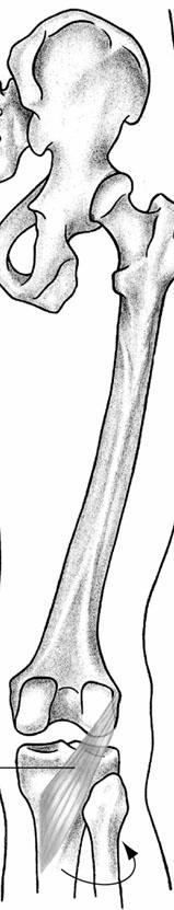
4 Movements Knee screws home to fully extend due to the shape of medial femoral condyle As approaches full extension tibia must externally rotate approximately 10 degrees to achieve proper alignment of tibial & femoral condyles In full extension close congruency of articular surfaces no appreciable of During initial flexion from full extension unlocks by tibia rotating internally, to a degree, from its externally rotated position to achieve flexion 2007 McGraw-Hill Higher Education. All rights reserved Flexion bending or decreasing angle between femur & leg, characterized by heel moving toward buttocks Extension straightening or increasing angle between femur & lower leg 2007 McGraw-Hill Higher Education. All rights reserved External Movements rotary movement of leg laterally away from midline Internal rotary movement of lower leg medially toward midline Neither will occur unless flexed degrees or > Quadriceps muscle group extends located in anterior compartment of thigh consists of 4 muscles rectus femoris vastus lateralis vastus intermedius vastus medialis 2007 McGraw-Hill Higher Education. All rights reserved McGraw-Hill Higher Education. All rights reserved Q angle Central line of pull for entire quadriceps runs from ASIS to the center of patella Line of pull of patella tendon runs from center of patella to center of tibial tuberosity Angle formed by the intersection of these two lines at the patella is the Q angle Normally, angle will be 15 degrees or less for males & 20 degrees or less in females Generally, females have higher angles due to a wider pelvis Q angle Higher Q angles generally predispose people in varying degrees to a variety of potential problems including lateral patellar subluxation or dislocation, patellar compression syndrome, chondromalacia, and ligamentous injuries For people with above normal Q angles, it is particularly important to maintain high levels of strength & endurance in vastus medialis so as to counteract lateral pull of vastus lateralis 2007 McGraw-Hill Higher Education. All rights reserved McGraw-Hill Higher Education. All rights reserved
5 Hamstring muscle group responsible for flexion located in posterior compartment of thigh consists of 3 muscles semitendinosus - medial, internal rotator semimembranosus - medial, internal rotator biceps femoris - lateral, external rotator Popliteus assist medial hamstrings in internal Two-joint muscles most effective when either origin or insertion is stabilized to prevent movement in direction of the contacting muscle To a degree, muscles are able to exert greater force when lengthened than when shortened Hamstring muscles & rectus femoris are biarticular (two-joint) muscles 2007 McGraw-Hill Higher Education. All rights reserved McGraw-Hill Higher Education. All rights reserved Ex. sartorius muscle increases its total length & becomes a better flexor at when pelvis is rotated posteriorly & stabilized by abdominal muscles exemplified by trying to flex & cross the legs in the sitting position one usually leans backward to flex legs at s Football kicker invariably leans well backward to raise & fix the rectus femoris origin to make it more effective as a extensor 2007 McGraw-Hill Higher Education. All rights reserved Gracilis, sartorius, & semitendinosus join together distally to form pes anserinus attaches to anteromedial aspect of proximal tibia below the level of tibial tuberosity Their attachment & posteromedially line of pull enable them to assist with flexion particularly once the is flexed & hip is externally rotated Medial & lateral gastrocnemius heads attach posteriorly on medial & lateral femoral condyles assist with flexion 2007 McGraw-Hill Higher Education. All rights reserved Knee joint muscles location Anterior - primarily extension Rectus femoris Vastus medialis Vastus intermedius Vastus lateralis Knee joint muscles location Posterior - primarily flexion Biceps femoris Semimembranosus Semitendinosus Sartorius Gracilis Popliteus Gastrocnemius 2007 McGraw-Hill Higher Education. All rights reserved McGraw-Hill Higher Education. All rights reserved
6 Nerves Femoral nerves innervates the extensors (quadriceps) rectus femoris vastus medialis vastus intermedius vastus lateralis Sciatic nerve tibial division Nerves semitendinosus, semimembranosus, biceps femoris (long head) common peroneal (fibular) division biceps femoris (short head) 2007 McGraw-Hill Higher Education. All rights reserved McGraw-Hill Higher Education. All rights reserved Quadriceps Quadriceps muscles - vital in jumping functions as a decelerator when decreasing speed to change direction when coming down from a jump eccentric contraction during decelerating actions controls slowing of movements initiated in previous phases of the sports skill Quadriceps Rectus femoris (two-joint), vastus medialis, vastus intermedius, vastus lateralis (largest) All attach to patella then to tibial tuberosity via patellar tendon All superficial & palpable except vastus intermedius (under rectus femoris) Strength or power may be indicated by vertical jump test Generally desired to be 25% to 33% stronger than hamstring group 2007 McGraw-Hill Higher Education. All rights reserved McGraw-Hill Higher Education. All rights reserved Quadriceps Rectus Femoris Muscle Strength & endurance is essential for maintenance of patellofemoral stability often a problem quads are particularly prone to atrophy when injuries occur may be developed by resisted extension activities from a seated position functional weight bearing activities such as step-ups or squats are particularly useful for strengthening & endurance Flexion of hip Extension of Anterior pelvic 2007 McGraw-Hill Higher Education. All rights reserved McGraw-Hill Higher Education. All rights reserved


7 Vastus Lateralis Muscle Vastus Intermedius Muscle Extension of Extension of 2007 McGraw-Hill Higher Education. All rights reserved McGraw-Hill Higher Education. All rights reserved Vastus Medialis Muscle Hamstring Extension of Hamstring muscle group Semitendinosus Biceps femoris Semimembranosus 2007 McGraw-Hill Higher Education. All rights reserved McGraw-Hill Higher Education. All rights reserved Hamstring Hamstring muscle strains very common Running muscles function in acceleration Antagonists to quadriceps muscles at Named for cordlike attachments at All originate on ischial tuberosity of pelvis Semitendinosus inserts on anteromedial tibia Semimembranosus inserts on posteromedial tibia Biceps femoris inserts on lateral tibial condyle & head of fibula Flexion of Extension of hip Internal of hip Internal of flexed Posterior pelvic Semitendinosus Muscle 2007 McGraw-Hill Higher Education. All rights reserved McGraw-Hill Higher Education. All rights reserved


8 Semimembranosus Muscle Biceps Femoris Muscle Flexion of Extension of hip Internal of hip Internal of flexed Posterior pelvic Flexion of Extension of hip External of hip External of flexed Posterior pelvic 2007 McGraw-Hill Higher Education. All rights reserved McGraw-Hill Higher Education. All rights reserved Flexion of Internal of flexed Popliteus Muscle Agonists Rectus Femoris Vastus Lateralis Vastus Intermedius Vastus Medialis Knee Extension 2007 McGraw-Hill Higher Education. All rights reserved McGraw-Hill Higher Education. All rights reserved Agonists Knee Flexion Biceps Femoris (Long & Short Head) Semitendinosus Semimembranosus Agonists Semitendinosus Semimembranosus Popliteus Knee Internal Rotation 2007 McGraw-Hill Higher Education. All rights reserved McGraw-Hill Higher Education. All rights reserved
9 Knee External Rotation Agonists Biceps Femoris 2007 McGraw-Hill Higher Education. All rights reserved Radiologic Anatomy Browser This site has numerous radiological views of the musculoskeletal system. University of Arkansas Medical School Gross Anatomy for Medical Students Dissections, anatomy tables, atlas images, links, etc. Loyola University Medical Center: Structure of the Human Body An excellent site with many slides, dissections, tutorials, etc. for the study of human anatomy Wheeless Textbook of Orthopaedics This site has an extensive index of links to the fractures, joints, muscles, nerves, trauma, medications, medical topics, lab tests, and links to orthopedic journals and other orthopedic and medical news McGraw-Hill Higher Education. All rights reserved Premiere Medical Search Engine This site allows the reader to enter any medical condition and it will search the net to find relevant articles. Arthroscopy.com Patient information on various musculoskeletal problems of the lower extremity Virtual Hospital Numerous slides, patient information, etc. Human Anatomy Online Interactive musculoskeletal anatomy The Hip and Knee Institute Arthritis of the Knee Joint 2007 McGraw-Hill Higher Education. All rights reserved Adam Healthcare Center Knee joint replacement American Academy of Orthopaedic Surgeons Patient education library on the Edheads Activities Allows you to perform virtual surgery Gross Anatomy: The Functional Anatomy of the Knee Joint Functional Anatomy of the Knee Knee Ligament Anatomy and Injury Anatomy and injuries of the Knee and its ligaments 2007 McGraw-Hill Higher Education. All rights reserved Duke Orthopaedics e joint Anatomy and Kinematics of the joint Knee Injury: Meniscus Understanding the joint and purpose of meniscus Smart Play: The Knee Anatomy, functions, injuries, etc. of the Patellofemoral Instability Patella Femoral Alignment Chiroweb.com Abnormal Q Angle and Orthotic Support The Physician and Sportsmedicine Acute Knee Injuries: When to Refer The Physician and Sportsmedicine Acute Knee Injuries: On-the-Field and Sideline Evaluation 2007 McGraw-Hill Higher Education. All rights reserved McGraw-Hill Higher Education. All rights reserved
The Knee. Clarification of Terms. Osteology of the Knee 7/28/2013. The knee consists of: The tibiofemoral joint Patellofemoral joint
 The Knee Clarification of Terms The knee consists of: The tibiofemoral joint Patellofemoral joint Mansfield, p273 Osteology of the Knee Distal Femur Proximal tibia and fibula Patella 1 Osteology of the
In the name of god. Knee. By: Tofigh Bahraminia Graduate Student of the Pathology Sports and corrective actions. Heat: Dr. Babakhani. Nov.
 In the name of god Knee By: Tofigh Bahraminia Graduate Student of the Pathology Sports and corrective actions Heat: Dr. Babakhani Nov. 2014 1 Anatomy-Bones Bones Femur Medial/lateral femoral condyles articulate
The Knee. Prof. Oluwadiya Kehinde
 The Knee Prof. Oluwadiya Kehinde www.oluwadiya.sitesled.com The Knee: Introduction 3 bones: femur, tibia and patella 2 separate joints: tibiofemoral and patellofemoral. Function: i. Primarily a hinge joint,
Sports Medicine 15. Unit I: Anatomy. The knee, Thigh, Hip and Groin. Part 4 Anatomies of the Lower Limbs
 Sports Medicine 15 Unit I: Anatomy Part 4 Anatomies of the Lower Limbs The knee, Thigh, Hip and Groin Anatomy of the lower limbs In Part 3 of this section we focused upon 11 of the 12 extrinsic muscles
Myology of the Knee. PTA 105 Kinesiology
 Myology of the Knee PTA 105 Kinesiology Objectives Describe the planes of motion and axes of rotation of the knee joint Visualize the origins and insertions of the muscles about the knee List the innervations
Knee Joint Assessment and General View
 Knee Joint Assessment and General View Done by; Mshari S. Alghadier BSc Physical Therapy RHPT 366 m.alghadier@sau.edu.sa http://faculty.sau.edu.sa/m.alghadier/ Functional anatomy The knee is the largest
The Lower Limb II. Anatomy RHS 241 Lecture 3 Dr. Einas Al-Eisa
 The Lower Limb II Anatomy RHS 241 Lecture 3 Dr. Einas Al-Eisa Tibia The larger & medial bone of the leg Functions: Attachment of muscles Transfer of weight from femur to skeleton of the foot Articulations
CHAPTER 8: THE BIOMECHANICS OF THE HUMAN LOWER EXTREMITY
 CHAPTER 8: THE BIOMECHANICS OF THE HUMAN LOWER EXTREMITY _ 1. The hip joint is the articulation between the and the. A. femur, acetabulum B. femur, spine C. femur, tibia _ 2. Which of the following is
Joints of the Lower Limb II
 Joints of the Lower Limb II Lecture Objectives Describe the components of the knee and ankle joint. List the ligaments associated with these joints and their attachments. List the muscles acting on these
The Knee. Tibio-Femoral
 The Knee Tibio-Femoral Osteology Distal Femur with Proximal Tibia Largest Joint Cavity in the Body A modified hinge joint with significant passive rotation Technically, one degree of freedom (Flexion/Extension)
Chapter 6 The Elbow and Radioulnar Joints
 The Elbow & Radioulnar Chapter 6 The Elbow and Radioulnar Manual of Structural Kinesiology R.T. Floyd, EdD, ATC, CSCS Most upper extremity movements involve the elbow & radioulnar joints Usually grouped
RN(EC) ENC(C) GNC(C) MN ACNP *** MECHANISM OF INJURY.. MOST IMPORTANT *** - Useful in determining mechanism of injury / overuse
 ENC(C) GNC(C) MN ACNP *** MECHANISM OF INJURY.. MOST IMPORTANT *** - Useful in determining mechanism of injury / overuse") HISTORY *** MECHANISM OF INJURY.. MOST IMPORTANT *** Age of patient Sport / Occupation - Certain conditions are more prevalent in particular age groups (Osgood Schlaters in youth / Degenerative Joint Disease
HISTORY *** MECHANISM OF INJURY.. MOST IMPORTANT *** Age of patient Sport / Occupation - Certain conditions are more prevalent in particular age groups (Osgood Schlaters in youth / Degenerative Joint Disease
Muscle Testing of Knee Extensors. Yasser Moh. Aneis, PhD, MSc., PT. Lecturer of Physical Therapy Basic Sciences Department
 Muscle Testing of Knee Extensors Yasser Moh. Aneis, PhD, MSc., PT. Lecturer of Physical Therapy Basic Sciences Department Muscle Testing of Knee Extensors othe Primary muscle Quadriceps Femoris -Rectus
Ligamentous and Meniscal Injuries: Diagnosis and Management
 Ligamentous and Meniscal Injuries: Diagnosis and Management Daniel K Williams, MD Franciscan Physician Network Orthopedic Specialists September 29, 2017 No Financial Disclosures INTRODUCTION Overview of
Prevention and Treatment of Injuries. Anatomy. Anatomy. Chapter 20 The Knee Westfield High School Houston, Texas
 Prevention and Treatment of Injuries Chapter 20 The Knee Westfield High School Houston, Texas Anatomy MCL, Medial Collateral Ligament LCL, Lateral Collateral Ligament PCL, Posterior Cruciate Ligament ACL,
Human Anatomy Biology 351
 Human Anatomy Biology 351 Lower Limb Please place your name on the back of the last page of this exam. You must answer all questions on this exam. Because statistics demonstrate that, on average, between
and K n e e J o i n t Is the most complicated joint in the body!!!!
 K n e e J o i n t K n e e J o i n t Is the most complicated joint in the body!!!! 1-Consists of two condylar joints between: A-The medial and lateral condyles of the femur and The condyles of the tibia
Main Menu. Joint and Pelvic Girdle click here. The Power is in Your Hands
 1 Hip Joint and Pelvic Girdle click here Main Menu K.6 http://www.handsonlineeducation.com/classes//k6entry.htm[3/23/18, 2:01:12 PM] Hip Joint (acetabular femoral) Relatively stable due to : Bony architecture
Copyright 2012 by The McGraw-Hill Companies, Inc. All rights reserved. McGraw-Hill/Irwin
 CHAPTER 8: THE LOWER EXTREMITY: KNEE, ANKLE, AND FOOT KINESIOLOGY Scientific Basis of Human Motion, 12 th edition Hamilton, Weimar & Luttgens Presentation Created by TK Koesterer, Ph.D., ATC Humboldt State
The thigh. Prof. Oluwadiya KS
 The thigh Prof. Oluwadiya KS www.oluwadiya.com The Thigh: Boundaries The thigh is the region of the lower limb that is approximately between the hip and knee joints Anteriorly, it is separated from the
The Hip (Iliofemoral) Joint. Presented by: Rob, Rachel, Alina and Lisa
 Joint. Presented by: Rob, Rachel, Alina and Lisa") The Hip (Iliofemoral) Joint Presented by: Rob, Rachel, Alina and Lisa Surface Anatomy: Posterior Surface Anatomy: Anterior Bones: Os Coxae Consists of 3 Portions: Ilium Ischium Pubis Bones: Pubis Portion
The Hip (Iliofemoral) Joint Presented by: Rob, Rachel, Alina and Lisa Surface Anatomy: Posterior Surface Anatomy: Anterior Bones: Os Coxae Consists of 3 Portions: Ilium Ischium Pubis Bones: Pubis Portion
Muscles of the Thigh. 6.1 Identify, describe the attachments of and deduce the actions of the muscles of the thigh: Anterior group
 Muscles of the Thigh 6.1 Identify, describe the attachments of and deduce the actions of the muscles of the thigh: Anterior group Sartorius: This is a long strap like muscle with flattened tendons at each
Muscles of the lower extremities. Dr. Nabil khouri MD, MSc, Ph.D
 Muscles of the lower extremities Dr. Nabil khouri MD, MSc, Ph.D Posterior leg Popliteal fossa Boundaries Biceps femoris (superior-lateral) Semitendinosis and semimembranosis (superior-medial) Gastrocnemius
To describe he knee joint, ligaments, structure & To list the main features of other lower limb joints
 To describe he knee joint, ligaments, structure & neurovascular supply To demonstrate the ankle joint anatomy To list the main features of other lower limb joints To list main groups of lymph nodes in
The Knee Joint By Prof. Dr. Muhammad Imran Qureshi
 The Knee Joint By Prof. Dr. Muhammad Imran Qureshi Structurally, it is the Largest and the most complex joint in the body because of the functions that it performs: Allows mobility (flexion/extension)
Recognizing common injuries to the lower extremity
 Recognizing common injuries to the lower extremity Bones Femur Patella Tibia Tibial Tuberosity Medial Malleolus Fibula Lateral Malleolus Bones Tarsals Talus Calcaneus Metatarsals Phalanges Joints - Knee
A Patient s Guide to Knee Anatomy
 A Patient s Guide to Knee Anatomy 15195 Heathcote Blvd Suite 334 Haymarket, VA 20169 Phone: 703-369-9070 Fax: 703-369-9240 DISCLAIMER: The information in this booklet is compiled from a variety of sources.
UNIT 7 JOINTS. Knee and Ankle Joints DR. ABDEL-MONEM A. HEGAZY
 UNIT 7 JOINTS Knee and Ankle Joints BY DR. ABDEL-MONEM A. HEGAZY (Degree in Bachelor of Medicine and Surgery with honor 1983, Dipl."Gynaecology and Obstetrics "1989, Master "Anatomy and Embryology "1994,
Muscles of the Hip 1. Tensor Fasciae Latae O: iliac crest I: lateral femoral condyle Action: abducts the thigh Nerve: gluteal nerve
 Muscles of the Hip 1. Tensor Fasciae Latae O: iliac crest I: lateral femoral condyle Action: abducts the thigh Nerve: gluteal nerve 2. Gluteus Maximus O: ilium I: femur Action: abduct the thigh Nerve:
A Patient s Guide to Knee Anatomy. Stephanie E. Siegrist, MD, LLC
 A Patient s Guide to Knee Anatomy Hands, shoulders, knees and toes (and elbows and ankles, too!) Most bone and joint conditions have several treatment options. The best treatment for you is based on your
The Knee. Two Joints: Tibiofemoral. Patellofemoral
 Evaluating the Knee The Knee Two Joints: Tibiofemoral Patellofemoral HISTORY Remember the questions from lecture #2? Girth OBSERVATION TibioFemoral Alignment What are the consequences of faulty alignment?
Knee Injury Assessment
 Knee Injury Assessment Clinical Anatomy p. 186 Femur Medial condyle Lateral condyle Femoral trochlea Tibia Intercondylar notch Tibial tuberosity Tibial plateau Fibula Fibular head Patella Clinical Anatomy
The Hip Joint. Shenequia Howard David Rivera
 The Hip Joint Shenequia Howard David Rivera Topics Of Discussion Movement Bony Anatomy Ligamentous Anatomy Muscular Anatomy Origin/Insertion/Action/Innervation Common Injuries MOVEMENT Flexion Extension
Human Anatomy Biology 351
 Human Anatomy Biology 351 Lower Limb Please place your name on the back of the last page of this exam. You must answer all questions on this exam. Because statistics demonstrate that, on average, between
DIAGNOSIS AND EARLY MANAGEMENT OF KNEE INJURIES
 DIAGNOSIS AND EARLY MANAGEMENT OF KNEE INJURIES INTRODUCTION: The knee is a common site of athletic injury. The knee injuries can be classified either into traumatic or acute and chronic, with overuse
Gluteal region DR. GITANJALI KHORWAL
 Gluteal region DR. GITANJALI KHORWAL Gluteal region The transitional area between the trunk and the lower extremity. The gluteal region includes the rounded, posterior buttocks and the laterally placed
Chapter 20 The knee and related structures
 Chapter 20 The knee and related structures Athletic Training Spring 2014 Jihong Park Bones & joints Femur, tibia, fibula, & patella Femur & tibia Weight bearing & muscle attachment Patella functions Anterior
The Muscular System. Chapter 10 Part D. PowerPoint Lecture Slides prepared by Karen Dunbar Kareiva Ivy Tech Community College
 Chapter 10 Part D The Muscular System Annie Leibovitz/Contact Press Images PowerPoint Lecture Slides prepared by Karen Dunbar Kareiva Ivy Tech Community College Table 10.14: Muscles Crossing the Hip and
KNEE INJURIES IN FEMALE SOCCER PLAYERS: A FOCUS ON THE ACL
 KNEE INJURIES IN FEMALE SOCCER PLAYERS: A FOCUS ON THE ACL Item Type text; Electronic Thesis Authors PEÑA, VANESSA NICOLE Publisher The University of Arizona. Rights Copyright is held by the author. Digital
Topic 7: Hip and pelvis. Parts of the hip. Parts of the femur
 Topic 7: Hip and pelvis Parts of the hip Parts of the femur Classifying the hip joint Ball and socket Synovial Multiaxial Movements of the hip: Abduction/adduction Flexion/extension Medial/lateral rotation
Practical 1 Worksheet
 Practical 1 Worksheet ANATOMICAL TERMS 1. Use the word bank to fill in the missing words. reference side stand body arms palms anatomical forward All anatomical terms have a(n) point which is called the
Objectives. The BIG Joint. Case 1. Boney Architecture. Presenter Disclosure Information. Common Knee Problems
 3:30 4:15 pm Common Knee Problems SPEAKER Christopher J. Visco, MD Presenter Disclosure Information The following relationships exist related to this presentation: Christopher J. Visco, MD: Speaker s Bureau
Understanding Leg Anatomy and Function THE UPPER LEG
 Understanding Leg Anatomy and Function THE UPPER LEG The long thigh bone is the femur. It connects to the pelvis to form the hip joint and then extends down to meet the tibia (shin bone) at the knee joint.
Balanced Body Movement Principles
 Balanced Body Movement Principles How the Body Works and How to Train it. Module 3: Lower Body Strength and Power Developing Strength, Endurance and Power The lower body is our primary source of strength,
DISSECTION SCHEDULE. Session I - Hip (Front) & Thigh (Superficial)
 & Thigh (Superficial)") DISSECTION SCHEDULE Session I - Hip (Front) & Thigh (Superficial) Surface anatomy Inguinal region Gluteal region Thigh Leg Foot bones Hip bone Femur Superficial fascia Great saphenous vein Superficial
DISSECTION SCHEDULE Session I - Hip (Front) & Thigh (Superficial) Surface anatomy Inguinal region Gluteal region Thigh Leg Foot bones Hip bone Femur Superficial fascia Great saphenous vein Superficial
rotation of the hip Flexion of the knee Iliac fossa of iliac Lesser trochanter Femoral nerve Flexion of the thigh at the hip shaft of tibia
 Anatomy of the lower limb Anterior & medial compartments of the thigh Dr. Hayder The fascia lata encloses the entire thigh like a sleeve/stocking. Three intramuscular fascial septa (lateral, medial, and
Sonography of Knee and Calf Pain: the differential considerations
 Sonography of Knee and Calf Pain: the differential considerations Dr. Lisa L. S.Wong Consultant Radiologist St Paul s Hospital Outline Ultrasound techniques Common pathologies in calf and posterior knee
Contents of the Posterior Fascial Compartment of the Thigh
 Contents of the Posterior Fascial Compartment of the Thigh 1-Muscles: B i c e p s f e m o r i s S e m i t e n d i n o s u s S e m i m e m b r a n o s u s a small part of the adductor magnus (h a m s t
The psoas minor is medial to the psoas major. The iliacus is a fan-shaped muscle that when contracted helps bring the swinging leg forward in walking
 1 p.177 2 3 The psoas minor is medial to the psoas major. The iliacus is a fan-shaped muscle that when contracted helps bring the swinging leg forward in walking and running. The iliopsoas and adductor
Human Anatomy Biology 255
 Human Anatomy Biology 255 Exam #4 Please place your name and I.D. number on the back of the last page of this exam. You must answer all questions on this exam. Because statistics demonstrate that, on average,
lesser trochanter of femur lesser trochanter of femur iliotibial tract (connective tissue) medial surface of proximal tibia
 medial surface of proximal tibia") LOWER LIMB MUSCLES OF THE APPENDICULAR SKELETON The muscles that act on the lower limb fall into three groups: those that move the thigh, those that move the lower leg, and those that move the ankle, foot,
LOWER LIMB MUSCLES OF THE APPENDICULAR SKELETON The muscles that act on the lower limb fall into three groups: those that move the thigh, those that move the lower leg, and those that move the ankle, foot,
Posterior compartment of the thigh. Dr. Heba Kalbouneh Associate Professor of Anatomy and Histology
 Posterior compartment of the thigh Dr. Heba Kalbouneh Associate Professor of Anatomy and Histology Posterior compartment of the thigh 1-Muscles: Biceps femoris Semitendinosus Semimembranosus Adductor magnus
Patellofemoral Instability
 Disclaimer This movie is an educational resource only and should not be used to manage Patellofemoral Instability. All decisions about the management of Patellofemoral Instability must be made in conjunction
Anatomage Table Instructors Guide- Lower Limb
 The Lower Limb Anatomage Table Instructors Guide- Lower Limb Table of Contents Lower Limb 1- The Skeletal System...3 1: Hip Bone...3 2: Hip Joint and Femur...4 3: Patella and Knee Joint...7 4: Tibia, Fibula,
Hip and Knee Approaches
 Hip and Knee Approaches Professor VLADIMIR STAVREV, MD, PhD, DMSc GEORGI MINEV MD DEPARTMENT OF ORTHOPEDICS AND TRAUMATOLOGY, MEDICAL UNIVERSITY -PLOVDIV Anterior View Hip Joint Posterior View - Hip Joint
MUSCLES OF THE LOWER LIMBS
 MUSCLES OF THE LOWER LIMBS Naming, location and general function Dr. Nabil khouri ROLES THAT SHOULD NOT BE FORGOTTEN Most anterior compartment muscles of the hip and thigh Flexor of the femur at the hip
Muscles of Lesson Five. Muscular Nomenclature and Kinesiology - Two. Muscles of Lesson Five, cont. Chapter 16
 Chapter 16 Muscular Nomenclature and Kinesiology - Two Lessons 5-6 Muscles of Lesson Five Iliopsoas (psoas major, iliacus) Hip outward rotators (piriformis, gemellus superior, gemellus inferior, obturator
Knee Joint Anatomy 101
 Knee Joint Anatomy 101 Bone Basics There are three bones at the knee joint femur, tibia and patella commonly referred to as the thighbone, shinbone and kneecap. The fibula is not typically associated with
Other Culprits in Knee Dysfunction
 Unraveling the Mystery of Knee Pain #6: Other Culprits in Knee Dysfunction 1 Webinar Goals Explore the assessment and treatment of other culprits in knee dysfunction. 2 Time: 60 minutes Schedule: Logistics
SOFT TISSUE INJURIES OF THE KNEE: Primary Care and Orthopaedic Management
 SOFT TISSUE INJURIES OF THE KNEE: Primary Care and Orthopaedic Management Gauguin Gamboa Australia has always been a nation where emphasis on health and fitness has resulted in an active population engaged
Ultrasound of the Knee Joint. Jun Sung Park,M.D. Bundang General Hospital Dept. of Rehabilitation Medicine
 Ultrasound of the Knee Joint Jun Sung Park,M.D. Bundang General Hospital Dept. of Rehabilitation Medicine Clinical History and P/E Chronic or Acute Symptoms Chronic Sx. : possible of systemic articular
Please differentiate an internal derangement from an external knee injury.
 Knee Orthopaedic Tests Sports and Knee Injuries James J. Lehman, DC, MBA, DABCO University of Bridgeport College of Chiropractic Knee Injury Strain, Sprain, Internal Derangement Anatomy of the Knee Please
By Matthew Donohoe, MA, ATC; Helen Aslanian, BS; and Kenneth Solomon, PhD, PE, Post PhD
 This article is approved by the following for continuing education credit: ACFEI provides this continuing education credit for Diplomates after June 2001 who are required to obtain 15 credits per year
ACL RECONSTRUCTION HAMSTRING METHOD. Presents ACL RECONSTRUCTION HAMSTRING METHOD. Multimedia Health Education
 HAMSTRING METHOD Presents HAMSTRING METHOD Multimedia Health Education Disclaimer Stephen J. Incavo MD This movie is an educational resource only and should not be used to make a decision on Anterior Cruciate
Overview Ligament Injuries. Anatomy. Epidemiology Very commonly injured joint. ACL Injury 20/06/2016. Meniscus Tears. Patellofemoral Problems
 Overview Ligament Injuries Meniscus Tears Pankaj Sharma MBBS, FRCS (Tr & Orth) Consultant Orthopaedic Surgeon Manchester Royal Infirmary Patellofemoral Problems Knee Examination Anatomy Epidemiology Very
Unicompartmental Knee Resurfacing
 Disclaimer This movie is an educational resource only and should not be used to manage knee pain. All decisions about the management of knee pain must be made in conjunction with your Physician or a licensed
Glenohumeral Joint. Glenohumeral Joint. Glenohumeral Joint. Glenohumeral Joint. Glenohumeral Joint. Glenohumeral Joint
 The Shoulder Joint Chapter 5 The Shoulder Joint Manual of Structural Kinesiology R.T. Floyd, EdD, ATC, CSCS McGraw-Hill Higher Education. All rights reserved. 5-1 Shoulder joint is attached to axial skeleton
Lower limb summary. Anterior compartment of the thigh. Done By: Laith Qashou. Doctor_2016
 Lower limb summary Done By: Laith Qashou Doctor_2016 Anterior compartment of the thigh Sartorius Anterior superior iliac spine Upper medial surface of shaft of tibia 1. Flexes, abducts, laterally rotates
A Patient s Guide to Pes Anserine Bursitis
 A Patient s Guide to Pes Anserine Bursitis 1436 Exchange Street Middlebury, VT 05753 Phone: 802-388-3194 Fax: 802-388-4881 cvo@champlainvalleyortho.com DISCLAIMER: The information in this booklet is compiled
1-Muscles: 2-Blood supply: Branches of the profunda femoris artery. 3-Nerve supply: Sciatic nerve
 1-Muscles: B i c e p s f e m o r i s S e m i t e n d i n o s u s S e m i m e m b r a n o s u s a small part of the adductor magnus (h a m s t r i n g p a r t o r i s c h i a l p a r t ) 2-Blood supply:
Evaluation of Knee Problems
 Evaluation of Knee Problems OBJECTIVES Review knee anatomy Explain tests to look for pathology Briefly introduce knee problems Only by a thorough knowledge of anatomy and functional testing can one make
ANATYOMY OF The thigh
 ANATYOMY OF The thigh 1- Lateral cutaneous nerve of the thigh Ι) Skin of the thigh Anterior view 2- Femoral branch of the genitofemoral nerve 1, 2 and 3 are From the lumber plexus 5- Intermediate cutaneous
this makes sense, however this is lower order thinking and does not solve the lower leg
 Functional Knee Valgus in a Barbell Squat 1 One of the most common lower leg dysfunction we see in athletes, particularly general population is functional knee valgus, or better referred to as the knees
Knee Anatomy Introduction Welcome to BodyZone Physiotherapy's patient resource about Knee problems.
 Knee Anatomy Introduction Welcome to BodyZone Physiotherapy's patient resource about Knee problems. To better understand how knee problems occur, it is important to understand some of the anatomy of thee
CLASSIFICATION OF JOINTS STRUCTURAL VS FUNCTIONAL
 CHAPTER 8 JOINTS CLASSIFICATION OF JOINTS STRUCTURAL VS FUNCTIONAL The most moveable type of joint is a 1) Synarthrosis 2) Amphiarthrosis 3) Diarthrosis FIBROUS JOINTS Figure 8.1 Fibrous joints. (a) Suture
Financial Disclosure. Medial Collateral Ligament
 Matthew Murray, M.D. UTHSCSA Sports Medicine Financial Disclosure Dr. Matthew Murray has no relevant financial relationships with commercial interests to disclose. Medial Collateral Ligament Most commonly
Distal Femoral Resection
 Distal Femoral Resection Annie Arteau, Bruno Fuchs Introduction This text is a general description of a distal femoral resection. Focus is on anatomical structures and muscle resection. Each femoral resection
In-Depth Foundations: Anatomy Terms to Know
 Be familiar with / able to identify and define all the following parts. The Spine Cranium Vertebrae Cervical, Thoracic, Lumbar Sacrum Coccyx Bones of Upper Body Cranium Mastoid process; Occipital condyle,
MUSCULOSKELETAL LOWER LIMB
 MUSCULOSKELETAL LOWER LIMB Spinal Cord Lumbar and Sacral Regions Spinal cord Dorsal root ganglion Conus medullaris Cauda equina Dorsal root ganglion of the fifth lumbar nerve End of subarachnoid space
THE LOWER EXTREMITY EXAM FOR THE FAMILY PRACTITIONER
 THE LOWER EXTREMITY EXAM FOR THE FAMILY PRACTITIONER Melinda A. Scott, D.O. Orthopedic Associates of Dayton Board Certified in Primary Care Sports Medicine GOALS Identify landmarks necessary for exam of
A Recreational Runner s Guide to Knee Pain
 Grand Valley State University ScholarWorks@GVSU Honors Projects Undergraduate Research and Creative Practice 2015 A Recreational Runner s Guide to Knee Pain Alexzanda Johnson Grand Valley State University
Human anatomy reference:
 Human anatomy reference: Weak Glut Activation Weak gluteal activation comes from poor biomechanics, poor awareness when training or prolonged exposure in deactivated positions such as sitting. Weak Glut
Exercise Science Section 4: Joint Mechanics and Joint Injuries
 Exercise Science Section 4: Joint Mechanics and Joint Injuries An Introduction to Health and Physical Education Ted Temertzoglou Paul Challen ISBN 1-55077-132-9 Types of Joints Fibrous joint Cartilaginous
UNIT 2 - CHAPTER 8: JOINTS OF THE SKELETAL SYSTEM LEARNING OUTCOMES:
 LEARNING OUTCOMES: 8.1 Introduction 1. List the functions of joints. 2. Explain how joints can be classified according to the type of tissue that binds the bones together and the degree of movement possible
Musculoskeletal Examination Benchmarks
 Musculoskeletal Examination Benchmarks _ The approach to examining the musculoskeletal system is the same no matter what joint or limb is being examined. The affected and contralateral region should both
Common Knee Injuries
 Common Knee Injuries In 2010, there were roughly 10.4 million patient visits to doctors' offices because of common knee injuries such as fractures, dislocations, sprains, and ligament tears. Knee injury
Your Practice Online
 Your Practice Online Disclaimer P R E S E N T S - PATELLAR TENDON This movie is an educational resource only and should not be used to make a decision on Anterior Cruciate Ligament (ACL) Reconstruction.
Learning IRM. The Knee: lateral ligaments and anatomical quadrants.
 Learning IRM. The Knee: lateral ligaments and anatomical quadrants. Poster No.: C-1733 Congress: ECR 2014 Type: Educational Exhibit Authors: A. Amador Gil, M. D. C. Jurado Gómez, V. de Lara Bendahan ;
The examination of the painful knee. Maja K Artandi, MD, FACP Clinical Associate Professor of Medicine Stanford University
 The examination of the painful knee Maja K Artandi, MD, FACP Clinical Associate Professor of Medicine Stanford University Objectives of the talk By the end of this talk you will know The important anatomy
BONES JOINTS MUSCLES OF THE LOWER LIMB
 BONES JOINTS MUSCLES OF THE LOWER LIMB LOWER LIMB: BONES LOWER LIMB GLUTEAL REGION consists of 6 major segments: FEMORAL REGION (THIGH) KNEE REGION LEG REGION TALOCRURAL REGION (ANKLE) FOOT REGION LOWER
Mohammad Ashraf. Abdulrahman Al-Hanbali. Ahmad Salman. 1 P a g e
 - 7 Mohammad Ashraf Abdulrahman Al-Hanbali Ahmad Salman 1 P a g e Structures under the cover of Gluteus Maximus: 1-Bones: Ileum, Femur (Head, greater trochanter and gluteal tuberosity), Ischium (ischial
Lateral knee injuries
 Created as a free resource by Clinical Edge Based on Physio Edge podcast episode 051 with Matt Konopinski Get your free trial of online Physio education at Orthopaedic timeframes Traditionally Orthopaedic
Patellofemoral Pain Syndrome
 43 Thames Street, St Albans, Christchurch 8013 Phone: (03) 356 1353. Website: philip-bayliss.com Patellofemoral Pain Syndrome Patellofemoral pain syndrome can be defined as a Retro-patellar (behind the
Imaging the Knee 17/10/2017. Friction syndrome Common in runners or cyclists Fluid between ITB and Lateral femoral condyle
 17/10/2017 Imaging the Knee Alicia M. Yochum RN, DC, DACBR, RMSK Iliotibial Band Syndrome Ligamentous Tears (ACL, PCL, MCL, LCL) Meniscal Tears Cartilage Degeneration Quadriceps/Patellar tendinosis Osteochondral
Identify the muscles associated with the medial compartment of the thigh. Identify the attachment points of the medial thigh muscles.
 L 8 A B O R A T O R Y Thigh MEDIAL THIGH Identify the muscles associated with the medial compartment of the thigh. Identify the attachment points of the medial thigh muscles. Identify the actions of these
HUMAN BODY COURSE LOWER LIMB NERVES AND VESSELS
 HUMAN BODY COURSE LOWER LIMB NERVES AND VESSELS October 22, 2010 D. LOWER LIMB MUSCLES 2. Lower limb compartments ANTERIOR THIGH COMPARTMENT General lfunction: Hip flexion, knee extension, other motions
Biomechanics of the Knee. Valerie Nuñez SpR Frimley Park Hospital
 Biomechanics of the Knee Valerie Nuñez SpR Frimley Park Hospital Knee Biomechanics Kinematics Range of Motion Joint Motion Kinetics Knee Stabilisers Joint Forces Axes The Mechanical Stresses to which