Kathleen R. Fink, MD Virginia Mason Medical Center. 6 th Nordic Emergency Radiology Course 2017
|
|
|
- Toby Parsons
- 5 years ago
- Views:
Transcription


1 Kathleen R. Fink, MD Virginia Mason Medical Center 6 th Nordic Emergency Radiology Course 2017
2 Disclosure My spouse receives research salary support from: Guerbet
3 Outline Indications for imaging CNS infections Extra axial Parenchymal Vascular complications
4 Indications for Imaging Suspected infection and: Altered mental status Seizures Focal neurologic deficits Immunocompromised patient with: New headache Any concerning sign
5 Imaging strategy Non contrast head CT first choice Rapid and widely available Well tolerated by critically ill patients Exclude life threatening conditions Contrast enhanced MR More sensitive for subtle findings - Leptomeningitis - Ventriculitis - Empyema - Infarction Consider strongly for immunocompromised patients Can be problematic in sick patients Contrast enhanced head CT if: MR not immediately available Contraindications to MR
6 Imaging before LP? Noncontrast CT can exclude contraindications CT more likely to show a contraindication in patient with (suspected meningitis) and: Age 60 Immunocompromise Recent seizure Focal neurological deficit Impaired consciousness Hasbun 2001 N Engl J Med 345:24,


7 Not safe to LP Cerebral edema: Poor gray-white differentiation Effaced sulci Effaced cisterns
8 Contraindications to LP No absolute consensus on imaging contraindications. General agreement on the following: Midline shift Effacement of the basal cisterns Posterior fossa mass effect. Clinical signs of herniation even with normal imaging.


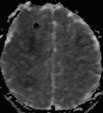
9 Cautionary tale 4 PM, comatose 8 PM, after LP
10 Extraaxial

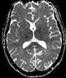
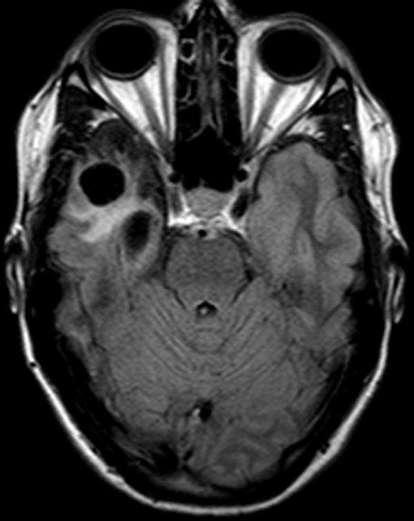
11 8 year old boy, sick one week

12 8 year old boy, sick one week Acute bacterial meningitis Post contrast
, empyema - Venous sinus")
13 Imaging in meningitis CSF evaluation is diagnostic Goal of imaging: 1) Exclude unexpected finding 2) Evaluate for complications: - Infarction - Hydrocephalus - Ventriculitis - Subdural effusions (kids), empyema - Venous sinus thrombosis
14 Meningitis: Imaging Imaging Findings: NORMAL Especially early Leptomeningeal enhancement Hemispheric Basilar Subdural effusions (especially children) Ddx leptomeningeal enhancement: Leptomeningeal spread of tumor Neurosarcoidosis CNS lymphoma


15 MRI: index case DWI/ADC T1 FLAIR

16 Meningitis: MRI Imaging Findings: FLAIR: high signal in subarachnoid space due to elevated protein May see arterial narrowing due to infectious arteritis with or without infarction Ddx: Subarachnoid FLAIR hyperintensities: Subarachnoid hemorrhage High inspired O2 Motion artifact Altered perfusion/blood brain barrier disruption Leakage of gad (renal failure, eg)

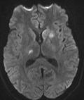
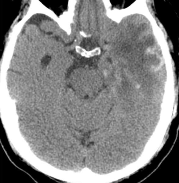
17 19 yo with worsening headache, nausea, and vomiting. NECT Hydrocephalus!!


18 MRI DWI/ADC FLAIR T1 post

19 Tuberculous meningitis Basilar meningitis: Can present with hydrocephalus due to thick inflammatory exudate Intracranial tuberculoma Granulomatous lesions Caseating or noncaseating +/- necrotic center Tuberculous abscess Complications: Vasculitis, infarcts Patkar Neuroimaging Clin N Am 22:4,


20 Key Imaging Features CT Normal Hydrocephalus Isodense exudate in basilar cisterns MR Enhancing basilar leptomeninges Infarcts Tuberculomas: Solid, nodular or ring enhancement


21 Image Gallery DWI T1 post Complications: Infarcts


22 Basilar meningitis T1 post T1 pre Ddx: basilar meningitis: Tuberculous meningitis Pyogenic meningitis Fungal meningitis Neurosarcoidosis Meningeal carcinomatosis


23 Tuberculous meningitis Basilar meningitis + infarcts: TB meningitis Fungal meningitis, including coccidioidomycosis Basilar meningitis + parenchymal lesions Think TB.
24 53 year old man with recurrent facial cellulitis, treated with antibiotics.


25 Subdural empyema T1 T2 T1 Post ADC
 T2 hyperintense +/- restricted diffusion (dark ADC) Peripheral and meningeal enhancement May see underlying cerebritis (as in this case)")
26 Subdural empyema: CT: Isodense collection Subdural empyema, DDX: Chronic Subdural hematoma Subdural effusion (sterile CSF collection associated with meningitis) Subdural hygroma Dural based mets MRI: T1 isointense (i.e proteinaceous material) T2 hyperintense +/- restricted diffusion (dark ADC) Peripheral and meningeal enhancement May see underlying cerebritis (as in this case)

27 Epidural abscess Usually associated with head and neck infection: Sinusitis Otomastoiditis Post trauma + Post Surgery * Subdural empyema and Epidural abscess can occur together. MRI may help differentiate.
28 Parenchymal


29 History: Feeling poorly for 3 weeks, bizarre behavior x 1 day, seizure

30 History: Feeling poorly for 3 weeks, bizarre behavior x 1 day, seizure FLAIR
31 Herpes Encephalitis Location: Anterior and medial temporal lobes Insula Lateral temporal lobes Inferior frontal lobes Cingulate
32 Key imaging features Normal -OR- Edema (low density) Hemorrhage Petechial Along brain surface Burned out: Gliosis CT Restricted diffusion may be first FLAIR MRI GRE for microhemorrhages May enhance Tien et al AJR Am J Roentgenol 161:1,


33 Image gallery FLAIR NECT DWI

34 Chronic changes of HSV encephalitis
35 Differential diagnosis: Ischemia (including venous infarction) Neoplasm Limbic encephalitis Other viral encephalitis (e.g. arboviral) Favor HSV: Bilateral Nonvascular distribution Normal basal ganglia
 Mitochondrial diseases Creutzfeldt Jacob Eastern equine encephalitis.")
36 Arbovirus infection Pathogenic viruses: Eastern equine Western Equine West Nile Japanese Tick-borne Basal ganglia and thalami lesions T2, FLAIR, DWI Ddx deep white matter: Anoxic/hypoxic injury CO2, toxic exposures Metabolic disorders (eg Wilson s disease) Mitochondrial diseases Creutzfeldt Jacob Eastern equine encephalitis. Case courtesy of Mahmoud Mossa-Basha, MD


37 History: 39 year old who fell Current Study Comparison from 9 months prior
38 MRI
39 HIV-associated neurocognitive disorders (HAND) Direct result of HIV on CNS Findings on CT and MRI do not predict cognitive dysfunction CT: Normal Volume loss: sulcal or ventricular enlargement Patchy white matter hypodensities
40 HIV-associated neurocognitive disorders (HAND) MRI: Symmetric white matter disease May resemble age-related volume loss or white matter lesions of vascular origin, but more than expected for age Spares Juxtacortical u- fibers


41 HIV-associated neurocognitive disorders (HAND) FLAIR T2


42 Key Imaging Features: HIV T1: occult DWI T1 post: Non-enhancing
43 Differential Diagnosis Age-related volume loss; white matter lesions of presumed vascular origin (chronic ischemic change) Hydrocephalus Progressive multifocal leukoencephalopathy

44 History: 43 year old with HIV and low CD4 count who presents with gait disturbance


45 T1 T1 post T2 DWI
46 Progressive Multifocal Leukoencephalopathy PML Seen in certain clinical scenarios: HIV Severe immunosuppression Multiple sclerosis on natalizumab
47 PML Imaging findings Low density CT and T2 hyperintense areas Little mass effect or contrast enhancement Parietal, occipital lobes Asymmetric

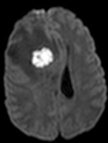
48 Features favoring a diagnosis of PML over HIV Involvement of subcortical u- fibers PML HIV
49 PML Confluent lesions, favors parietooccipital or CC Involves juxtacortical U fibers HIV Normal or patchy periventricular centrum semiovale lesions Spares U fibers Asymmetric Symmetric Low on T1 Low on DWI unless active demyelination Does not enhance unless IRIS (immune reconstitution inflammatory syndrome) Usually isointense on T1 Isointense on DWI No enhancement Sahraian. European Journal of Neurology 2012, 19: doi: /j x


50 Image Gallery Posterior fossa involvement


51 Image Gallery PML IRIS DWI NECT ADC T2 T1 post

52 History: 33 year old with nausea, vomiting and right sided weakness

53 Neurocysticercosis Taenia solium, pork tapeworm Cyst with central dot Central dot is scolex Four pathologic stages: Simple cyst complex cystic lesion calcification
54 Stage CT Findings MR Findings Noncystic (Active asymptomatic) Normal Normal Vesicular (Cyst or cluster of cysts with scolex) Colloidal vesicular (Larva degenerates, inflammatory response begins) Granular nodular (Cyst retracts and granulomatous reaction ensues) Calcified nodular (Inactive) 1-2 cm cyst Simple appearing fluid No edema. Scolex Cyst may be dense Enhances ± Edema Edema increases. Thick ring enhancement Calcific nodules without edema or enhancement Thin-walled cyst Follows CSF Little enhancement. Scolex Proteinaceous cyst Thick walled Edema Enhancement Edema increases. Thick ring enhancement Hypointense nodules without edema or enhancement Kimura-Hayama Radiographics 2010 Oct;30(6): doi: /rg


55 Key Imaging Features DWI NECT ADC T2 T1 post


56 Image Gallery Subarachnoid Calcified nodular phase Intraventricular cysts can cause hydrocephalus.
57 Differential Diagnosis Pyogenic abscess (no scolex) Ring enhancing mass: Metastasis Glioblastoma multiforme Lymphoma in immunocompromised patient Etc.


58 History: 38 year old with recurrent sinus infections, worsening headache, nausea and vomiting + Contrast


59 MRI: DWI T1 ADC FLAIR T1 post
60 Pyogenic abscess Focal pus collection with surrounding capsule. Direct extension Sinusitis Otomastoiditis Odontogenic Hematogenous IVDA Endocarditis Pulmonary AVF

61 Pyogenic abscess Parenchymal mass Gray-white junction Low T2 ring Hyperintense necrotic core Rim enhancement Thick smooth Thinned medial wall Restricted diffusion of central necrotic core Daughter cells Look for ventricular extension

62 Image gallery


63 Cerebritis 2 days later DWI FLAIR FLAIR T1 post T1 post


:972-89.")
64 Abscess development 13 days later Early cerebritis Ill defined edema Late cerebritis Central low density Early capsule Thin rim enhancement Britt J Neurosurg 1983 December;59(6): Late capsule Thick rim enhancement


65 History: 40 yo with HIV and 2 days of headache, blurry vision, gait disturbance


66 MRI: Vital in Immunocompromised

67 Toxoplasmosis Toxoplasma gondii Reactivation of latent infection in immunocompromised patient Masses: Eccentric target sign: Specific not sensitive Ring enhancing T2 heterogeneous No restricted DWI of central necrotic portion Location: Basal ganglia Thalamus Gray-white junction Akgoz et al. Neuroimaging Clin N Am 22:4,

68 HIV patient with mental status change DWI FLAIR ADC
69 Cryptococcus Cryptococcus neoformans Associated with HIV infections Can affect immunocompetent patients Presents as Meningitis Meningoencephalitis Cerebral vasculitis Imaging may be normal.
70 Cryptococcus Imaging patterns: Meningeal enhancement Basilar meningitis Masses: cryptococcomas Granulomas Basal ganglia predominant May enhance (immunocompetent) Choroid plexus Gelatinous exudate: Dilated perivascular spaces Pseudocysts


71 Image Gallery DWI T1 FLAIR ADC T1 post T2


72 Image gallery: C. gatti Dilated VR spaces Cryptococcomas of choroid plexus
73 Differential Diagnosis Tuberculous meningitis Cryptococcus meningitis Coccidioidal meningitis
74 Vascular complications


75 54 yo, aortic valve replacement, new headache NECT CTA


76 Conventional Angiogram 2 months prior Current Right ICA injection
77 Mycotic aneurysm New peripheral (distal MCA) aneurysm Unusual location for saccular aneurysm Treatment is resection * methicillin-sensitive staphylococcus aureus

78 Infectious vasculitis: S. pneumo meningitis Initial T2 2 wks later

79 Septic emboli
80 Outline Indications for imaging CNS infections Extra axial Parenchymal Vascular complications
81 Thank you! Kathleen Fink
Disclosure. + Outline. Case-based approach to neurological emergencies that might present to the ED
 Kathleen R. Fink, MD University of Washington 5 th Nordic Emergency Radiology Course May 21, 2015 Disclosure My spouse receives research salary support from: Bracco BayerHealthcare Guerbet Outline Case-based
An Approach. to Brain. Infection. 37F found down. Disclosures. Approach to CNS Infection. Objectives. Parenchymal. None.
 An Approach Disclosures to Brain None. Infection Jason Shewchuk, MD Clinical Associate Professor Head of Neuroradiology UBC European Course in Neuroradiology 2018 Objectives Following this session the
RINGS N THINGS: Imaging Patterns in Differential Diagnosis. Anne G. Osborn, M.D.
 RINGS N THINGS: Imaging Patterns in Differential Diagnosis Anne G. Osborn, M.D. ExpDDxs: Intra-axial (Parenchymal) Lesions Ring-enhancing lesions, solitary 1 Ring-enhancing lesion crossing corpus callosum
Brain Pain Infections of the CNS
 FRIDAY, OCTOBER 28, 2016 Brain Pain Infections of the CNS Suyash Mohan MD, PDCC Assistant Professor of Radiology & Neurosurgery Division of Neuroradiology, Department of Radiology Perelman School of Medicine
IMAGING OF INTRACRANIAL INFECTIONS
 IMAGING OF INTRACRANIAL INFECTIONS Dr Carolina Kachramanoglou LYSHOLM DEPARTMENT OF NEURORADIOLOGY NATIONAL HOSPITAL FOR NEUROLOGY AND NEUROSURGERY Plan Introduce MR sequences that are useful in the diagnosis
Imaging in a confused patient: Infections and Inflammation
 American Society of Neuroimaging Imaging in a confused patient: Infections and Inflammation January 21, 2017 Los Angeles, California Joshua P. Klein, MD, PhD, FANA, FAAN, FASN Chief, Division of Hospital
MR neuroimaging of HIV infected patients : A pictorial review
 MR neuroimaging of HIV infected patients : A pictorial review Poster No.: R-0198 Congress: 2014 CSM Type: Scientific Exhibit Authors: P. F. Kwan, R. Thomas, A. Dixon; SOUTH YARRA/AU Keywords: Neuroradiology
Central Nervous System Infection
 Central Nervous System Infection Ashley H. Aiken KEYWORDS CNS infections Meningitis Abscess Encephalitis Subdural empyema Infections of the brain and its linings pose a growing, worldwide health problem.
NEURO IMAGING 2. Dr. Said Huwaijah Chairman of radiology Dep, Damascus Univercity
 NEURO IMAGING 2 Dr. Said Huwaijah Chairman of radiology Dep, Damascus Univercity I. EPIDURAL HEMATOMA (EDH) LOCATION Seventy to seventy-five percent occur in temporoparietal region. CAUSE Most likely caused
Cerebro-vascular stroke
 Cerebro-vascular stroke CT Terminology Hypodense lesion = lesion of lower density than the normal brain tissue Hyperdense lesion = lesion of higher density than normal brain tissue Isodense lesion = lesion
RING ENCHANCING LESION BY M.S. HEMHNATH
 RING ENCHANCING LESION BY M.S. HEMHNATH A 21 YRS FEMALE CAME WITH H/O HEADACHE AND SEIZURE FOR THE PAST ONE MONTH. NO OTHER FOCAL NEUROLOGICAL DEFICIT. DIFFERENTIAL DIAGNOSIS For this case are Neurocysticerosis
Non-Traumatic Neuro Emergencies
 Department of Radiology University of California San Diego Non-Traumatic Neuro Emergencies John R. Hesselink, M.D. Nontraumatic Neuroemergencies 1. Acute focal neurological deficit 2. Worst headache of
CT and MR findings of systemic lupus erythematosus involving the brain: Differential diagnosis based on lesion distribution
 CT and MR findings of systemic lupus erythematosus involving the brain: Differential diagnosis based on lesion distribution Poster No.: C-2723 Congress: ECR 2010 Type: Educational Exhibit Topic: Neuro
Role of imaging (images) in my practice. Dr P Senthur Nambi Consultant Infectious Diseases
 in my practice. Dr P Senthur Nambi Consultant Infectious Diseases") Role of imaging (images) in my practice Dr P Senthur Nambi Consultant Infectious Diseases Medical images: My thoughts Images are just images Subject to the intellect of the interpreter View it in conjuction
Role of imaging (images) in my practice Dr P Senthur Nambi Consultant Infectious Diseases Medical images: My thoughts Images are just images Subject to the intellect of the interpreter View it in conjuction
The central nervous system
 Sectc.qxd 29/06/99 09:42 Page 81 Section C The central nervous system CNS haemorrhage Subarachnoid haemorrhage Cerebral infarction Brain atrophy Ring enhancing lesions MRI of the pituitary Multiple sclerosis
Head CT Scan Interpretation: A Five-Step Approach to Seeing Inside the Head Lawrence B. Stack, MD
 Head CT Scan Interpretation: A Five-Step Approach to Seeing Inside the Head Lawrence B. Stack, MD Five Step Approach 1. Adequate study 2. Bone windows 3. Ventricles 4. Quadrigeminal cistern 5. Parenchyma
CNS infections (1 of 2)
") CNS infections (1 of 2) How can microbes enter the nervous system? Hematogenous the most common mostly arterial can be from facial veins (through anastomoses with venous sinuses of the skull) Direct implantation
CNS infections (1 of 2) How can microbes enter the nervous system? Hematogenous the most common mostly arterial can be from facial veins (through anastomoses with venous sinuses of the skull) Direct implantation
Imaging of CNS Infections in Immunocompetent Hosts
 NS Infections, O rien Imaging of NS Infections in Immunocompetent Hosts William T. O rien, Sr., D.O. Division of Neuroradiology, Wilford Hall mbulatory Surgical enter, San ntonio, TX Infections of the
Benign brain lesions
 Benign brain lesions Diagnostic and Interventional Radiology Hung-Wen Kao Department of Radiology, Tri-Service General Hospital, National Defense Medical Center Computed tomography Hounsfield unit (HU)
An Introduction to Imaging the Brain. Dr Amy Davis
 An Introduction to Imaging the Brain Dr Amy Davis Common reasons for imaging: Clinical scenarios: - Trauma (NICE guidelines) - Stroke - Tumours - Seizure - Neurological degeneration memory, motor dysfunction,
Outline. Neuroradiology. Diffusion Imaging in. Clinical Applications of. Basics of Diffusion Imaging. Basics of Diffusion Imaging
 Clinical Applications of Diffusion Imaging in Neuroradiology No disclosures Stephen F. Kralik Assistant Professor of Radiology Indiana University School of Medicine Department of Radiology and Imaging
NEURO IMAGING OF ACUTE STROKE
 1 1 NEURO IMAGING OF ACUTE STROKE ALICIA RICHARDSON, MSN, RN, ACCNS-AG, ANVP-BC WENDY SMITH, MA, RN, MBA, SCRN, FAHA LYNN HUNDLEY, APRN, CNRN, CCNS, ANVP-BC 2 2 1 DISCLOSURES Alicia Richardson: Stryker
Bacterial, viral, protoozal and fungal infections of the CNS
 Bacterial, viral, protoozal and fungal infections of the CNS Prof. Isidro Ferrer, Institut Neuropatologia, Servei Anatomia Patològica, IDIBELL-Hospital Universitari de Bellvitge, Universitat de Barcelona,
Masses of the Corpus Callosum
 Masses of the Corpus Callosum Kesav Raghavan, HMS Year III Dr. Agenda Corpus Callosum Development and Anatomy Our Patient: Clinical Presentation Differential Diagnosis of Masses in the Corpus Callosum
ISCHEMIC STROKE IMAGING
 ISCHEMIC STROKE IMAGING ผศ.พญ พญ.จ ร ร ตน ธรรมโรจน ภาคว ชาร งส ว ทยา คณะแพทยศาสตร มหาว ทยาล ยขอนแก น A case of acute hemiplegia Which side is the abnormality, right or left? Early Right MCA infarction
Imaging findings in CNS infections and differential diagnosis. M. Lequin
 Imaging findings in CNS infections and differential diagnosis M. Lequin OUTLINE Introduction and terminology Diagnosis & Differential diagnosis Pediatric brain infections viral infections Meningitis Encephalitis
HEAD AND NECK IMAGING. James Chen (MS IV)
") HEAD AND NECK IMAGING James Chen (MS IV) Anatomy Course Johns Hopkins School of Medicine Sept. 27, 2011 OBJECTIVES Introduce cross sectional imaging of head and neck Computed tomography (CT) Review head
HEAD AND NECK IMAGING James Chen (MS IV) Anatomy Course Johns Hopkins School of Medicine Sept. 27, 2011 OBJECTIVES Introduce cross sectional imaging of head and neck Computed tomography (CT) Review head
Interactive Cases: Demyelinating Diseases and Mimics. Disclosures. Case 1 25 yo F with nystagmus; look for tumor 4/14/2017
 Interactive Cases: Demyelinating Diseases and Mimics Disclosures None Brad Wright, MD 27 March 2017 Case 1 25 yo F with nystagmus; look for tumor What do you suspect? A. Demyelinating disease B. Malignancy
Essentials of Clinical MR, 2 nd edition. 14. Ischemia and Infarction II
 14. Ischemia and Infarction II Lacunar infarcts are small deep parenchymal lesions involving the basal ganglia, internal capsule, thalamus, and brainstem. The vascular supply of these areas includes the
Stroke School for Internists Part 1
 Stroke School for Internists Part 1 November 4, 2017 Dr. Albert Jin Dr. Gurpreet Jaswal Disclosures I receive a stipend for my role as Medical Director of the Stroke Network of SEO I have no commercial
Index. aneurysm, 92 carotid occlusion, 94 ICA stenosis, 95 intracranial, 92 MCA, 94
 A ADC. See Apparent diffusion coefficient (ADC) Aneurysm cerebral artery aneurysm, 93 CT scan, 93 gadolinium, 93 Angiography, 13 Anoxic brain injury, 25 Apparent diffusion coefficient (ADC), 7 Arachnoid
Neuroradiology of AIDS
 Neuroradiology of AIDS Frank Minja,, HMS IV Gillian Lieberman MD September 2002 AIDS 90% of HIV patients have CNS involvement 1 10% of AIDS patients present first with neurological symptoms 2 73-80% of
SWI including phase and magnitude images
 On-line Table: MRI imaging recommendation and summary of key features Sequence Pathologies Visible Key Features T1 volumetric high-resolution whole-brain reformatted in axial, coronal, and sagittal planes
For Emergency Doctors. Dr Suzanne Smallbane November 2011
 For Emergency Doctors Dr Suzanne Smallbane November 2011 A: Orbit B: Sphenoid Sinus C: Temporal Lobe D: EAC E: Mastoid air cells F: Cerebellar hemisphere A: Frontal lobe B: Frontal bone C: Dorsum sellae
brain MRI for neuropsychiatrists: what do you need to know
 brain MRI for neuropsychiatrists: what do you need to know Christoforos Stoupis, MD, PhD Department of Radiology, Spital Maennedorf, Zurich & Inselspital, University of Bern, Switzerland c.stoupis@spitalmaennedorf.ch
MRI OF THE THALAMUS. Mohammed J. Zafar, MD, FAAN Kalamazoo, MI
 1 MRI OF THE THALAMUS Mohammed J. Zafar, MD, FAAN Kalamazoo, MI Objectives: The thalamic nuclei can be involved in a wide variety of conditions. A systematic imaging approach would be useful for narrowing
Disclosure. + Outline. What is a stroke? Role of imaging in stroke Ischemic stroke Venous infarct Current topics
 + Kathleen R. Fink, MD University of Washington 5 th Nordic Emergency Radiology Course May 21, 2015 + Disclosure My spouse receives research salary support from: Bracco BayerHealthcare Guerbet Thank you
[(PHY-3a) Initials of MD reviewing films] [(PHY-3b) Initials of 2 nd opinion MD]
![[(PHY-3a) Initials of MD reviewing films] [(PHY-3b) Initials of 2 nd opinion MD]](/thumbs/89/98619893.jpg "[(PHY-3a) Initials of MD reviewing films] [(PHY-3b) Initials of 2 nd opinion MD]") 2015 PHYSICIAN SIGN-OFF (1) STUDY NO (PHY-1) CASE, PER PHYSICIAN REVIEW 1=yes 2=no [strictly meets case definition] (PHY-1a) CASE, IN PHYSICIAN S OPINION 1=yes 2=no (PHY-2) (PHY-3) [based on all available
2015 PHYSICIAN SIGN-OFF (1) STUDY NO (PHY-1) CASE, PER PHYSICIAN REVIEW 1=yes 2=no [strictly meets case definition] (PHY-1a) CASE, IN PHYSICIAN S OPINION 1=yes 2=no (PHY-2) (PHY-3) [based on all available
Ashton Lehmann, Harvard Medical School Year III. Gillian Lieberman, MD
 May, 2012 Ashton Lehmann, Harvard Medical School Year III Our patient presents to ED with acute onset, worsening headache: worst headache of my life Evaluation for subarachnoid hemorrhage with noncontrast
Vasculitides in Surgical Neuropathology Practice
 Vasculitides in Surgical Neuropathology Practice USCAP requires that all faculty in a position to influence or control the content of CME disclose any relevant financial relationship WITH COMMERCIAL INTERESTS
The Many Faces of Central Nervous System Tuberculosis
 The Many Faces of Central Nervous System Tuberculosis Poster No.: C-2347 Congress: ECR 2013 Type: Educational Exhibit Authors: B. Alami, F. Belhoussine, O. Addou, M. Y. Alaoui Lamrani, M. 1 1 1 1 1 1 2
Fundamental Clinical Brain MR Imaging Applications and Protocols
 Continuing Education Seminar for Radiologic Technologists Fundamental Clinical Brain MR Imaging Applications and Protocols Darren P. O Neill, MD Indiana University Neuroradiology Objectives Review fundamental
Cerebral Toxoplasmosis in HIV-Infected Patients. Ahmed Saad,MD,FACP
 Cerebral Toxoplasmosis in HIV-Infected Patients Ahmed Saad,MD,FACP Introduction Toxoplasmosis: Caused by the intracellular protozoan, Toxoplasma gondii. Immunocompetent persons with primary infection
Unit VIII Problem 6 Pathology: Meningitis
 Unit VIII Problem 6 Pathology: Meningitis - Important terms: Meningitis: it is inflammation of meninges (coverings of the central nervous system) caused by infection. They are classified to: Pachymeningitis:
Vascular Malformations of the Brain: A Review of Imaging Features and Risks
 Vascular Malformations of the Brain: A Review of Imaging Features and Risks Comprehensive Neuroradiology: Best Practices October 27-30, 2016 Sudhakar R. Satti, MD Associate Director Neurointerventional
Demyelinating Diseases of the Brain
 Department of Radiology University of California San Diego Demyelinating Diseases of the Brain John R. Hesselink, M.D. T1-Weighted Images Normal White Matter Contents Axons with envelope of myelin Neuroglia
Moath Darweesh. Zaid Emad. Anas Abu -Humaidan
 3 Moath Darweesh Zaid Emad Anas Abu -Humaidan Introduction: First two lectures we talked about acute and chronic meningitis, which is considered an emergency situation. If you remember, CSF examination
Pathologic Analysis of CNS Surgical Specimens
 2015 Kenneth M. Earle Memorial Neuropathology Review Pathologic Analysis of CNS Surgical Specimens Peter C. Burger, MD Interdisciplinary Quality Control Familiarity with entities Use of diagnostic algorithm
Kathleen R. Fink, MD Virginia Mason Medical Center. 6 th Nordic Emergency Radiology Course 2017
 Kathleen R. Fink, MD Virginia Mason Medical Center 6 th Nordic Emergency Radiology Course 2017 Disclosure My spouse receives research salary support from: Guerbet Outline Acute neck and back pain Acute
10 May Disclosure. + Outline. Case-based approach to nontraumatic intracranial hemorrhage. Kathleen R. Fink, MD University of Washington
 Kathleen R. Fink, MD University of Washington 5 th Nordic Emergency Radiology Course May 21, 2015 Disclosure My spouse receives research salary support from: Bracco BayerHealthcare Guerbet Outline Case-based
CNS Infections in the Pediatric Age Group
 CNS Infections in the Pediatric Age Group Introduction CNS infections are frequently life-threatening In the Philippines, bacterial meningitis is one of the top leading causes of mortality in children
Disclosure. Learner Objectives. Congenital Infections. Question. Main Categories 4/26/2016
 Communicating Communicability: Imaging of CNS Infections Aaron P. Kamer, MD Assistant Professor of Clinical Radiology Neuroradiology Section April 26, 2016 Disclosure Within the past 12 months: I have
Meninges and Ventricles
 Meninges and Ventricles Irene Yu, class of 2019 LEARNING OBJECTIVES Describe the meningeal layers, the dural infolds, and the spaces they create. Name the contents of the subarachnoid space. Describe the
Automated Identification of Neoplasia in Diagnostic Imaging text reports
 Automated Identification of Neoplasia in Diagnostic Imaging text reports "This work has been funded in whole or in part with Federal funds from the National Cancer Institute, National Institutes of Health,
Intracranial spontaneous hemorrhage mechanisms, imaging and management
 Intracranial spontaneous hemorrhage mechanisms, imaging and management Dora Zlatareva Department of Diagnostic Imaging Medical University, Sofia, Bulgaria Intracranial hemorrhage (ICH) ICH 15% of strokes
Case 7391 Intraventricular Lesion
 Case 7391 Intraventricular Lesion Bastos Lima P1, Marques C1, Cabrita F2, Barbosa M2, Rebelo O3, Rio F1. 1Neuroradiology, 2Neurosurgery, 3Neuropathology, Coimbra University Hospitals, Portugal. University
MRI and differential diagnosis in patients suspected of having MS
 Andrea Falini Italy MRI and differential diagnosis in patients suspected of having MS IMPROVING THE PATIENT S LIFE THROUGH MEDICAL EDUCATION www.excemed.org Outline of presentation - Diagnostic criteria
Reassessment: Neuroimaging in the Emergency Patient Presenting with Seizure
 Reassessment: Neuroimaging in the Emergency Patient Presenting with Seizure This evidence-based report provides clinicians with information to identify which seizure patients in the emergency department
How to Analyse Difficult Chest CT
 How to Analyse Difficult Chest CT Complex diseases are:- - Large lesion - Unusual or atypical pattern - Multiple discordant findings Diffuse diseases are:- - Numerous findings in both sides 3 basic steps
A pictorial review of neurological complications of systemic lupus erythematosus and antiphospholipid syndrome
 A pictorial review of neurological complications of systemic lupus erythematosus and antiphospholipid syndrome Poster No.: C-2780 Congress: ECR 2010 Type: Educational Exhibit Topic: Neuro Authors: E. Tavernaraki,
Classical CNS Disease Patterns
 Classical CNS Disease Patterns Inflammatory Traumatic In response to the trauma of having his head bashed in GM would have experienced some of these features. NOT TWO LITTLE PEENY WEENY I CM LACERATIONS.
Pediatric CNS Tumors. Disclosures. Acknowledgements. Introduction. Introduction. Posterior Fossa Tumors. Whitney Finke, MD
 Pediatric CNS Tumors Disclosures Whitney Finke, MD Neuroradiology Fellow PGY-6 University of Utah Health Sciences Center Salt Lake City, Utah None Acknowledgements Introduction Nicholas A. Koontz, MD Luke
Pearls and Pitfalls in Neuroradiology of Cerebrovascular Disease The Essentials with MR and CT
 Pearls and Pitfalls in Neuroradiology of Cerebrovascular Disease The Essentials with MR and CT Val M. Runge, MD Wendy R. K. Smoker, MD Anton Valavanis, MD Control # 823 Purpose The focus of this educational
Attenuation value in HU From -500 To HU From -10 To HU From 60 To 90 HU. From 200 HU and above
 Brain Imaging Common CT attenuation values Structure Air Fat Water Brain tissue Recent hematoma Calcifications Bone Brain edema and infarction Normal liver parenchyma Attenuation value in HU From -500
Intracranial Infections: Clinical and Imaging Characteristics
 Acta Radiologica ISSN: 0284-1851 (Print) 1600-0455 (Online) Journal homepage: http://www.tandfonline.com/loi/iard20 Intracranial Infections: Clinical and Imaging Characteristics B. R. Foerster, M. M. Thurnher,
41 year old female with headache. Elena G. Violari MD and Leo Wolansky MD
 41 year old female with headache Elena G. Violari MD and Leo Wolansky MD ? Dural Venous Sinus Thrombosis with Hemorrhagic Venous Infarct Acute intraparenchymal hematoma measuring ~3 cm in diameter centered
Acute Ischaemic Stroke
 Acute Ischaemic Stroke CT or MR SCA READIG FORM SCA ID: DATE OF READIG: SCA QUALIT: Good Moderate Poor Comment: READER ID: TPE OF SCA: CT: Without contrast: With contrast: MR: Diffusion: Perfusion ote
CASE OF THE WEEK PROFESSOR YASSER METWALLY
 CLINICAL PICTURE CLINICAL PICTURE 26 years old male patient presented clinically with a grand male fit, confusion, fever, headache, and nausea. Examination showed bilateral papilledema and left sided extensor
AMSER Case of the Month July 2018 Complicated Headache with Fever
 AMSER Case of the Month July 2018 Complicated Headache with Fever Benjamin Park, MS IV Dr. Karen Xie Department of Radiology University of Illinois College of Medicine at Chicago Patient Presentation CC:
DES 9 janvier P. David. Clinic of Neuroradiology Erasme Hospital Université Libre de Bruxelles Belgium
 DES 9 janvier 2015 P. David Clinic of Neuroradiology Erasme Hospital Université Libre de Bruxelles Belgium CNS Infections Early recognition in children, infants Longterm effects on the brain :devastating
Case 9 10/29/2018. CJD (Creutzfeldt -Jakob Disease) CJD (Creutzfeldt -Jakob Disease) CJD (Creutzfeldt -Jakob Disease)
 CJD (Creutzfeldt -Jakob Disease) CJD (Creutzfeldt -Jakob Disease)") CJD (Creutzfeldt -Jakob Disease) Rare fatal neurodegen dz caused by infectious protein Prion (lacks nucleic acid)- causes spongiform changes of the brain and neuronal death. 4 types: scjd- 85% of cases
CJD (Creutzfeldt -Jakob Disease) Rare fatal neurodegen dz caused by infectious protein Prion (lacks nucleic acid)- causes spongiform changes of the brain and neuronal death. 4 types: scjd- 85% of cases
Cerebrovascular diseases-2
 Cerebrovascular diseases-2 Primary angiitis of CNS - Other causes of infarction i. Hypercoagulable states ii. Drug-abuse such as amphetamine, heroin and cocain Note - The venous side of the circulation
Neuroradiological Findings in Non- Accidental Trauma Educational Pictorial Review
 Neuroradiological Findings in Non- Accidental Trauma Educational Pictorial Review M B Moss, MD; L Lanier, MD; R Slater; C L Sistrom, MD; R G Quisling, MD; I M Schmalfuss, MD; and D Rajderkar, MD Contact:
A challenging neurological complication in a young HIV-infected woman
 A challenging neurological complication in a young HIV-infected woman Ianache Irina-Cristiana Vi tor Ba es Clini al Hospital for Infectious and Tropical Diseases Bucharest - HIV/AIDS department Assessment
Applicable Neuroradiology
 For the Clinical Neurology Clerkship LSU Medical School New Orleans Amy W Voigt, MD Clerkship Director Introduction The field of Radiology first developed following the discovery of X-Rays by Wilhelm Roentgen
In-Training Examination for Diagnostic Radiology Residents Rationales
 28th Annual In-Training Examination for Diagnostic Radiology Residents Rationales Sponsored by: Commission on Education Committee on Residency Training in Diagnostic Radiology February 3, 2005 The American
Brain Imaging. IC calcifications. Mamdouh mahfouz MD
 Brain Imaging IC calcifications www.ssregypt.com Mamdouh mahfouz MD mamdouh.m5@gmail.com CT Hyper dense [ more than100 HU ] MRI Low signal in T1 and T2 WIs [non mobile protons] Exceptions Minute calcifications
CNS TUMORS. D r. Ali Eltayb ( U. of Omdurman. I ). M. Path (U. of Alexandria)
. M. Path (U. of Alexandria)") CNS TUMORS D r. Ali Eltayb ( U. of Omdurman. I ). M. Path (U. of Alexandria) CNS TUMORS The annual incidence of intracranial tumors of the CNS ISmore than intraspinal tumors May be Primary or Secondary
CNS TUMORS D r. Ali Eltayb ( U. of Omdurman. I ). M. Path (U. of Alexandria) CNS TUMORS The annual incidence of intracranial tumors of the CNS ISmore than intraspinal tumors May be Primary or Secondary
An Introduction to Radiology for TB Nurses
 An Introduction to Radiology for TB Nurses Garold O. Minns, MD September 14, 2017 TB Nurse Case Management September 12 14, 2017 EXCELLENCE EXPERTISE INNOVATION Garold O. Minns, MD has the following disclosures
C. Douglas Phillips, MD FACR Director of Head and Neck Imaging Weill Cornell Medical College NewYork-Presbyterian Hospital
 C. Douglas Phillips, MD FACR Director of Head and Neck Imaging Weill Cornell Medical College NewYork-Presbyterian Hospital I have no financial disclosures Understand range of pathology that may present
Characteristic features of CNS pathology. By: Shifaa AlQa qa
 Characteristic features of CNS pathology By: Shifaa AlQa qa Normal brain: - The neocortex (gray matter): six layers: outer plexiform, outer granular, outer pyramidal, inner granular, inner pyramidal, polymorphous
MR Imaging of Acute Coccidioidal Meningitis
 AJNR Am J Neuroradiol 2:59 514, March 1999 MR Imaging of Acute Coccidioidal Meningitis William K. Erly, Richard J. Bellon, Joachim F. Seeger, and Raymond F. Carmody BACKGROUND AND PURPOSE: Our purpose
FOCAL NEUROLOGICAL DEFICIT in HIV PATIENTS -a case based approach. Dr Jency Maria Koshy, CMC, Ludhiana
 FOCAL NEUROLOGICAL DEFICIT in HIV PATIENTS -a case based approach Dr Jency Maria Koshy, CMC, Ludhiana Case 1 Middle aged gentleman Diagnosed to have HIV 5 months prior to admission CD4 at the time of detection-132
Discovering the hippocampus with cranial-ct.
 Discovering the hippocampus with cranial-ct. Poster No.: C-0378 Congress: ECR 2018 Type: Educational Exhibit Authors: F. Pozo Piñon, A. B. Barba Arce, E. herrera romero, V. 1 2 3 1 3 3 Fernández Lobo,
The Neurology of HIV Infection. Carolyn Barley Britton, MD, MS Associate Professor of Clinical Neurology Columbia University
 The Neurology of HIV Infection Carolyn Barley Britton, MD, MS Associate Professor of Clinical Neurology Columbia University HIV/AIDS Epidemiology World-wide pandemic, 40 million affected U.S.- Disproportionate
What Are We Going to Do? Fourth Year Meds Clinical Neuroanatomy. Hydrocephalus and Effects of Interruption of CSF Flow. Tube Blockage Doctrine
 Fourth Year Meds Clinical Neuroanatomy Ventricles, CSF, Brain Swelling etc. David A. Ramsay, Neuropathologist, LHSC What Are We Going to Do? Hydrocephalus and some effects of the interruption of CSF flow
Supplementary Appendix
 Supplementary Appendix This appendix has been provided by the authors to give readers additional information about their work. Supplement to: Carrera J-P, Forrester N, Wang E, et al. Eastern equine encephalitis
Introduction. Brain Abscess. Stages of Abscess Formation. Pathogenesis of Hematogneous Bacterial CNS Infection. Entry of CNS Infections
 Bacterial and Fungal Disease of the CNS Introduction Simon R Platt BVM&S MRCVS Dipl. ACVIM (Neurology) Dipl. ECVN College of Veterinary Medicine University of Georgia, Athens, USA n Meningitis / Encephalitis
EEG IN FOCAL ENCEPHALOPATHIES: CEREBROVASCULAR DISEASE, NEOPLASMS, AND INFECTIONS
 246 Figure 8.7: FIRDA. The patient has a history of nonspecific cognitive decline and multiple small WM changes on imaging. oligodendrocytic tumors of the cerebral hemispheres (11,12). Electroencephalogram
NEURORADIOLOGY Angela Lignelli, MD
 Neuroradiology NEURORADIOLOGY Angela Lignelli, MD Plain radiographs CT MRI Cerebral Angiogram Myelograms Neuroradiology Computerized Axial Tomography (CT) CT without and with contrast CTA CT angiogram
NEURORADIOLOGY Angela Lignelli, MD
 NEURORADIOLOGY Angela Lignelli, MD Neuroradiology Plain radiographs CT MRI Cerebral Angiogram Myelograms 1 Neuroradiology Computerized Axial Tomography (CT) CT without and with contrast CTA CT angiogram
TB Intensive Houston, Texas
 TB Intensive Houston, Texas October 15-17, 17 2013 Diagnosis of TB: Radiology Rosa M Estrada-Y-Martin, MD MSc FCCP October 16, 2013 Rosa M Estrada-Y-Martin, MD MSc FCCP, has the following disclosures to
Structural and functional imaging for the characterization of CNS lymphomas
 Structural and functional imaging for the characterization of CNS lymphomas Cristina Besada Introduction A few decades ago, Primary Central Nervous System Lymphoma (PCNSL) was considered as an extremely
Unusual Presentation of Central Nervous System Cryptococcal Infection in an Immunocompetent Patient
 AJNR Am J Neuroradiol 26:2522 2526, November/December 2005 Case Report Unusual Presentation of Central Nervous System Cryptococcal Infection in an Immunocompetent Patient Gaurav Saigal, M. Judith Donovan
Kathleen R. Fink, MD Virginia Mason Medical Center. 6 th Nordic Emergency Radiology Course 2017
 Kathleen R. Fink, MD Virginia Mason Medical Center 6 th Nordic Emergency Radiology Course 2017 Disclosure My spouse has a financial relationship with a commercial organization that may have a direct or
Acute stroke imaging
 Acute stroke imaging Aims Imaging modalities and differences Why image acute stroke Clinical correlation to imaging appearance What is stroke Classic definition: acute focal injury to the central nervous
CENTRAL NERVOUS SYSTEM TRAUMA and Subarachnoid Hemorrhage. By: Shifaa AlQa qa
 CENTRAL NERVOUS SYSTEM TRAUMA and Subarachnoid Hemorrhage By: Shifaa AlQa qa Subarachnoid Hemorrhage Causes: Rupture of a saccular (berry) aneurysm Vascular malformation Trauma Hematologic disturbances
intracranial anomalies
 Chapter 5: Fetal Central Nervous System 84 intracranial anomalies Hydrocephaly Dilatation of ventricular system secondary to an increase in the amount of CSF. Effects of hydrocephalus include flattening
PITUITARY PARASELLAR LESIONS. Kim Learned, MD
 PITUITARY PARASELLAR LESIONS Kim Learned, MD DIFFERENTIALS Pituitary Sella Clivus, Sphenoid Sinus Suprasellar Optic chiasm, Hypothalamus, Circle of Willis Parasellar Cavernous Sinus Case 1 17 YEAR-OLD