CONTINUING EDUCATION IN TOXICOLOGIC PATHOLOGY REPRODUCTIVE SYSTEM
|
|
|
- Solomon Oliver
- 5 years ago
- Views:
Transcription
 OCTOBER 29-31, 2010 The Atria Hotel, # 1, Palace Road,")
1 CONTINUING EDUCATION IN TOXICOLOGIC PATHOLOGY REPRODUCTIVE SYSTEM ORGANIZED BY SOCIETY FOR TOXICOLOGIC PATHOLOGY IN INDIA (STPI) OCTOBER 29-31, 2010 The Atria Hotel, # 1, Palace Road, Bangalore

2 Harlan Laboratories Neoplasms of the Female Reproductive System of Rodents Klaus Weber, PhD, DVM, MSBiol Harlan Laboratories Ltd. Switzerland
3 Classification Harmonization: Dixon et al., 1999, Proliferative Lesions of the Ovary, Uterus, Vagina, Cervix and Oviducts in Rats. In: Guides for Toxicologic Pathology, STP/ARP/AFIP. Washington, DC Mann, P.C. et al., 1996, Proliferative Lesions of the Mammary Glands in Rats. In: Guides for Toxicologic Pathology, STP/ARP/AFIP. Washington, DC Boorman et al., Pathology of the Fisher Rat. Academic Press Maronot et al., Pathology of the Mouse, Cache River Press Mohr, U (ed): International Classification if Rodent Tumors. The Mouse. Springer Verlag Harlan Laboratories 2
4 Classification: Ovary Recommendations Hyperplasia: tubulostromal, cystic/papillary, sex cord stromal [diffuse mixed type; granulosa cell; mixed; Sertoli cell; theca cell] Adenoma, tubulostromal and Adenocarcinoma, tubulostromal Mesothelioma, malignant Cystadenoma and Cystadenocarcinoma Tumor, granulosa cell, benign and malignant Thecoma, benign and malignant Luteoma, benign Tumor, Sertoli cell, benign and malignant Tumor, sex cord stromal, mixed, benign and malignant Dysgerminoma (teratoma, benign or malignant) Carcinoma, yolk sac Choriocarcinoma Harlan Laboratories 3
5 Classification: Ovarian Lesions Only organ specific tumors are recommanded (harmonized), i.e. neoplasms arising in mesenchymal tissues are not listed under particular organs in the International Harmonization of Rat Nomenclature but under Soft Tissues and Others Differentiation between hyperplastic and neoplastic stromal sex cord lesions: - Focal hyperplasia: focal small, 2-3 mm - Diffuse hyperplasia: 2-3fold increased size of a normal ovary Harlan Laboratories 4
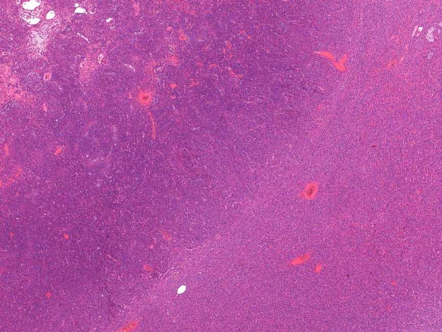
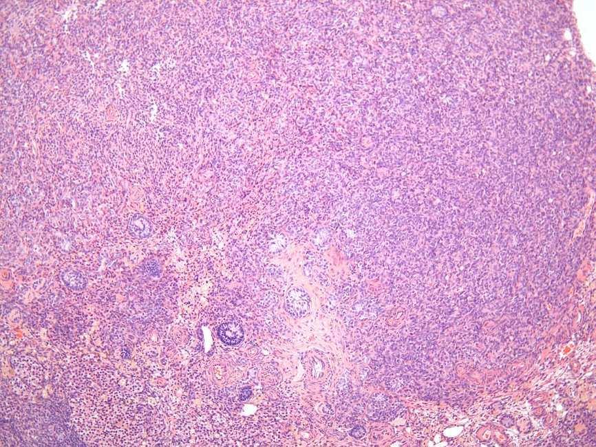
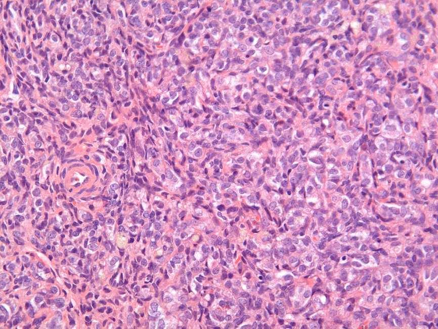
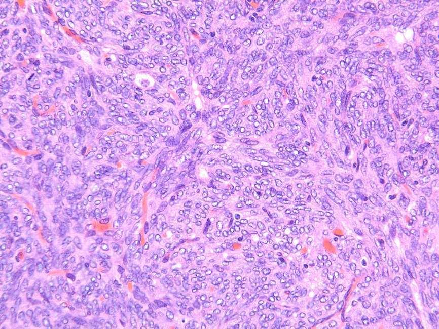
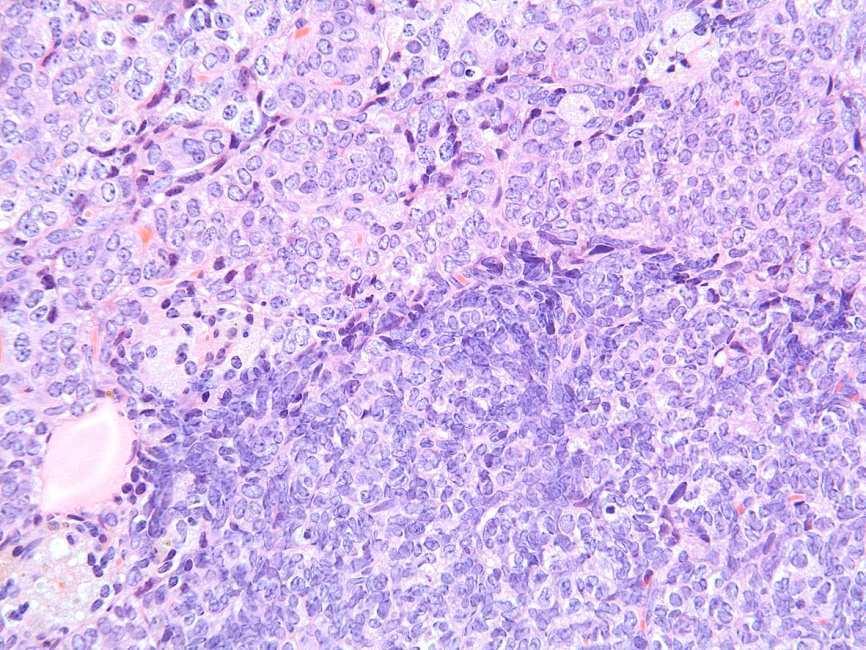
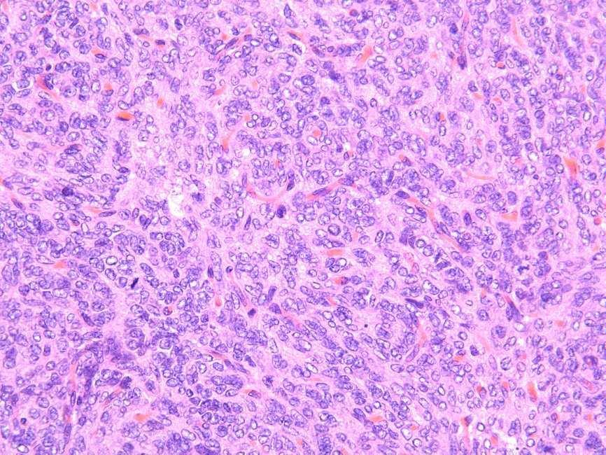
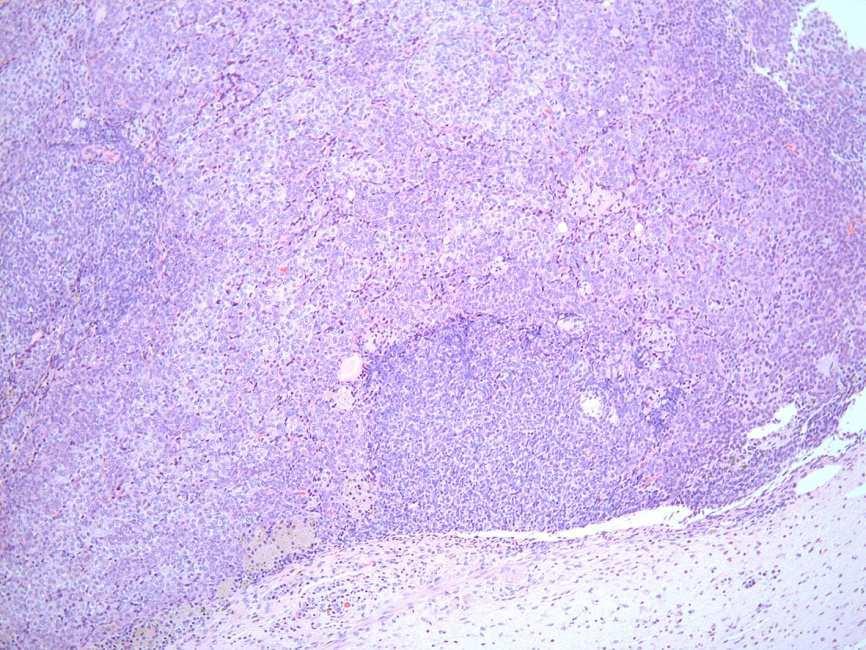
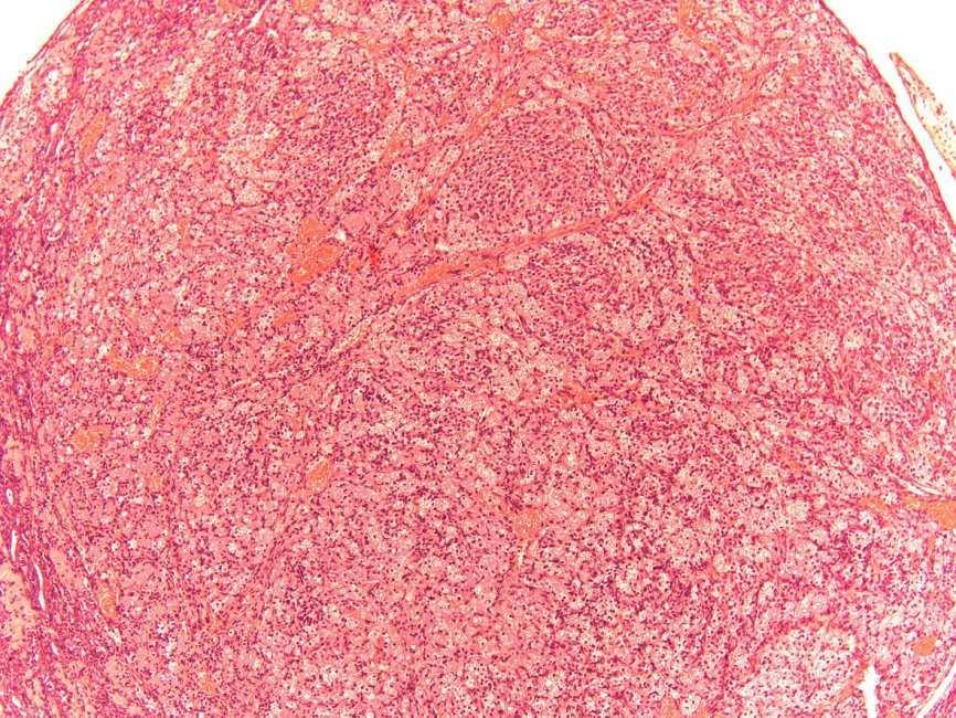
6 Ovary-Sex Cord Neoplasms: Granulosa Cell Tumor Variety of patterns: solid, cystic, tubular, occasionally sertoliform Resemble normal granulosa cells with round nucleus and coarse stippled chromatin Cytoplasm may become luteinized Malignancy: pleomorphism, increased mitosis, necrosis, innasion Differential Diagnose: Hyperplasia, Luteoma, Thecoma Harlan Laboratories 5


7 Rat: Granulosa Cell Tumor, benign Harlan Laboratories 6


8 Mouse: Granulosa Cell Tumor, benign Harlan Laboratories 7


9 Rat: Granulosa Cell Tumor, malignant Harlan Laboratories 8


10 Mouse: Granulosa Cell Tumor, malignant Harlan Laboratories 9
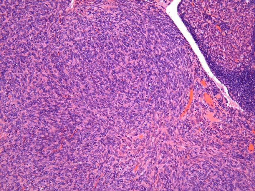
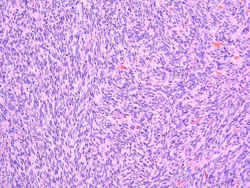
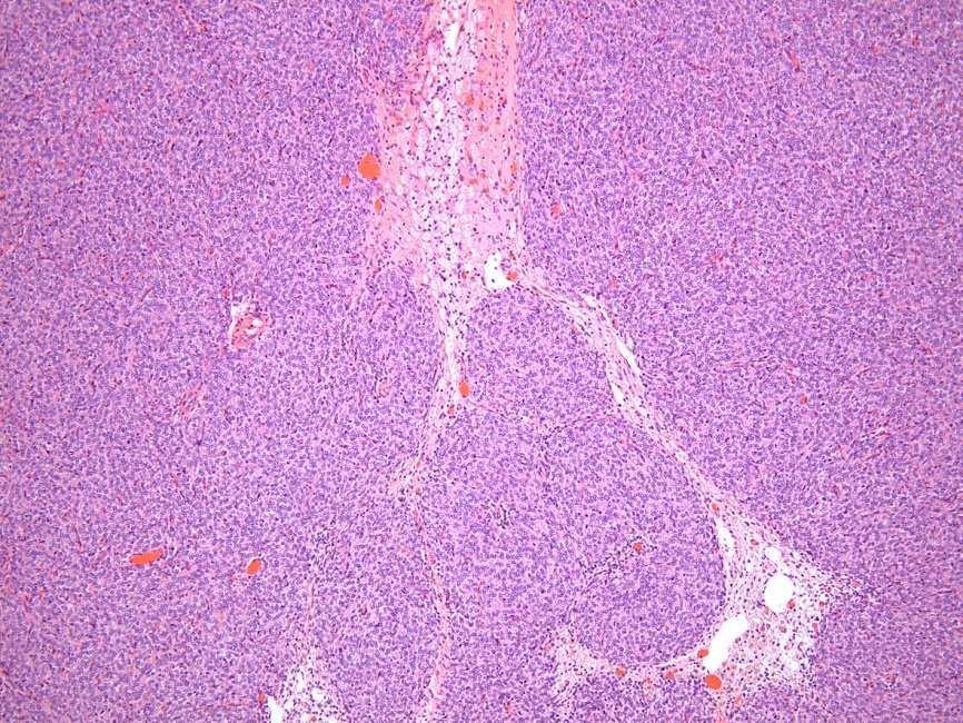
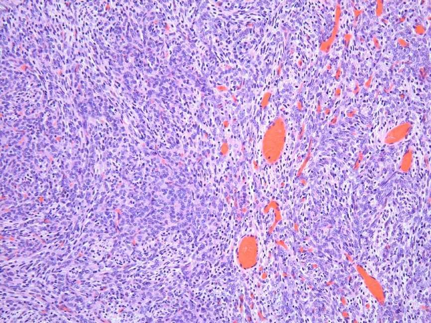
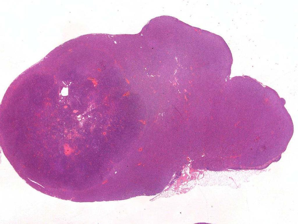
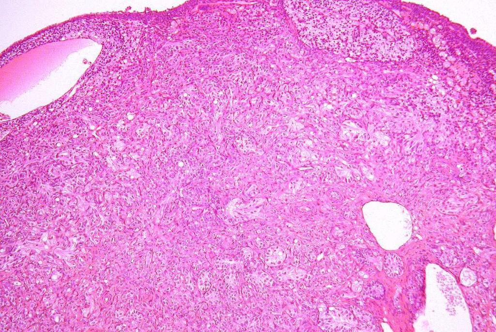
11 Ovary-Sex Cord Neoplasms: Thecoma Densely packed masses Fusiform, spindleshaped cells in whorls and nodular Areas of vacuolated cells may be present Malignancy: infiltrative growth pattern, invasion, atypia Differential Diagnose: Hyperplasia, Fibroma, Granulosa cell tumor, Luteoma Harlan Laboratories 10


12 Rat: Thecoma, benign Harlan Laboratories 11


13 Mouse: Thecoma, benign Harlan Laboratories 12


14 Hamster: Thecoma, benign Harlan Laboratories 13


15 Hamster: Thecoma, malignant Harlan Laboratories 14
16 Ovary-Sex Cord Neoplasms: Granulosa-Theca Cell Tumor Mixed neoplasm Partly dense areas of a particular cell type Differential Diagnose: Hyperplasia, Granulosa cell tumor, Thecoma Harlan Laboratories 15
17 Rat: Granulosa-Theca Cell Tumor, benign Harlan Laboratories 16


18 Mouse: Granulosa-Theca Cell Tumor, benign Harlan Laboratories 17
19 Hamster: Granulosa-Theca Cell Tumor, benign Harlan Laboratories 18
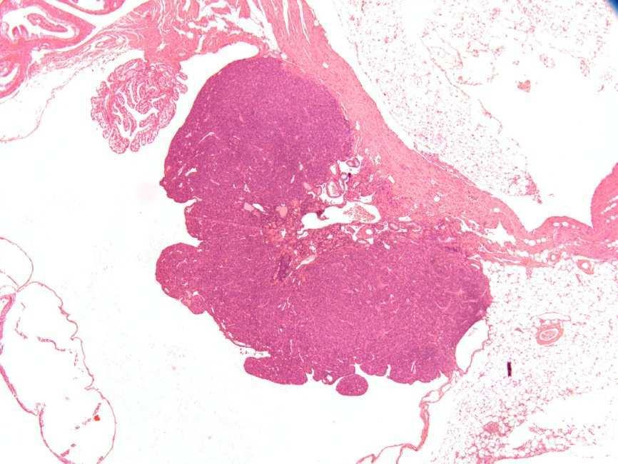
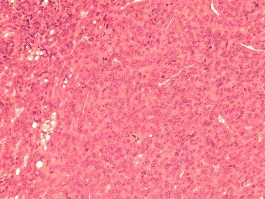
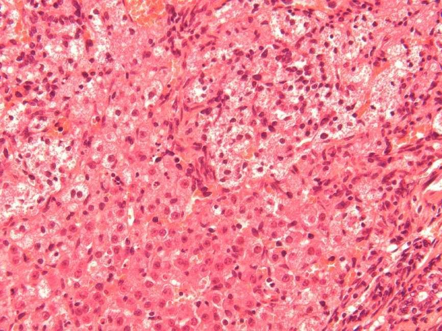
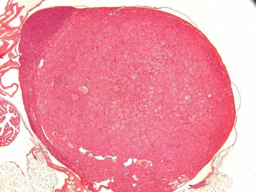
20 Ovary-Sex Cord Neoplasms: Luteoma Similar to granulosa cell tumor Neoplastic cells with abundant eosinophilic or vacuolated cytoplasm Intranuclear cytoplasmic invaginations Lobulation of tumor mass Differential Diagnose: Granulosa cell tumor, Thecoma Harlan Laboratories 19


21 Rat: Luteoma, benign Harlan Laboratories 20


22 Rat: Luteoma, benign Harlan Laboratories 21


23 Mouse: Luteoma, benign Harlan Laboratories 22
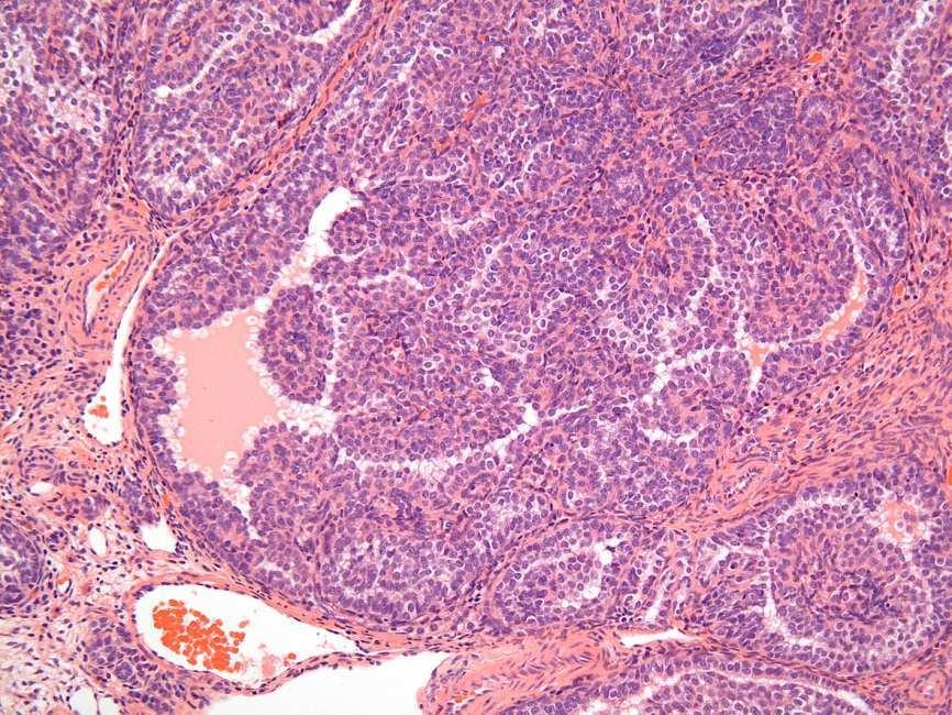
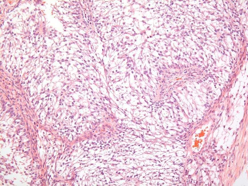
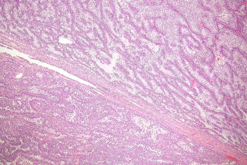
24 Ovary-Sex Cord Neoplasms: Sertoli s Cell Tumor Usually well circumscribed Irregular lobules Tubules composed of pale, vacuolated cells with indistict cell boundaries, seminiferous-like Malignancy: pleomorphism, increased mitosis, necrosis, invasion Differential Diagnose: Hyperplasia, Granulosa cell tumor,thecoma Harlan Laboratories 23


25 Rat: Sertoli s Cell Tumor, benign Harlan Laboratories 24


26 Rat: Sertoli s Cell Tumor, benign Harlan Laboratories 25


27 Mouse: Sertoli s Cell Tumor, benign Harlan Laboratories 26


28 Rat: Sertoli s Cell Tumor, malignant Harlan Laboratories 27


29 Rat: Sertoli s Cell Tumor, malignant Harlan Laboratories 28
Ovarian Neoplasms in F344 Rats and B6C3F1 Mice
 Environmental Health Perspectives Vol. 73, pp. 91-106, 1987 Ovarian Neoplasms in F344 Rats and B6C3F1 Mice by Roger H. Alison* and Kevin T. Morgant The National Toxicology Program (NTP) classification
Dr Sanjiv Manek Oxford. Oxford Pathology Course 2010 for FRCPath Illustration-Cellular Pathology. Oxford Radcliffe NHS Trust
 Dr Sanjiv Manek Oxford Oxford Pathology Course 2010 for FRCPath Illustration-Cellular Pathology. Oxford Radcliffe NHS Trust Ovarian Endometrial Vulvo-vaginal Cervical Illustration-Cellular Pathology. Oxford
Pathology of Ovarian Tumours. Dr. Jyothi Ranganathan MD ( Path) AFMC Pune PDCC (Cytopathology) PGI Chandigarh
 AFMC Pune PDCC (Cytopathology) PGI Chandigarh") Pathology of Ovarian Tumours Dr. Jyothi Ranganathan MD ( Path) AFMC Pune PDCC (Cytopathology) PGI Chandigarh Outline Incidence Risk factors Classification Pathology of tumours Tumour markers Prevention
Pathology of Ovarian Tumours Dr. Jyothi Ranganathan MD ( Path) AFMC Pune PDCC (Cytopathology) PGI Chandigarh Outline Incidence Risk factors Classification Pathology of tumours Tumour markers Prevention
3 cell types in the normal ovary
 Ovarian tumors 3 cell types in the normal ovary Surface (coelomic epithelium) the origin of the great majority of ovarian tumors 90% of malignant ovarian tumors Totipotent germ cells Sex cord-stromal cells
Salivary Glands 3/7/2017
 Salivary Glands 3/7/2017 Goals and objectives Focus on the entities unique to H&N Common board type facts Information for your future practice Salivary Glands Salivary Glands Major gland. Paratid. Submandibular.
Institute of Pathology First Faculty of Medicine Charles University. Ovary
 Ovary Barrett esophagus ph in vagina between 3.8 and 4.5 ph of stomach varies from 1-2 (hydrochloric acid) up to 4-5 BE probably results from upward migration of columnar cells from gastroesophageal junction
3 cell types in the normal ovary
 Ovarian tumors 3 cell types in the normal ovary Surface (coelomic epithelium) the origin of the great majority of ovarian tumors (neoplasms) 90% of malignant ovarian tumors Totipotent germ cells Sex cord-stromal
Note: The cause of testicular neoplasms remains unknown
 - In the 15- to 34-year-old age group, they are the most common tumors of men. - Tumors of the testis are a heterogeneous group of neoplasms that include: I. Germ cell tumors : 95%; all are malignant.
Pathology of the female genital tract
 Pathology of the female genital tract Common illnesses of the female genital tract Before menarche Developmental anomalies Tumors (ovarial teratoma) Amenorrhea Fertile years PCOS, ovarian cysts Endometriosis
Female Reproduc.ve System. Kris.ne Kra7s, M.D.
 Female Reproduc.ve System Kris.ne Kra7s, M.D. Female Reproduc.ve System Outline Cervix Uterus Ovaries Breast Cervical Carcinoma Once the most common cancer in women now not even in top 10. Decrease due
Diagnostically Challenging Cases in Gynecologic Pathology
 Diagnostically Challenging Cases in Gynecologic Pathology Eric C. Huang, M.D., Ph.D. Department of Pathology and Laboratory Medicine University of California, Davis Medical Center Case 1 Presentation 38
Case year female. Routine Pap smear
 Case 1 57 year female Routine Pap smear Diagnosis? 1. Atypical glandular cells of unknown significance (AGUS) 2. Endocervical AIS 3. Endocervical adenocarcinoma 4. Endometrial adenocarcinoma 5. Adenocarcinoma
Tinh hoàn
 Tinh hoàn Tinh hoàn Tinh hoàn Tiền liệt tuyến Tiền liệt tuyến Mào tinh hoàn Mào tinh hoàn Túi tinh Túi tinh Túi tinh Túi tinh So-called cystadenoma of seminal vesicle. Gross appearance of granulomatous
Mammary Nodular Hyperplasia in Intact R hesus Monkeys
 Vet. Path. 10: 130-134 (1973) Mammary Nodular Hyperplasia in Intact R hesus Monkeys L. W NELSON and L. D. SHOTT Department of Pathology and Toxicology, Mead Johnson Research Center, Evansville, Ind., and
Female Reproduc.ve System. Kris.ne Kra7s, M.D.
 Female Reproduc.ve System Kris.ne Kra7s, M.D. Female Reproduc.ve System Outline Cervix Uterus Ovaries Breast Female Reproduc.ve System Outline Cervix Cervical carcinoma Cervical Carcinoma Once the most
Pathology Slides. [Pathology]
![Pathology Slides. [Pathology]](/thumbs/94/120604575.jpg "Pathology Slides. [Pathology]") Pathology Slides MedicoNotes provides real laboratory pathological slides to aid you to differentiate between different pathological structures under microscope. www.mediconotes.com Histology slides example
Pathology Slides MedicoNotes provides real laboratory pathological slides to aid you to differentiate between different pathological structures under microscope. www.mediconotes.com Histology slides example
Circulation: X Case number: 501 Number of responses: 84 Date: 4 MAY 12
 Circulation: X Case number: 500 Number of responses: 81 Date: 4 MAY 12 Female, aged 65 TAH and BSO for G1 endometrioid adenocarcinoma. Tumour positive with inhibin, vimentin, CD56 and SMA. Negative with
Gross appearance of nodular hyperplasia in material obtained from suprapubic prostatectomy. Note the multinodular appearance and the admixture of
 Tiền liệt tuyến Tiền liệt tuyến Gross appearance of nodular hyperplasia in material obtained from suprapubic prostatectomy. Note the multinodular appearance and the admixture of solid and microcystic areas.
Exam 4 Review Outline
 Exam 4 Review Exam 4 Review Outline This review covers the following: Nervous System Female and Male Reproduc
Spontaneous Neoplastic Lesions in the CrI:CD-1(ICR) Mouse in Control Groups from 18 Month to 2 year Studies. March, 2005
 Mouse in Control Groups from 18 Month to 2 year Studies. March, 2005") Spontaneous Neoplastic Lesions in the CrI:CD-1(ICR) Mouse in Control Groups from 18 Month to 2 year Studies March, 2005 Information Prepared by Mary L.A. Giknis Ph.D Charles B. Clifford D.V.M, Ph.D 1063
Spontaneous Neoplastic Lesions in the CrI:CD-1(ICR) Mouse in Control Groups from 18 Month to 2 year Studies March, 2005 Information Prepared by Mary L.A. Giknis Ph.D Charles B. Clifford D.V.M, Ph.D 1063
7 Mousa. Obada Zalat. Mohammad Badi
 7 Mousa Obada Zalat Mohammad Badi Tumors of the ovaries Last lecture we talked about surface epithelial tumors of the ovaries (the most common type). But there are many other types of tumors of germ cell
Spontaneous Neoplastic Lesions in the Crl:CD-1 (ICR)BR Mouse. March, 2000
BR Mouse. March, 2000") Spontaneous Neoplastic Lesions in the Crl:CD-1 (ICR)BR Mouse March, 2000 Information Prepared by Mary L. A. Giknis, Ph.D. Charles B. Clifford, D.V.M., Ph.D. CHARLES RIVER LABORATORIES TABLE OF CONTENTS
Spontaneous Neoplastic Lesions in the Crl:CD-1 (ICR)BR Mouse March, 2000 Information Prepared by Mary L. A. Giknis, Ph.D. Charles B. Clifford, D.V.M., Ph.D. CHARLES RIVER LABORATORIES TABLE OF CONTENTS
-The cause of testicular neoplasms remains unknown
 - In the 15- to 34-year-old age group, they are the most common tumors of men. - include: I. Germ cell tumors : (95%); all are malignant. II. Sex cord-stromal tumors: from Sertoli or Leydig cells; usually
Mammary analogue secretory carcinoma of salivary gland A case report of new entity
 Case Report Mammary analogue secretory carcinoma of salivary gland A case report of new entity Vaibhav Bhika Bari 1*, Sandhya Unmesh Bholay 2 1 Assistant Professor, 2 Associate Professor Rajiv Gandhi Medical
A neoplasm is defined as "an abnormal tissue proliferation, which exceeds that of adjacent normal tissue. This proliferation continues even after
 NEOPLASIA Neoplasia is a very important topic in pathology because neoplasms are both common and serious diseases. A neoplasm literally means a new growth, and this term is used interchangeably with a
Cytology and Surgical Pathology of Gynecologic Neoplasms
 Cytology and Surgical Pathology of Gynecologic Neoplasms Current Clinical Pathology ANTONIO GIORDANO, MD, PHD SERIES EDITOR For further titles published in this series, go to http://www.springer.com/springer/series/7632
Gynaecological Malignancies
 Gynaecological Malignancies Dr Rodney Itaki Lecturer Anatomical Pathology Discipline University of Papua New Guinea Division of Pathology School of Medicine & Health Sciences Overview Genital tract tumors
OVARIES. MLS Basic histological diagnosis MLS HIST 422 Semester 8- batch 7 L13 Dr: Ali Eltayb.
 OVARIES MLS Basic histological diagnosis MLS HIST 422 Semester 8- batch 7 L13 Dr: Ali Eltayb. OBJECTIVES Recognize different disease of ovaries Classify ovarian cyst Describe the pathogenesis, morphology
Pancreas. Atrophy, acinar cell. Pathogenesis: Diagnostic key features:
 Pancreas Atrophy, acinar cell Pathogenesis: Decrease in number and/or size of acinar cells may be due to spontaneous or experimentally induced degenerative changes, apoptosis, or a sequel of chronic inflammation.
PATHOLOGY OF LIVER TUMORS
 PATHOLOGY OF LIVER TUMORS Pathobasic, 31.05.2016 WHO Classification Approach to a Liver Mass Lesion in a patient with chronic liver disease? Lesion in a patient without chronic liver disease? Malignant
TUMOR,NEOPLASM. Pathology Department, Zhejiang University School of Medicine,
 TUMOR,NEOPLASM Pathology Department, Zhejiang University School of Medicine, 马丽琴,maliqin198@zju.edu.cn The points in this chapter What is a neoplasm (conception) Morphology of neoplasm Macroscopy of Neoplasm
Endometrial Stromal Tumors
 Endometrial Stromal Tumors WHO Categories: Endometrial Stromal Nodule (ESN) Endometrial Stromal Sarcoma, low grade (LGESS) Endometrial Stromal Sarcoma, high grade (HGESS) Undifferentiated Uterine Sarcoma
XIII. Tumours of the liver and biliary system
 XIII. Tumours of the liver and biliary system V. PONOMARKOV 1 & L. J. MACKEY 2 In this histological classification of liver and gall bladder tumours the tumour types largely correspond to those found in
Neoplasia literally means "new growth.
 NEOPLASIA Neoplasia literally means "new growth. A neoplasm, defined as "an abnormal mass of tissue the growth of which exceeds and is uncoordinated with that of the normal tissues and persists in the
NEOPLASIA-I CANCER. Nam Deuk Kim, Ph.D.
 NEOPLASIA-I CANCER Nam Deuk Kim, Ph.D. 1 2 Tumor in the hieroglyphics of the Edwin Smith papyrus (1,600 B.C., Breasted s translation 1930) 3 War on Cancer (National Cancer Act, 1971) 4 Cancer Acts in Korea
Springer Healthcare. Understanding and Diagnosing Ovarian Cancer. Concise Reference: Krishnansu S Tewari, Bradley J Monk
 Concise Reference: Understanding and Diagnosing Ovarian Cancer Krishnansu S Tewari, Bradley J Monk Extracted from: The 21 st Century Handbook of Clinical Ovarian Cancer Published by Springer Healthcare
the urinary system pathology Dr. Fairoz A Eltorgman
 the urinary system pathology Dr. Fairoz A Eltorgman Tumors of the renal pelvis & kidney Benign tumors of the renal pelvis: Hemangioma Leiomyoma Malignant tumors: Transitional cell carcinoma Squamous cell
Biliary tract tumors
 Short Course 2010 Annual Fall Meeting of the Korean Society for Pathologists Biliary tract tumors Joon Hyuk Choi, M.D., Ph.D. Professor, Department of Pathology, Yeungnam Univ. College of Medicine, Daegu,
2/9/2015. Bartholin Cyst. Vulva: Squamous epithelium skin. Vagina: Squamous epithelium mucosa. Cervix: Ectocervix: squamous Endocervix: glandular
 Vulva: Squamous epithelium skin Bartholin Cyst Vagina: Squamous epithelium mucosa Cervix: Ectocervix: squamous Endocervix: glandular Slide courtesy of Dr. Lodge Rigal Slide courtesy of Dr. Lodge Rigal
PATHOLOGY OF THE FEMALE GENITAL TRACT
 MBBS 2 nd Yr. Lecture Dr. Annie Cheung September 30, 2002 9:30 am LT2,G/F, Academic and Administration Block Faculty of Medicine Building UROGENITAL SYSTEM PATHOLOGY OF THE FEMALE GENITAL TRACT Learning
Diagnostic accuracy of ultrasonography with color doppler imaging techniques in adnexal masses and correlation with histopathological analysis
 Original Article Diagnostic accuracy of ultrasonography with color doppler imaging techniques in adnexal masses and correlation with histopathological analysis Neha Gupta 1*, Poonam Gupta 2, Omvati Gupta
ONCOLOGY. Csaba Bödör. Department of Pathology and Experimental Cancer Research november 19., ÁOK, III.
 ONCOLOGY Csaba Bödör Department of Pathology and Experimental Cancer Research 2018. november 19., ÁOK, III. bodor.csaba1@med.semmelweis-univ.hu ONCOLOGY Characteristics of Benign and Malignant Neoplasms
International Journal of Medical and Health Sciences
 International Journal of Medical and Health Sciences Journal Home Page: http://www.ijmhs.net ISSN:2277-4505 Case Report Histomorphological Specrtum of Malignant Germ Cell Tumours: An Overview and Report
Disorders of Cell Growth & Neoplasia. Histopathology Lab
 Disorders of Cell Growth & Neoplasia Histopathology Lab Paul Hanna April 2010 Case #84 Clinical History: 5 yr-old, West Highland White terrier. skin mass from axillary region. has been present for the
Spontaneous Neoplasms and Survival in Wistar Han Rats: Compilation of Control Group Data. March, 2003
 Spontaneous Neoplasms and Survival in Wistar Han Rats: Compilation of Control Group Data March, 2003 Information Prepared by Mary L.A. Giknis Ph.D Charles B. Clifford D.V.M, Ph.D TABLE OF CONTENTS INTRODUCTION...1
The Adnexal Mass. Handout NCUS 3/18/2017 Suzanne Dixon, MD
 The Adnexal Mass Handout NCUS 3/18/2017 Suzanne Dixon, MD Objectives: Pelvic mass differential Characteristics of the normal ovary Standard terminology for ovarian masses Benign vs. malignant features
XX. Tumours of the nasal cavity *
 XX. Tumours of the nasal cavity * H. STONZI 1 & B. HAUSER2 Tumours of the nasal cavity are rare in domestic animals, most cases occurring in the dog. Epithelial tumours are the most common type in carnivores
Ovarian fibromas/thecomas are uncommon
 Sonography of Ovarian s/thecomas Patricia A. Athey, MD, Robert S. Malone, MD The sonographic findings in 14 patients with ovarian fibromas/thecomas are described. A broad spectrum of sonographic features
Disclosures. Parathyroid Pathology. Objectives. The normal parathyroid 11/10/2012
 Disclosures Parathyroid Pathology I have nothing to disclose Annemieke van Zante MD/PhD Assistant Professor of Clinical Pathology Associate Chief of Cytopathology Objectives 1. Review the pathologic features
Synonyms. Nephrogenic metaplasia Mesonephric adenoma
 Nephrogenic Adenoma Synonyms Nephrogenic metaplasia Mesonephric adenoma Definition Benign epithelial lesion of urinary tract with tubular, glandular, papillary growth pattern Most frequently in the urinary
Thyroid follicular neoplasms in cytology. Ulrika Klopčič Institute of Oncology, Department of Cytopathology, Ljubljana, Slovenia
 Thyroid follicular neoplasms in cytology Ulrika Klopčič Institute of Oncology, Department of Cytopathology, Ljubljana, Slovenia Lecture overview importance of FNAB in assessing thyroid lesions follicular
Oogenesis. Key Concepts. Female Reproductive Tract
 Oogenesis 1 Key Concepts Female Reproductive Tract Ovary Oogenesis Follicles Ovulation Corpus Luteum Molecular Activity Primordial Germ Cells (PGCs) 2 Female Reproductive Tract Ovary Oviduct Uterus Vagina
Diseases of the breast (1 of 2)
") Diseases of the breast (1 of 2) Introduction A histology introduction Normal ducts and lobules of the breast are lined by two layers of cells a layer of luminal cells overlying a second layer of myoepithelial
Diseases of the breast (1 of 2) Introduction A histology introduction Normal ducts and lobules of the breast are lined by two layers of cells a layer of luminal cells overlying a second layer of myoepithelial
Ovarian Tumors: A Study of 2146 Cases at AFIP, Rawalpindi, Pakistan.
 Ovarian Tumors: A Study of 2146 Cases at AFIP, Rawalpindi, Pakistan. 1 Muhammad Zubair, 2 Shoaib Naiyar Hashmi, 3 Saeed Afzal, 4 Iqbal Muhammad, 5 Hafeez Ud Din, 6 Syed Naeem Raza Hamdani, 7 Rabia Ahmad.
Endosalpingiosis. Case report
 Case report Endosalpingiosis Michael D. Holmes, M.D. Howard S. Levin M.D. Department of Pathology Lester A. Ballard, Jr., M.D. Department of Gynecology Endosalpingiosis, a term referring to tuballike epithelium
Lesions Mimicking Adenoid Cystic Carcinoma. Diagnostic Problems in Salivary Gland Pathology An Update 5/29/2009
 Diagnostic Problems in Salivary Gland Pathology An Update Lesions Mimicking Adenoid Cystic Carcinoma Stacey E. Mills, M.D. W.S. Royster Professor of Pathology Director of Surgical and Cytopathology University
Endometrial Stromal Sarcoma
 May 26, 2011 By Sushila Ladumor, MD [1] Endometrial stromal sarcoma (ESS) is a rare malignant tumor of the endometrium, occurring in the age group of 40-50 years. History The 50-year-old, female patient
Presenter: Yeh-Han Wang M.D.
 Korea-Taiwan-Japan Joint Meeting for Gynecological Pathology Mini-lecture Female Adnexal Tumor of Probable Wolffian Origin (FATWO) in Taiwan: A Small Case Series and Literature Review Presenter: Yeh-Han
number Done by Corrected by Doctor Maha Shomaf
 number 16 Done by Waseem Abo-Obeida Corrected by Zeina Assaf Doctor Maha Shomaf MALIGNANT NEOPLASMS The four fundamental features by which benign and malignant tumors can be distinguished are: 1- differentiation
Neoplasia 2018 Lecture 2. Dr Heyam Awad MD, FRCPath
 Neoplasia 2018 Lecture 2 Dr Heyam Awad MD, FRCPath ILOS 1. List the differences between benign and malignant tumors. 2. Recognize the histological features of malignancy. 3. Define dysplasia and understand
Guide to Small Animal Reproductive Imaging using the Vevo 770
 Guide to Small Animal Reproductive Imaging using the Vevo 770 Course Objectives: After completion of this module, the participant will be able to accomplish the following: Recognize reproductive female
Epithelial tumors. Dr. F.F. Khuzin, PhD Dr. M.O. Mavlikeev
 Epithelial tumors Dr. F.F. Khuzin, PhD Dr. M.O. Mavlikeev Epithelial tumors Tumors from the epithelium are the most frequent among tumors. There are 2 group features of these tumors: The presence in most
Abdulrahman Alhanbali. Bahaa Najjar. Maha shomaf
 14 Abdulrahman Alhanbali Bahaa Najjar Maha shomaf 1 Neoplasia In this lecture we will talk about neoplasia, its features and the nomenclature of different types of tumors. Neoplasia (neo: new and plasia:
Normal endometrium: A, proliferative. B, secretory.
 Normal endometrium: A, proliferative. B, secretory. Nội mạc tử cung Nội mạc tử cung Cyclic changes in endometrium.. Approximate relationship of useful microscopic changes. Arias-Stella reaction in endometrial
Outline 11/2/2017. Pancreatic EUS-FNA general aspects. Cytomorphologic features of solid neoplasms/lesions of the pancreas
 ENDOSCOPIC ULTRASOUND GUIDED-FINE NEEDLE ASPIRATION CYTOLOGY OF PANCREAS Khalid Amin M.D. Assistant Professor Department of Laboratory Medicine and Pathology University of Minnesota Outline Pancreatic
Icd 10 ovarian stroma
 Icd 10 ovarian stroma Struma ovarii; Micrograph of a struma ovarii. Characteristic thyroid follicles are seen on the right, and ovarian stroma on the left. H&E stain. Classification and. Free, official
Breast Pathology. Breast Development
 Breast Pathology Lecturer: Hanina Hibshoosh, M.D. Reading: Kumar, Cotran, Robbins, Basic Pathology, 6th Edition, pages 623-635 Breast Development 5th week - thickening of the epidermis - milk line 5th
Review of the AP Part II Practical Examination. Dr David Clift Co Chief Examiner
 Review of the AP Part II Practical Examination Dr David Clift Co Chief Examiner General Remarks The part II practical examination involved 15 cases which were presented with sufficient clinical data to
Prepared By Jocelyn Palao and Layla Faqih
 Prepared By Jocelyn Palao and Layla Faqih The structure of the suspected atypical cell should always be compared to the structure of other similar, benign, cells which are present in the smears. The diagnosis
2/24/19. Ovarian pathology: IOTA ADNEXAL MASSES. Content. IOTA terms for description of an adnexal mass. IOTA terms for description of an adnexal mass
 Content Ovarian pathology: IOTA ADNEXAL MASSES X SIMPLE COMPLEX Dr DESCRIBE WHAT YOU SEE FRANZCOG, MPH, DDU, COGU Sonologist Clinically useful Benign Malignant Communication between clinicians/research
My Journey into the World of Salivary Gland Sebaceous Neoplasms
 My Journey into the World of Salivary Gland Sebaceous Neoplasms Douglas R. Gnepp Warren Alpert Medical School at Brown University Rhode Island Hospital Pathology Department Providence RI Asked to present
CINtec p16 INK4a Staining Atlas
 CINtec p16 INK4a Staining Atlas Rating Rating Positive The rating positive will be assigned if the p16 INK4a -stained slide shows a continuous staining of cells of the basal and parabasal cell layers of
TUMOR,NEOPLASM. Pathology Department, Zhejiang University School of Medicine,
 TUMOR,NEOPLASM Pathology Department, Zhejiang University School of Medicine, 马丽琴,maliqin198@zju.edu.cn Precancerous lesions, dysplasia, IN and carcinoma in situ 1. precancerous lesions: certain clinical
Differential Diagnosis of Oral Masses. Palatal Lesions
 Differential Diagnosis of Oral Masses Palatal Lesions Palatal Masses Periapical Abscess Torus Palatinus Mucocele Lymphoid Hyperplasia Adenomatous Hyperplasia Benign Salivary Neoplasms Malignant Salivary
PLEOMORPHIC ADENOMA ( BENIGN MIXED TUMOR )
") ( BENIGN MIXED TUMOR ) Grossly, the tumor is freely movable, solid, sometimes lobulated and occasionally cystic. If recurrent, multinodular masses are common. Histologically, within a fibrous capsule,
( BENIGN MIXED TUMOR ) Grossly, the tumor is freely movable, solid, sometimes lobulated and occasionally cystic. If recurrent, multinodular masses are common. Histologically, within a fibrous capsule,
FNA of Thyroid. Toward a Uniform Terminology With Management Guidelines. NCI NCI Thyroid FNA State of the Science Conference
 FNA of Thyroid NCI NCI Thyroid FNA State of the Science Conference Toward a Uniform Terminology With Management Guidelines Thyroid Thyroid FNA Cytomorphology NCI Thyroid FNA State of the Science Conference
SESSION 1: GENERAL (BASIC) PATHOLOGY CONCEPTS Thursday, October 16, :30am - 11:30am FACULTY COPY
 PATHOLOGY CONCEPTS Thursday, October 16, :30am - 11:30am FACULTY COPY") SESSION 1: GENERAL (BASIC) PATHOLOGY CONCEPTS Thursday, October 16, 2008 9:30am - 11:30am FACULTY COPY GOAL: Describe the basic morphologic (structural) changes which occur in various pathologic conditions.
SESSION 1: GENERAL (BASIC) PATHOLOGY CONCEPTS Thursday, October 16, 2008 9:30am - 11:30am FACULTY COPY GOAL: Describe the basic morphologic (structural) changes which occur in various pathologic conditions.
Pleomorphic adenoma of breast - a case report and distinction with metaplastic carcinoma D Gupta, S Agrawal, N Trivedi, A Tewari
 of breast - a case report and distinction with metaplastic carcinoma D Gupta, S Agrawal, N Trivedi, A Tewari Introduction, also known as mixed tumour, is a benign tumour which typically presents as a painless,
Neoplasms of the Canine, Feline and Lemur Liver:
 Neoplasms of the Canine, Feline and Lemur Liver: Classification and Prognosis Annual Seminar of the French Society of Veterinary Pathology John M. Cullen VMD PhD DACVP North Carolina State University Primary
Adenocarcinoma of the Cervix
 Question 1. Each of the following statements about cervical adenocarcinoma is true except: Adenocarcinoma of the Cervix SAMS a) A majority of women with cervical adenocarcinoma have stage I tumors at diagnosis.
Journal of Rawalpindi Medical College (JRMC); 2016;20(4):
; 2016;20(4):") Original Article Morphological Profile of Ovarian Tumours Bilquis Begum, Iram Nadeem Rana, Nadeem Ikram Department of Pathology, Rawalpindi Medical College, Rawalpindi Abstract Background: To study the
Original Article Morphological Profile of Ovarian Tumours Bilquis Begum, Iram Nadeem Rana, Nadeem Ikram Department of Pathology, Rawalpindi Medical College, Rawalpindi Abstract Background: To study the
Oncocytic carcinoma: A rare malignancy of the parotid gland
 ISPUB.COM The Internet Journal of Pathology Volume 8 Number 2 Oncocytic carcinoma: A rare malignancy of the parotid gland K Mardi, J Sharma Citation K Mardi, J Sharma.. The Internet Journal of Pathology.
H&E, IHC anti- Cytokeratin
 Cat No: OVC2281 - Ovary cancer tissue array Lot# Cores Size Cut Format QA/QC OVC228101 228 1.1mm 4um 12X19 H&E, IHC anti- Cytokeratin Recommended applications: For Research use only. RNA or protein ovary
Histological pattern of ovarian tumors and their age distribution
 Original Article Nepal Med Coll J 2008; 10(2): 81-85 Histological pattern of ovarian s and their age distribution R Jha and S Karki Department of Pathology, TUTH, Maharajgunj, Kathmandu, Nepal Corresponding
Diagnostic Cytology of Cancer Cases
 Diagnostic Cytology of Cancer Cases Somporn Techangamsuwan Companion Animal Cancer Research Unit (CAC-RU) Department of Pathology, Faculty of Veterinary Science, Chulalongkorn University 1 Tumor or Non-tumor
Mody. AIS vs. Invasive Adenocarcinoma of the Cervix
 Common Problems in Gynecologic Pathology Michael T. Deavers, M.D. Houston Methodist Hospital, Houston, Texas Common Problems in Gynecologic Pathology Adenocarcinoma in-situ (AIS) of the Cervix vs. Invasive
Gynecologic Cytopathology: Glandular lesions
 Gynecologic Cytopathology: Glandular lesions Lin Wai Fung (MSc, MPH, CMIAC) 17/4/2014 Glandular lesions of the uterus Endocervix Endometrium Normal endocervical cells Sheets, strips well-preserved architecture:
Clinicopathological and Histological Features of Ovarian Tumour- A Study
 IOSR Journal of Dental and Medical Sciences (IOSR-JDMS) e-issn: 2279-0853, p-issn: 2279-0861.Volume 16, Issue 9 Ver. IX (September. 2017), PP 56-60 www.iosrjournals.org Clinicopathological and Histological
Top Tips for Gynaecological Ultrasound. Catherine Kirkpatrick Consultant Sonographer Dublin Oct 2018
 Top Tips for Gynaecological Ultrasound Catherine Kirkpatrick Consultant Sonographer Dublin Oct 2018 We can all scan a pelvis so what can we do to improve? Uterus, endometrium and ovaries, got it covered!
Salivary gland cytology. Salivary gland cytology. Triage helps the clinician. Salivary gland tumors. Diagnostic difficulties
 Salivary gland cytology Salivary Gland Cytology Pınar Fırat, MD Professor of Pathology İ.U. İstanbul Faculty of Medicine Çapa, İstanbul It is a reliable diagnostic test However, definitive subclassification
Histopathological Study of Ovarian Lesions
 Histopathological Study of Ovarian Lesions Nirali N. Thakkar 1, Shaila N. Shah 2 1 Resident doctor, Pathology department, Government Medical College, Bhavnagar-364001, India 2 Head of the department, Pathology
ARTHUR PURDY STOUT SOCIETY COMPANION MEETING: DIFFICULT NEW DIFFERENTIAL DIAGNOSES IN PROSTATE PATHOLOGY. Jonathan I. Epstein.
 1 ARTHUR PURDY STOUT SOCIETY COMPANION MEETING: DIFFICULT NEW DIFFERENTIAL DIAGNOSES IN PROSTATE PATHOLOGY Jonathan I. Epstein Professor Pathology, Urology, Oncology The Reinhard Professor of Urological
1 NORMAL HISTOLOGY AND METAPLASIAS
 1 NORMAL HISTOLOGY AND METAPLASIAS, MD Anatomy and Histology 1 Metaplasias 2 ANATOMY AND HISTOLOGY The female breast is composed of a branching duct system, which begins at the nipple with the major lactiferous
Testicular Germ Cell Tumors; A Simplistic Approach
 Testicular Germ Cell Tumors; A Simplistic Approach Merce Jorda, MD, PhD, MBA Professor and Vice Chair, Director of Anatomic Pathology Director of Genitourinary Pathology Service Interim Director of Cytopathology
Multiple Synchronous Lesions in the Genital Tract of a Female: A Rare Combination with Unrelated Histogenesis
 pissn: 2288-6478, eissn: 2288-6761 Case Report Multiple Synchronous Lesions in the Genital Tract of a Female: Rare Combination with Unrelated Histogenesis Pattomthadathil Sankaran Jayalakshmy 1, Faseela
بسم هللا الرحمن الرحيم. Prof soha Talaat
 بسم هللا الرحمن الرحيم Ovarian tumors The leading indication for gynecologic surgery. Preoperative characterization of complex solid and cystic adnexal masses is crucial for informing patients about possible
MVST BOD & NST PART IB Thurs. 2 nd & Fri. 3 rd March 2017 Pathology Practical Class 23
 MVST BOD & NST PART IB Thurs. 2 nd & Fri. 3 rd March 2017 Pathology Practical Class 23 Neoplasia I Neoplasia I: Benign and malignant neoplasms in glandular epithelium and mesenchyme 1.0. Aims 1. To understand
South East England General Histopathology EQA Scheme
 South East England General Round g Final Case Analyses Cases 707 to 718 Circulated January February 2018 135 responses (85.44%) Prepared April 2018 Authorised by: Prof J Schofield Date: 23 rd April 2018
Spontaneous Neoplastic Lesions in the CrI:CD-I (ICR)BR Mouse. March, 2000
BR Mouse. March, 2000") Spontaneous Neoplastic Lesions in the CrI:CD-I (ICR)BR Mouse March, 000 Information Prepared by Mary L. A. Giknis, Ph.D. Charles B. Clifford, D.V.M., Ph. D. CHARLES RIVER LABORATORIES I Dewayne Johnson
Spontaneous Neoplastic Lesions in the CrI:CD-I (ICR)BR Mouse March, 000 Information Prepared by Mary L. A. Giknis, Ph.D. Charles B. Clifford, D.V.M., Ph. D. CHARLES RIVER LABORATORIES I Dewayne Johnson
Abid Irshad, MD Director Breast Imaging. Medical University of South Carolina Charleston
 Abid Irshad, MD Director Breast Imaging Medical University of South Carolina Charleston Cases Financial disclosure: I or my family have no financial interest related to the material discussed in this presentation